Library › Lung tumors: adenocarcinoma, squamous and neuroendocrine carcinomas, staging, biomarkers and other lung neoplasms
Lung tumors: adenocarcinoma, squamous and neuroendocrine carcinomas, staging, biomarkers and other lung neoplasms
Last reviewed 2026-11-10 by a board-certified pathologist. 251 references.
Lung Adenocarcinoma: Precursor Lesions, Invasive Patterns, IASLC Grading, Mucinous Adenocarcinoma and Spread Through Air Spaces
Definition. Lung adenocarcinoma is the most common histologic type of lung cancer. The IASLC/ATS/ERS classification divides it into preinvasive lesions, minimally invasive adenocarcinoma, invasive adenocarcinoma classified by predominant pattern, and variants (Travis et al. 2011). The current published classification is the WHO Classification of Thoracic Tumours, 5th edition (2021). Its principles are morphology first, supported by immunohistochemistry and then by molecular techniques (Nicholson et al. 2022). The 6th edition of the thoracic volume is in progress and has no published summary (IARC 2026). Entity-level statements follow the WHO 5th edition.
Related page: Squamous Cell, Adenosquamous, Large Cell and Sarcomatoid Carcinomas of the Lung, NUT Carcinoma and Thoracic SMARCA4-Deficient Undifferentiated Tumor (squamous, adenosquamous, large cell and sarcomatoid carcinomas; small-biopsy terminology) Related page: Lung Neuroendocrine Neoplasms: Typical and Atypical Carcinoid, Large Cell Neuroendocrine Carcinoma, Small Cell Carcinoma and DIPNECH (carcinoid tumors, large cell neuroendocrine carcinoma, small cell carcinoma) Related page: Lung Cancer Staging and Reporting: AJCC Version 9, Visceral Pleural Invasion, Multiple Tumors, Neoadjuvant Response and the CAP Protocol (AJCC Version 9 T categories, visceral pleural invasion, multiple tumors, CAP synoptic elements) Related page: Lung Cancer Predictive Biomarkers: Driver Genes, PD-L1 Immunohistochemistry and Specimen Handling (driver gene testing and PD-L1) Related page: Other Lung Tumors: Hamartoma, Sclerosing Pneumocytoma, Adenomas, Salivary Gland-Type, Mesenchymal and Lymphoid Tumors, and Metastases (adenomas, salivary gland-type tumors and metastases) Related page: Lung Adenocarcinoma (Cytology) (cytomorphology of lung adenocarcinoma) Related page: Metastatic Lung Adenocarcinoma in Serous Effusions (lung adenocarcinoma in effusions)
WHO 5th edition classification of lung carcinomas
The CAP resection protocol (version 5.1.0.0) lists these histologic types and recommends the WHO classification for reporting (CAP 2025). Benign epithelial tumors, salivary gland-type tumors in detail, mesenchymal tumors and lymphomas are covered in Other Lung Tumors: Hamartoma, Sclerosing Pneumocytoma, Adenomas, Salivary Gland-Type, Mesenchymal and Lymphoid Tumors, and Metastases.
| Group (WHO 5th edition) | Types listed for resection reporting | Source |
|---|---|---|
| Glandular precursor lesions | Atypical adenomatous hyperplasia; adenocarcinoma in situ, nonmucinous and mucinous | Travis et al. 2011; CAP 2025 |
| Adenocarcinoma | Minimally invasive adenocarcinoma (nonmucinous, mucinous); invasive lepidic, acinar, papillary, micropapillary and solid adenocarcinoma; invasive mucinous adenocarcinoma; mixed invasive mucinous and nonmucinous adenocarcinoma; colloid, fetal and enteric-type adenocarcinoma | CAP 2025 |
| Squamous precursor and carcinoma | Squamous cell carcinoma in situ; keratinizing, nonkeratinizing and basaloid squamous cell carcinoma; lymphoepithelial carcinoma | CAP 2025; Nicholson et al. 2022 |
| Large cell and adenosquamous carcinoma | Large cell carcinoma; adenosquamous carcinoma | CAP 2025 |
| Sarcomatoid carcinomas | Pleomorphic carcinoma (spindle cell and giant cell components); pulmonary blastoma; carcinosarcoma | CAP 2025 |
| Other epithelial tumors | NUT carcinoma; thoracic SMARCA4-deficient undifferentiated tumor | CAP 2025; Nicholson et al. 2022 |
| Salivary gland-type tumors | Adenoid cystic, epithelial-myoepithelial, mucoepidermoid, hyalinizing clear cell and myoepithelial carcinoma | CAP 2025 |
| Neuroendocrine neoplasms | Typical carcinoid/neuroendocrine tumor grade 1; atypical carcinoid/neuroendocrine tumor grade 2; carcinoid tumor, NOS; small cell carcinoma and combined small cell carcinoma; large cell neuroendocrine carcinoma and combined large cell neuroendocrine carcinoma | CAP 2025 |
Changes in the WHO 5th edition classification of lung tumors
- Genetic testing. Genetic testing receives broader emphasis, and a section is dedicated to small diagnostic samples (Nicholson et al. 2022).
- Adenocarcinoma. Percentages of histologic patterns continue to be recorded in invasive nonmucinous adenocarcinoma and form the basis of a formal grading system. Only the invasive size gives the T size in part-lepidic nonmucinous adenocarcinoma (Nicholson et al. 2022).
- Spread through air spaces. STAS is recognized as a histologic feature with prognostic significance (Nicholson et al. 2022).
- Other tumor types. Lymphoepithelial carcinoma is reclassified as a squamous cell carcinoma, bronchiolar adenoma/ciliated muconodular papillary tumor is a new adenoma, and thoracic SMARCA4-deficient undifferentiated tumor is recognized. Neuroendocrine concepts are updated, and each tumor has essential and desirable diagnostic criteria (Nicholson et al. 2022).
- Previous rule (2011). The IASLC/ATS/ERS classification retired the terms bronchioloalveolar carcinoma (BAC) and mixed subtype adenocarcinoma and introduced AIS, MIA and the micropapillary subtype (Travis et al. 2011).
Epidemiology of lung adenocarcinoma
- Burden. Lung cancer was the most frequently diagnosed cancer worldwide in 2022, with almost 2.5 million new cases (12.4% of all cancers). It was the leading cause of cancer death, with about 1.8 million deaths (18.7%) (Bray et al. 2024).
- Sex. In the United States in 2021, lung cancer incidence in women younger than 65 years exceeded that in men (15.7 compared with 15.4 per 100,000) (Siegel et al. 2025).
- Smoking and other risk factors. About 85% of the 124,730 lung cancer deaths projected in the United States for 2025 are caused directly by cigarette smoking. A further 3,500 are attributed to second-hand smoke and 15,100 to a combination of other combustible tobacco products, radon, occupational exposures, air pollution and other environmental exposures. Cigarette smoking prevalence fell from 42% in 1965 to 12% in 2022 (Siegel et al. 2025).
- Precursor lesions. Atypical adenomatous hyperplasia is found more often in lungs bearing cancer, especially adenocarcinoma, and is more common in women (Kerr 2001).
Atypical adenomatous hyperplasia, adenocarcinoma in situ and minimally invasive adenocarcinoma
Atypical adenomatous hyperplasia
- Morphology. AAH is a localized proliferation, usually 0.5 cm or less, of mildly to moderately atypical type II pneumocytes or club (Clara) cells lining alveolar walls and sometimes respiratory bronchioles. Gaps are usually present between the cells, which are rounded, cuboidal, low columnar or peg-shaped. Intranuclear inclusions are frequent (Travis et al. 2011).
- Grading. A spectrum of cellularity and atypia occurs, and grading of AAH is not recommended. Separation of cellular AAH from AIS can be difficult histologically and is impossible cytologically (Travis et al. 2011).
Adenocarcinoma in situ
- Definition. AIS is a localized adenocarcinoma of 3 cm or less with growth restricted to neoplastic cells along preexisting alveolar structures (lepidic growth). Stromal, vascular and pleural invasion are absent. Papillary and micropapillary patterns and intra-alveolar tumor cells are absent (Travis et al. 2011).
- Nonmucinous and mucinous AIS. Virtually all AIS is nonmucinous, composed of type II pneumocytes or club cells. Rare mucinous AIS consists of tall columnar cells with basal nuclei and abundant cytoplasmic mucin. Nuclear atypia is absent or inconspicuous in both, and septal widening with sclerosis is common in nonmucinous AIS (Travis et al. 2011).
- Outcome. In 236 resected adenocarcinomas of 2 cm or less, Noguchi types A and B (localized BAC with or without alveolar collapse) showed no nodal metastasis and 100% 5-year survival (Noguchi et al. 1995). These tumors meet criteria for AIS (Travis et al. 2011).
Minimally invasive adenocarcinoma
- Definition. MIA is a solitary adenocarcinoma of 3 cm or less with a predominantly lepidic pattern and invasion of 5 mm or less in greatest dimension in any one focus (Travis et al. 2011).
- Measured invasive component. The invasive component consists of histologic subtypes other than lepidic (acinar, papillary, micropapillary or solid) or tumor cells infiltrating myofibroblastic stroma. When several invasive foci are present, the largest focus is measured, and the foci are not summed (Travis et al. 2011).
- Exclusion criteria. MIA is excluded when the tumor invades lymphatics, blood vessels or pleura, contains tumor necrosis or shows STAS (CAP 2025). Tumors of 3 cm or less with 0.5 cm or less of invasion and any of these features are classified as lepidic-predominant adenocarcinoma (CAP 2025).
- Criteria of 2011. The IASLC/ATS/ERS criteria excluded MIA for lymphatic, vascular or pleural invasion and tumor necrosis (Travis et al. 2011). Under CAP 5.1.0.0, which follows the WHO 5th edition, STAS also excludes MIA (CAP 2025).
- Sampling. When AIS or MIA is being considered, the lesion is submitted entirely for histologic examination (CAP 2025).
- Outcome. In 1038 stage I adenocarcinomas, patients with AIS (n=2) and MIA (n=34) had no recurrences (Kadota et al. 2014a). In 514 stage I adenocarcinomas, AIS and MIA had 100% 5-year disease-free survival (Yoshizawa et al. 2011).
| Lesion | Size | Growth pattern | Invasion | Exclusions | Pathologic T (AJCC) | Source |
|---|---|---|---|---|---|---|
| Atypical adenomatous hyperplasia | Usually ≤0.5 cm | Lepidic, mild to moderate atypia, gaps between cells | None | Not applicable | Not staged | Travis et al. 2011 |
| Adenocarcinoma in situ | ≤3 cm, solitary | Pure lepidic | None | Papillary, micropapillary or intra-alveolar cells | pTis (AIS) | Travis et al. 2011; CAP 2025 |
| Minimally invasive adenocarcinoma | ≤3 cm, solitary | Lepidic predominant | ≤5 mm in the largest focus | Lymphatic, vascular or pleural invasion; necrosis; STAS | pT1mi | Travis et al. 2011; CAP 2025 |
| Lepidic-predominant adenocarcinoma | Any | Lepidic predominant | >5 mm, or ≤5 mm with an exclusion feature | Not applicable | T by invasive size | Travis et al. 2011; CAP 2025 |
Lepidic growth and invasion in nonmucinous adenocarcinoma
- Definition of invasion. The WHO defines invasion in adenocarcinoma as any histologic subtype other than lepidic, tumor cells infiltrating myofibroblastic stroma, vascular or pleural invasion, or STAS (CAP 2025).
- Collapse. AIS with collapse can be difficult to distinguish from the acinar pattern. Loss of alveolar architecture or the presence of myofibroblastic stroma indicates invasive acinar adenocarcinoma (Travis et al. 2011).
- Papillary pattern. Tangential sectioning of alveolar walls in AIS mimics papillae. Alveolar spaces filled with papillary structures are classified as papillary adenocarcinoma (Travis et al. 2011).
- Reproducibility. Among 28 pulmonary pathologists, kappa for invasive compared with noninvasive patterns was 0.55 for typical and 0.08 for difficult images (Thunnissen et al. 2012).
- IASLC criteria for invasion. In a Delphi study, the coefficient of variation was 6.9% for total size and 54% for invasive size. Collapsed alveoli and transition zones with extensive epithelial proliferation were the main sources of variability. Extensive epithelial proliferation is multilayered luminal epithelial growth, usually with high-grade cytology, in several alveolar spaces within collapsed lepidic areas. It is regarded as likely invasive (Thunnissen et al. 2023).
- Elastic stains. Elastin staining is used to variable extents in different countries to separate true invasion from iatrogenic collapse of alveoli. It highlights distorted but preexisting alveolar architecture, and its interpretation differs between collapsed lepidic patterns and true papillary patterns (Thunnissen et al. 2022). In 120 lesions, AIS and the in situ components of MIA and invasive adenocarcinoma showed a continuous, thickened and recoiled elastic framework. Elastic fibers were decreased or absent in all invasive components of MIA and in most invasive adenocarcinomas, and elastic staining improved diagnostic accuracy (Zhou et al. 2026).
Prognostic value of invasive size in part-lepidic nonmucinous adenocarcinoma
- Evidence. Invasive size adjusted for the percentage of lepidic growth was associated with disease-free survival slightly more strongly than gross size (Yoshizawa et al. 2011). In stage I adenocarcinoma, the difference in recurrence between tumors of ≤2 cm and >2 to 3 cm was larger for invasive size (12% compared with 27%) than for total size (13% compared with 21%) (Kadota et al. 2014a).
Related page: Lung Cancer Staging and Reporting: AJCC Version 9, Visceral Pleural Invasion, Multiple Tumors, Neoadjuvant Response and the CAP Protocol (invasive-size rule for pT, STAS as a staging descriptor, visceral pleural invasion and multiple tumors)
Invasive nonmucinous adenocarcinoma
Histologic patterns of invasive nonmucinous adenocarcinoma
- Comprehensive subtyping. Invasive nonmucinous adenocarcinoma is classified by its predominant pattern. Nonpredominant patterns are recorded semiquantitatively in 5% increments totaling 100% (CAP 2025).
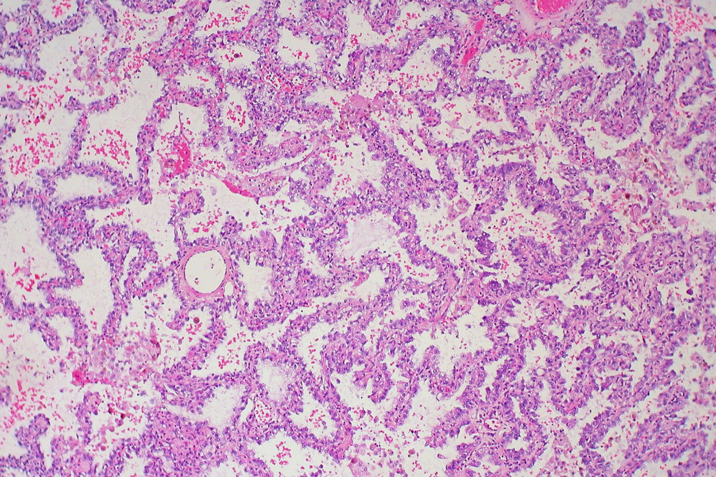
- Lepidic. Lepidic-predominant adenocarcinoma consists of bland pneumocytic cells along alveolar walls with at least one invasive focus larger than 5 mm (Travis et al. 2011).
- Acinar. Round to oval glands with a central lumen form the major component. Rounded aggregates with peripheral nuclear polarization and no clear lumen also count as acinar (Travis et al. 2011).
- Papillary. Glandular cells grow along central fibrovascular cores. Myofibroblastic stroma is not needed for this pattern (Travis et al. 2011).
- Micropapillary. Tumor cells form papillary tufts without fibrovascular cores, detached or attached to alveolar walls. Ring-like structures may float in alveolar spaces, and vascular and stromal invasion are frequent (Travis et al. 2011).
- Solid. Polygonal cells form sheets without other patterns. A 100% solid tumor requires intracellular mucin in at least five tumor cells in each of two high-power fields, confirmed with mucin stains (Travis et al. 2011).
- Complex glands. Cribriform glands are nests with sieve-like perforations. Fused glands are poorly formed glands without intervening stroma, or ribbon-like glands with irregular borders and single cells infiltrating desmoplastic stroma (Moreira et al. 2020).
| Pattern | Defining feature | Prognostic group | IASLC high-grade pattern | Source |
|---|---|---|---|---|
| Lepidic | Bland cells along intact alveolar walls | Favorable | No | Travis et al. 2011; Moreira et al. 2020 |
| Acinar | Glands with central lumens in stroma | Intermediate | No | Travis et al. 2011; Moreira et al. 2020 |
| Papillary | Cells on fibrovascular cores | Intermediate | No | Travis et al. 2011; Moreira et al. 2020 |
| Micropapillary | Tufts without fibrovascular cores, floating rings | Poor | Yes | Travis et al. 2011; Moreira et al. 2020 |
| Solid | Sheets with intracellular mucin | Poor | Yes | Travis et al. 2011; Moreira et al. 2020 |
| Cribriform | Nests with sieve-like spaces | Poor | Yes (complex gland) | Kadota et al. 2014b; Moreira et al. 2020 |
| Fused glands | Fused or ribbon-like glands; single cells in desmoplastic stroma | Poor | Yes (complex gland) | Moreira et al. 2020; CAP 2025 |
Prognostic evidence for adenocarcinoma patterns
- Stage I series. In 514 stage I tumors, 5-year disease-free survival was 90% for lepidic-predominant, 83% for papillary-predominant and 84% for acinar-predominant tumors. It was 67% for micropapillary-predominant and 75% for invasive mucinous adenocarcinoma (Yoshizawa et al. 2011).
- All stages. In 500 adenocarcinomas of stages I to IV, the predominant pattern independently predicted survival (Warth et al. 2012).
- Lepidic extent. Tumors with >50% lepidic pattern had no recurrences (n=84). Tumors with ≤10% lepidic pattern had a 5-year cumulative incidence of recurrence of 22% (Kadota et al. 2014a).
- Micropapillary component. In 35 adenocarcinomas with a micropapillary component, 33 patients (94%) developed metastases. Most metastases showed a prominent micropapillary component regardless of its extent in the primary tumor (Amin et al. 2002).
- Cribriform pattern. Cribriform-predominant tumors had a 5-year recurrence-free probability of 70%, comparable to micropapillary and solid tumors. A cribriform component of ≥10% independently predicted recurrence in acinar-predominant tumors (Kadota et al. 2014b).
IASLC grading of invasive nonmucinous adenocarcinoma
- Scheme. The IASLC grade combines the predominant pattern with the proportion of high-grade patterns (solid, micropapillary and complex glands) at a cutoff of 20% (Moreira et al. 2020). The WHO 5th edition and the CAP protocol use it (CAP 2025).
- Derivation. The 20% cutoff gave the best combination of sensitivity and specificity for recurrence (Moreira et al. 2020).
- Comparison. Separation of recurrence-free survival between grades was clearer with the IASLC grade than with a predominant-pattern grade (Moreira et al. 2020).
- Reproducibility. Five observers reached a kappa of 0.617 on whole-slide images. Most disagreements were between grades 1 and 2 (lepidic compared with papillary) (Moreira et al. 2020).
- Scope. The grade has not been validated in invasive mucinous adenocarcinoma or other variants (Moreira et al. 2020). The CAP protocol states that no grading scheme is established for invasive mucinous adenocarcinoma or squamous cell carcinoma (CAP 2025).
- CAP reporting. Histologic grade is an optional element in the CAP template. When reported, G1 includes typical carcinoid, G2 includes atypical carcinoid and G4 includes small cell and large cell carcinoma (CAP 2025).
- Previous rule. Before the IASLC grade, predominant-pattern grouping placed lepidic tumors in a low grade, acinar and papillary tumors in an intermediate grade, and solid and micropapillary tumors in a high grade (Sica et al. 2010; CAP 2025). The Sica system scored the grades of the two most prevalent patterns (Sica et al. 2010).
| IASLC grade | Criteria | Differentiation | Source |
|---|---|---|---|
| Grade 1 | Lepidic predominant with no or <20% high-grade patterns | Well differentiated | Moreira et al. 2020; CAP 2025 |
| Grade 2 | Acinar or papillary predominant with no or <20% high-grade patterns | Moderately differentiated | Moreira et al. 2020; CAP 2025 |
| Grade 3 | Any tumor with ≥20% high-grade patterns (solid, micropapillary, cribriform or fused glands) | Poorly differentiated | Moreira et al. 2020; CAP 2025 |
| Not applicable | Invasive mucinous adenocarcinoma and other variants | No established scheme | Moreira et al. 2020; CAP 2025 |
Invasive mucinous adenocarcinoma of the lung
- Morphology. Tumor cells have goblet or columnar morphology with abundant intracytoplasmic mucin and inconspicuous or absent atypia. Alveolar spaces often contain mucin. Lepidic, acinar, papillary, micropapillary and solid growth occur as in nonmucinous tumors (Travis et al. 2011).
- Separation from mucinous AIS and MIA. Invasive mucinous adenocarcinoma differs by size >3 cm, invasion >0.5 cm, multiple nodules, or lack of a circumscribed border with miliary spread into adjacent lung (Travis et al. 2011).
- Distribution. The tumors tend to be multicentric, multilobar and bilateral (Travis et al. 2011). Pneumonic-type adenocarcinoma is diffusely distributed through a region of lung without discrete nodules and usually corresponds to invasive mucinous adenocarcinoma (CAP 2025).
- Mixed mucinous and nonmucinous adenocarcinoma. A tumor with at least 10% of each mucinous and nonmucinous component is classified as mixed mucinous and nonmucinous adenocarcinoma. Adenocarcinomas with mucin but without goblet or columnar cell morphology are reported "with mucinous features" (Travis et al. 2011).
- Immunohistochemistry. Most invasive mucinous adenocarcinomas are TTF-1 negative (Travis et al. 2011). HNF4α was expressed in 33 of 37 resected tumors and was associated with TTF-1 negativity, CDX2 expression and KRAS mutation. ALK-positive tumors with mucinous morphology were HNF4α negative (Sugano et al. 2013). HNF4α does not separate lung mucinous adenocarcinoma from gastrointestinal or pancreatic primaries (Yatabe et al. 2019).
- Molecular findings. KRAS mutations were present in 62% of 90 tumors, and NRG1, ERBB4, BRAF and RET fusions occurred in KRAS-negative tumors (Nakaoku et al. 2014). TP53 mutations were rare, and mutation burden was low (Shim et al. 2015).
- Mixed tumors. All ALK-rearranged tumors in one series were mixed mucinous and nonmucinous adenocarcinomas (Boland et al. 2018).
- Outcome. Recurrence-free survival matched that of stage-matched nonmucinous tumors, and all recurrences after complete resection were in the lungs (Shim et al. 2015).
Related page: Lung Cancer Predictive Biomarkers: Driver Genes, PD-L1 Immunohistochemistry and Specimen Handling (KRAS and NRG1 testing)
Colloid, fetal and enteric-type adenocarcinoma of the lung
- Colloid adenocarcinoma. Extracellular mucin pools distend alveolar spaces and destroy their walls, and the tumor is classified as colloid when this pattern predominates. Tumors formerly called mucinous cystadenocarcinoma are classified as colloid adenocarcinoma with cystic change (Travis et al. 2011). All 11 goblet cell-type colloid carcinomas expressed CDX2 and MUC2, and 8 expressed TTF-1 (Rossi et al. 2004). In 13 colloid adenocarcinomas, CK7, CK20 and CDX2 were expressed in all, and TTF-1 was absent or focal in most. Two tumors carried KRAS mutations, and none had EGFR mutation or ALK rearrangement (Zenali et al. 2015). Clustering by immunophenotype and targeted sequencing grouped colloid and invasive mucinous adenocarcinoma together, apart from KRAS-mutant and ALK-rearranged nonmucinous adenocarcinomas (Sonzogni et al. 2017).
- Fetal adenocarcinoma. Tubules of glycogen-rich, nonciliated cells with subnuclear vacuoles resemble fetal lung tubules, and squamoid morules may be present. Most are low grade, and patients are younger than those with other adenocarcinomas (Travis et al. 2011). Four low-grade tumors all carried CTNNB1 and DICER1 mutations. Two high-grade tumors were genetically related to conventional adenocarcinoma (Li et al. 2021).
- Enteric-type adenocarcinoma. An enteric component exceeding 50% defines the tumor. Glands lined by tall columnar cells with pseudostratification, luminal necrosis and nuclear debris resemble colorectal carcinoma. At least one enteric marker (CDX2, CK20 or MUC2) is expressed. CK7 positivity and TTF-1 expression in about half of cases favor a lung primary (Travis et al. 2011).
- Enteric-type adenocarcinoma series. Seven tumors were CK7 positive in 100% and napsin A positive in none, and all 14 metastatic colorectal carcinomas were CK7 negative (Inamura et al. 2005). In 16 tumors, CK7 and CDX2 were each positive in 88% (Okada et al. 2024).
| Variant | Defining morphology | Immunophenotype | Molecular finding | Source |
|---|---|---|---|---|
| Invasive mucinous adenocarcinoma | Goblet or columnar cells with abundant mucin; multifocal or pneumonic spread | TTF-1 usually negative; HNF4α positive; CDX2 and CK20 in a subset | KRAS 62–76%; NRG1 fusions | Travis et al. 2011; Sugano et al. 2013; Nakaoku et al. 2014; Boland et al. 2018 |
| Colloid adenocarcinoma | Mucin pools distending alveoli; sparse tumor cells | CDX2 and MUC2 in goblet cell-type; TTF-1 positive or focal | KRAS in 2 of 13; EGFR and ALK negative | Travis et al. 2011; Rossi et al. 2004; Zenali et al. 2015 |
| Fetal adenocarcinoma, low grade | Glycogen-rich tubules, subnuclear vacuoles, morules | Nuclear and cytoplasmic β-catenin | CTNNB1, DICER1 | Nakatani et al. 2004; Li et al. 2021 |
| Fetal adenocarcinoma, high grade | Fetal-like areas mixed with conventional adenocarcinoma | Membranous β-catenin; diffuse p53 | Profile of conventional adenocarcinoma | Li et al. 2021 |
| Enteric-type adenocarcinoma | >50% colorectal-like glands with dirty necrosis | CDX2, CK20 or MUC2; CK7 positive; TTF-1 in about half | KRAS 44%; TP53 56% | Travis et al. 2011; Inamura et al. 2005; Okada et al. 2024 |
Immunohistochemistry of lung adenocarcinoma
- TTF-1 and napsin A. In 95 adenocarcinomas, napsin A was positive in 83% and TTF-1 in 73%, and 85% expressed at least one. All squamous cell carcinomas, mesotheliomas and colonic, pancreatic and breast adenocarcinomas were negative for both (Bishop et al. 2010).
- Napsin A in other tumors. Napsin A also stained renal cell and some papillary thyroid carcinomas (Bishop et al. 2010), and marker panels by primary site are given in Other Lung Tumors: Hamartoma, Sclerosing Pneumocytoma, Adenomas, Salivary Gland-Type, Mesenchymal and Lymphoid Tumors, and Metastases. Polyclonal napsin A can stain nonpulmonary mucinous adenocarcinomas, so a monoclonal antibody is preferred (Yatabe et al. 2019).
- TTF-1 interpretation. Focal TTF-1 positivity is a positive result for pulmonary adenocarcinoma. Adenocarcinomas with mucinous features tend to be TTF-1 negative (Yatabe et al. 2019).
- TTF-1 clones. Clone 8G7G3/1 is the most specific clone for lung adenocarcinoma (Yatabe et al. 2019), and clone differences are given in Lung Adenocarcinoma (Cytology). Both SPT24 and 8G7G3/1 stained 5.1% of invasive urothelial carcinomas (Matoso et al. 2010).
Related page: Immunohistochemistry on Cytology Specimens (immunohistochemistry validation on cytology specimens) Related page: Metastatic Tumors to the Lung (Cytology) (immunohistochemical panels by primary site)
Spread through air spaces (STAS) in lung adenocarcinoma: definition, morphology and artifacts
- Definition. STAS consists of micropapillary clusters, solid nests or single tumor cells extending beyond the edge of the tumor into air spaces of the surrounding lung (CAP 2025). The edge of the main tumor is its smooth surface recognizable grossly or at low power. STAS counts even in the first alveolar layer beyond the edge (Kadota et al. 2015).
- Distance from the tumor. In 411 stage I adenocarcinomas of 2 cm or less, the farthest STAS lay a median of 1.5 mm (range 0.2–8.5 mm) from the tumor edge (Kadota et al. 2015).
- Macrophages. Alveolar macrophages have pigmented or foamy cytoplasm and small, regular nuclei with folds. STAS cells lack pigment, form cohesive clusters and have atypical, hyperchromatic nuclei with nucleoli. Keratin and CD68 immunostains separate the two in difficult cases (Kadota et al. 2015).
- Artifacts. Clusters scattered randomly or at the edges of the section, jagged clusters suggesting knife cuts, and linear strips lifted off alveolar walls favor artifact. Tumor cells distant from the tumor count as STAS only when a continuum of involved air spaces leads back to the tumor edge (Kadota et al. 2015).
- Subtyping. STAS is not included in comprehensive histologic subtyping (Kadota et al. 2015).
- Prognosis and staging. STAS is a histologic descriptor in the 9th edition TNM classification and does not change the T category (Travis et al. 2024). Its frequency, prognostic value and frozen-section performance are given in Lung Cancer Staging and Reporting: AJCC Version 9, Visceral Pleural Invasion, Multiple Tumors, Neoadjuvant Response and the CAP Protocol.
Differential diagnosis of lung adenocarcinoma patterns and variants
| Entity | Mimic | Distinguishing finding | Source |
|---|---|---|---|
| Adenocarcinoma in situ | Atypical adenomatous hyperplasia | AAH is usually ≤0.5 cm with gaps between cells; separation can be impossible on cytology | Travis et al. 2011 |
| Adenocarcinoma in situ with collapse | Invasive acinar adenocarcinoma | Preserved alveolar architecture and no myofibroblastic stroma | Travis et al. 2011 |
| Lepidic pattern with tangential cuts | Papillary adenocarcinoma | True fibrovascular cores filling alveolar spaces define papillary pattern | Travis et al. 2011 |
| Micropapillary adenocarcinoma | STAS artifact | Floating rings within the tumor; artifacts are random, jagged or at section edges | Travis et al. 2011; Kadota et al. 2015 |
| Solid adenocarcinoma | Large cell carcinoma, nonkeratinizing squamous cell carcinoma | Intracellular mucin in ≥5 cells in each of two high-power fields; TTF-1 and p40 as the first-line panel | Travis et al. 2011; Yatabe et al. 2019 |
| Invasive mucinous adenocarcinoma | Metastatic pancreatic or gastrointestinal adenocarcinoma | Clinical correlation; focal CDX2 and CK20 occur in both; TTF-1 staining of entrapped pneumocytes; panels in Other Lung Tumors: Hamartoma, Sclerosing Pneumocytoma, Adenomas, Salivary Gland-Type, Mesenchymal and Lymphoid Tumors, and Metastases | Yatabe et al. 2019 |
| Enteric-type adenocarcinoma | Metastatic colorectal adenocarcinoma | CK7 positive, TTF-1 in about half, lepidic component | Travis et al. 2011; Inamura et al. 2005 |
| Colloid adenocarcinoma | Invasive mucinous adenocarcinoma | Mucin pools destroying alveolar walls; CDX2 and MUC2 positive | Rossi et al. 2004 |
| Low-grade fetal adenocarcinoma | High-grade fetal adenocarcinoma | Nuclear β-catenin; absent p53; CTNNB1 and DICER1 | Li et al. 2021 |
| STAS | Alveolar macrophages | Cohesive clusters, atypical nuclei, keratin positive and CD68 negative | Kadota et al. 2015 |
Squamous Cell, Adenosquamous, Large Cell and Sarcomatoid Carcinomas of the Lung, NUT Carcinoma and Thoracic SMARCA4-Deficient Undifferentiated Tumor
Definition. This group comprises the non-small cell carcinomas of the lung other than adenocarcinoma and two undifferentiated tumors defined by molecular events. The CAP resection protocol (version 5.1.0.0) lists squamous cell carcinoma in situ, keratinizing, nonkeratinizing and basaloid squamous cell carcinoma, lymphoepithelial carcinoma, large cell carcinoma, adenosquamous carcinoma, pleomorphic carcinoma, pulmonary blastoma, carcinosarcoma, NUT carcinoma and thoracic SMARCA4-deficient undifferentiated tumor (CAP 2025). The full WHO 5th edition list is in Lung Adenocarcinoma: Precursor Lesions, Invasive Patterns, IASLC Grading, Mucinous Adenocarcinoma and Spread Through Air Spaces.
Related page: Lung Adenocarcinoma: Precursor Lesions, Invasive Patterns, IASLC Grading, Mucinous Adenocarcinoma and Spread Through Air Spaces (WHO 5th edition list, adenocarcinoma patterns, solid adenocarcinoma) Related page: Lung Neuroendocrine Neoplasms: Typical and Atypical Carcinoid, Large Cell Neuroendocrine Carcinoma, Small Cell Carcinoma and DIPNECH (small cell and large cell neuroendocrine carcinoma) Related page: Lung Cancer Predictive Biomarkers: Driver Genes, PD-L1 Immunohistochemistry and Specimen Handling (MET exon 14 skipping and other driver testing) Related page: Non-Small Cell Carcinoma, NOS, and Favor Terminology in Small Samples (small-sample terminology, the minimal TTF-1/p40 panel and tissue conservation in cytology) Related page: Lung Squamous Cell Carcinoma (Cytology) (squamous cell carcinoma on cytology) Related page: Thymoma and Thymic Carcinoma on FNA (NUT carcinoma in the mediastinum) Related page: Mediastinal Germ Cell Tumors on FNA (SMARCA4-deficient tumors and germ cell markers) Related page: Lung Cancer Staging and Reporting: AJCC Version 9, Visceral Pleural Invasion, Multiple Tumors, Neoadjuvant Response and the CAP Protocol (STAS and AJCC staging of non-adenocarcinoma lung carcinomas)
Squamous dysplasia and carcinoma in situ of the bronchus
- Grading. Squamous dysplasia and carcinoma in situ are graded as mild, moderate and severe dysplasia and carcinoma in situ, based on the distribution of atypical cells and mitotic figures. Most affected airways show a range of grades (Kerr 2001).
- Natural history. Progression to invasive central carcinoma may take 1 to 10 years, and lesions may regress if carcinogen exposure stops (Kerr 2001).
- Persistence. In 188 high-risk subjects followed by serial bronchoscopy, a higher frequency of sites persisting or progressing to high-grade dysplasia was associated with incident invasive squamous cell carcinoma (adjusted hazard ratio 7.84) (Merrick et al. 2016).
- Staging. Squamous cell carcinoma in situ is staged as pTis (SCIS) (CAP 2025).
Squamous cell carcinoma of the lung
Morphology and subtypes of squamous cell carcinoma
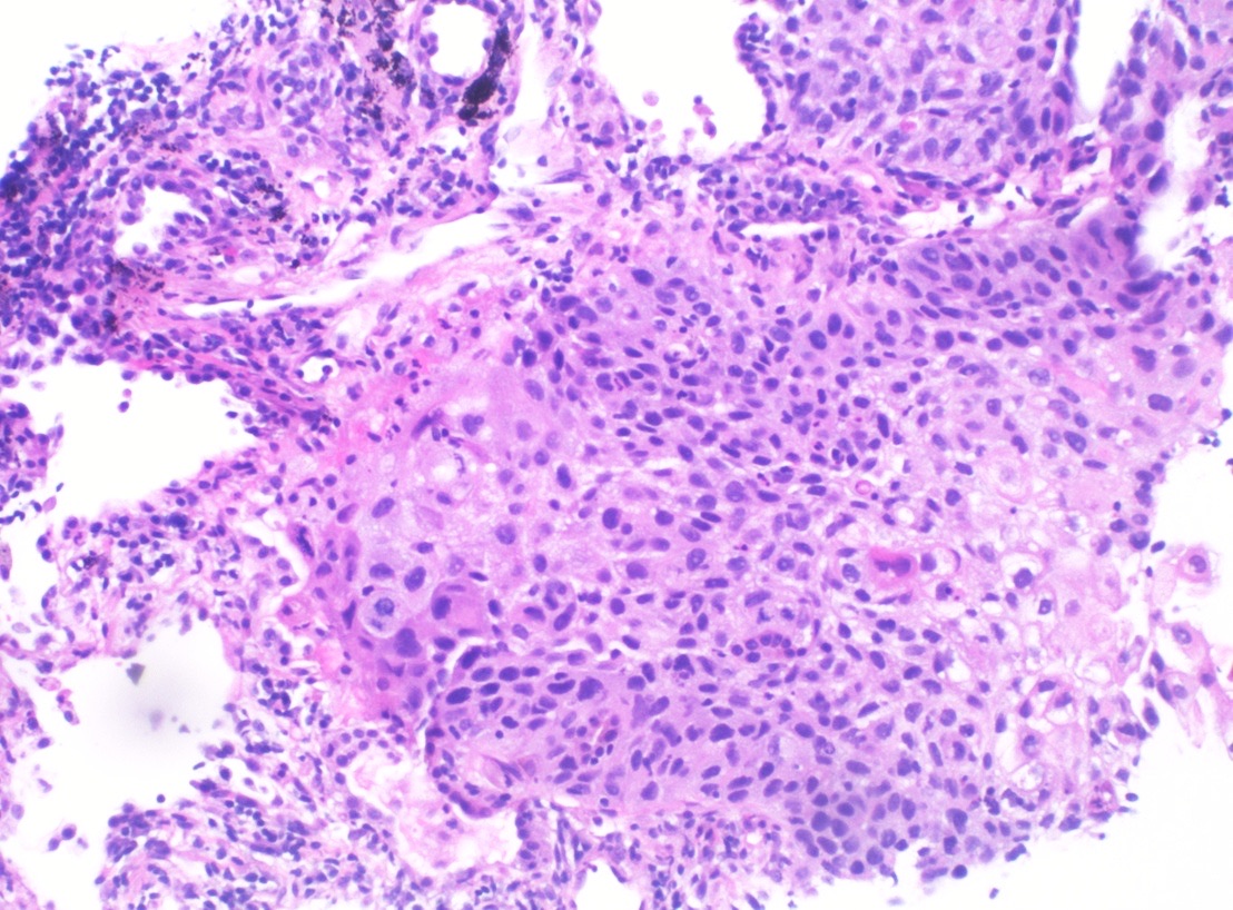
- Squamous differentiation. Keratinization and intercellular bridges define squamous differentiation by light microscopy (Yatabe et al. 2019).
- Subtypes. The CAP protocol lists keratinizing, nonkeratinizing and basaloid squamous cell carcinoma (CAP 2025). Immunohistochemistry is particularly useful in the diagnosis of nonkeratinizing squamous cell carcinoma (CAP 2025).
- Basaloid squamous cell carcinoma. Lobules of small cells with moderately hyperchromatic nuclei, absent nucleoli, scant cytoplasm, peripheral palisading and a high mitotic rate define the tumor. Of 38 cases, 19 were pure and 19 were mixed with other carcinomas. Neuroendocrine markers were infrequent, and median survival in stage I and II disease was 22 months (Brambilla et al. 1992).
- Basaloid squamous cell carcinoma compared with large cell neuroendocrine carcinoma. Lack of p40 expression supports large cell neuroendocrine carcinoma over basaloid squamous cell carcinoma (Beasley et al. 2026).
Immunohistochemistry of squamous cell carcinoma
- p40 compared with p63. In 470 tumors, p40 and p63 were positive in 100% of squamous cell carcinomas. p63 also stained 31% of adenocarcinomas and 54% of large cell lymphomas, whereas p40 stained 3% of adenocarcinomas and no lymphomas. Specificity was 98% for p40 and 60% for p63 (Bishop et al. 2012).
- Coexpression profiles. Squamous cell carcinoma was consistently TTF-1 negative and diffusely positive for p63, CK5/6 and 34βE12. Adenocarcinomas expressed p63 in 32%, CK5/6 in 18% and 34βE12 in 82%. A TTF-1 and p63 first-line panel with CK5/6 for indeterminate cases predicted the resection type with 100% accuracy in 38 small specimens (Rekhtman et al. 2011).
- Thresholds. Focal TTF-1 positivity is a positive result, and focal or weak p40 is not diagnostic of squamous cell carcinoma (Yatabe et al. 2019). The p40 scoring thresholds are given in Non-Small Cell Carcinoma, NOS, and Favor Terminology in Small Samples.
Molecular findings in squamous cell carcinoma
- Genomics. In 178 squamous cell carcinomas, TP53 was mutated in nearly all. The NFE2L2 and KEAP1 pathway was altered in 34%, squamous differentiation genes in 44%, the phosphatidylinositol 3-kinase pathway in 47%, and CDKN2A and RB1 in 72% (TCGA 2012).
- EGFR and KRAS. Ninety-five biomarker-verified squamous cell carcinomas (p40 positive, TTF-1 negative) had no EGFR or KRAS mutations. Of 16 EGFR- or KRAS-mutant tumors originally diagnosed as squamous cell carcinoma, 10 were reclassified as adenosquamous carcinoma and 5 as adenocarcinoma with squamoid morphology (Rekhtman et al. 2012).
- Squamous transformation. EGFR-mutant adenocarcinoma can recur after targeted therapy as p40-positive, TTF-1-negative squamous cell carcinoma that retains the original EGFR mutation (Yatabe et al. 2019).
Lymphoepithelial carcinoma of the lung
- Classification. The WHO 5th edition classifies lymphoepithelial carcinoma as a squamous cell carcinoma (Nicholson et al. 2022).
- Morphology. Syncytial sheets of poorly differentiated tumor cells are accompanied by marked lymphocytic infiltration, as in undifferentiated nasopharyngeal carcinoma. Among 61 Epstein-Barr virus (EBV)-associated tumors, a continuous spectrum extended to tumors with little lymphocytic infiltration that resembled nonkeratinizing squamous cell carcinoma. Focal keratinization, granulomatous inflammation, STAS and lepidic spread occurred (Yeh et al. 2019).
- Patients. EBV-associated pulmonary carcinoma usually occurs in women and nonsmokers (Yeh et al. 2019). Six cases from Western patients were all EBV negative (Castro et al. 2001).
- Genomics. Pulmonary lymphoepithelioma-like carcinoma has a low somatic mutation burden and widespread copy number changes and resembles nasopharyngeal carcinoma genetically (Hong et al. 2019).
- Biomarkers. In 66 tumors, PD-L1 overexpression was present in 75.8% and EGFR mutation in 12.1%. No KRAS, BRAF, ALK or ROS1 alterations were found (Chang et al. 2015).
Adenosquamous carcinoma of the lung
- Definition. Adenosquamous carcinoma requires a resection specimen showing at least 10% of each component, adenocarcinoma and squamous cell carcinoma (Travis et al. 2011), the definition used in the WHO 2015 classification (Illini et al. 2024). Tumors meeting these criteria form less than 5% of resected NSCLC (Travis et al. 2011). The open summary of the WHO 5th edition does not restate the threshold (Nicholson et al. 2022), and its full text could not be checked.
- Resection requirement. The diagnosis requires a resection specimen. In small biopsies, the possibility can be raised when two distinct cell populations are present (Yatabe et al. 2019).
- Immunohistochemistry. When a component has solid growth, TTF-1 and p40 must mark different cells or areas. Coexpression of TTF-1 and p40 or p63 in the same cells does not define adenosquamous carcinoma (Yatabe et al. 2019).
- Outcome and drivers. In a Japanese registry of 326 resected tumors, 5-year overall survival was 57.5%, compared with 83.9% for adenocarcinoma and 62.3% for squamous cell carcinoma. A sensitizing EGFR mutation was found in 21.5% of 130 screened tumors (Hishida et al. 2024). In 66 patients, EGFR alterations were found in 17% and ALK in 2% (Illini et al. 2024).
Large cell carcinoma of the lung
- Resection-only diagnosis. The term large cell carcinoma is restricted to resection specimens that are thoroughly sampled to exclude a differentiated component. It is not used in small biopsy or cytology specimens (Travis et al. 2011).
- Exclusion of solid adenocarcinoma. Intracytoplasmic mucin in a purely solid tumor indicates solid adenocarcinoma, and its absence was the historical basis for large cell carcinoma (Travis et al. 2011). Focal TTF-1 positivity also indicates adenocarcinoma (Yatabe et al. 2019).
- Immunophenotypic subsets. Of 102 large cell carcinomas, TTF-1 and p40 classified 60% as adenocarcinoma-like, 20% as squamous-like and 20% as marker-null. Adenocarcinoma-type mutations occurred in 50% of adenocarcinoma-like and 30% of marker-null tumors and in no squamous-like tumors. Marker-null tumors had worse disease-free and overall survival (Rekhtman et al. 2013).
- CAP grade. Large cell carcinoma is grouped with small cell carcinoma as G4, undifferentiated (CAP 2025).
| Immunoprofile of a solid tumor without morphologic differentiation | Classification in a resection | Source |
|---|---|---|
| TTF-1 positive (any extent), p40 negative | Solid adenocarcinoma | Yatabe et al. 2019; Rekhtman et al. 2013 |
| p40 diffuse (>50% of nuclei), TTF-1 negative | Nonkeratinizing squamous cell carcinoma | Yatabe et al. 2019 |
| TTF-1 negative, p40 negative or focal, mucin negative | Large cell carcinoma with null or unclear immunophenotype | Travis et al. 2011; Yatabe et al. 2019 |
| Neuroendocrine morphology with neuroendocrine marker expression | Large cell neuroendocrine carcinoma | Yatabe et al. 2019 |
| Keratin negative | Melanoma, lymphoma, sarcoma or epithelioid hemangioendothelioma to be excluded | Yatabe et al. 2019 |
Sarcomatoid carcinomas of the lung
Pleomorphic, spindle cell and giant cell carcinoma of the lung
- Concept. Sarcomatoid carcinoma is an umbrella term for spindle cell, giant cell and pleomorphic carcinomas, carcinosarcoma and pulmonary blastoma. Pleomorphic carcinoma combines spindle or giant cells with one or more conventional carcinoma components (Yatabe et al. 2019). Under the WHO 5th edition, pleomorphic carcinoma is a poorly differentiated non-small cell carcinoma containing at least 10% spindle or giant cells, or a carcinoma consisting only of spindle and giant cells (Hong et al. 2025).
- Clinical series. In 78 pleomorphic carcinomas, the male-to-female ratio was 2.7:1 and mean age 62 years. Adenocarcinoma accompanied 45%, large cell carcinoma 25% and squamous cell carcinoma 8%, and 22% were purely spindle or giant cell. Nodal metastasis was the strongest prognostic factor, and median survival was 10 months (Fishback et al. 1994).
- Immunohistochemistry. In tumors composed only of spindle or giant cells, TTF-1 was positive in 55% and CK7 in 70% (Rossi et al. 2003). Keratin expression is often weak and focal, and CK18 is a sensitive marker. A conventional carcinoma component makes immunohistochemical proof of epithelial differentiation unnecessary (Yatabe et al. 2019).
- Sarcoma and mesothelioma. Sarcomatoid mesotheliomas are commonly negative or only focally positive for WT1, calretinin and D2-40. Some high-grade sarcomas express keratin focally, and lack of keratin in a small sample does not favor sarcoma. Primary pulmonary spindle and giant cell sarcomas are extremely rare (Yatabe et al. 2019).
- Molecular findings. MET exon 14 skipping was present in 8 of 36 pulmonary sarcomatoid carcinomas (22%) (Liu et al. 2016). In 125 sarcomatoid carcinomas, MET exon 14 alterations occurred in 12% and 20% had high tumor mutational burden (Schrock et al. 2017).
Carcinosarcoma and pulmonary blastoma
- Carcinosarcoma. Carcinoma is mixed with sarcoma containing differentiated elements such as malignant cartilage, bone or skeletal muscle. In 66 cases, the male-to-female ratio was 7.25:1, mean age 65 years and 5-year survival 21.3%. The epithelial component was squamous in 46% and adenocarcinoma in 31% (Koss et al. 1999).
- Pulmonary blastoma. The tumor combines fetal adenocarcinoma with a primitive mesenchymal stromal component (CAP 2025). Biphasic tumors were larger than pure fetal adenocarcinomas, and 52% of patients with biphasic tumors died of disease (Koss et al. 1991).
- Pulmonary blastoma molecular findings. All four classic pulmonary blastomas showed nuclear and cytoplasmic β-catenin in both components, and three had CTNNB1 exon 3 mutations. Blastomatoid carcinosarcomas lacked this pattern (Nakatani et al. 2004). Biallelic somatic DICER1 variants were found in 7 of 8 pulmonary blastomas and CTNNB1 hotspot variants in 6 of 8 (Alirezaie et al. 2024).
| Type | Components | Immunohistochemistry | Molecular finding | Source |
|---|---|---|---|---|
| Pleomorphic carcinoma | Spindle or giant cells with adenocarcinoma, squamous or large cell carcinoma | Keratin often focal; TTF-1 and CK7 in a subset | MET exon 14 skipping 12–22% | Yatabe et al. 2019; Rossi et al. 2003; Liu et al. 2016; Schrock et al. 2017 |
| Spindle cell or giant cell carcinoma | Spindle cells only, or giant cells only | CK18 sensitive; TTF-1 in about half | As for pleomorphic carcinoma | Yatabe et al. 2019; Rossi et al. 2003 |
| Carcinosarcoma | Carcinoma plus heterologous sarcoma (cartilage, bone, muscle) | Sarcomatous part negative for CK7 and TTF-1 | Not established | Koss et al. 1999; Rossi et al. 2003 |
| Pulmonary blastoma | Low-grade fetal adenocarcinoma plus primitive mesenchyme | Nuclear β-catenin in both components | DICER1, CTNNB1 | Nakatani et al. 2004; Alirezaie et al. 2024 |
NUT carcinoma of the lung
- Definition. NUT carcinoma is a poorly differentiated carcinoma defined by rearrangement of NUTM1. Partners include BRD4, BRD3, BRD2, NSD3, ZNF532, ZNF592 and ZMYND8 (Luo et al. 2025). BRD4::NUTM1 was the first fusion identified, in carcinomas with t(15;19) (French et al. 2003).
- Patients and outcome. In 141 registry patients, the median age was 23.6 years and 51% of tumors were thoracic. Fusions were BRD4::NUTM1 in 78%, BRD3::NUTM1 in 15% and NSD3::NUTM1 in 6%. Median overall survival was 6.5 months overall and 4.4 months for thoracic primaries (Chau et al. 2020).
- Morphology. The tumor is monomorphic and poorly differentiated, with focal abrupt keratinization in 33–42% of cases. Pleomorphism typical of tobacco-related squamous cell carcinoma is lacking (Luo et al. 2025).
- Immunophenotype. Keratins are expressed in 85%, CK5 in 71–83%, p63 in 87% and p40 in 65–86% (Luo et al. 2025).
- NUT immunohistochemistry. The C52 monoclonal antibody had 87% sensitivity and 100% specificity against NUTM1 rearrangement, and detected two cryptic fusions missed by conventional FISH. Weak staining occurred in some germ cell tumors (Haack et al. 2009). NUT carcinoma shows diffuse speckled nuclear staining. Seminoma staining is focal and lacks the speckled pattern (Yatabe et al. 2019).
- Misdiagnosis. Eight of 13 patients in one series were initially misdiagnosed (Jung et al. 2019). Misdiagnoses include poorly differentiated carcinoma, small cell carcinoma, Ewing sarcoma and leukemia (Luo et al. 2025).
- Classification proposal. Luo et al. propose reclassifying NUT carcinoma of the lung as a subtype of squamous cell carcinoma. This is an author proposal and not a WHO criterion (Luo et al. 2025).
Related page: Thymoma and Thymic Carcinoma on FNA (NUT carcinoma on mediastinal cytology)
Thoracic SMARCA4-deficient undifferentiated tumor
- Recognition. The WHO 5th edition recognizes thoracic SMARCA4-deficient undifferentiated tumor (Nicholson et al. 2022).
- Original description. Nineteen tumors with SMARCA4 inactivation presented as compressive mediastinal and pulmonary masses in adults aged 30 to 35 years, with a median survival of 7 months. They were transcriptionally related to malignant rhabdoid tumors and small cell carcinoma of the ovary, hypercalcemic type, and expressed SOX2 (Le Loarer et al. 2015).
- Clinical features. In 30 cases, median age was 48 years, the male-to-female ratio was 9:1 and patients were usually smokers. Median overall survival was 6 months (Perret et al. 2019). In 12 cases, heavy smoking and emphysema were common (Yoshida et al. 2017).
- Morphology. Diffuse sheets of mildly dyscohesive, monotonous epithelioid cells with prominent nucleoli and frequent rhabdoid features are typical (Yoshida et al. 2017; Perret et al. 2019).
- Immunophenotype. All tested tumors lost SMARCA4 and SMARCA2. Keratin or EMA was expressed in 29 of 30, SOX2 in 26 of 27, CD34 in 17 of 27, SALL4 in 7 of 21 and claudin-4 in 2 of 19. p40 and NUT were negative, and SMARCB1 was retained (Perret et al. 2019).
- Relationship to carcinoma. In 22 SMARCA4-deficient thoracic sarcomatoid tumors, smoking history, a smoking mutational signature, high tumor mutational burden and focal NSCLC components supported origin as undifferentiated carcinoma. Compared with SMARCA4-deficient NSCLC, the undifferentiated tumors were larger, occurred in younger patients and had worse survival. They lacked claudin-4, showed SMARCA2 codeficiency and frequently expressed stem cell markers (Rekhtman et al. 2020).
SMARCA4-deficient non-small cell lung carcinoma
- Frequency. About 10% of lung carcinomas carry SMARCA4 mutations (Le Loarer et al. 2015). BRG1 loss was found in 4 of 100 NSCLC (Nambirajan et al. 2021).
- Phenotype. The four BRG1-deficient NSCLC were solid adenocarcinoma or large cell carcinoma, negative for TTF-1, p40, CD34 and SOX2 and lacking EGFR, ALK and ROS1 alterations (Nambirajan et al. 2021).
- Signature. The immunohistochemical signature of SMARCA4-deficient undifferentiated tumor combines SMARCA2 coinactivation with SOX2 and SALL4 overexpression (Sesboue and Le Loarer 2021).
Related page: Mediastinal Germ Cell Tumors on FNA (SALL4 and other germ cell markers in SMARCA4-deficient tumors)
| Feature | NUT carcinoma | Thoracic SMARCA4-deficient undifferentiated tumor | SMARCA4-deficient NSCLC | Basaloid squamous cell carcinoma | Source |
|---|---|---|---|---|---|
| Patients | Median age 23.6 years | Median age 48 years; male smokers | Older than patients with SMARCA4-deficient undifferentiated tumor | Not reported | Chau et al. 2020; Perret et al. 2019; Rekhtman et al. 2020 |
| Morphology | Monomorphic cells; abrupt keratinization | Dyscohesive epithelioid or rhabdoid sheets | Solid adenocarcinoma or large cell carcinoma | Lobules with peripheral palisading | Luo et al. 2025; Perret et al. 2019; Nambirajan et al. 2021; Brambilla et al. 1992 |
| Keratin | 85% | Keratin or EMA in 29 of 30 | Variable | Low-level low-molecular-weight keratins | Luo et al. 2025; Perret et al. 2019; Nambirajan et al. 2021 |
| p40 | 65–86% | Negative | Negative | Positive | Luo et al. 2025; Perret et al. 2019; Nambirajan et al. 2021; Beasley et al. 2026 |
| NUT | Diffuse speckled nuclear | Negative | Not reported | Not reported | Yatabe et al. 2019; Perret et al. 2019 |
| SMARCA4 and SMARCA2 | Not reported | Both lost | SMARCA4 lost; SMARCA2 retained or reduced, without complete loss | Not reported | Perret et al. 2019; Nambirajan et al. 2021; Rekhtman et al. 2020 |
| SOX2, CD34, SALL4 | Not reported | Frequently positive | Negative in a small series | Not reported | Perret et al. 2019; Nambirajan et al. 2021 |
| Claudin-4 | Not reported | Usually negative | Expressed | Not reported | Rekhtman et al. 2020 |
| Defining event | NUTM1 fusion | SMARCA4 inactivation | SMARCA4 mutation | Squamous lineage | Luo et al. 2025; Le Loarer et al. 2015 |
Small biopsy and cytology terminology for non-small cell carcinoma
- Scope. About 70% of lung cancers are diagnosed in small biopsies or cytology (Travis et al. 2011). Small-sample terms, the TTF-1 and p40 panel and tissue conservation are covered in Non-Small Cell Carcinoma, NOS, and Favor Terminology in Small Samples.
- Resection-only diagnoses. Large cell carcinoma and adenosquamous carcinoma are not diagnosed in small biopsy or cytology specimens (Travis et al. 2011; Yatabe et al. 2019).
- Sarcomatoid features. A small sample with pleomorphism, giant cells or spindle cells and no glandular or squamous features is reported as poorly differentiated non-small cell carcinoma with giant or spindle cell features (Travis et al. 2011).
- Resection. The designation non-small cell carcinoma is not acceptable in resection specimens (CAP 2025).
Differential diagnosis of non-adenocarcinoma lung carcinomas
| Entity | Mimic | Distinguishing finding | Source |
|---|---|---|---|
| Nonkeratinizing squamous cell carcinoma | Solid adenocarcinoma | Diffuse p40 in >50% of nuclei; TTF-1 negative | Yatabe et al. 2019; Rekhtman et al. 2011 |
| Squamous cell carcinoma | Large cell lymphoma | p40 negative in lymphoma; p63 positive in 54% of lymphomas | Bishop et al. 2012 |
| Basaloid squamous cell carcinoma | Large cell neuroendocrine carcinoma | p40 positive; neuroendocrine markers infrequent | Brambilla et al. 1992; Beasley et al. 2026 |
| Lymphoepithelial carcinoma | Nonkeratinizing squamous cell carcinoma | EBV association; syncytial growth with lymphocytes | Yeh et al. 2019 |
| Adenosquamous carcinoma | Adenocarcinoma with p40 and TTF-1 coexpression | Separate glandular and squamous cell populations, each ≥10% | Yatabe et al. 2019; Illini et al. 2024 |
| Pleomorphic carcinoma | Sarcoma or sarcomatoid mesothelioma | Conventional carcinoma component; keratin, TTF-1 or MET exon 14 skipping | Yatabe et al. 2019 |
| Pulmonary blastoma | Blastomatoid carcinosarcoma | Nuclear β-catenin; DICER1 and CTNNB1 | Nakatani et al. 2004; Alirezaie et al. 2024 |
| NUT carcinoma | Small cell carcinoma, Ewing sarcoma, germ cell tumor | Speckled nuclear NUT staining | Luo et al. 2025; Yatabe et al. 2019 |
| SMARCA4-deficient undifferentiated tumor | Malignant rhabdoid tumor | SMARCB1 retained; adult smokers | Perret et al. 2019; Sesboue and Le Loarer 2021 |
| SMARCA4-deficient undifferentiated tumor | SMARCA4-deficient NSCLC | SMARCA2 codeficiency; claudin-4 absent; SOX2 and SALL4 | Rekhtman et al. 2020; Sesboue and Le Loarer 2021 |
Lung Neuroendocrine Neoplasms: Typical and Atypical Carcinoid, Large Cell Neuroendocrine Carcinoma, Small Cell Carcinoma and DIPNECH
Definition. Lung neuroendocrine neoplasms range from low-grade typical carcinoid and intermediate-grade atypical carcinoid to the high-grade large cell neuroendocrine carcinoma (LCNEC) and small cell carcinoma (SCLC) (Travis et al. 1998a). The CAP resection protocol lists typical carcinoid/neuroendocrine tumor grade 1, atypical carcinoid/neuroendocrine tumor grade 2, carcinoid tumor NOS, small cell carcinoma, combined small cell carcinoma, LCNEC and combined LCNEC (CAP 2025). An IARC and WHO consensus framework separates well-differentiated neuroendocrine tumors (NETs), also called carcinoid tumors, from poorly differentiated neuroendocrine carcinomas (NECs) (Rindi et al. 2018). The WHO 5th edition (2021) retained typical and atypical carcinoid as the primary terms, corresponding to NET G1 and G2 (Beasley et al. 2026). The WHO 5th edition updated evolving concepts in the classification of lung neuroendocrine neoplasms (Nicholson et al. 2022). Entity-level criteria are those of the WHO 5th edition. The 6th edition of the thoracic volume is in progress (IARC 2026).
Related page: Typical and Atypical Carcinoid Tumor of the Lung (Cytology) (carcinoid tumor on cytology and the 2026 IASLC carcinoid/NET proposals) Related page: Small Cell Lung Carcinoma (Cytology) (small cell carcinoma on cytology) Related page: Lung Adenocarcinoma: Precursor Lesions, Invasive Patterns, IASLC Grading, Mucinous Adenocarcinoma and Spread Through Air Spaces (WHO 5th edition list of lung tumors) Related page: Squamous Cell, Adenosquamous, Large Cell and Sarcomatoid Carcinomas of the Lung, NUT Carcinoma and Thoracic SMARCA4-Deficient Undifferentiated Tumor (basaloid squamous cell carcinoma, large cell carcinoma and small-biopsy terms) Related page: Lung Cancer Staging and Reporting: AJCC Version 9, Visceral Pleural Invasion, Multiple Tumors, Neoadjuvant Response and the CAP Protocol (AJCC staging, which also applies to small cell carcinoma and carcinoid tumors) Related page: Lung Cancer Predictive Biomarkers: Driver Genes, PD-L1 Immunohistochemistry and Specimen Handling (EGFR testing and resistance mechanisms)
Epidemiology and clinical features of lung neuroendocrine neoplasms
- Frequency. Bronchopulmonary neuroendocrine tumors form about 20% of lung cancers. SCLC is the most common of these tumors, and LCNEC forms 1% or less of lung cancers (Gustafsson et al. 2008). SCLC forms about 14–15% of lung cancers (Travis 2012; Rudin et al. 2021).
- Carcinoid incidence. The incidence of bronchopulmonary carcinoids in the United States was 1.57 per 100,000 in 2003 (Gustafsson et al. 2008).
- Symptoms. Cough, hemoptysis and obstructive pneumonia are common. Fewer than 5% show hormonal syndromes such as carcinoid syndrome, Cushing syndrome, acromegaly or inappropriate antidiuretic hormone secretion (Gustafsson et al. 2008).
- Cushing syndrome. In 68 patients with lung neuroendocrine tumors causing Cushing syndrome, median age was 41 years and nodal metastasis was present in 37% of evaluable patients (Seastedt et al. 2021).
- Carcinoid stage. Most carcinoids present at an early stage, and fewer than 10% metastasize to distant sites, most often the liver (Beasley et al. 2026).
- SCLC. SCLC is strongly associated with tobacco exposure, and most patients have metastatic disease at diagnosis (Rudin et al. 2021).
- Survival. Five-year survival was 87% for typical carcinoid, 56% for atypical carcinoid, 27% for LCNEC and 9% for SCLC. After stratification for stage, LCNEC and SCLC did not differ (Travis et al. 1998a).
Neuroendocrine cell hyperplasia, tumorlets and DIPNECH
- Tumorlets. Lesions smaller than 5 mm with typical carcinoid morphology are carcinoid tumorlets. They are often peribronchiolar, and the size limit is arbitrary (Beasley et al. 2026).
- Preinvasive lesion. Diffuse idiopathic pulmonary neuroendocrine cell hyperplasia (DIPNECH) is one of the three preinvasive lesions of the lung, with squamous dysplasia and atypical adenomatous hyperplasia. It is associated with multiple carcinoid tumors (Kerr 2001).
- Definition. The WHO definition of DIPNECH is histologic. DIPNECH occurs in symptomatic patients with airway disease and in asymptomatic patients with neuroendocrine cell hyperplasia and multiple tumorlets or carcinoids. It is commonly found with peripheral carcinoid tumors (Rossi et al. 2016).
- Patients. DIPNECH affects mainly middle-aged and older women. In a multinational series of 258 patients, the mean age was 63.3 years and 93.4% were female, and diffuse pulmonary nodules (98.8%) and mosaic attenuation (59.1%) were the most common CT findings (O'Callaghan et al. 2026). All 32 patients in another series were women, 69% had never smoked, and asthma was the initial clinical diagnosis in 41% (Little et al. 2020). Among 311 patients with well-differentiated lung NETs, 20% had DIPNECH, and 95% of them were female (Hayes et al. 2022).
- DIPNECH syndrome. Respiratory symptoms, airflow obstruction, mosaic attenuation with air trapping and constrictive obliterative bronchiolitis characterize the proposed DIPNECH syndrome. Surgical lung biopsy is the diagnostic standard, and transbronchial biopsy may suffice in a compatible clinical and radiologic setting (Rossi et al. 2016).
- Minimum pathologic criteria. In 70 specimens with multifocal neuroendocrine proliferations, carcinoids occurred in 72.4% of cases with neuroendocrine cell hyperplasia plus more than two tumorlets, compared with 22.8% of cases with tumorlets alone. Multifocal neuroendocrine cell hyperplasia with three or more tumorlets was proposed as the minimum criterion for DIPNECH (Marchevsky et al. 2015).
- Secondary proliferations. Neuroendocrine cell proliferations also occur with interstitial fibrosis, bronchiectasis and other lung disease. DIPNECH requires the proliferation to be idiopathic (Marchevsky et al. 2015).
Typical and atypical carcinoid tumors of the lung
Diagnostic criteria for carcinoid tumors (WHO 5th edition)
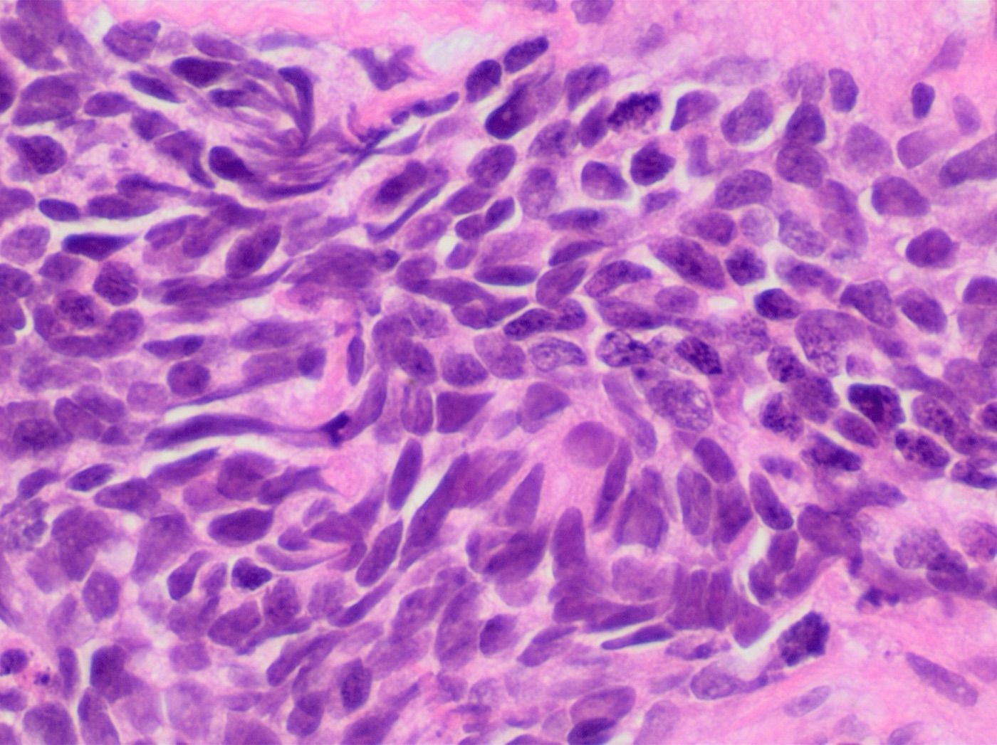
- Definition. Carcinoids are tumors at least 5 mm in size with carcinoid morphology and a mitotic rate of 10 or fewer per 2 mm² (Beasley et al. 2026).
- Typical carcinoid. Fewer than 2 mitoses per 2 mm² and no necrosis (Beasley et al. 2026).
- Atypical carcinoid. 2–10 mitoses per 2 mm² or necrosis, which is often punctate (Beasley et al. 2026).
- Mitotic counting. Mitoses have been counted per uniform 2-mm² area since the 1999 WHO classification, because high-power field size varies between microscopes (Beasley et al. 2026). The atypical carcinoid range of 2 to 10 mitoses per 2 mm² of viable tumor corresponded to 10 high-power fields in the original survival analysis (Travis et al. 1998a).
- Derivation. In 200 neuroendocrine tumors, mitotic count was the only independent predictor of prognosis. The optimal mitotic range for atypical carcinoid was 2 to 10 per 2 mm² (Travis et al. 1998a).
- Carcinoid tumor, NOS. The 2021 WHO classification introduced carcinoid tumor/NET, NOS for most small biopsy and cytology specimens, metastatic sites and incompletely sampled resections (Beasley et al. 2026).
- Neuroendocrine markers. Chromogranin A and synaptophysin expression is a desirable criterion. It is needed in limited, crushed or poorly fixed material and in tumors with unusual architecture (Beasley et al. 2026).
- CAP grade. Typical carcinoid is reported as G1 and atypical carcinoid as G2 (CAP 2025).
- Reproducibility. Five lung pathologists agreed unanimously in 58% of typical carcinoids and 50% of atypical carcinoids. For atypical carcinoid, 15% of diagnoses were rendered as typical carcinoid (Travis et al. 1998b).
- IASLC proposals (2026). The IASLC Pathology Committee proposed a carcinoid/NET classification that no longer recommends the terms typical and atypical carcinoid, grades all tumors as carcinoid/NET G1, G2 or G3, adds Ki-67 as a diagnostic criterion and recognizes carcinoid/NET G3, a category classified as LCNEC under the WHO 5th edition (Beasley et al. 2026; Rekhtman et al. 2026). These proposals lie outside the WHO 5th edition criteria and are summarized in Typical and Atypical Carcinoid Tumor of the Lung (Cytology).
| Tumor (WHO 5th edition) | Size and morphology | Mitoses per 2 mm² | Necrosis | Source |
|---|---|---|---|---|
| Carcinoid tumorlet | <5 mm; carcinoid morphology; peribronchiolar | Not applicable | Absent | Beasley et al. 2026 |
| Typical carcinoid (NET G1) | ≥5 mm; carcinoid morphology | <2 | Absent | Beasley et al. 2026 |
| Atypical carcinoid (NET G2) | ≥5 mm; carcinoid morphology | 2–10 | Present (often punctate) or absent | Beasley et al. 2026 |
| Carcinoid tumor, NOS | Carcinoid morphology in a biopsy, cytology sample, metastasis or incompletely sampled resection | Not assessable | Not assessable | Beasley et al. 2026 |
| Large cell neuroendocrine carcinoma | Neuroendocrine architecture; cells larger than three lymphocytes; vesicular chromatin, nucleoli | >10 (median 70) | Virtually always, often extensive | Beasley et al. 2026 |
| Small cell carcinoma | Small cells, scant cytoplasm; nested, trabecular, palisading or rosette patterns | >10 (typically >60) | Prominent | Beasley et al. 2026; Nicholson et al. 2002 |
Immunohistochemistry and molecular findings in carcinoid tumors
- TTF-1. TTF-1 is most often expressed in peripheral carcinoids, which commonly show spindle cell histology. It is present in up to 40% of all lung carcinoids (Beasley et al. 2026).
- OTP and CD44. OTP expression is generally restricted to pulmonary carcinoids, is higher in typical than atypical carcinoids and is mostly absent in NECs (Beasley et al. 2026). In 292 carcinoids, absence of CD44 and loss of nuclear OTP were associated with poor 20-year survival (Swarts et al. 2013).
- Ki-67. Reported Ki-67 ranges are 2.3–4.15% for typical and 9–17.8% for atypical carcinoid (Yatabe et al. 2019). Its use in crushed samples and its status in the WHO 5th edition are covered in Typical and Atypical Carcinoid Tumor of the Lung (Cytology).
- Rb. Carcinoids consistently have wild-type RB1 and retain Rb protein, so Rb loss supports SCLC over carcinoid (Beasley et al. 2026).
- Genomics. Covalent histone modifiers were mutated in 40% and SWI/SNF subunits in 22.2% of carcinoids, with MEN1, PSIP1 and ARID1A recurrently affected. TP53 and RB1 mutations were rare (Fernandez-Cuesta et al. 2014).
- Molecular groups. ASCL1, HNF1A/4A and OTP expression define three carcinoid molecular groups, whose clinical role is under investigation (Beasley et al. 2026).
- Metastatic carcinoid. No histologic criterion identifies a pulmonary carcinoid at a metastatic site. Spindle cell histology suggests pulmonary origin, and gastroenteropancreatic NETs frequently express CDX2 or Islet-1 (Beasley et al. 2026). Site-of-origin panels are given in Gastrointestinal neuroendocrine neoplasms on cytology.
Large cell neuroendocrine carcinoma of the lung
- Original criteria. LCNEC was proposed in 1991 for high-grade non-small cell tumors with neuroendocrine appearance, large polygonal cells with a low nuclear-to-cytoplasmic ratio, coarse chromatin and frequent nucleoli, a high mitotic rate, frequent necrosis and neuroendocrine features by immunohistochemistry or electron microscopy (Travis et al. 1991).
- WHO 5th edition description. Organoid nesting, peripheral palisading and rosette-like structures are present. Tumor cells are larger than three small resting lymphocytes and have visible cytoplasm, often with vesicular chromatin and prominent nucleoli. Necrosis is present in virtually all tumors. Mitoses exceed 10 per 2 mm², with a median of 70 (Beasley et al. 2026).
- Neuroendocrine markers. Expression of at least one neuroendocrine marker is required. Most tumors express two or three of synaptophysin, chromogranin A and CD56, with diffuse expression of at least one (Beasley et al. 2026). Isolated CD56 positivity is interpreted with caution (Beasley et al. 2026).
- Combined LCNEC. About 25–30% of LCNECs include adenocarcinoma, squamous cell carcinoma, large cell carcinoma or sarcomatoid carcinoma, and no minimum percentage is required for combined LCNEC. When SCLC forms at least 10%, the tumor is classified as combined SCLC and LCNEC (Beasley et al. 2026).
- Small biopsies. LCNEC can be diagnosed on a biopsy that allows assessment of neuroendocrine morphology and markers. Crush artifact and necrosis can prevent separation from SCLC, and the diagnosis is then high-grade neuroendocrine carcinoma, NOS (Beasley et al. 2026). A small biopsy with neuroendocrine morphology and positive markers is reported as non-small cell carcinoma, possible LCNEC (Travis et al. 2011; Yatabe et al. 2019).
- Markers without morphology. Neuroendocrine immunostains are performed only when neuroendocrine morphology is present. Resected non-small cell carcinomas with marker expression but no neuroendocrine morphology are classified by their non-neuroendocrine type (Yatabe et al. 2019).
- Separation from other NSCLC. Solid adenocarcinoma with a cribriform pattern and basaloid squamous cell carcinoma require both neuroendocrine morphology and markers for reclassification. Absence of p40 expression supports LCNEC over basaloid squamous cell carcinoma (Beasley et al. 2026).
- Molecular subsets. In 45 LCNECs, TP53 (78%), RB1 (38%), STK11 (33%), KEAP1 (31%) and KRAS (22%) were the most commonly altered genes. Tumors segregated into SCLC-like (TP53 and RB1 comutation or loss), NSCLC-like (STK11, KRAS, KEAP1) and rare carcinoid-like (MEN1) subsets (Rekhtman et al. 2016). In 75 LCNECs, type I tumors (37%) had TP53 with STK11 or KEAP1 alterations and type II tumors (42%) had TP53 and RB1 inactivation (George et al. 2018).
- Rb and chemotherapy. RB1 mutation was found in 47% and Rb protein loss in 72%. Patients with RB1 wild-type or Rb-expressing LCNEC lived longer with NSCLC-type chemotherapy than with platinum-etoposide (Derks et al. 2018).
Small cell carcinoma of the lung
Morphology of small cell lung carcinoma
- Diagnosis. SCLC is defined by light microscopy, and most cases are diagnosed on a good-quality H&E section. Most patients are diagnosed on small biopsy and cytology specimens, and cytology can be more definitive than a crushed or necrotic biopsy (Travis 2012).
- Surgical specimens. In 100 surgical specimens, nested and trabecular growth with peripheral palisading and rosettes was common. Necrosis and apoptotic debris were present in all cases, and crush artifact was infrequent. Cells were larger in surgical biopsies than in bronchoscopic biopsies, and occasional prominent nucleoli did not preclude the diagnosis (Nicholson et al. 2002).
- Combined SCLC. Combinations with NSCLC occurred in 28% of cases, most often with large cell carcinoma. At least 10% large cell carcinoma is required for combined small cell and large cell carcinoma, and no percentage is required for adenocarcinoma or squamous cell carcinoma components (Nicholson et al. 2002). The current subclassification recognizes pure and combined SCLC (Travis 2012).
- Previous rule. The 1962 Kreyberg scheme separated oat cell and polygonal cell types. These subtypes are no longer used (Travis 2012).
Immunohistochemistry of small cell lung carcinoma
- INSM1. INSM1 was positive in 94.9% of SCLC and 91.3% of LCNEC, compared with 74.4% and 78.3% for the combined synaptophysin, chromogranin and CD56 panel (Rooper et al. 2017). Its rates in non-small cell carcinomas are given in Small Cell Lung Carcinoma (Cytology).
- Classic markers. CD56 is the most sensitive marker for SCLC and has low specificity. About 5–10% of SCLCs are negative for all three classic markers. Chromogranin A staining is often focal in SCLC and diffuse in carcinoids (Yatabe et al. 2019).
- Rb. RB1 genomic inactivation or protein loss is present in 90% of SCLC. Rb immunohistochemistry is not used to separate SCLC from NSCLC, because 10% of SCLC retain Rb and 10–20% of NSCLC show RB1 alterations (Beasley et al. 2026).
- Ki-67. The Ki-67 index in SCLC is 70–100%, with a minimum above 50% (Beasley et al. 2026).
- TTF-1. TTF-1 was expressed in 85.5% of 55 pure SCLCs and 49% of 64 pure LCNECs, and in none of 50 carcinoids, 23 tumorlets or 15 hyperplasias (Sturm et al. 2002). Another series found TTF-1 in 95% of 21 SCLCs, 75% of 8 LCNECs, all 9 atypical carcinoids and 35% of 51 typical carcinoids (Folpe et al. 1999). TTF-1 does not prove pulmonary origin of a small cell carcinoma, because 44% of extrapulmonary small cell carcinomas were positive (Agoff et al. 2000). TTF-1 was negative in all basaloid carcinomas (Sturm et al. 2001).
Molecular findings in small cell lung carcinoma
- Genome. Inactivating NOTCH family mutations occurred in 25% of 110 SCLC genomes (George et al. 2015). Biallelic TP53 and RB1 inactivation and the ASCL1, NEUROD1 and POU2F3 transcriptional subtypes are covered in Small Cell Lung Carcinoma (Cytology).
- Inflamed subtype. An inflamed subtype, SCLC-I, benefited most from adding immunotherapy to chemotherapy (Gay et al. 2021). Testing for subtype markers is not indicated in routine practice (Beasley et al. 2026).
Transformation of EGFR-mutant adenocarcinoma to small cell carcinoma
- Frequency. About 3–10% of EGFR-mutant NSCLCs transform to SCLC (Marcoux et al. 2019). Other resistance mechanisms are covered in Lung Cancer Predictive Biomarkers: Driver Genes, PD-L1 Immunohistochemistry and Specimen Handling, and transformation on cytology in Small Cell Lung Carcinoma (Cytology).
- Course. In 67 patients, median time to transformation was 17.8 months. All 59 genotyped tumors kept the founder EGFR mutation, and 15 of 19 with prior EGFR T790M had lost it. TP53, RB1 and PIK3CA mutations recurred, and none of 17 patients responded to immunotherapy (Marcoux et al. 2019).
- Rb loss. Rb was lost in 100% of SCLC-transformed resistant cancers and rarely in tumors that remained NSCLC (Niederst et al. 2015).
Immunohistochemistry of lung neuroendocrine neoplasms and mimics
| Marker | Typical and atypical carcinoid | LCNEC | SCLC | Basaloid squamous cell carcinoma | Source |
|---|---|---|---|---|---|
| INSM1 | Positive (100%) | Positive (91.3%) | Positive (94.9%) | Positive in 4.2% of squamous cell carcinomas overall | Rooper et al. 2017 |
| Chromogranin A | Typical 97%, atypical 79%; usually diffuse | 41% (range 9–85%) | 47% (range 4–58%); often focal | Infrequent | Yatabe et al. 2019; Brambilla et al. 1992 |
| TTF-1 | Up to 40%, mostly peripheral spindle cell tumors | 49–75% | 85–95% | Negative | Beasley et al. 2026; Sturm et al. 2002; Folpe et al. 1999; Sturm et al. 2001 |
| Rb | Retained | Lost in 72% | Lost or inactivated in 90% | Not reported | Beasley et al. 2026; Derks et al. 2018 |
| p53 and TP53 | Wild type | TP53 altered in 78% | TP53 inactivated in nearly all | Not reported | Fernandez-Cuesta et al. 2014; Rekhtman et al. 2016; George et al. 2015 |
| Ki-67 | Typical 2.3–4.15%; atypical 9–17.8% | 30–80% | 70–100% | Not reported | Yatabe et al. 2019; Beasley et al. 2026 |
| OTP | Positive, higher in typical carcinoid | Mostly negative | Mostly negative | Not reported | Beasley et al. 2026 |
| p40 | Not reported | Negative | Not reported | Positive | Beasley et al. 2026 |
Differential diagnosis of lung neuroendocrine neoplasms
| Entity | Mimic | Distinguishing finding | Source |
|---|---|---|---|
| Carcinoid in a crushed biopsy | SCLC | Low Ki-67; retained Rb | Yatabe et al. 2019; Beasley et al. 2026 |
| Carcinoid tumorlet | Typical carcinoid | Size <5 mm | Beasley et al. 2026 |
| DIPNECH | Secondary neuroendocrine proliferation | Idiopathic multifocal hyperplasia with ≥3 tumorlets (proposed minimum criterion); no fibrosis or bronchiectasis | Marchevsky et al. 2015 |
| LCNEC | SCLC | Cells larger than three lymphocytes, vesicular chromatin, prominent nucleoli; high-grade NEC, NOS if crushed | Beasley et al. 2026 |
| LCNEC | Basaloid squamous cell carcinoma | Neuroendocrine markers positive; p40 negative | Beasley et al. 2026 |
| LCNEC | Solid adenocarcinoma with neuroendocrine marker expression | Neuroendocrine morphology required | Yatabe et al. 2019; Beasley et al. 2026 |
| SCLC, neuroendocrine-low | Other small round cell tumors | POU2F3 expression; keratin; RB1 and TP53 inactivation; panels in Small Cell Lung Carcinoma (Cytology) | Beasley et al. 2026 |
| Transformed SCLC | De novo SCLC | Prior EGFR-mutant adenocarcinoma; founder EGFR mutation retained | Marcoux et al. 2019 |
| Metastatic gastroenteropancreatic NET | Pulmonary carcinoid | CDX2 or Islet-1; OTP negative | Beasley et al. 2026 |
Lung Cancer Staging and Reporting: AJCC Version 9, Visceral Pleural Invasion, Multiple Tumors, Neoadjuvant Response and the CAP Protocol
Definition. Pathologic staging of lung cancer records the anatomic extent of tumor in a resection specimen, its lymph nodes and any submitted metastases. The CAP Protocol for the Examination of Resection Specimens from Patients with Primary Non-Small Cell Carcinoma, Small Cell Carcinoma, or Carcinoid Tumor of the Lung, version 5.1.0.0 (posted September 2025), applies to wedge resection, segmentectomy, lobectomy and pneumonectomy. Its staging standard is AJCC Version 9. It is not required for biopsies, cytologic specimens or resections with no residual cancer after neoadjuvant therapy. Mesothelioma, lymphoma and sarcoma are reported with other protocols (CAP 2025). The ninth edition TNM classification took effect on 1 January 2025 (AJCC Version 9; Kifjak et al. 2025). The AJCC 8th edition is the previous rule. The TNM system is used for non-small cell carcinoma, small cell carcinoma, and typical and atypical carcinoid tumors (CAP 2025).
Related page: Lung Adenocarcinoma: Precursor Lesions, Invasive Patterns, IASLC Grading, Mucinous Adenocarcinoma and Spread Through Air Spaces (AIS and MIA criteria, IASLC grading and STAS morphology) Related page: Lung Neuroendocrine Neoplasms: Typical and Atypical Carcinoid, Large Cell Neuroendocrine Carcinoma, Small Cell Carcinoma and DIPNECH (carcinoid tumors, LCNEC and small cell carcinoma) Related page: Lung Cancer Predictive Biomarkers: Driver Genes, PD-L1 Immunohistochemistry and Specimen Handling (biomarker testing) Related page: Lung FNA Adequacy and Rapid On-Site Evaluation (EBUS-TBNA nodal staging) Related page: Metastatic Lung Adenocarcinoma in Serous Effusions (malignant pleural effusion, classified as M1a)
Grossing of lung resection specimens
| Procedure (CAP 5.1.0.0) | Surgical margins present | Source |
|---|---|---|
| Wedge resection | Parenchymal margin at the staple line | CAP 2025 |
| Lobectomy, segmentectomy, completion or sleeve lobectomy, bilobectomy | Bronchial and vascular margins; parenchymal staple lines depending on the completeness of the fissures and other anatomic factors | CAP 2025 |
| Pneumonectomy | Bronchial and vascular margins | CAP 2025 |
| En bloc resection with chest wall or other structures | Additional margins designated by the surgeon (parietal pleura, chest wall) | CAP 2025 |
- Margins. The visceral pleura is not a surgical margin (CAP 2025).
- Inflation and fixation. Lung specimens are inflated through the bronchi and fixed in 10% neutral buffered formalin for 6 to 48 hours. (Travis et al. 2020).
- AIS and MIA. A lesion considered for adenocarcinoma in situ (AIS) or minimally invasive adenocarcinoma (MIA) is submitted entirely. AIS and MIA are diagnosed only on solitary lesions ≤3 cm (CAP 2025).
- Lymph nodes. Nodes are reported by IASLC station. Mediastinoscopy nodes are often fragmented. The number of nodes is reported only when it is known, and nodal sites may be reported without a count (CAP 2025).
Intraoperative consultation on lung resection specimens
- Frozen section to guide sublobar resection. In 803 clinical stage I peripheral adenocarcinomas, frozen section and final diagnosis agreed in 84.4%, and in 95.9% when AAH, AIS and MIA were grouped as low risk. Most discrepancies underestimated AIS and MIA (Liu et al. 2016).
- Bronchial margin. In 268 bronchial margin frozen sections, 1.9% were false negatives, and the missed foci were submucosal, lymphatic or peribronchial. Three of 6 adenoid cystic and mucoepidermoid carcinomas had positive margins (Maygarden et al. 2004).
- STAS on frozen section. Five pathologists reached 71% sensitivity and 92% specificity (Eguchi et al. 2019). In 100 stage I adenocarcinomas, sensitivity was 44% and specificity 91%, interobserver agreement was moderate, and artifacts were the only predictor of discordance (Villalba et al. 2021). In 48 adenocarcinomas, sensitivity was 50% and the negative predictive value 8% (Walts and Marchevsky 2018).
Tumor size, invasive size and STAS in the pT category of lung cancer
- Invasive size rule. For nonmucinous adenocarcinoma with a lepidic component, only the invasive size assigns the T category. Invasive tumor size equals total size for every other tumor type, including invasive mucinous adenocarcinoma (CAP 2025). The rule was proposed for the 8th edition, along with Tis (AIS), Tis (SCIS) and T1mi for MIA (Travis et al. 2016).
- Estimating invasive size. The invasive component includes nonlepidic patterns (acinar, papillary, micropapillary, solid) and tumor cells infiltrating myofibroblastic stroma. When invasion is not one discrete focus, the percentage of invasive tumor is multiplied by the total size (CAP 2025). Criteria that separate invasive from lepidic growth are given in Lung Adenocarcinoma: Precursor Lesions, Invasive Patterns, IASLC Grading, Mucinous Adenocarcinoma and Spread Through Air Spaces.
- Status in Version 9. The data were insufficient to analyze the invasive-size rule for the 9th edition, so the 8th-edition rule continues and both sizes remain reportable (Van Schil et al. 2024; CAP 2025). In CAP 5.1.0.0, invasive size and in situ carcinoma size are conditionally reported elements (CAP 2025).
- Lepidic tumors >3 cm. A lepidic-predominant tumor >3.0 cm with invasion ≤0.5 cm, or none, is classified as lepidic-predominant adenocarcinoma, pT1a (CAP 2025).
- STAS. STAS consists of micropapillary clusters, solid nests or single cells extending beyond the tumor edge into air spaces of the surrounding lung. It is not included in the tumor size (CAP 2025).
- Superficial spreading tumor. A tumor of any size whose invasive component is limited to the bronchial wall, even if it extends proximal to the main bronchus, is pT1a (CAP 2025).
- Atelectasis and obstructive pneumonitis. When recognized only by pathology, these T2 descriptors are not used for staging (CAP 2025).
Visceral pleural invasion in lung cancer
Visceral pleural invasion (VPI) raises a tumor ≤3 cm from pT1 to pT2a. Invasion is present when tumor cells cross the thickest visceral pleural elastic layer, usually the outer (external) layer, or reach the pleural surface. Penetration of the thinner, often discontinuous inner elastic layer does not qualify. Elastic stains are used when invasion is indeterminate on H&E (CAP 2025). The PL notation was proposed for the 7th edition (Travis et al. 2008). Routine elastic staining of 100 peripheral stage IA carcinomas changed the stage to IB in 19 (19%) (Taube et al. 2007).
| PL category | Definition | T effect (8th and Version 9) | Source |
|---|---|---|---|
| PL0 | No penetration beyond the visceral pleural elastic layer | None | Travis et al. 2008; CAP 2025 |
| PL1 | Invasion beyond the elastic layer | T2a for a tumor ≤3 cm | Travis et al. 2008; CAP 2025 |
| PL2 | Invasion to the visceral pleural surface | T2a for a tumor ≤3 cm | Travis et al. 2008; CAP 2025 |
| PL3 | Invasion of the parietal pleura | T3 | Travis et al. 2008; CAP 2025 |
- Fissure. Direct invasion across a fissure, or across an incomplete fissure, into an adjacent ipsilateral lobe is T2a unless other criteria give a higher category (CAP 2025).
- Pleural nodules. Pleural tumor nodules that are not continuous with the primary tumor are M1a, including same-lobe visceral pleural nodules (CAP 2025).
- Parietal pleura in Version 9. In the 9th-edition database, pathologic chest wall or parietal pleural (PL3) invasion carried worse survival than other single T3 descriptors (5-year overall survival 53% compared with 60% in the N-any cohort), similar to T4. This difference was not seen in clinical staging. Because of the inconsistency, chest wall and PL3 invasion remain T3 (Van Schil et al. 2024). The evidence was insufficient to classify parietal pleural invasion as T4 (CAP 2025).
Direct invasion of structures adjacent to the lung
| Structure invaded | Category | Source |
|---|---|---|
| Hilar fat or soft tissue alone | T2a | CAP 2025 |
| Main bronchus, up to the carina | T2 | CAP 2025 |
| Parietal pleura, chest wall (including superior sulcus), phrenic nerve, parietal pericardium, azygos vein, thoracic nerve roots or stellate ganglion | T3 | CAP 2025 |
| Visceral pericardium (epicardium), heart, great vessels, trachea, carina, esophagus, recurrent laryngeal or vagus nerve, diaphragm, vertebral body, mediastinum | T4 | CAP 2025 |
AJCC Version 9 and AJCC 8th edition TNM categories for lung cancer
T category. The IASLC proposed no change to the 8th-edition T descriptors for the 9th edition (Van Schil et al. 2024). Version 9 keeps them (CAP 2025). The size steps are T1a ≤1 cm, T1b >1 to ≤2 cm, T1c >2 to ≤3 cm, T2a >3 to ≤4 cm, T2b >4 to ≤5 cm, T3 >5 to ≤7 cm and T4 >7 cm (CAP 2025).
N category. N0, N1 and N3 are unchanged. N2 is divided into N2a and N2b (Huang et al. 2024). Five-year survival by pathologic N in 35,009 resected patients was 83% for N0, 58% for N1, 47% for N2 and 28% for N3. Single-station N2 had better survival than multistation N2 in every subgroup, including after neoadjuvant therapy. Subdivision of N1 and a separate skip-metastasis group were not adopted (Huang et al. 2024).
M category. M1c is divided because patients with one extrathoracic organ system involved had better survival than those with several (Fong et al. 2024). The skeleton is one organ system, so several metastases in one bone or in several bones are M1c1 (CAP 2025). When the number of involved N2 stations or organ systems cannot be established, the CAP template offers pN2 (subgroup cannot be determined) and pM1c (subgroup cannot be determined) (CAP 2025). Nonregional lymph node metastasis is M1b when single and M1c when multiple (CAP 2025).
| Category | AJCC 8th edition | AJCC Version 9 | Source |
|---|---|---|---|
| N1 | Ipsilateral peribronchial, hilar or intrapulmonary nodes, including direct extension | Unchanged | CAP 2025; Kifjak et al. 2025 |
| N2 | Ipsilateral mediastinal and/or subcarinal nodes, one category | N2a: a single ipsilateral mediastinal station or the subcarinal station | Huang et al. 2024; CAP 2025 |
| N2b: multiple ipsilateral mediastinal stations, with or without the subcarinal station | Huang et al. 2024; CAP 2025 | ||
| N3 | Contralateral mediastinal or hilar, or any scalene or supraclavicular nodes | Unchanged | CAP 2025; Kifjak et al. 2025 |
| M1a | Pleural or pericardial nodules, malignant pleural or pericardial effusion, contralateral lobe nodule | Unchanged | CAP 2025; Fong et al. 2024 |
| M1b | Single extrathoracic metastasis, including a single nonregional node | Unchanged | CAP 2025; Fong et al. 2024 |
| M1c | Multiple extrathoracic metastases in one or several organs | M1c1: multiple metastases in one organ system | Fong et al. 2024; CAP 2025 |
| M1c2: multiple metastases in multiple organ systems | Fong et al. 2024; CAP 2025 |
Pleural effusion. When repeated cytologic examinations are negative and the fluid is nonbloody and not an exudate, the effusion can be excluded as a staging descriptor (CAP 2025).
Stage groups in AJCC Version 9 compared with the AJCC 8th edition
The stage groups were derived from 76,518 analyzable patients. T1N1 moves to IIA, T1N2a to IIB and T3N2a to IIIA. T2aN2b and T2bN2b are IIIB. M1c1 and M1c2 both remain IVB (Rami-Porta et al. 2024).
| T or M | N0 (8th / V9) | N1 (8th / V9) | N2 (8th) | N2a (V9) | N2b (V9) | N3 (8th / V9) |
|---|---|---|---|---|---|---|
| T1a | IA1 / IA1 | IIB / IIA | IIIA | IIB | IIIA | IIIB / IIIB |
| T1b | IA2 / IA2 | IIB / IIA | IIIA | IIB | IIIA | IIIB / IIIB |
| T1c | IA3 / IA3 | IIB / IIA | IIIA | IIB | IIIA | IIIB / IIIB |
| T2a | IB / IB | IIB / IIB | IIIA | IIIA | IIIB | IIIB / IIIB |
| T2b | IIA / IIA | IIB / IIB | IIIA | IIIA | IIIB | IIIB / IIIB |
| T3 | IIB / IIB | IIIA / IIIA | IIIB | IIIA | IIIB | IIIC / IIIC |
| T4 | IIIA / IIIA | IIIA / IIIA | IIIB | IIIB | IIIB | IIIC / IIIC |
| M1a or M1b | IVA / IVA | IVA / IVA | IVA | IVA | IVA | IVA / IVA |
| M1c (8th); M1c1 or M1c2 (V9) | IVB / IVB | IVB / IVB | IVB | IVB | IVB | IVB / IVB |
Bold cells differ from the 8th edition. V9, AJCC Version 9. Sources: 8th edition, Erasmus et al. 2023 (from Goldstraw et al. 2016); Version 9, Kifjak et al. 2025 and Rami-Porta et al. 2024.
Survival by stage at diagnosis. In the United States, 5-year relative survival for lung cancer diagnosed in 2014–2020 was 27%, and 43% of cases were diagnosed at a distant stage. Three-year relative survival increased from 26% in 2004 to 43% in 2018 for NSCLC and from 9% to 12% for small cell lung cancer (Siegel et al. 2025).
Regional lymph node stations and N categories
The IASLC map has 14 stations and remains unchanged in the 9th edition (Rusch et al. 2009; Kifjak et al. 2025). Positive stations 4R and 10R are N2a, and positive stations 4L, 10L and 7 are N2b (Kifjak et al. 2025).
| Station (IASLC map) | Name | N category when ipsilateral | Source |
|---|---|---|---|
| 1 | Low cervical, supraclavicular and sternal notch | N3 | CAP 2025 |
| 2R, 2L | Upper paratracheal | N2 | CAP 2025 |
| 3a, 3p | Prevascular, retrotracheal | N2 | CAP 2025 |
| 4R, 4L | Lower paratracheal | N2 | CAP 2025 |
| 5, 6 | Subaortic (aortopulmonary window); para-aortic | N2 | CAP 2025 |
| 7 | Subcarinal | N2 | CAP 2025 |
| 8, 9 | Paraesophageal; pulmonary ligament | N2 | CAP 2025 |
| 10, 11 | Hilar; interlobar | N1 | CAP 2025 |
| 12, 13, 14 | Lobar, segmental, subsegmental (intrapulmonary) | N1 | CAP 2025 |
- Extranodal extension. Extension of tumor beyond the nodal capsule may be unfavorable but does not change pN or stage. Direct extension of the primary tumor into a node is not extranodal extension (CAP 2025).
Multiple lung tumors: second primary, separate tumor nodule, multifocal and pneumonic-type disease
The IASLC defined four patterns of multiple pulmonary sites of lung cancer for the 8th edition, and CAP 5.1.0.0 applies them with AJCC Version 9 (Detterbeck et al. 2016a; CAP 2025).
| Pattern | Pathologic features | TNM classification | Source |
|---|---|---|---|
| Second primary lung cancer | Different histologic type, or different morphology on comprehensive histologic assessment | Separate cTNM and pTNM for each cancer; separate synoptic reports | Detterbeck et al. 2016a; CAP 2025 |
| Separate tumor nodule (intrapulmonary metastasis) | Same morphology on comprehensive histologic assessment | T3 same lobe, T4 different ipsilateral lobe, M1a contralateral lobe; single N and M | Detterbeck et al. 2016c; CAP 2025 |
| Multifocal ground-glass/lepidic adenocarcinoma | Adenocarcinomas with a prominent lepidic component (AIS, MIA, lepidic-predominant adenocarcinoma) | Highest T lesion with (m) or the number in parentheses, for example T1b(m) or T1a(3); single N and M | Detterbeck et al. 2016b; CAP 2025 |
| Pneumonic-type adenocarcinoma | Diffuse consolidation with the same histology throughout, most often invasive mucinous adenocarcinoma | T by size or T3 in one lobe, T4 in another ipsilateral lobe, M1a contralateral; single N and M | Detterbeck et al. 2016b; CAP 2025 |
- Criteria for a second primary. Few criteria are reliable alone: different histologic types, matching DNA breakpoints, and comprehensive histologic assessment. Biomarkers, imaging and nodal status are suggestive only (Detterbeck et al. 2016d).
- Comprehensive histologic assessment. The comparison records pattern percentages, cytologic features, stroma, necrosis and variant patterns. It agreed with molecular classification in 20 of 22 pairs (91%), and Martini-Melamed criteria disagreed with molecular data in 7 of 22 (32%) (Girard et al. 2009).
- Comparative sequencing. In 76 tumor pairs, broad-panel NGS separated 51 second primaries from 25 intrapulmonary metastases. Prospective histologic prediction disagreed with NGS in 22% overall and in 44% of intrapulmonary metastases (Chang et al. 2019). Whole-exome sequencing of multifocal squamous cell carcinomas agreed with individual morphologic assessment in 58.3%, with a kappa of 0.232 between pathologists (Dacic et al. 2024).
- IASLC recommendation. Molecular profiling is performed when possible, especially for tumors with similar morphology (Chou et al. 2025).
- Multifocal tumors of other types. For multiple tumors of the same type that are not intrapulmonary metastases, such as numerous carcinoid tumors, the T category follows the largest tumor with the m suffix. Foci of atypical adenomatous hyperplasia are not counted for TNM (CAP 2025).
STAS and lymphovascular invasion as reporting elements in lung resections
- STAS in Version 9. STAS is a histologic descriptor in the 9th edition and does not change the T category; it is a core CAP element (Travis et al. 2024; CAP 2025). STAS was present in 930 of 4,061 pathologic stage I R0 NSCLC (22.9%). It was associated with worse recurrence-free and overall survival in all NSCLC, in adenocarcinoma and in other types, after lobar and sublobar resection, and independently of VPI (Travis et al. 2024).
- STAS and limited resection. In 411 adenocarcinomas ≤2 cm, STAS was present in 38%. After limited resection, 5-year recurrence was 42.6% with STAS and 10.9% without it. STAS was not associated with recurrence after lobectomy (Kadota et al. 2015). Limited resection of stage I tumors with STAS carried a higher risk of locoregional recurrence than lobectomy (Kadota et al. 2019). After propensity matching of T1 adenocarcinomas, sublobar resection was associated with recurrence (subhazard ratio 2.84) and lung cancer-specific death in patients with STAS but not in those without STAS (Eguchi et al. 2019).
- STAS in other histologic types. STAS was present in 30% of squamous cell carcinomas and independently predicted recurrence (Lu et al. 2017). It occurred in 16% of typical carcinoids, 37% of atypical carcinoids, 43% of LCNEC and 46% of small cell carcinomas (Aly et al. 2019).
- Artifact. In a prospective study, tumor clusters increased from the first clean-knife cut to later sections, and 93% of loose tissue fragments were attributed to handling (Blaauwgeers et al. 2017). Morphologic criteria for artifact are given in Lung Adenocarcinoma: Precursor Lesions, Invasive Patterns, IASLC Grading, Mucinous Adenocarcinoma and Spread Through Air Spaces.
- Lymphovascular invasion. Lymphatic invasion is unfavorable. LVI excludes AIS and MIA. It does not alter pT, pN or stage group (CAP 2025).
Residual tumor (R) classification after lung resection
| Category | Definition | Source |
|---|---|---|
| R0 | Complete resection with negative margins and adequate nodal assessment | Detterbeck et al. 2024; Rami-Porta 2021 |
| R(un), uncertain | Negative margins, with nodal assessment below systematic or lobe-specific systematic dissection, positive highest mediastinal node removed, carcinoma in situ at the bronchial margin, or positive pleural lavage cytology | Rami-Porta 2021 |
| R1 | Microscopic tumor at a surgical margin | Detterbeck et al. 2024 |
| R2 | Gross residual tumor | Detterbeck et al. 2024 |
- Nodal adequacy. The IASLC definition requires at least six nodes, three from intrapulmonary or hilar stations and three from mediastinal stations, always including the subcarinal station (Rami-Porta 2021). Extracapsular extension in a node removed separately or at the specimen margin is a criterion of incomplete resection (Rami-Porta 2021).
- Survival data. In 14,712 resected patients, inadequate nodal staging moved 56% of cases from R0 to R(un). In node-positive patients, R(un) survival lay between R0 and R1 (Edwards et al. 2020).
- Version 9. The 9th edition keeps the R categories and their descriptors and defines them more precisely (Detterbeck et al. 2024).
Pathologic response of lung cancer after neoadjuvant therapy
The IASLC 2020 recommendations standardize tumor-bed assessment after neoadjuvant therapy (Travis et al. 2020). CAP 5.1.0.0 requires the percentage of residual viable tumor and lists necrosis, stroma and inflammation as optional elements (CAP 2025).
| Step or category | Rule | Source |
|---|---|---|
| Tumor bed ≤3 cm | Cross section through the maximum dimension; submit the bed entirely | Travis et al. 2020; CAP 2025 |
| Tumor bed >3 cm | One complete cross section about 0.5 cm thick, submitted entirely and mapped to a gross photograph | Travis et al. 2020 |
| Components | Viable tumor, necrosis and stroma (fibrosis and inflammation) total 100%, in 10% increments; single-digit estimates below 5% | Travis et al. 2020; CAP 2025 |
| Major pathologic response (MPR) | ≤10% viable tumor; proposed as a surrogate end point | Travis et al. 2020; Hellmann et al. 2014 |
| Complete pathologic response | No viable tumor on H&E in the primary and all sampled regional nodes (ypT0N0) | Travis et al. 2020 |
| Residual node disease | No viable tumor in the primary with positive nodes is ypT0 ypN1–N3 | Travis et al. 2020 |
- Evidence for MPR. In 192 patients after neoadjuvant chemotherapy, ≤10% viable tumor gave 5-year overall survival of 85% compared with 40% for >10% (Pataer et al. 2012).
- Immunotherapy. The regression bed after anti-PD-1 therapy contains lymphocytes, cholesterol clefts and proliferative fibrosis (Cottrell et al. 2018).
- CheckMate 816. After neoadjuvant nivolumab and chemotherapy, 0% residual viable tumor in the primary was associated with better event-free survival (hazard ratio 0.18) (Deutsch et al. 2024).
- ypT size. When viable tumor is scattered, its size can be estimated as the percentage of viable tumor multiplied by the tumor-bed dimension. In tumors with a lepidic component, the invasive size is used. Fibrosis extending into the chest wall without viable tumor does not raise ypT (Travis et al. 2020).
- Prefixes. The y prefix records classification during or after multimodality therapy. If no viable tumor remains, the category is ypT0. The r prefix records recurrent tumor after a disease-free interval (CAP 2025).
CAP lung resection protocol 5.1.0.0: synoptic elements
| Element | Status | Note | Source |
|---|---|---|---|
| Synchronous tumors | Required if separate primaries are present | One synoptic report per primary | CAP 2025 |
| Tumor focality | Core | Single, separate nodules (pT3, pT4, pM1a), multifocal (m), pneumonic type | CAP 2025 |
| Invasive tumor size | Conditional (5.1.0.0) | Equals total size except for nonmucinous adenocarcinoma with a lepidic component | CAP 2025 |
| Histologic type | Core | WHO classification; "non-small cell carcinoma" is not acceptable in resections | CAP 2025 |
| Histologic grade | Optional (+) | IASLC grade for nonmucinous adenocarcinoma | CAP 2025 |
| STAS | Core | Not identified, present, cannot be determined, not applicable | CAP 2025 |
| Visceral pleural invasion | Core | Elastic stain when indeterminate | CAP 2025 |
| Treatment effect | Core | Percentage of residual viable tumor; necrosis, stroma and inflammation optional | CAP 2025 |
| Margins | Core | Bronchial, vascular, parenchymal, chest wall; in situ carcinoma and lepidic component at margins | CAP 2025 |
| Regional lymph nodes | Core | Number positive and examined; stations involved and examined | CAP 2025 |
| Distant metastasis | Core when examined | Separate options for single and multiple extrathoracic sites | CAP 2025 |
| pTNM (AJCC Version 9) | Core | y and r prefixes; m suffix; (sn) and (f) N suffixes | CAP 2025 |
Biomarker results are reported with the CAP Lung Biomarker template (CAP 2025).
Differential diagnosis of staging findings in lung resections
| Finding | Mimic | Distinguishing feature | Source |
|---|---|---|---|
| Separate tumor nodule (intrapulmonary metastasis) | Second primary lung cancer | Same morphology on comprehensive histologic assessment; shared somatic mutations | Girard et al. 2009; Chang et al. 2019 |
| PL1 invasion | PL0 | Tumor beyond the thick outer elastic layer; inner layer penetration does not count | CAP 2025 |
| STAS | Knife or handling artifact | Clusters that increase from the first clean-knife section to later sections favor handling artifact; artifacts were the only predictor of frozen-section discordance | Blaauwgeers et al. 2017; Villalba et al. 2021 |
Lung Cancer Predictive Biomarkers: Driver Genes, PD-L1 Immunohistochemistry and Specimen Handling
Definition. Predictive biomarker testing in non-small cell lung carcinoma (NSCLC) identifies genomic alterations and protein expression that select patients for targeted therapy or immune checkpoint inhibitors. The CAP/IASLC/AMP molecular testing guideline of 2018 updated and largely reaffirmed the 2013 guideline (Lindeman et al. 2018; CAP 2018). Results are reported with the CAP Template for Reporting Results of Biomarker Testing of Specimens from Patients with Non-Small Cell Carcinoma of the Lung, version 2.2.0.0 (posted September 2025). The template is not required for accreditation (CAP 2025). Each targeted drug is prescribed for an alteration "as detected by an FDA-approved test" or an FDA-authorized test, and the FDA keeps a public list of authorized companion diagnostic devices (FDA CDx list).
Related page: Molecular Testing and Predictive Biomarkers on Cytology Specimens (CAP/IASLC/AMP 2018 statements on cytology specimens, cell blocks, supernatant cell-free DNA and PD-L1 on cytology) Related page: Non-Small Cell Carcinoma, NOS, and Favor Terminology in Small Samples (small-sample terminology and tissue conservation) Related page: Lung Adenocarcinoma: Precursor Lesions, Invasive Patterns, IASLC Grading, Mucinous Adenocarcinoma and Spread Through Air Spaces (adenocarcinoma patterns, invasive mucinous adenocarcinoma and the one-line driver associations of each variant) Related page: Lung Neuroendocrine Neoplasms: Typical and Atypical Carcinoid, Large Cell Neuroendocrine Carcinoma, Small Cell Carcinoma and DIPNECH (transformation to small cell carcinoma after EGFR tyrosine kinase inhibitor therapy) Related page: Lung Cancer Staging and Reporting: AJCC Version 9, Visceral Pleural Invasion, Multiple Tumors, Neoadjuvant Response and the CAP Protocol (stage groups that define adjuvant and perioperative eligibility) Related page: Breast Cancer Biomarkers: ER, PR, HER2, Ki-67 and Multigene Assays (HER2 immunohistochemistry scoring and in situ hybridization)
Selection of lung tumors for predictive biomarker testing
The 2013 guideline statements on patient selection were reaffirmed in 2018 (Lindeman et al. 2013; Lindeman et al. 2018).
- Histology. EGFR and ALK testing is recommended for adenocarcinomas and mixed carcinomas with an adenocarcinoma component, regardless of histologic grade. In fully excised tumors, testing is not recommended for pure squamous cell carcinoma, pure small cell carcinoma or large cell carcinoma without immunohistochemical evidence of adenocarcinoma differentiation (Lindeman et al. 2013).
- Small samples. In biopsies and cytology where an adenocarcinoma component cannot be excluded, tumors with squamous or small cell histology may be tested, and young age or absence of a smoking history is used to select them (Lindeman et al. 2013). The 2018 update states that biomarker testing may be used in other histologies when clinical features indicate a higher probability of an oncogenic driver (Lindeman et al. 2018).
- Clinical characteristics. Age, sex, ethnicity and smoking history are not sensitive or specific enough to select or exclude patients. Adenocarcinoma subtype is not a criterion for testing (Lindeman et al. 2013).
- Primary or metastasis. Primary tumors and metastatic lesions are equally suitable for initial testing, and each of multiple separate primary adenocarcinomas may be tested (Lindeman et al. 2013).
- Stage. EGFR and ALK testing is ordered at diagnosis of advanced-stage disease, or at recurrence or progression in a patient not tested earlier. Testing at stage I–III was encouraged as a local decision in 2013 (Lindeman et al. 2013). Current labels make adjuvant and perioperative therapy depend on biomarker status, so resection specimens are also tested (FDA osimertinib label; FDA alectinib label; FDA atezolizumab label).
- Turnaround. EGFR and ALK results are expected within 2 weeks (10 working days) of receipt in the testing laboratory. Specimens are sent to an outside molecular laboratory within 3 working days of a request and to an intramural laboratory within 24 hours (Lindeman et al. 2013).
Biomarker requirements for adjuvant and perioperative therapy of resected NSCLC
| Setting | Agent | Biomarker requirement in the label | Source |
|---|---|---|---|
| Adjuvant after resection | Osimertinib | EGFR exon 19 deletion or exon 21 L858R | FDA osimertinib label |
| Adjuvant after resection | Alectinib | ALK-positive tumor ≥4 cm or node positive | FDA alectinib label |
| Adjuvant after resection and platinum chemotherapy, stage II–IIIA | Atezolizumab | PD-L1 on ≥1% of tumor cells | FDA atezolizumab label |
| Neoadjuvant chemoimmunotherapy then adjuvant, tumor ≥4 cm or node positive | Durvalumab | No known EGFR mutation or ALK rearrangement | FDA durvalumab label |
| Neoadjuvant chemoimmunotherapy then adjuvant, tumor ≥4 cm or node positive | Pembrolizumab | No biomarker requirement in the indication | FDA pembrolizumab label |
Tumor content, fixation and specimen requirements for lung biomarker testing
| Requirement | Statement | Source |
|---|---|---|
| Tumor cell content | Assays detect alterations in specimens with as little as 20% cancer cells. The 2013 floor was 50%, with 10% encouraged | Lindeman et al. 2018; Lindeman et al. 2013 |
| Adequacy | Pathologists assess cancer cell content and DNA quantity and quality, and guide microdissection | Lindeman et al. 2013 |
| Fixation for PCR-based EGFR testing | Formalin-fixed paraffin-embedded, fresh, frozen or alcohol-fixed tissue; acidic or heavy-metal fixatives and decalcifying solutions avoided | Lindeman et al. 2013 |
| Cytology | Cell blocks or other cytologic preparations are suitable | Lindeman et al. 2018 |
| Tissue priority | Tissue is prioritized for EGFR and ALK testing | Lindeman et al. 2013 |
| PD-L1 IHC 22C3 pharmDx | 10% neutral buffered formalin for 12–72 hours; fixation ≤3 hours gives variable detection; blocks ≥5 years old may lose reactivity; sections stained within 6 months; decalcified tissue not validated | FDA P150013 labeling |
| PD-L1 adequacy | At least 100 viable tumor cells (22C3 and 28-8 pharmDx) | FDA P150013 labeling; FDA P150025 labeling |
| Discordant results | Unexpected, discordant, equivocal or low-confidence results are confirmed with another method or sample | Lindeman et al. 2018 |
Assay types for lung cancer driver alterations
- Panels. Multiplexed genetic sequencing panels are preferred over multiple single-gene tests to find options beyond EGFR, ALK and ROS1 (Lindeman et al. 2018).
- RNA-based fusion testing. In 254 lung adenocarcinomas with no driver on DNA sequencing, RNA sequencing found a previously undetected alteration in 14%. Fusions were found in 31% of these tumors with tumor mutational burden 0–5 mutations/megabase and in 7% above 5 (Benayed et al. 2019).
- NRG1 fusions. In the eNRGy1 registry, RNA-based testing identified 74% of NRG1 fusions (Drilon et al. 2021).
- EGFR protein. Total EGFR expression by immunohistochemistry is not used to select patients for EGFR tyrosine kinase inhibitors (strong recommendation). EGFR copy number by FISH or CISH is not recommended for this purpose (Lindeman et al. 2018; Lindeman et al. 2013).
- EGFR mutation-specific immunohistochemistry. Antibodies against L858R and the 15-bp exon 19 deletion (E746_A750del) showed high sensitivity and specificity for those mutations and reduced sensitivity for other exon 19 deletions. They are too insensitive to be a stand-alone test, and negative cases require molecular testing. The evidence was insufficient for a recommendation (Lindeman et al. 2013). The CAP template provides optional fields for L858R (clone 43B2) and E746_A750del (clone 6B6) immunohistochemistry (CAP 2025).
- ALK immunohistochemistry. In 2013, ALK FISH with dual-labeled break-apart probes was the selection method, with carefully validated immunohistochemistry as a screen (Lindeman et al. 2013). In 2018, immunohistochemistry became an equivalent alternative to FISH (Lindeman et al. 2018).
- ALK FISH scoring. In the FDA-approved Vysis break-apart assay, a cell is positive when orange and green signals are separated by at least two signal diameters or only a single orange (3′) signal remains. Fifty cells are scored first: fewer than 5 positive is negative, more than 25 is positive, and 5–25 is equivocal and leads a second reader to score 50 more cells, with ≥15% of the 100 cells positive (FDA P110012 labeling). The 2013 guideline gives the same 15% cutoff for 50 nuclei in a tumor-rich area (Lindeman et al. 2013). The cytology unit Molecular Testing and Predictive Biomarkers on Cytology Specimens applies the same kit algorithm.
- ALK D5F3 immunohistochemistry. The VENTANA ALK (D5F3) CDx Assay is binary. Strong granular cytoplasmic staining in any percentage of tumor cells is positive, and its absence is negative. Light granular stippling may be seen in alveolar macrophages (FDA P140025 labeling).
- ROS1 immunohistochemistry. ROS1 immunohistochemistry may be used as a screen in adenocarcinoma, and positive results are confirmed by a molecular or cytogenetic method (expert consensus opinion) (Lindeman et al. 2018).
- Pan-TRK immunohistochemistry. Pan-TRK immunohistochemistry had 95.2% sensitivity and 100% specificity for transcribed NTRK fusions across tumor types (Hechtman et al. 2017). In the CAP template, NTRK staining with negative fusion testing is not associated with response to NTRK inhibitors (CAP 2025).
Driver alterations in NSCLC and their tests
| Gene | Alteration | Frequency and association | Testing | Source |
|---|---|---|---|---|
| EGFR | Exon 19 deletions and exon 21 L858R (about 90% of EGFR mutations); G719X, S768I, L861Q; exon 20 insertions | Pooled prevalence 32.3% of NSCLC (38.4% China, 14.1% Europe); 38.0% of adenocarcinomas; 43.7% in women and 49.3% in never-smokers | DNA sequencing or PCR covering exons 18–21; plasma cell-free DNA when tissue is limited | Zhang et al. 2016; Lindeman et al. 2013; Lindeman et al. 2018 |
| ALK | Fusions, most often EML4::ALK | 2%–7% of NSCLC in the United States; younger patients and never- or light smokers; solid or signet ring histology in Western series | IHC (D5F3) or break-apart FISH; DNA or RNA sequencing | Lindeman et al. 2013; Shaw et al. 2009; Soda et al. 2007 |
| ROS1 | Fusions | 18 of 1,073 NSCLC (1.7%); younger patients, never-smokers, all adenocarcinoma | IHC screen with molecular or FISH confirmation | Bergethon et al. 2012; Lindeman et al. 2018 |
| KRAS | Missense mutations; the template lists codons 12, 13 and 61, including p.G12C | About 30% of adenocarcinomas; 90–95% of patients have a smoking history. p.G12C is about 40% of KRAS mutations and 10–13% of advanced nonsquamous NSCLC (8.9–19.5% in the US, 1.4–4.3% in Asia); 15.5% of stage IV nonsquamous NSCLC in a Dutch national cohort | DNA sequencing or PCR | Lindeman et al. 2013; Lim et al. 2023; Garcia et al. 2022; CAP 2025 |
| BRAF | p.V600E; the template also lists p.V600K, p.V600R and p.V600D | About 1–2% of adenocarcinomas | DNA sequencing | Planchard et al. 2016; CAP 2025 |
| MET | Exon 14 skipping by diverse splice-site alterations; amplification | Exon 14 in 3% of adenocarcinomas; 3–4% of NSCLC; amplification in 1–6% | Comprehensive DNA or RNA sequencing; copy number with ratio to centromere 7 | Frampton et al. 2015; Wolf et al. 2020; CAP 2025 |
| RET | Fusions, often KIF5B::RET | 1–2% of adenocarcinomas | DNA or RNA sequencing | Kohno et al. 2012; Drilon et al. 2020 |
| NTRK1–3 | Fusions | 0.23% of 4,872 NSCLC; all ages, sexes, smoking histories and histologies | Multiplexed fusion assay; pan-TRK IHC screen | Farago et al. 2018; Hechtman et al. 2017 |
| ERBB2 (HER2) | Kinase-domain mutations, including exon 20 insertions; amplification; protein overexpression | Mutations in 65 of 3,800 (1.7%); 69% women and 52% never-smokers; all adenocarcinoma | DNA sequencing; amplification and IHC recorded separately | Mazieres et al. 2013; CAP 2025 |
| NRG1 | Fusions | 0.3% of NSCLC tested by RNA-based assay; among fusion-positive cases, never-smokers 57% and invasive mucinous adenocarcinoma 57% | RNA-based sequencing | Jonna et al. 2019; Drilon et al. 2021 |
EGFR mutation testing and resistance
- Assay coverage. Testing limited to the 2 major mutations is not acceptable. Exon 19 assays detect 9- to 27-bp deletions and rare insertions. Exon 18 is analyzed for E709 and G719, exon 20 for S768, T790M and insertions, and exon 21 for L858R, T854 and L861Q. Pretreatment T790M and most exon 20 insertions are associated with lack of response to first-generation inhibitors. Up to 50% of pretreatment T790M mutations may be germline (Lindeman et al. 2013).
- Exon 20 insertions. Amivantamab is labeled for NSCLC with EGFR exon 20 insertion mutations (FDA amivantamab label).
- T790M at progression. T790M testing is required when selecting third-generation EGFR inhibitor therapy after progression (strong recommendation). Assays detect T790M in as little as 5% of viable cells (Lindeman et al. 2018).
- Plasma testing. Plasma cell-free DNA may identify EGFR mutations when tissue is limited and T790M at progression (Lindeman et al. 2018). The guideline statements on plasma and cytology specimens are covered in Molecular Testing and Predictive Biomarkers on Cytology Specimens.
- Other resistance mechanisms. In 37 EGFR-mutant tumors biopsied at resistance, mechanisms included T790M, MET amplification and PIK3CA mutation, and 5 (14%) transformed to small cell lung carcinoma (Sequist et al. 2011). In plasma from 15 osimertinib-resistant patients, 6 acquired EGFR C797S (Thress et al. 2015). Small cell transformation is described in Lung Neuroendocrine Neoplasms: Typical and Atypical Carcinoid, Large Cell Neuroendocrine Carcinoma, Small Cell Carcinoma and DIPNECH.
Testing for KRAS, BRAF, MET, RET, NTRK and NRG1 alterations
- Panel inclusion. BRAF, RET, ERBB2, KRAS and MET are not stand-alone tests. They are included in larger panels performed initially or after negative EGFR, ALK and ROS1 testing (expert consensus opinion) (Lindeman et al. 2018).
- KRAS. KRAS and EGFR mutations are mutually exclusive (Lindeman et al. 2013). Sotorasib and adagrasib are labeled for previously treated KRAS G12C-mutated NSCLC (FDA sotorasib label; FDA adagrasib label).
- MET exon 14 skipping. More than 100 distinct sequence variants cause exon 14 skipping, so detection requires comprehensive genomic profiling (Frampton et al. 2015).
- MET amplification. The CAP template records MET copy number and the ratio to centromere 7 (CAP 2025).
- NTRK. Larotrectinib and entrectinib are labeled for solid tumors with an NTRK gene fusion without a known acquired resistance mutation (FDA larotrectinib label; FDA entrectinib label).
- NRG1. Zenocutuzumab, a bispecific HER2- and HER3-directed antibody, is labeled for advanced NSCLC with an NRG1 gene fusion that progressed after prior systemic therapy (FDA zenocutuzumab label). NRG1 fusion status in the registration trial was determined prospectively by next-generation sequencing at local or central laboratories. The label states that no FDA-approved test for NRG1 fusions is currently available (FDA zenocutuzumab label).
HER2 in lung cancer: mutation, amplification and protein overexpression
- Three alteration types. The CAP template reports ERBB2 mutation (p.S310F, p.L755S, p.Y772_A775dup and others), amplification (copy number and ratio to centromere 17) and HER2 immunohistochemistry (0–1, 2+, 3+) as separate fields (CAP 2025).
- Mutation as the target. Trastuzumab deruxtecan produced responses in 55% of 91 patients with HER2-mutant NSCLC. Responses occurred in tumors with no detectable HER2 expression or amplification (Li et al. 2022). Its NSCLC indication specifies activating HER2 (ERBB2) mutations detected in tumor or plasma (FDA trastuzumab deruxtecan label).
- Tumor-agnostic overexpression. Trastuzumab deruxtecan also carries a tumor-agnostic indication for HER2-positive (IHC 3+) solid tumors (FDA trastuzumab deruxtecan label). HER2 scoring criteria are described in Breast Cancer Biomarkers: ER, PR, HER2, Ki-67 and Multigene Assays.
- Zongertinib. Zongertinib is labeled for unresectable or metastatic nonsquamous NSCLC with HER2 (ERBB2) tyrosine kinase domain activating mutations. Tumor or plasma may be tested, and tumor tissue is tested when plasma is negative (FDA zongertinib label).
- Sevabertinib. Sevabertinib is labeled, under accelerated approval, for locally advanced or metastatic nonsquamous NSCLC with HER2 (ERBB2) tyrosine kinase domain activating mutations detected by an FDA-authorized test on tumor specimens (FDA sevabertinib label). The FDA lists the Oncomine Dx Target Test and the Oncomine Dx Express Test (tissue) as its companion diagnostics (FDA CDx list).
c-Met protein overexpression and telisotuzumab vedotin
- Indication. Telisotuzumab vedotin received US accelerated approval on 14 May 2025 (Blair 2025). It is labeled for previously treated locally advanced or metastatic nonsquamous NSCLC with high c-Met protein overexpression, defined as ≥50% of tumor cells with strong (3+) staining (FDA telisotuzumab vedotin label).
- Assay. The companion diagnostic is the VENTANA MET (SP44) RxDx Assay on tissue (FDA CDx list).
- Distinction from MET genomic alterations. c-Met overexpression is a protein biomarker scored by immunohistochemistry. MET exon 14 skipping and MET amplification are genomic alterations detected by sequencing or copy-number analysis (FDA CDx list; CAP 2025).
FDA-labeled targets in NSCLC and examples of companion diagnostics
| Target | Drugs labeled in NSCLC (examples) | Companion diagnostic examples (sample) | Source |
|---|---|---|---|
| EGFR exon 19 deletion or L858R | Osimertinib, gefitinib, erlotinib, afatinib, dacomitinib, lazertinib | cobas EGFR Mutation Test v2 (tissue or plasma); FoundationOne CDx (tissue); FoundationOne Liquid CDx (plasma) | FDA osimertinib label; FDA CDx list |
| EGFR T790M | Osimertinib | cobas EGFR Mutation Test v2 (tissue or plasma); Guardant360 CDx (plasma) | FDA osimertinib label; FDA CDx list |
| EGFR exon 20 insertion | Amivantamab | Oncomine Dx Target Test (tissue); Guardant360 CDx (plasma) | FDA amivantamab label; FDA CDx list |
| ALK | Alectinib, lorlatinib, crizotinib, brigatinib, ceritinib | VENTANA ALK (D5F3) CDx (IHC); Vysis ALK Break Apart FISH | FDA alectinib label; FDA lorlatinib label; FDA CDx list |
| ROS1 | Crizotinib, entrectinib | Oncomine Dx Target Test; FoundationOne CDx | FDA crizotinib label; FDA entrectinib label; FDA CDx list |
| BRAF V600E | Dabrafenib with trametinib; encorafenib with binimetinib | Oncomine Dx Target Test; FoundationOne CDx | FDA dabrafenib label; FDA CDx list |
| KRAS G12C | Sotorasib, adagrasib | therascreen KRAS RGQ PCR (tissue); Guardant360 CDx (plasma) | FDA sotorasib label; FDA adagrasib label; FDA CDx list |
| MET exon 14 skipping | Capmatinib, tepotinib | FoundationOne CDx (tissue); FoundationOne Liquid CDx (plasma) | FDA capmatinib label; FDA tepotinib label; FDA CDx list |
| RET fusion | Selpercatinib, pralsetinib | Oncomine Dx Target Test; TruSight Oncology Comprehensive | FDA selpercatinib label; FDA pralsetinib label; FDA CDx list |
| NTRK fusion | Larotrectinib, entrectinib | FDA-approved test required by the label | FDA larotrectinib label; FDA entrectinib label |
| HER2 mutation | Trastuzumab deruxtecan, zongertinib, sevabertinib | Oncomine Dx Target Test (tissue); Guardant360 CDx (plasma); Oncomine Dx Express Test (tissue, sevabertinib) | FDA trastuzumab deruxtecan label; FDA zongertinib label; FDA sevabertinib label; FDA CDx list |
| c-Met protein (≥50% at 3+) | Telisotuzumab vedotin | VENTANA MET (SP44) RxDx (IHC) | FDA telisotuzumab vedotin label; FDA CDx list |
| NRG1 fusion | Zenocutuzumab | No FDA-approved test available (label) | FDA zenocutuzumab label |
PD-L1 immunohistochemistry in NSCLC
PD-L1 assays, scoring and labeled cutoffs
| Assay (platform) | Scoring | Labeled NSCLC cutoff and drug | Source |
|---|---|---|---|
| PD-L1 IHC 22C3 pharmDx (Dako Autostainer Link 48) | Tumor proportion score (TPS) | TPS ≥1% for pembrolizumab; TPS ≥50% for cemiplimab | FDA pembrolizumab label; FDA cemiplimab label; FDA CDx list |
| PD-L1 IHC 28-8 pharmDx (Dako Autostainer Link 48) | Percentage of tumor cells with membrane staining | ≥1% for nivolumab with ipilimumab, first line | FDA nivolumab label; FDA P150025 labeling |
| VENTANA PD-L1 (SP263) (BenchMark ULTRA) | Percentage of tumor cells with membrane staining | ≥1% tumor cells for adjuvant atezolizumab; ≥50% for cemiplimab | FDA atezolizumab label; FDA CDx list |
| VENTANA PD-L1 (SP142) (BenchMark ULTRA) | Tumor cells (TC) and tumor-infiltrating immune cells (IC) as proportion of tumor area | TC ≥50% or IC ≥10% for first-line atezolizumab | FDA atezolizumab label; FDA CDx list |
- TPS definition. TPS is the percentage of viable tumor cells with partial or complete membrane staining at any intensity (≥1+). Cytoplasmic staining is nonspecific. Tumor-associated immune cells, such as lymphocytes and macrophages, and normal cells are excluded. At least 100 viable tumor cells are required (FDA P150013 labeling). In the 28-8 insert, all viable tumor cells on the slide are evaluated (FDA P150025 labeling).
- SP142 immune cell scoring. Tumor-infiltrating immune cells are scored as the proportion of tumor area, including intratumoral and contiguous peritumoral stroma, occupied by staining immune cells of any intensity (FDA P160002 labeling).
- Trials behind the cutoffs. KEYNOTE-024 enrolled untreated advanced NSCLC with PD-L1 on ≥50% of tumor cells (Reck et al. 2016). KEYNOTE-042 extended first-line pembrolizumab to TPS ≥1% (Mok et al. 2019). IMpower110 used SP142 to select patients with expression on ≥1% of tumor cells or immune cells (Herbst et al. 2020).
- Driver status. First-line single-agent pembrolizumab and first-line atezolizumab indications specify no EGFR or ALK genomic tumor aberrations (FDA pembrolizumab label; FDA atezolizumab label). Single-agent cemiplimab also requires no ROS1 aberration (FDA cemiplimab label).
Comparability of PD-L1 assays (Blueprint)
- Blueprint phase 1. In 39 NSCLC, 22C3, 28-8 and SP263 stained comparable percentages of tumor cells, and SP142 stained fewer. In 14 of 38 cases (37%), PD-L1 classification depended on the assay and scoring system (Hirsch et al. 2017).
- Blueprint phase 2. In 81 clinical samples, 22C3, 28-8 and SP263 were highly comparable, SP142 was less sensitive, and 73-10 was more sensitive for tumor cells. Pathologist reliability was high for tumor cell scoring (intraclass correlation coefficient 0.86–0.93) and poor for immune cell scoring (0.18–0.19). Cytology cell blocks gave good agreement (0.78–0.85) (Tsao et al. 2018).
- Multi-institutional comparison. In 90 NSCLC scored by 13 pathologists, SP142 gave lower tumor and immune cell scores, and concordance between antibodies was high for tumor cells (0.813) and low for immune cells (0.277) (Rimm et al. 2017).
- Laboratory-developed tests. Many laboratories use laboratory-developed PD-L1 tests, and the IASLC Pathology Committee addresses their validation, external quality assurance and training (Lantuejoul et al. 2020). The CAP template records whether an FDA-cleared test or a laboratory-developed test was used (CAP 2025).
Tumor mutational burden and mismatch repair in NSCLC
- TMB. In CheckMate 227, tumor mutational burden ≥10 mutations/megabase (FoundationOne CDx) identified patients with longer progression-free survival on nivolumab plus ipilimumab than on chemotherapy (Hellmann et al. 2018). The pembrolizumab label effective October 2026 defines TMB-high as ≥13 mutations/megabase for its tumor-agnostic indication (FDA pembrolizumab label).
- MMR and MSI. Pembrolizumab is labeled for previously treated unresectable or metastatic MSI-high or mismatch repair deficient solid tumors (FDA pembrolizumab label). The CAP lung template records MLH1, MSH2, MSH6 and PMS2 immunohistochemistry with internal control status, and MSI as stable, low or high (CAP 2025). Interpretation of MMR immunohistochemistry is covered in Hereditary Colorectal Cancer, Polyposis Syndromes and Mismatch Repair Testing.
CAP Lung Biomarker Template 2.2.0.0: reporting elements
| Section | Elements (all optional, "+") | Source |
|---|---|---|
| Specimen | Adequacy; percent tumor cellularity; untreated or relapse specimen | CAP 2025 |
| EGFR | G719X, exon 19 deletion, exon 20 insertion, S768I, T790M, L858R, L861Q; L858R (clone 43B2) and E746_A750del (clone 6B6) immunohistochemistry; interpretation of sensitizing and resistance mutations | CAP 2025 |
| ALK | EML4::ALK (variant), KIF5B::ALK, KLC1::ALK, other; ALK immunohistochemistry | CAP 2025 |
| ROS1, RET, NTRK1–3, NRG1 | Rearrangement by molecular methods with optional fusion partner; ROS1 and NTRK immunohistochemistry | CAP 2025 |
| KRAS | G12C, G12D, G12V, G12S, G12A, G12R, G13D, G13C, Q61L | CAP 2025 |
| BRAF | V600E, V600K, V600R, V600D | CAP 2025 |
| ERBB2 | Mutation; amplification (copy number, ratio to centromere 17); HER2 IHC 0–1, 2+, 3+ | CAP 2025 |
| MET | Exon 14 splice-site mutations and deletion; amplification (copy number, ratio to centromere 7) | CAP 2025 |
| MMR, MSI and TMB | Four MMR proteins; MSS, MSI-L, MSI-H; TMB value and level | CAP 2025 |
| PD-L1 (22C3, 28-8, SP142, SP263, each separate) | Interpretation; TPS; CPS; percentage of immune cells staining; tumor area occupied by immune cells; controls; FDA-cleared or laboratory-developed test; quantitative image analysis | CAP 2025 |
| Other | Variants with potential pathologic relevance; variants of unknown significance | CAP 2025 |
- Changes in version 2.2.0.0. The September 2025 version added an NRG1 section, optional fusion-partner fields and BRAF p.V600K, p.V600R and p.V600D, and separated PD-L1 fields by clone (CAP 2025).
Differential interpretation of lung biomarker results
| Result | Possible misinterpretation | Correct interpretation | Source |
|---|---|---|---|
| Pan-TRK IHC positive, fusion testing negative | NTRK inhibitor candidate | Not associated with response to NTRK inhibitors | CAP 2025 |
| c-Met IHC ≥50% at 3+ | MET exon 14 skipping | Protein overexpression; genomic MET status requires sequencing | FDA telisotuzumab vedotin label; FDA CDx list |
| HER2 IHC 0 with ERBB2 mutation | Ineligible for HER2-directed therapy | Mutation-selected indication; responses occurred without HER2 expression | Li et al. 2022; FDA trastuzumab deruxtecan label |
| PD-L1-positive macrophages in TPS | Included in numerator | Immune cells excluded from TPS | FDA P150013 labeling |
NSCLC driver target and companion diagnostic lookup
Other Lung Tumors: Hamartoma, Sclerosing Pneumocytoma, Adenomas, Salivary Gland-Type, Mesenchymal and Lymphoid Tumors, and Metastases
Definition. Other primary lung tumors are the benign epithelial and mixed tumors (hamartoma, sclerosing pneumocytoma, papillomas and adenomas), salivary gland-type carcinomas, mesenchymal tumors, pleuropulmonary blastoma and primary pulmonary lymphomas. The WHO 5th edition (2021) classification of lung tumors uses morphology first, supported by immunohistochemistry and then molecular techniques, and it gives essential and desirable diagnostic criteria for each tumor (Nicholson et al. 2022).
Related page: Lung Adenocarcinoma: Precursor Lesions, Invasive Patterns, IASLC Grading, Mucinous Adenocarcinoma and Spread Through Air Spaces (WHO 5th edition lung tumor list, and enteric-type adenocarcinoma versus metastatic colorectal carcinoma) Related page: Lung Cancer Predictive Biomarkers: Driver Genes, PD-L1 Immunohistochemistry and Specimen Handling (ALK, ROS1 and other driver testing in lung carcinoma) Related page: Lymphoproliferative and Spindle Cell Lesions on Lung FNA (hamartoma, solitary fibrous tumor, inflammatory myofibroblastic tumor, synovial sarcoma and lymphoma on FNA) Related page: Metastatic Tumors to the Lung (Cytology) (immunohistochemical panels by primary site and their pitfalls) Related page: Molecular Testing and Predictive Biomarkers on Cytology Specimens (molecular and fusion testing on cytology specimens and cell blocks) Related page: Round Cell, Spindle Cell and Pleomorphic Sarcomas on FNA (synovial sarcoma and other sarcomas on FNA)
Pulmonary hamartoma: clinical, gross and microscopic features
- Frequency. Hamartoma is the most common benign lung tumor and accounts for about 3% of lung tumors and 6% of solitary pulmonary nodules (Guo et al. 2008).
- Clinical series. In 39 resected hamartomas, 71.8% of patients were 40 to 60 years old. Calcification was seen on chest radiographs in 38.5%, and only 15.4% had the typical popcorn pattern (Guo et al. 2008). In 316 hamartomas, cartilaginous tumors made up 72.8% (Dong et al. 2025).
- Morphology. The tumor is a well-circumscribed peripheral nodule composed of cartilage, fat, fibrous tissue and smooth muscle, with branching clefts lined by non-neoplastic bronchial-type epithelium. Cartilage is usually the predominant component (Guo et al. 2008).
- Genetics. HMGI-C (HMGA2) or HMGI(Y) (HMGA1) protein expression is common in pulmonary chondroid hamartomas and correlates with 12q15 and 6p21 rearrangements. In these biphasic lesions the genetic alteration is located in the mesenchymal component (Tallini et al. 2000). The HMGA2::LPP fusion results from t(3;12)(q27–q28;q14–q15). It was found in only 1 of 61 karyotypically normal hamartomas (Lemke et al. 2002). Next-generation sequencing of 15 hamartomas found HMGA2-related fusions among other alterations (Dong et al. 2025). Nuclear HMGA2 immunostaining identified hamartoma or lipoma in 7 of 13 bronchial biopsies containing fat (Piton et al. 2017).
Sclerosing pneumocytoma: two cell types and AKT1 mutation
- Clinical features. In the AFIP series of 100 tumors (then called sclerosing hemangioma), the lesion was peripheral in 95%, solitary in 96% and smaller than 3 cm in 74%. Patients were mostly women (83%), with a mean age of 46.2 years, and most were asymptomatic (Devouassoux-Shisheboran et al. 2000).
- Two cell types. The tumor is composed of cuboidal surface cells and round (stromal) cells. Both cell types express TTF-1 and EMA in more than 90% of cases. The round cells are uniformly negative for pancytokeratin and express CK7 in only 31%. Surfactant proteins A and B and Clara cell antigen stain surface cells and are absent from round cells (Devouassoux-Shisheboran et al. 2000).
- Growth patterns. In 68 tumors, 85.4% showed two or more of the papillary, sclerotic, solid and hemorrhagic patterns (Wang et al. 2023).
- Genetics. AKT1 internal tandem duplications, point mutations and short indels occur in almost all sclerosing pneumocytomas, with activation of the PI3K/AKT/mTOR pathway. SOX9 expression is prominent in the round cells (Yeh et al. 2024). BRAF V600E mutation was not detected in 51 tumors tested (Wang et al. 2023).
- Behavior. One of 68 tumors (1.47%) had lymph node metastasis, with the same AKT1 p.E17K mutation in the lung and nodal tumor. Most tumors are benign (Wang et al. 2023).
| Marker | Surface cells | Round (stromal) cells | Source |
|---|---|---|---|
| TTF-1 | Positive (100%) | Positive (100%) | Wang et al. 2023 |
| EMA | Positive (100%) | Positive (93.9%) | Wang et al. 2023 |
| Pancytokeratin | Positive (100%) | Usually negative (13.5%) | Wang et al. 2023; Devouassoux-Shisheboran et al. 2000 |
| CK7 | Positive (100%) | Usually negative (13.8%) | Wang et al. 2023 |
| Napsin A | Positive (90%) | Negative (0%) | Wang et al. 2023 |
| Surfactant proteins A and B | Positive in a variable number | Negative | Devouassoux-Shisheboran et al. 2000 |
Bronchial papillomas, bronchiolar adenoma and alveolar adenoma
Solitary papillomas of the lower airways. In 32 patients with solitary respiratory papillomas, squamous papillomas accounted for 65.6%, glandular papillomas for 18.75% and mixed squamous and glandular papillomas for 15.6%. Malignant transformation occurred in 5 patients (15.7%), most often to squamous cell carcinoma (Tryfon et al. 2012).
Bronchiolar adenoma and ciliated muconodular papillary tumor
- WHO 5th edition. Bronchiolar adenoma/ciliated muconodular papillary tumor is a new entity within the adenoma subgroup (Nicholson et al. 2022).
- Definition. Bronchiolar adenoma is a nodular proliferation of bland bilayered bronchiolar-type epithelium with a continuous layer of p40-positive and CK5/6-positive basal cells. The classic ciliated muconodular papillary tumor is the papillary, proximal-type subset of this family (Chang et al. 2018).
- Proximal and distal types. Proximal-type lesions contain moderate to abundant mucinous and ciliated cells, and their luminal cells are TTF-1 negative or weak. Distal-type lesions have scant or absent mucinous and ciliated cells, and TTF-1 stains both luminal and basal cells (Chang et al. 2018).
- Size and mutations. The median size was 0.5 cm (range 0.2–2.0 cm). Mutations included BRAF V600E (38%), KRAS (24%), EGFR exon 19 deletions (10%), EGFR exon 20 insertions (10%) and HRAS (5%). No lesion recurred after surgery (Chang et al. 2018).
- Frozen section. Seven of 9 lesions submitted for frozen section were diagnosed as adenocarcinoma (Chang et al. 2018). In 208 lesions, the frozen-section error rate was 21.33%, and most errors were diagnoses of invasive adenocarcinoma in distal-type lesions (Ding et al. 2023). The continuous basal cell layer and the absence of atypia and invasive patterns separate bronchiolar adenoma from adenocarcinoma (Ding et al. 2023).
- Relation to glandular papilloma. Glandular and mixed papillomas and bronchiolar adenomas form a morphologic continuum along the bronchial tree. BRAF (83.3%) and AKT1 (66.6%) mutations predominated in glandular and mixed papillomas, while bronchiolar adenomas without papillary structures more often carried EGFR or KRAS mutations (Piao et al. 2026).
Alveolar adenoma. Alveolar adenoma is a well-circumscribed benign tumor composed of cuboidal or flattened alveolar epithelial cells lining glandular or cystic spaces, with a spindle cell stroma. In three tumors, the stromal cells expressed CD34 strongly and diffusely, and CD10 was diffusely positive in two (Chen et al. 2026).
Salivary gland-type tumors of the lung
- Overall. In 88 patients from China, 69 had mucoepidermoid carcinoma, 12 adenoid cystic carcinoma and 7 epithelial-myoepithelial carcinoma. Overall survival at 5 and 10 years was 86% and 80.6% (Zhu et al. 2013).
- Exclusion of a salivary primary and lung lineage. A metastasis from a salivary gland primary is excluded clinically. Salivary gland-type tumors lack TTF-1 and napsin A, and entrapped TTF-1-positive pneumocytes can be extensive in tumors that infiltrate the interstitium (Yatabe et al. 2019).
Mucoepidermoid carcinoma of the lung
- Morphology and immunophenotype. The tumor contains mucinous, epidermoid (squamoid) and intermediate cells and is graded as low or high grade. Tumor cells express CK7, p40, p63 and CK5/6 (Bai et al. 2025). Intracytoplasmic mucin is demonstrated with mucin stains, and SOX10 is usually negative (Yatabe et al. 2019).
- Clinical features. In 26 patients, 73.1% of tumors were central. Lymph node metastasis independently predicted shorter survival (hazard ratio 12.73), and high-grade tumors had worse survival than low-grade tumors (Bai et al. 2025). In 16 patients, 88% of tumors were low grade (Salem et al. 2017).
- MAML2 rearrangement. The t(11;19)(q21;p13) translocation fuses CRTC1 (formerly MECT1) and MAML2 (Achcar et al. 2009). FISH detected MAML2 rearrangement in 13 of 17 tumors (77%), including all 10 low-grade and 3 of 7 high-grade tumors. No adenosquamous, squamous cell or adenocarcinoma case was rearranged (Achcar et al. 2009). In 42 tumors, rearrangement was present in 50%, was confined to low- and intermediate-grade tumors, occurred in younger patients (median 33 versus 60 years) and was associated with better survival (Zhu et al. 2014).
- Mucoepidermoid versus adenosquamous carcinoma. In studies that tested both tumors, MAML2 rearrangement was found in 52 of 88 mucoepidermoid carcinomas (59%) and in none of 110 adenosquamous carcinomas. EGFR mutations occurred in 3.3% of mucoepidermoid carcinomas and 38.7% of adenosquamous carcinomas (White et al. 2022). TTF-1 and napsin A in the glandular component support adenosquamous carcinoma, and MAML2 rearrangement supports mucoepidermoid carcinoma (Yatabe et al. 2019).
Adenoid cystic carcinoma of the lung
- Immunophenotype. Luminal cells express low-molecular-weight keratins and c-Kit, and abluminal myoepithelial cells express p63/p40, SMA and S100. The tumor is SOX10 positive. Most tumors are MYB positive by immunohistochemistry and harbor MYB fusions (Yatabe et al. 2019). The main differential diagnoses are basaloid squamous cell carcinoma and other salivary gland-type tumors (Yatabe et al. 2019).
- MYB status. In 21 primary pulmonary adenoid cystic carcinomas (18 typical and 3 solid-basaloid), 15 (71.4%) showed MYB staining and MYB rearrangement. Tumors without rearrangement had more lymph node metastases (75% versus 8.3%) (Yao et al. 2025).
Hyalinizing clear cell carcinoma and epithelial-myoepithelial carcinoma
- Hyalinizing clear cell carcinoma. The first primary pulmonary case was a 2.6-cm peribronchial tumor that expressed CK7, AE1/AE3, p63 and p40 and lacked TTF-1, napsin A, S100 and SMA. FISH showed EWSR1 rearrangement, and RT-PCR confirmed the EWSR1::ATF1 fusion. The tumor was considered to arise from bronchial submucosal glands (García et al. 2015).
- Epithelial-myoepithelial carcinoma. In 5 patients, the tumors were obstructing endobronchial masses 2.5 to 5.0 cm in size. All showed bilayered duct-like structures of keratin-positive epithelial cells and actin-positive and S100-positive myoepithelial cells. One tumor metastasized to a lymph node (Nguyen et al. 2009).
- Genetics of epithelial-myoepithelial carcinoma. In 87 epithelial-myoepithelial carcinomas from various sites, HRAS mutations were found at exon 3 (p.Q61R) in 55% and at exon 2 (p.G13R) in 6%, and PIK3CA mutations in 20.7% (Urano et al. 2019, cited by Okada et al. 2025). A peripheral 20-mm pulmonary tumor in a woman in her 70s carried HRAS p.G13R and PIK3CA p.H1047R by next-generation sequencing. Another reported pulmonary tumor showed no pathogenic mutation by Sanger sequencing and pyrosequencing (Okada et al. 2025). A further pulmonary case carried HRAS p.Q61R with BCOR and TET2 mutations (Chen et al. 2022).
| Tumor | Defining morphology | Immunohistochemistry | Molecular finding | Source |
|---|---|---|---|---|
| Mucoepidermoid carcinoma | Mucinous, intermediate and epidermoid cells; low or high grade | p40, p63, CK5/6, CK7; TTF-1 and napsin A negative | CRTC1::MAML2; MAML2 rearrangement in 50–77% | Achcar et al. 2009; Zhu et al. 2014; Bai et al. 2025; Yatabe et al. 2019 |
| Adenoid cystic carcinoma | Luminal and abluminal myoepithelial cells; typical and solid-basaloid types | Luminal: keratin, c-Kit; abluminal: p63/p40, SMA, S100; SOX10 and MYB positive | MYB rearrangement in 71.4% | Yatabe et al. 2019; Yao et al. 2025 |
| Hyalinizing clear cell carcinoma | Peribronchial tumor with the morphology of salivary hyalinizing clear cell carcinoma | CK7, p63, p40; TTF-1 and napsin A negative | EWSR1::ATF1 | García et al. 2015 |
Perivascular epithelioid cell tumor (clear cell tumor) and lymphangioleiomyomatosis
- PEComa of the lung. Clear cell tumor of the lung ("sugar tumor") is a perivascular epithelioid cell tumor. Fewer than 100 cases had been reported by 2023, and most are benign. The cells are round or oval with clear, PAS-positive glycogen-rich cytoplasm and a prominent sinusoidal vascular network. Mitoses and necrosis are absent in benign tumors (Campos et al. 2023).
- Immunophenotype. HMB-45 stains about 90% of tumors. Cytokeratins (AE1/AE3, CK7, CK8, CK20) and EMA are negative in 90%. Cathepsin K was diffusely positive in the reported cases tested. Co-expression of HMB-45 and cathepsin K supports the diagnosis (Campos et al. 2023).
- Mimics. Metastatic clear cell renal cell carcinoma expresses EMA, cytokeratins, CD10 and RCC, which separate it from the clear cell tumor. HMB-45 and S100 are shared with melanoma (Campos et al. 2023).
- Genetics. Most PEComas carry loss-of-function TSC1 or TSC2 mutations. In 38 PEComas from soft tissue and visceral sites including the lung, 9 (23%) had TFE3 rearrangement, and these lacked TSC2 mutations and showed nested or alveolar morphology. TSC2 mutations were found in 80% of the TFE3 fusion-negative tumors tested (Agaram et al. 2015). TFE3-rearranged PEComas are HMB-45 positive and usually negative for muscle markers. mTOR inhibitors benefit TSC-mutated PEComas, and little evidence supports their activity against TFE3-rearranged tumors (Argani et al. 2024).
- Lymphangioleiomyomatosis (differential diagnosis). Lymphangioleiomyomatosis is a rare cystic lung disease that primarily affects women. The ATS/JRS guideline recommends vascular endothelial growth factor D testing in diagnosis and sirolimus treatment (McCormack et al. 2016). In 10 pulmonary lymphangioleiomyomatosis cases, TSC2 immunohistochemical loss and strong cathepsin K staining were present in all, while HMB-45 was focally positive in 5 (Alfayez et al. 2026).
- Lymphangioleiomyomatosis: tuberous sclerosis and serum VEGF-D. Lymphangioleiomyomatosis occurs sporadically or with tuberous sclerosis complex, a heritable syndrome caused by germline TSC1 or TSC2 mutations. In 108 Japanese women with lymphangioleiomyomatosis, renal angiomyolipoma was present in 19.4% of sporadic and 81.3% of tuberous sclerosis-associated cases. Median serum VEGF-D was 1,568 pg/mL in sporadic disease, 4,485 pg/mL in tuberous sclerosis-associated disease, 399 pg/mL in other cystic or lymphatic lung diseases and 392 pg/mL in healthy women. At 800 pg/mL, the diagnostic threshold consistent with the ATS/JRS guideline, sensitivity was 72% and specificity 100%. At 645 pg/mL, sensitivity was 83% and specificity 97%. Serum VEGF-D fell markedly during sirolimus treatment (Hirose et al. 2019).
Inflammatory myofibroblastic tumor and other mesenchymal tumors of the lung
Inflammatory myofibroblastic tumor
- Clinical features. In 59 tumors, the median age was 11 years, and the lung was the site in 22%. Cytoplasmic ALK reactivity was present in 56%. All 6 metastatic tumors were ALK negative (Coffin et al. 2007).
- Kinase fusions. In 62 tumors, 56% had ALK rearrangement and 10% had ROS1 rearrangement, and one adult tumor had a RET rearrangement. ROS1-rearranged tumors occurred mostly in children, in the lung and abdomen. More than 90% of pediatric tumors had a gene rearrangement, and 90% of fusion-negative tumors occurred in adults (Antonescu et al. 2015).
Pulmonary myxoid sarcoma
In 10 tumors, patients were 27 to 67 years old (mean 45 years), and 8 tumors had a predominant endobronchial component. The tumors were lobulated and composed of cords of polygonal, spindle or stellate cells in myxoid stroma, resembling extraskeletal myxoid chondrosarcoma. They expressed only vimentin and focal weak EMA. EWSR1::CREB1 fusion was found in 7 of 9 tumors tested. One patient died of brain metastasis (Thway et al. 2011).
Primary pleuropulmonary synovial sarcoma
In 26 genetically confirmed tumors, the lung was the site in 76.9%, and the median age was 36.5 years. Most were monophasic (84.6%). Diffuse TLE1 staining was present in 91.7%, and EMA was focally positive in 84.6%. Fusion transcripts were SS18::SSX1 (68.2%), SS18::SSX2 (27.3%) and SS18::SSX4 (4.5%). The median survival was 14.5 months (Lan et al. 2016).
Pulmonary artery intimal sarcoma
- Clinical features. Intimal sarcoma of the pulmonary artery presents with nonspecific symptoms that can resemble thromboembolic disease (Koelsche et al. 2021).
- Morphology and immunohistochemistry. In four pulmonary vessel tumors, atypical spindle cells in lobules and bundles with myxoid change, hemorrhage and necrosis expressed MDM2 and vimentin. CD31, FLI-1 and ERG were negative, and the Ki-67 index was 30–60% (Wang et al. 2021).
- MDM2. In 100 primary cardiac sarcomas, MDM2 overexpression was present in all intimal sarcomas and also in 10 of 22 undifferentiated sarcomas and 5 of 26 angiosarcomas. MDM2 amplification was demonstrated only in intimal sarcomas (Neuville et al. 2014).
- Other amplifications. In 25 pulmonary artery intimal sarcomas, 1 renal artery tumor and 9 left atrial undifferentiated pleomorphic sarcomas, amplifications of MDM2 (25 of 35), MDM4 (2) and CDK6 (2) were mutually exclusive. Co-amplification of PDGFRA (21 of 35) and CDK4 (15 of 35) was frequent (Koelsche et al. 2021).
| Tumor | Typical patient and site | Immunohistochemistry | Genetic finding | Source |
|---|---|---|---|---|
| Inflammatory myofibroblastic tumor | Children and young adults; lung in 22% | Cytoplasmic ALK in 56% | ALK (56%), ROS1 (10%), rarely RET | Coffin et al. 2007; Antonescu et al. 2015 |
| Pulmonary myxoid sarcoma | Adults; endobronchial | Vimentin; focal weak EMA | EWSR1::CREB1 | Thway et al. 2011 |
| Synovial sarcoma | Median age 36.5 years | TLE1, BCL-2, CD99; focal EMA and keratin | SS18::SSX1, SS18::SSX2, SS18::SSX4 | Lan et al. 2016 |
| Intimal sarcoma | Pulmonary artery lumen | MDM2; CD31 and ERG negative | MDM2 amplification; PDGFRA and CDK4 co-amplification | Neuville et al. 2014; Koelsche et al. 2021; Wang et al. 2021 |
| Clear cell tumor (PEComa) | Solitary nodule | HMB-45, cathepsin K; keratin negative | TSC2 mutation in most; TFE3 rearrangement in a subset | Campos et al. 2023; Agaram et al. 2015 |
Pleuropulmonary blastoma and the DICER1 syndrome
- DICER1. Germline heterozygous DICER1 mutations were found in 11 families with pleuropulmonary blastoma. DICER1 protein was absent in the epithelium and retained in the malignant mesenchyme (Hill et al. 2009). In the International Pleuropulmonary Blastoma Registry, 66% of 97 patients tested had a germline DICER1 mutation (Messinger et al. 2015).
- Types. Type I is purely cystic, type II is cystic and solid, and type III is purely solid. Type I has septa lined by low cuboidal epithelium, with a subepithelial cambium layer-like band of small primitive cells with or without rhabdomyoblastic differentiation. Type Ir (regressed or nonprogressed) is purely cystic and lacks the primitive cell component. Anaplasia is defined by the Wilms tumor criteria (Messinger et al. 2015).
- Outcome. Of 350 centrally confirmed tumors, 33% were type I or Ir, 35% type II and 32% type III or II/III. Median ages at diagnosis were 8, 35 and 41 months for types I, II and III. Five-year overall survival was 91% for type I/Ir, 71% for type II and 53% for type III, and all deaths in the type I/Ir group followed progression to type II or III. Central review reclassified 20% of submitted cases as another entity (Messinger et al. 2015).
| Type | Architecture | Median age at diagnosis | 5-year overall survival | Source |
|---|---|---|---|---|
| I | Cystic, with subepithelial primitive cells | 8 months | 91% (with Ir) | Messinger et al. 2015 |
| Ir | Cystic, without primitive cells | 46.5 months | 91% (with I) | Messinger et al. 2015 |
| II | Cystic and solid | 35 months | 71% | Messinger et al. 2015 |
| III | Solid | 41 months | 53% | Messinger et al. 2015 |
Primary pulmonary lymphomas
Pulmonary MALT lymphoma
- Frequency and clinical features. Extranodal marginal zone lymphoma of mucosa-associated lymphoid tissue (MALT lymphoma) is the most frequent primary pulmonary lymphoma (Borie et al. 2009).
- Presentation and cytology. The clinical presentation and FNA findings are described in Lymphoproliferative and Spindle Cell Lesions on Lung FNA.
- Outcome. In 63 patients, 46% had extrapulmonary lymphoma, and 5- and 10-year overall survival was 90% and 72%. Extrapulmonary involvement did not affect survival (Borie et al. 2009).
- Cytogenetics. In 133 North American MALT lymphomas, translocations were present in 26%. BIRC3::MALT1 (API2::MALT1) was more frequent in lung and intestine, and IGH::MALT1 and IGH::BCL10 occurred almost exclusively in the lung (Remstein et al. 2006). In 20 pulmonary MALT lymphomas, BIRC3::MALT1 fusion from t(11;18)(q21;q21) was found in 9, trisomy 3 in 5 and trisomy 18 in 4 (Xia et al. 2011).
Lymphomatoid granulomatosis
Lymphomatoid granulomatosis is an angiocentric and angiodestructive EBV-associated B-cell lymphoproliferative disorder. In 55 patients, all had lung involvement and 38% had central nervous system involvement, and no patient had nodal or marrow disease. Lesions were rich in T cells, contained large atypical EBV-positive B cells and showed necrosis in all grades. Grade 1 was found in 30%, grade 2 in 22% and grade 3 in 48%. Clonal immunoglobulin rearrangement was found in 8% of grade 1, 50% of grade 2 and 69% of grade 3 lesions (Song et al. 2015). Grade is based on the number and density of EBV-positive atypical B cells. Nodal or marrow involvement suggests another diagnosis (Melani et al. 2020).
Large B-cell lymphomas of the lung
Intravascular large B-cell lymphoma. Intravascular large B-cell lymphoma is an aggressive lymphoma with growth of large cells almost exclusively within the lumens of blood vessels of all sizes (Ponzoni et al. 2018). It affects mainly older patients with poor performance status, elevated lactate dehydrogenase, anemia and B symptoms, and it has an aggressive course (Ponzoni et al. 2007). Marked lymphadenopathy is absent, and diagnosis is often delayed (Shimada et al. 2009). The classical variant, mainly in Western countries, presents with fever and central nervous system and skin involvement. The hemophagocytic variant, mainly in Asian countries, shows hemophagocytic syndrome, hepatosplenic involvement and cytopenia. The cutaneous variant, limited to the skin, has a better prognosis. Rituximab-based immunochemotherapy improved the outcome, but relapse is frequent, especially with central nervous system involvement (Ponzoni et al. 2018).
Diagnosis of intravascular large B-cell lymphoma. Intravascular large B-cell lymphoma forms 1% of B-cell lymphomas in the Japanese Society of Hematology registry. Large tumor cells lie in the lumens of small vessels or along the sinusoids of organs. CD20 and CD34 immunostains confirm tumor cells within vessels or sinusoids. Ground-glass opacity is a frequent lung finding. Random skin biopsy combined with bone marrow examination establishes the diagnosis in most patients. In 21 patients studied by whole-exome sequencing, MYD88 mutations were found in 57% and CD79B mutations in 67%, similar to activated B-cell-type diffuse large B-cell lymphoma, and CDKN2A/CDKN2B loss in 86% (Shimada and Kiyoi 2021).
Primary pulmonary diffuse large B-cell lymphoma. Primary pulmonary lymphoma forms about 0.5–1% of primary pulmonary malignancies. Among 2,782 patients with primary pulmonary lymphoma in the SEER database (2000–2014), MALT lymphoma formed 35.8% and diffuse large B-cell lymphoma 33.7%. MALT lymphoma is typically indolent and localized, and diffuse large B-cell lymphoma behaves aggressively (Zhang et al. 2026).
| Lymphoma | Key features | Molecular or ancillary finding | Source |
|---|---|---|---|
| MALT lymphoma | Most frequent primary pulmonary lymphoma; consolidation or nodules; indolent | BIRC3::MALT1 in 9 of 20; IGH::MALT1 and IGH::BCL10 almost exclusively in lung | Borie et al. 2009; Xia et al. 2011; Remstein et al. 2006 |
| Lymphomatoid granulomatosis | Angiocentric, T-cell-rich, necrosis; lung in all patients | EBV-positive large B cells; grade by EBV-positive B-cell density | Song et al. 2015; Melani et al. 2020 |
| Intravascular large B-cell lymphoma | Older patients; elevated lactate dehydrogenase, anemia and B symptoms; no marked lymphadenopathy | Large B cells within vessel lumens; classical, hemophagocytic and cutaneous variants | Ponzoni et al. 2007; Shimada et al. 2009; Ponzoni et al. 2018 |
Metastatic tumors in the lung: frequency, patterns and endobronchial metastasis
- Frequency. In 332,195 Japanese autopsy cases, the lung was the most frequently involved metastatic organ (113,678 cases), followed by the liver. Parathyroid (41.9%), thyroid (24.1%) and liver and intrahepatic bile duct (23.7%) malignancies had the highest proportions of lung metastases (Hara et al. 2025).
- Usual pattern. Pulmonary metastases are most commonly hematogenous and appear as multiple peripheral, well-defined nodules of variable size, with or without lymphatic interstitial thickening (Shroff et al. 2025).
- Endobronchial metastasis. In 174 endobronchial metastases, the primaries were breast (30%), colorectal (24%), renal (14%), gastric (6%), prostate (4.5%) and melanoma (4.5%). The mean latency was 136 months (Marchioni et al. 2014).
- Pulmonary tumor thrombotic microangiopathy. Tumor cell emboli in small pulmonary arteries induce coagulation and fibrocellular intimal proliferation that cause pulmonary hypertension. It was found in 21 of 630 autopsies with carcinoma (3.3%). Most were adenocarcinomas, chiefly of gastric origin (von Herbay et al. 1990). In 51 gastric cancer autopsies, 8 had tumor thrombotic microangiopathy, with small nests or single cells and intimal proliferation. Simple tumor emboli contained large nests without intimal proliferation (Abe et al. 2013).
- Immunohistochemistry by primary site. Panels by primary site and their pitfalls, including CDX2, SATB2 and CK20 in mucinous lung adenocarcinoma and lepidic growth of pancreatic metastases, are described in Metastatic Tumors to the Lung (Cytology). HMB-45 and S100 are shared by melanoma and clear cell tumor (PEComa) (Campos et al. 2023).
Differential diagnosis of other lung tumors
| Lesion | Main mimic | Distinguishing feature | Source |
|---|---|---|---|
| Bronchiolar adenoma | Invasive adenocarcinoma at frozen section | Continuous p40/CK5/6-positive basal cell layer; no atypia | Chang et al. 2018; Ding et al. 2023 |
| Sclerosing pneumocytoma | Papillary adenocarcinoma | TTF-1-positive, pancytokeratin-negative round cells | Devouassoux-Shisheboran et al. 2000; Wang et al. 2023 |
| Mucoepidermoid carcinoma | Adenosquamous carcinoma | MAML2 rearrangement; TTF-1 and napsin A negative | Achcar et al. 2009; White et al. 2022 |
| Adenoid cystic carcinoma | Basaloid squamous cell carcinoma | Dual luminal and myoepithelial cells; SOX10 and MYB | Yatabe et al. 2019 |
| Clear cell tumor (PEComa) | Metastatic clear cell renal cell carcinoma | HMB-45 and cathepsin K; keratin and EMA negative | Campos et al. 2023 |
| Pulmonary artery intimal sarcoma | Chronic thromboembolism; angiosarcoma | MDM2 and MDM2 amplification; CD31 and ERG negative | Koelsche et al. 2021; Neuville et al. 2014; Wang et al. 2021 |
| Type Ir pleuropulmonary blastoma | Type I pleuropulmonary blastoma | No subepithelial primitive cells | Messinger et al. 2015 |
| Metastatic pancreatic adenocarcinoma | Invasive mucinous adenocarcinoma | Clinical history and comparison with the primary | Yatabe et al. 2019 |
References
- Abbott Molecular. Vysis ALK Break Apart FISH Probe Kit package insert. FDA PMA P110012. https://www.accessdata.fda.gov/cdrh_docs/pdf11/P110012C.pdf (cited as FDA P110012 labeling)
- Abe H, Hino R, Fukayama M. Platelet-derived growth factor-A and vascular endothelial growth factor-C contribute to the development of pulmonary tumor thrombotic microangiopathy in gastric cancer. Virchows Arch. 2013;462(5):523–531. doi:10.1007/s00428-013-1403-7. PMID 23536282
- Achcar Rde O, Nikiforova MN, Dacic S, et al. Mammalian mastermind like 2 11q21 gene rearrangement in bronchopulmonary mucoepidermoid carcinoma. Hum Pathol. 2009;40(6):854–860. doi:10.1016/j.humpath.2008.11.007. PMID 19269006
- Adagrasib (KRAZATI) prescribing information, revised August 2026. DailyMed. https://dailymed.nlm.nih.gov/dailymed/drugInfo.cfm?setid=0b8bf078-34c2-4f45-9012-38a8ac082b01
- Agaram NP, Sung YS, Zhang L, et al. Dichotomy of Genetic Abnormalities in PEComas With Therapeutic Implications. Am J Surg Pathol. 2015;39(6):813–825. doi:10.1097/PAS.0000000000000389. PMID 25651471
- Agoff SN, Lamps LW, Philip AT, et al. Thyroid transcription factor-1 is expressed in extrapulmonary small cell carcinomas but not in other extrapulmonary neuroendocrine tumors. Mod Pathol. 2000;13(3):238–242. doi:10.1038/modpathol.3880044. PMID 10757334
- Alectinib (ALECENSA) prescribing information, revised August 2026. DailyMed. https://dailymed.nlm.nih.gov/dailymed/drugInfo.cfm?setid=42c49deb-713b-427a-9670-08af08adcffb
- Alfayez A, Belanger E, Cheung S, et al. TSC2, cathepsin K, and β-catenin as immunohistochemical markers for lymphangioleiomyomatosis. Hum Pathol. 2026;174:106142. doi:10.1016/j.humpath.2026.106142. PMID 42102978
- Alirezaie N, Chong AL, Kommoss FKF, et al. Exomic and epigenomic analysis of pulmonary blastoma. Lung Cancer. 2024;195:107916. doi:10.1016/j.lungcan.2024.107916. PMID 39121796
- Aly RG, Rekhtman N, Li X, et al. Spread Through Air Spaces (STAS) Is Prognostic in Atypical Carcinoid, Large Cell Neuroendocrine Carcinoma, and Small Cell Carcinoma of the Lung. J Thorac Oncol. 2019;14(9):1583–1593. doi:10.1016/j.jtho.2019.05.009. PMID 31121325
- American College of Surgeons. AJCC Version 9 Cancer Staging System. https://www.facs.org/quality-programs/cancer-programs/american-joint-committee-on-cancer/version-9/ (cited as AJCC Version 9)
- Amin MB, Tamboli P, Merchant SH, et al. Micropapillary component in lung adenocarcinoma: a distinctive histologic feature with possible prognostic significance. Am J Surg Pathol. 2002;26(3):358–364. doi:10.1097/00000478-200203000-00010. PMID 11859208
- Amivantamab (RYBREVANT) prescribing information, revised November 2025. DailyMed. https://dailymed.nlm.nih.gov/dailymed/drugInfo.cfm?setid=1466c070-9f97-4fa4-a955-6a6b59981fb8
- Antonescu CR, Suurmeijer AJ, Zhang L, et al. Molecular characterization of inflammatory myofibroblastic tumors with frequent ALK and ROS1 gene fusions and rare novel RET rearrangement. Am J Surg Pathol. 2015;39(7):957–967. doi:10.1097/PAS.0000000000000404. PMID 25723109
- Argani P, Gross JM, Baraban E, et al. TFE3-Rearranged PEComa/PEComa-like Neoplasms: Report of 25 New Cases Expanding the Clinicopathologic Spectrum and Highlighting its Association With Prior Exposure to Chemotherapy. Am J Surg Pathol. 2024;48(7):777–789. doi:10.1097/PAS.0000000000002218. PMID 38597260
- Atezolizumab (TECENTRIQ) prescribing information, revised October 2026. DailyMed. https://dailymed.nlm.nih.gov/dailymed/drugInfo.cfm?setid=6fa682c9-a312-4932-9831-f286908660ee
- Bai J, Yan M, Guo L, et al. [Clinical Characteristics, MAML2 Gene Rearrangement and Prognosis of Pulmonary Mucoepidermoid Carcinoma]. Zhongguo Fei Ai Za Zhi. 2025;28(6):441–449. doi:10.3779/j.issn.1009-3419.2025.101.10. PMID 40640094 (English abstract)
- Beasley MB, Yatabe Y, Papotti M, et al. IASLC Update on Classification of Pulmonary Neuroendocrine Neoplasms. J Thorac Oncol. 2026;21(10):103977. doi:10.1016/j.jtho.2026.103977. PMID 42251892
- Benayed R, Offin M, Mullaney K, et al. High Yield of RNA Sequencing for Targetable Kinase Fusions in Lung Adenocarcinomas with No Mitogenic Driver Alteration Detected by DNA Sequencing and Low Tumor Mutation Burden. Clin Cancer Res. 2019;25(15):4712–4722. doi:10.1158/1078-0432.CCR-19-0225. PMID 31028088
- Bergethon K, Shaw AT, Ou SH, et al. ROS1 rearrangements define a unique molecular class of lung cancers. J Clin Oncol. 2012;30(8):863–870. doi:10.1200/JCO.2011.35.6345. PMID 22215748
- Bishop JA, Sharma R, Illei PB. Napsin A and thyroid transcription factor-1 expression in carcinomas of the lung, breast, pancreas, colon, kidney, thyroid, and malignant mesothelioma. Hum Pathol. 2010;41(1):20–25. doi:10.1016/j.humpath.2009.06.014. PMID 19740516
- Bishop JA, Teruya-Feldstein J, Westra WH, et al. p40 (ΔNp63) is superior to p63 for the diagnosis of pulmonary squamous cell carcinoma. Mod Pathol. 2012;25(3):405–415. doi:10.1038/modpathol.2011.173. PMID 22056955
- Blaauwgeers H, Flieder D, Warth A, et al. A Prospective Study of Loose Tissue Fragments in Non-Small Cell Lung Cancer Resection Specimens: An Alternative View to "Spread Through Air Spaces". Am J Surg Pathol. 2017;41(9):1226–1230. doi:10.1097/PAS.0000000000000889. PMID 28622180
- Blair HA. Telisotuzumab Vedotin: First Approval. Drugs. 2025;85(9):1171–1176. doi:10.1007/s40265-025-02210-z. PMID 40736649
- Boland JM, Maleszewski JJ, Wampfler JA, et al. Pulmonary invasive mucinous adenocarcinoma and mixed invasive mucinous/nonmucinous adenocarcinoma-a clinicopathological and molecular genetic study with survival analysis. Hum Pathol. 2018;71:8–19. doi:10.1016/j.humpath.2017.08.002. PMID 28823574
- Borie R, Wislez M, Thabut G, et al. Clinical characteristics and prognostic factors of pulmonary MALT lymphoma. Eur Respir J. 2009;34(6):1408–1416. doi:10.1183/09031936.00039309. PMID 19541720
- Brambilla E, Moro D, Veale D, et al. Basal cell (basaloid) carcinoma of the lung: a new morphologic and phenotypic entity with separate prognostic significance. Hum Pathol. 1992;23(9):993–1003. doi:10.1016/0046-8177(92)90260-a. PMID 1381335
- Bray F, Laversanne M, Sung H, et al. Global cancer statistics 2022: GLOBOCAN estimates of incidence and mortality worldwide for 36 cancers in 185 countries. CA Cancer J Clin. 2024;74(3):229–263. doi:10.3322/caac.21834. PMID 38572751
- Campos MAG, Vasques LF, de Medeiros RG, et al. Malignant lung PEComa (clear cell tumor): rare case report and literature review. Front Oncol. 2023;13:1260844. doi:10.3389/fonc.2023.1260844. PMID 37799476
- Cancer Genome Atlas Research Network. Comprehensive genomic characterization of squamous cell lung cancers. Nature. 2012;489(7417):519–525. doi:10.1038/nature11404. PMID 22960745 (cited as TCGA 2012)
- Capmatinib (TABRECTA) prescribing information, revised October 2026. DailyMed. https://dailymed.nlm.nih.gov/dailymed/drugInfo.cfm?setid=455892c3-d144-4ba8-9ab4-79cabff9876d
- Castro CY, Ostrowski ML, Barrios R, et al. Relationship between Epstein-Barr virus and lymphoepithelioma-like carcinoma of the lung: a clinicopathologic study of 6 cases and review of the literature. Hum Pathol. 2001;32(8):863–872. doi:10.1053/hupa.2001.26457. PMID 11521232
- Cemiplimab (LIBTAYO) prescribing information, revised August 2026. DailyMed. https://dailymed.nlm.nih.gov/dailymed/drugInfo.cfm?setid=4347ae1f-d397-4f18-8b70-03897e1c054a
- Chang JC, Alex D, Bott M, et al. Comprehensive Next-Generation Sequencing Unambiguously Distinguishes Separate Primary Lung Carcinomas From Intrapulmonary Metastases: Comparison with Standard Histopathologic Approach. Clin Cancer Res. 2019;25(23):7113–7125. doi:10.1158/1078-0432.CCR-19-1700. PMID 31471310
- Chang JC, Montecalvo J, Borsu L, et al. Bronchiolar Adenoma: Expansion of the Concept of Ciliated Muconodular Papillary Tumors With Proposal for Revised Terminology Based on Morphologic, Immunophenotypic, and Genomic Analysis of 25 Cases. Am J Surg Pathol. 2018;42(8):1010–1026. doi:10.1097/PAS.0000000000001086. PMID 29846186
- Chang YL, Yang CY, Lin MW, et al. PD-L1 is highly expressed in lung lymphoepithelioma-like carcinoma: A potential rationale for immunotherapy. Lung Cancer. 2015;88(3):254–259. doi:10.1016/j.lungcan.2015.03.017. PMID 25862146
- Chau NG, Ma C, Danga K, et al. An Anatomical Site and Genetic-Based Prognostic Model for Patients With Nuclear Protein in Testis (NUT) Midline Carcinoma: Analysis of 124 Patients. JNCI Cancer Spectr. 2020;4(2):pkz094. doi:10.1093/jncics/pkz094. PMID 32328562
- Chen J, Liu Z, Zheng W, et al. Clinicopathological characteristics of alveolar adenoma. Front Oncol. 2026;16:1832957. doi:10.3389/fonc.2026.1832957. PMID 42311240
- Chen L, Fan Y, Lu H. Pulmonary Epithelial-Myoepithelial Carcinoma. J Oncol. 2022;2022:4559550. doi:10.1155/2022/4559550. PMID 36268279
- Chou TY, Dacic S, Wistuba I, et al. Differentiating Separate Primary Lung Adenocarcinomas From Intrapulmonary Metastases With Emphasis on Pathological and Molecular Considerations: Recommendations From the International Association for the Study of Lung Cancer Pathology Committee. J Thorac Oncol. 2025;20(3):311–330. doi:10.1016/j.jtho.2024.11.016. PMID 39579981
- Coffin CM, Hornick JL, Fletcher CD. Inflammatory myofibroblastic tumor: comparison of clinicopathologic, histologic, and immunohistochemical features including ALK expression in atypical and aggressive cases. Am J Surg Pathol. 2007;31(4):509–520. doi:10.1097/01.pas.0000213393.57322.c7. PMID 17414097
- College of American Pathologists. Protocol for the Examination of Resection Specimens From Patients With Primary Non-Small Cell Carcinoma, Small Cell Carcinoma, or Carcinoid Tumor of the Lung, version 5.1.0.0. Posted September 2025. https://www.cap.org/protocols-and-guidelines/cancer-reporting-tools/cancer-protocol-templates (cited as CAP 2025)
- College of American Pathologists. Template for Reporting Results of Biomarker Testing of Specimens from Patients with Non-Small Cell Carcinoma of the Lung, version 2.2.0.0. Posted September 2025. https://www.cap.org/protocols-and-guidelines/cancer-reporting-tools/cancer-protocol-templates (cited as CAP 2025)
- College of American Pathologists. Updated Molecular Testing Guideline for the Selection of Lung Cancer Patients for Treatment With Targeted Tyrosine Kinase Inhibitors: Statements and Strength of Recommendations. 2018. https://www.cap.org/wp-content/uploads/documents/2018-lung-update-summary.pdf (cited as CAP 2018)
- Cottrell TR, Thompson ED, Forde PM, et al. Pathologic features of response to neoadjuvant anti-PD-1 in resected non-small-cell lung carcinoma: a proposal for quantitative immune-related pathologic response criteria (irPRC). Ann Oncol. 2018;29(8):1853–1860. doi:10.1093/annonc/mdy218. PMID 29982279
- Crizotinib (XALKORI) prescribing information, revised October 2026. DailyMed. https://dailymed.nlm.nih.gov/dailymed/drugInfo.cfm?setid=2a51b0de-47d6-455e-a94c-d2c737b04ff7
- Dabrafenib (TAFINLAR) prescribing information, revised May 2026. DailyMed. https://dailymed.nlm.nih.gov/dailymed/drugInfo.cfm?setid=fee1e6b1-e1a5-4254-9f2e-a70e0f8dbdea
- Dacic S, Cao X, Bota-Rabassedas N, et al. Genomic Staging of Multifocal Lung Squamous Cell Carcinomas Is Independent of the Comprehensive Morphologic Assessment. J Thorac Oncol. 2024;19(2):273–284. doi:10.1016/j.jtho.2023.09.275. PMID 37717856
- Dako North America. PD-L1 IHC 22C3 pharmDx package insert. FDA PMA P150013. https://www.accessdata.fda.gov/cdrh_docs/pdf15/P150013c.pdf; original 2015 labeling, with the TPS ≥1% cutoff per P150013/S012 (FDA CDx list) (cited as FDA P150013 labeling)
- Dako North America. PD-L1 IHC 28-8 pharmDx package insert. FDA PMA P150025/S013. https://www.accessdata.fda.gov/cdrh_docs/pdf15/P150025S013C.pdf (cited as FDA P150025 labeling)
- Derks JL, Leblay N, Thunnissen E, et al. Molecular Subtypes of Pulmonary Large-cell Neuroendocrine Carcinoma Predict Chemotherapy Treatment Outcome. Clin Cancer Res. 2018;24(1):33–42. doi:10.1158/1078-0432.CCR-17-1921. PMID 29066508
- Detterbeck FC, Bolejack V, Arenberg DA, et al. The IASLC Lung Cancer Staging Project: Background Data and Proposals for the Classification of Lung Cancer with Separate Tumor Nodules in the Forthcoming Eighth Edition of the TNM Classification for Lung Cancer. J Thorac Oncol. 2016;11(5):681–692. doi:10.1016/j.jtho.2015.12.114. PMID 26940530 (cited as Detterbeck et al. 2016c)
- Detterbeck FC, Franklin WA, Nicholson AG, et al. The IASLC Lung Cancer Staging Project: Background Data and Proposed Criteria to Distinguish Separate Primary Lung Cancers from Metastatic Foci in Patients with Two Lung Tumors in the Forthcoming Eighth Edition of the TNM Classification for Lung Cancer. J Thorac Oncol. 2016;11(5):651–665. doi:10.1016/j.jtho.2016.01.025. PMID 26944304 (cited as Detterbeck et al. 2016d)
- Detterbeck FC, Marom EM, Arenberg DA, et al. The IASLC Lung Cancer Staging Project: Background Data and Proposals for the Application of TNM Staging Rules to Lung Cancer Presenting as Multiple Nodules with Ground Glass or Lepidic Features or a Pneumonic Type of Involvement in the Forthcoming Eighth Edition of the TNM Classification. J Thorac Oncol. 2016;11(5):666–680. doi:10.1016/j.jtho.2015.12.113. PMID 26940527 (cited as Detterbeck et al. 2016b)
- Detterbeck FC, Nicholson AG, Franklin WA, et al. The IASLC Lung Cancer Staging Project: Summary of Proposals for Revisions of the Classification of Lung Cancers with Multiple Pulmonary Sites of Involvement in the Forthcoming Eighth Edition of the TNM Classification. J Thorac Oncol. 2016;11(5):639–650. doi:10.1016/j.jtho.2016.01.024. PMID 26940528 (cited as Detterbeck et al. 2016a)
- Detterbeck FC, Ostrowski M, Hoffmann H, et al. The International Association for the Study of Lung Cancer Lung Cancer Staging Project: Proposals for Revision of the Classification of Residual Tumor After Resection for the Forthcoming (Ninth) Edition of the TNM Classification of Lung Cancer. J Thorac Oncol. 2024;19(7):1052–1072. doi:10.1016/j.jtho.2024.03.021. PMID 38569931
- Deutsch JS, Cimino-Mathews A, Thompson E, et al. Association between pathologic response and survival after neoadjuvant therapy in lung cancer. Nat Med. 2024;30(1):218–228. doi:10.1038/s41591-023-02660-6. PMID 37903504
- Devouassoux-Shisheboran M, Hayashi T, Linnoila RI, et al. A clinicopathologic study of 100 cases of pulmonary sclerosing hemangioma with immunohistochemical studies: TTF-1 is expressed in both round and surface cells, suggesting an origin from primitive respiratory epithelium. Am J Surg Pathol. 2000;24(7):906–916. doi:10.1097/00000478-200007000-00002. PMID 10895813
- Ding B, Shang Z, Xiang Z, et al. Clinicopathologic Features and Frozen Diagnostic Pitfalls of Bronchiolar Adenoma/Ciliated Muconodular Papillary Tumors (BA/CMPTs). Am J Surg Pathol. 2023;47(4):431–439. doi:10.1097/PAS.0000000000002016. PMID 36764678
- Dong Y, Meng FQ, Hu HD, et al. [Pulmonary hamartoma: a clinicopathological analysis of 316 cases]. Zhonghua Bing Li Xue Za Zhi. 2025;54(11):1172–1179. doi:10.3760/cma.j.cn112151-20250402-00227. PMID 41183818 (English abstract)
- Drilon A, Duruisseaux M, Han JY, et al. Clinicopathologic Features and Response to Therapy of NRG1 Fusion-Driven Lung Cancers: The eNRGy1 Global Multicenter Registry. J Clin Oncol. 2021;39(25):2791–2802. doi:10.1200/JCO.20.03307. PMID 34077268
- Drilon A, Oxnard GR, Tan DSW, et al. Efficacy of Selpercatinib in RET Fusion-Positive Non-Small-Cell Lung Cancer. N Engl J Med. 2020;383(9):813–824. doi:10.1056/NEJMoa2005653. PMID 32846060
- Durvalumab (IMFINZI) prescribing information, revised August 2026. DailyMed. https://dailymed.nlm.nih.gov/dailymed/drugInfo.cfm?setid=8baba4ea-2855-42fa-9bd9-5a7548d4cec3
- Edwards JG, Chansky K, Van Schil P, et al. The IASLC Lung Cancer Staging Project: Analysis of Resection Margin Status and Proposals for Residual Tumor Descriptors for Non-Small Cell Lung Cancer. J Thorac Oncol. 2020;15(3):344–359. doi:10.1016/j.jtho.2019.10.019. PMID 31731014
- Eguchi T, Kameda K, Lu S, et al. Lobectomy Is Associated with Better Outcomes than Sublobar Resection in Spread through Air Spaces (STAS)-Positive T1 Lung Adenocarcinoma: A Propensity Score-Matched Analysis. J Thorac Oncol. 2019;14(1):87–98. doi:10.1016/j.jtho.2018.09.005. PMID 30244070
- Entrectinib (ROZLYTREK) prescribing information, revised October 2026. DailyMed. https://dailymed.nlm.nih.gov/dailymed/drugInfo.cfm?setid=c7c71b0c-2549-4495-86b6-c2807fa54908
- Erasmus LT, Strange TA, Agrawal R, et al. Lung Cancer Staging: Imaging and Potential Pitfalls. Diagnostics (Basel). 2023;13(21):3359. doi:10.3390/diagnostics13213359. PMID 37958255
- Fam-trastuzumab deruxtecan (ENHERTU) prescribing information, revised May 2026. DailyMed. https://dailymed.nlm.nih.gov/dailymed/drugInfo.cfm?setid=7e67e73e-ddf4-4e4d-8b50-09d7514910b6
- Farago AF, Taylor MS, Doebele RC, et al. Clinicopathologic Features of Non-Small-Cell Lung Cancer Harboring an NTRK Gene Fusion. JCO Precis Oncol. 2018;2:PO.18.00037. doi:10.1200/PO.18.00037. PMID 30215037
- Fernandez-Cuesta L, Peifer M, Lu X, et al. Frequent mutations in chromatin-remodelling genes in pulmonary carcinoids. Nat Commun. 2014;5:3518. doi:10.1038/ncomms4518. PMID 24670920
- Fishback NF, Travis WD, Moran CA, et al. Pleomorphic (spindle/giant cell) carcinoma of the lung. A clinicopathologic correlation of 78 cases. Cancer. 1994;73(12):2936–2945. doi:10.1002/1097-0142(19940615)73:12<2936::aid-cncr2820731210>3.0.co;2-u. PMID 8199991
- Folpe AL, Gown AM, Lamps LW, et al. Thyroid transcription factor-1: immunohistochemical evaluation in pulmonary neuroendocrine tumors. Mod Pathol. 1999;12(1):5–8. PMID 9950155
- Fong KM, Rosenthal A, Giroux DJ, et al. The International Association for the Study of Lung Cancer Staging Project for Lung Cancer: Proposals for the Revision of the M Descriptors in the Forthcoming Ninth Edition of the TNM Classification for Lung Cancer. J Thorac Oncol. 2024;19(5):786–802. doi:10.1016/j.jtho.2024.01.019. PMID 38320664
- Frampton GM, Ali SM, Rosenzweig M, et al. Activation of MET via diverse exon 14 splicing alterations occurs in multiple tumor types and confers clinical sensitivity to MET inhibitors. Cancer Discov. 2015;5(8):850–859. doi:10.1158/2159-8290.CD-15-0285. PMID 25971938
- French CA, Miyoshi I, Kubonishi I, et al. BRD4-NUT fusion oncogene: a novel mechanism in aggressive carcinoma. Cancer Res. 2003;63(2):304–307. PMID 12543779
- Garcia BNC, van Kempen LC, Kuijpers CCHJ, et al. Prevalence of KRAS p.(G12C) in stage IV NSCLC patients in the Netherlands; a nation-wide retrospective cohort study. Lung Cancer. 2022;167:1–7. doi:10.1016/j.lungcan.2022.03.015. PMID 35349855
- García JJ, Jin L, Jackson SB, et al. Primary pulmonary hyalinizing clear cell carcinoma of bronchial submucosal gland origin. Hum Pathol. 2015;46(3):471–475. doi:10.1016/j.humpath.2014.11.010. PMID 25543160
- Gay CM, Stewart CA, Park EM, et al. Patterns of transcription factor programs and immune pathway activation define four major subtypes of SCLC with distinct therapeutic vulnerabilities. Cancer Cell. 2021;39(3):346–360.e7. doi:10.1016/j.ccell.2020.12.014. PMID 33482121
- George J, Lim JS, Jang SJ, et al. Comprehensive genomic profiles of small cell lung cancer. Nature. 2015;524(7563):47–53. doi:10.1038/nature14664. PMID 26168399
- George J, Walter V, Peifer M, et al. Integrative genomic profiling of large-cell neuroendocrine carcinomas reveals distinct subtypes of high-grade neuroendocrine lung tumors. Nat Commun. 2018;9(1):1048. doi:10.1038/s41467-018-03099-x. PMID 29535388
- Girard N, Deshpande C, Lau C, et al. Comprehensive histologic assessment helps to differentiate multiple lung primary nonsmall cell carcinomas from metastases. Am J Surg Pathol. 2009;33(12):1752–1764. doi:10.1097/PAS.0b013e3181b8cf03. PMID 19773638
- Goldstraw P, Chansky K, Crowley J, et al. The IASLC Lung Cancer Staging Project: Proposals for Revision of the TNM Stage Groupings in the Forthcoming (Eighth) Edition of the TNM Classification for Lung Cancer. J Thorac Oncol. 2016;11(1):39–51. doi:10.1016/j.jtho.2015.09.009. PMID 26762738
- Guo W, Zhao YP, Jiang YG, et al. Surgical treatment and outcome of pulmonary hamartoma: a retrospective study of 20-year experience. J Exp Clin Cancer Res. 2008;27(1):8. doi:10.1186/1756-9966-27-8. PMID 18577258
- Gustafsson BI, Kidd M, Chan A, et al. Bronchopulmonary neuroendocrine tumors. Cancer. 2008;113(1):5–21. doi:10.1002/cncr.23542. PMID 18473355
- Haack H, Johnson LA, Fry CJ, et al. Diagnosis of NUT midline carcinoma using a NUT-specific monoclonal antibody. Am J Surg Pathol. 2009;33(7):984–991. doi:10.1097/PAS.0b013e318198d666. PMID 19363441
- Hara T, Oka S, Ito S, et al. Metastatic Patterns: Insights from Japanese Pathological Autopsy Registry Analysis. Oncology. 2025;103(8):655–666. doi:10.1159/000542684. PMID 39571552
- Hayes AR, Luong TV, Banks J, et al. Diffuse idiopathic pulmonary neuroendocrine cell hyperplasia (DIPNECH): Prevalence, clinicopathological characteristics and survival outcome in a cohort of 311 patients with well-differentiated lung neuroendocrine tumours. J Neuroendocrinol. 2022;34(10):e13184. doi:10.1111/jne.13184. PMID 36121922
- Hechtman JF, Benayed R, Hyman DM, et al. Pan-Trk Immunohistochemistry Is an Efficient and Reliable Screen for the Detection of NTRK Fusions. Am J Surg Pathol. 2017;41(11):1547–1551. doi:10.1097/PAS.0000000000000911. PMID 28719467
- Hellmann MD, Chaft JE, William WN Jr, et al. Pathological response after neoadjuvant chemotherapy in resectable non-small-cell lung cancers: proposal for the use of major pathological response as a surrogate endpoint. Lancet Oncol. 2014;15(1):e42–e50. doi:10.1016/S1470-2045(13)70334-6. PMID 24384493
- Hellmann MD, Ciuleanu TE, Pluzanski A, et al. Nivolumab plus Ipilimumab in Lung Cancer with a High Tumor Mutational Burden. N Engl J Med. 2018;378(22):2093–2104. doi:10.1056/NEJMoa1801946. PMID 29658845
- Herbst RS, Giaccone G, de Marinis F, et al. Atezolizumab for First-Line Treatment of PD-L1-Selected Patients with NSCLC. N Engl J Med. 2020;383(14):1328–1339. doi:10.1056/NEJMoa1917346. PMID 32997907
- Hill DA, Ivanovich J, Priest JR, et al. DICER1 mutations in familial pleuropulmonary blastoma. Science. 2009;325(5943):965. doi:10.1126/science.1174334. PMID 19556464
- Hirose M, Matsumuro A, Arai T, et al. Serum vascular endothelial growth factor-D as a diagnostic and therapeutic biomarker for lymphangioleiomyomatosis. PLoS One. 2019;14(2):e0212776. doi:10.1371/journal.pone.0212776. PMID 30818375
- Hirsch FR, McElhinny A, Stanforth D, et al. PD-L1 Immunohistochemistry Assays for Lung Cancer: Results from Phase 1 of the Blueprint PD-L1 IHC Assay Comparison Project. J Thorac Oncol. 2017;12(2):208–222. doi:10.1016/j.jtho.2016.11.2228. PMID 27913228
- Hishida T, Okami J, Asamura H, et al. Clinicopathological Features and Survival Outcomes of Resected Lung Adenosquamous Carcinoma: Results From a Nationwide Japanese Registry Data. Clin Lung Cancer. 2024;25(6):519–528.e3. doi:10.1016/j.cllc.2024.05.010. PMID 38906754
- Hong L, Di Federico A, Liu B, et al. Distinct Clinicogenomic Features and Immunotherapy Associations in Pulmonary Sarcomatoid Carcinoma: A Multicenter Retrospective Study. J Thorac Oncol. 2025;20(12):1763–1777. doi:10.1016/j.jtho.2025.07.121. PMID 40716572
- Hong S, Liu D, Luo S, et al. The genomic landscape of Epstein-Barr virus-associated pulmonary lymphoepithelioma-like carcinoma. Nat Commun. 2019;10(1):3108. doi:10.1038/s41467-019-10902-w. PMID 31311932
- Huang J, Osarogiagbon RU, Giroux DJ, et al. The International Association for the Study of Lung Cancer Staging Project for Lung Cancer: Proposals for the Revision of the N Descriptors in the Forthcoming Ninth Edition of the TNM Classification for Lung Cancer. J Thorac Oncol. 2024;19(5):766–785. doi:10.1016/j.jtho.2023.10.012. PMID 37866624
- Illini O, Fabikan H, Fischer E, et al. Adenosquamous Carcinoma of the Lung: Survival, Radiologic Findings, PD-L1, and Driver Mutations. J Clin Med. 2024;13(19):5711. doi:10.3390/jcm13195711. PMID 39407771
- Inamura K, Satoh Y, Okumura S, et al. Pulmonary adenocarcinomas with enteric differentiation: histologic and immunohistochemical characteristics compared with metastatic colorectal cancers and usual pulmonary adenocarcinomas. Am J Surg Pathol. 2005;29(5):660–665. doi:10.1097/01.pas.0000160438.00652.8b. PMID 15832091
- International Agency for Research on Cancer. WHO Classification of Tumours: future titles (Thoracic Tumours, 6th edition, in progress). https://whobluebooks.iarc.who.int/future-titles/. Accessed 10 November 2026 (cited as IARC 2026)
- Jonna S, Feldman RA, Swensen J, et al. Detection of NRG1 Gene Fusions in Solid Tumors. Clin Cancer Res. 2019;25(16):4966–4972. doi:10.1158/1078-0432.CCR-19-0160. PMID 30988082
- Jung M, Kim S, Lee JK, et al. Clinicopathological and Preclinical Findings of NUT Carcinoma: A Multicenter Study. Oncologist. 2019;24(8):e740–e748. doi:10.1634/theoncologist.2018-0477. PMID 30696721
- Kadota K, Kushida Y, Kagawa S, et al. Limited Resection Is Associated With a Higher Risk of Locoregional Recurrence than Lobectomy in Stage I Lung Adenocarcinoma With Tumor Spread Through Air Spaces. Am J Surg Pathol. 2019;43(8):1033–1041. doi:10.1097/PAS.0000000000001285. PMID 31107717
- Kadota K, Nitadori JI, Sima CS, et al. Tumor Spread through Air Spaces is an Important Pattern of Invasion and Impacts the Frequency and Location of Recurrences after Limited Resection for Small Stage I Lung Adenocarcinomas. J Thorac Oncol. 2015;10(5):806–814. doi:10.1097/JTO.0000000000000486. PMID 25629637
- Kadota K, Villena-Vargas J, Yoshizawa A, et al. Prognostic significance of adenocarcinoma in situ, minimally invasive adenocarcinoma, and nonmucinous lepidic predominant invasive adenocarcinoma of the lung in patients with stage I disease. Am J Surg Pathol. 2014;38(4):448–460. doi:10.1097/PAS.0000000000000134. PMID 24472852 (cited as Kadota et al. 2014a)
- Kadota K, Yeh YC, Sima CS, et al. The cribriform pattern identifies a subset of acinar predominant tumors with poor prognosis in patients with stage I lung adenocarcinoma: a conceptual proposal to classify cribriform predominant tumors as a distinct histologic subtype. Mod Pathol. 2014;27(5):690–700. doi:10.1038/modpathol.2013.188. PMID 24186133 (cited as Kadota et al. 2014b)
- Kerr KM. Pulmonary preinvasive neoplasia. J Clin Pathol. 2001;54(4):257–271. doi:10.1136/jcp.54.4.257. PMID 11304841
- Kifjak D, Mura R, Khenkina N, et al. Something old something new-introduction to the ninth edition TNM classification of lung cancer. Br J Radiol. 2025;98(1174):1543–1555. doi:10.1093/bjr/tqaf161. PMID 40663374
- Koelsche C, Benhamida JK, Kommoss FKF, et al. Intimal sarcomas and undifferentiated cardiac sarcomas carry mutually exclusive MDM2, MDM4, and CDK6 amplifications and share a common DNA methylation signature. Mod Pathol. 2021;34(12):2122–2129. doi:10.1038/s41379-021-00874-y. PMID 34312479
- Kohno T, Ichikawa H, Totoki Y, et al. KIF5B-RET fusions in lung adenocarcinoma. Nat Med. 2012;18(3):375–377. doi:10.1038/nm.2644. PMID 22327624
- Koss MN, Hochholzer L, Frommelt RA. Carcinosarcomas of the lung: a clinicopathologic study of 66 patients. Am J Surg Pathol. 1999;23(12):1514–1526. doi:10.1097/00000478-199912000-00009. PMID 10584705
- Koss MN, Hochholzer L, O'Leary T. Pulmonary blastomas. Cancer. 1991;67(9):2368–2381. doi:10.1002/1097-0142(19910501)67:9<2368::aid-cncr2820670926>3.0.co;2-g. PMID 1849449
- Lan T, Chen H, Xiong B, et al. Primary pleuropulmonary and mediastinal synovial sarcoma: a clinicopathologic and molecular study of 26 genetically confirmed cases in the largest institution of southwest China. Diagn Pathol. 2016;11(1):62. doi:10.1186/s13000-016-0513-3. PMID 27401493
- Lantuejoul S, Sound-Tsao M, Cooper WA, et al. PD-L1 Testing for Lung Cancer in 2019: Perspective From the IASLC Pathology Committee. J Thorac Oncol. 2020;15(4):499–519. doi:10.1016/j.jtho.2019.12.107. PMID 31870882
- Larotrectinib (VITRAKVI) prescribing information, revised May 2026. DailyMed. https://dailymed.nlm.nih.gov/dailymed/drugInfo.cfm?setid=0c8ca614-58b2-4aa4-83d3-0387a8f782fd
- Le Loarer F, Watson S, Pierron G, et al. SMARCA4 inactivation defines a group of undifferentiated thoracic malignancies transcriptionally related to BAF-deficient sarcomas. Nat Genet. 2015;47(10):1200–1205. doi:10.1038/ng.3399. PMID 26343384
- Lemke I, Rogalla P, Grundmann F, et al. Expression of the HMGA2-LPP fusion transcript in only 1 of 61 karyotypically normal pulmonary chondroid hamartomas. Cancer Genet Cytogenet. 2002;138(2):160–164. doi:10.1016/s0165-4608(02)00595-2. PMID 12505264
- Li BT, Smit EF, Goto Y, et al. Trastuzumab Deruxtecan in HER2-Mutant Non-Small-Cell Lung Cancer. N Engl J Med. 2022;386(3):241–251. doi:10.1056/NEJMoa2112431. PMID 34534430
- Li Y, Xi SY, Yong JJ, et al. Morphologic, Immunohistochemical, and Genetic Differences Between High-grade and Low-grade Fetal Adenocarcinomas of the Lung. Am J Surg Pathol. 2021;45(11):1464–1475. doi:10.1097/PAS.0000000000001744. PMID 34138800
- Lim TKH, Skoulidis F, Kerr KM, et al. KRAS G12C in advanced NSCLC: Prevalence, co-mutations, and testing. Lung Cancer. 2023;184:107293. doi:10.1016/j.lungcan.2023.107293. PMID 37683526
- Lindeman NI, Cagle PT, Aisner DL, et al. Updated Molecular Testing Guideline for the Selection of Lung Cancer Patients for Treatment With Targeted Tyrosine Kinase Inhibitors: Guideline From the College of American Pathologists, the International Association for the Study of Lung Cancer, and the Association for Molecular Pathology. Arch Pathol Lab Med. 2018;142(3):321–346. doi:10.5858/arpa.2017-0388-CP. PMID 29355391
- Lindeman NI, Cagle PT, Beasley MB, et al. Molecular testing guideline for selection of lung cancer patients for EGFR and ALK tyrosine kinase inhibitors: guideline from the College of American Pathologists, International Association for the Study of Lung Cancer, and Association for Molecular Pathology. Arch Pathol Lab Med. 2013;137(6):828–860. doi:10.5858/arpa.2012-0720-OA. PMID 23551194
- Little BP, Junn JC, Zheng KS, et al. Diffuse Idiopathic Pulmonary Neuroendocrine Cell Hyperplasia: Imaging and Clinical Features of a Frequently Delayed Diagnosis. AJR Am J Roentgenol. 2020;215(6):1312–1320. doi:10.2214/AJR.19.22628. PMID 33021835
- Liu S, Wang R, Zhang Y, et al. Precise Diagnosis of Intraoperative Frozen Section Is an Effective Method to Guide Resection Strategy for Peripheral Small-Sized Lung Adenocarcinoma. J Clin Oncol. 2016;34(4):307–313. doi:10.1200/JCO.2015.63.4907. PMID 26598742
- Liu X, Jia Y, Stoopler MB, et al. Next-Generation Sequencing of Pulmonary Sarcomatoid Carcinoma Reveals High Frequency of Actionable MET Gene Mutations. J Clin Oncol. 2016;34(8):794–802. doi:10.1200/JCO.2015.62.0674. PMID 26215952
- Lorlatinib (LORBRENA) prescribing information, revised June 2026. DailyMed. https://dailymed.nlm.nih.gov/dailymed/drugInfo.cfm?setid=2b34d62d-e02a-4af3-bc0d-1571dd4ee76d
- Lu S, Tan KS, Kadota K, et al. Spread through Air Spaces (STAS) Is an Independent Predictor of Recurrence and Lung Cancer-Specific Death in Squamous Cell Carcinoma. J Thorac Oncol. 2017;12(2):223–234. doi:10.1016/j.jtho.2016.09.129. PMID 27693541
- Luo J, Bishop JA, DuBois SG, et al. Hiding in plain sight: NUT carcinoma is an unrecognized subtype of squamous cell carcinoma of the lungs and head and neck. Nat Rev Clin Oncol. 2025;22(4):292–306. doi:10.1038/s41571-025-00986-3. PMID 39900969
- Marchevsky AM, Wirtschafter E, Walts AE. The spectrum of changes in adults with multifocal pulmonary neuroendocrine proliferations: what is the minimum set of pathologic criteria to diagnose DIPNECH? Hum Pathol. 2015;46(2):176–181. doi:10.1016/j.humpath.2014.10.019. PMID 25532694
- Marchioni A, Lasagni A, Busca A, et al. Endobronchial metastasis: an epidemiologic and clinicopathologic study of 174 consecutive cases. Lung Cancer. 2014;84(3):222–228. doi:10.1016/j.lungcan.2014.03.005. PMID 24681280
- Marcoux N, Gettinger SN, O'Kane G, et al. EGFR-Mutant Adenocarcinomas That Transform to Small-Cell Lung Cancer and Other Neuroendocrine Carcinomas: Clinical Outcomes. J Clin Oncol. 2019;37(4):278–285. doi:10.1200/JCO.18.01585. PMID 30550363
- Matoso A, Singh K, Jacob R, et al. Comparison of thyroid transcription factor-1 expression by 2 monoclonal antibodies in pulmonary and nonpulmonary primary tumors. Appl Immunohistochem Mol Morphol. 2010;18(2):142–149. doi:10.1097/PAI.0b013e3181bdf4e7. PMID 19887917
- Maygarden SJ, Detterbeck FC, Funkhouser WK. Bronchial margins in lung cancer resection specimens: utility of frozen section and gross evaluation. Mod Pathol. 2004;17(9):1080–1086. doi:10.1038/modpathol.3800154. PMID 15133477
- Mazières J, Peters S, Lepage B, et al. Lung cancer that harbors an HER2 mutation: epidemiologic characteristics and therapeutic perspectives. J Clin Oncol. 2013;31(16):1997–2003. doi:10.1200/JCO.2012.45.6095. PMID 23610105
- McCormack FX, Gupta N, Finlay GR, et al. Official American Thoracic Society/Japanese Respiratory Society Clinical Practice Guidelines: Lymphangioleiomyomatosis Diagnosis and Management. Am J Respir Crit Care Med. 2016;194(6):748–761. doi:10.1164/rccm.201607-1384ST. PMID 27628078
- Melani C, Jaffe ES, Wilson WH. Pathobiology and treatment of lymphomatoid granulomatosis, a rare EBV-driven disorder. Blood. 2020;135(16):1344–1352. doi:10.1182/blood.2019000933. PMID 32107539
- Merrick DT, Gao D, Miller YE, et al. Persistence of Bronchial Dysplasia Is Associated with Development of Invasive Squamous Cell Carcinoma. Cancer Prev Res (Phila). 2016;9(1):96–104. doi:10.1158/1940-6207.CAPR-15-0305. PMID 26542061
- Messinger YH, Stewart DR, Priest JR, et al. Pleuropulmonary blastoma: a report on 350 central pathology-confirmed pleuropulmonary blastoma cases by the International Pleuropulmonary Blastoma Registry. Cancer. 2015;121(2):276–285. doi:10.1002/cncr.29032. PMID 25209242
- Mok TSK, Wu YL, Kudaba I, et al. Pembrolizumab versus chemotherapy for previously untreated, PD-L1-expressing, locally advanced or metastatic non-small-cell lung cancer (KEYNOTE-042): a randomised, open-label, controlled, phase 3 trial. Lancet. 2019;393(10183):1819–1830. doi:10.1016/S0140-6736(18)32409-7. PMID 30955977
- Moreira AL, Ocampo PSS, Xia Y, et al. A Grading System for Invasive Pulmonary Adenocarcinoma: A Proposal From the International Association for the Study of Lung Cancer Pathology Committee. J Thorac Oncol. 2020;15(10):1599–1610. doi:10.1016/j.jtho.2020.06.001. PMID 32562873
- Nakaoku T, Tsuta K, Ichikawa H, et al. Druggable oncogene fusions in invasive mucinous lung adenocarcinoma. Clin Cancer Res. 2014;20(12):3087–3093. doi:10.1158/1078-0432.CCR-14-0107. PMID 24727320
- Nakatani Y, Miyagi Y, Takemura T, et al. Aberrant nuclear/cytoplasmic localization and gene mutation of beta-catenin in classic pulmonary blastoma: beta-catenin immunostaining is useful for distinguishing between classic pulmonary blastoma and a blastomatoid variant of carcinosarcoma. Am J Surg Pathol. 2004;28(7):921–927. doi:10.1097/00000478-200407000-00012. PMID 15223963
- Nambirajan A, Singh V, Bhardwaj N, et al. SMARCA4/BRG1-Deficient Non-Small Cell Lung Carcinomas: A Case Series and Review of the Literature. Arch Pathol Lab Med. 2021;145(1):90–98. doi:10.5858/arpa.2019-0633-OA. PMID 33367658
- Neuville A, Collin F, Bruneval P, et al. Intimal sarcoma is the most frequent primary cardiac sarcoma: clinicopathologic and molecular retrospective analysis of 100 primary cardiac sarcomas. Am J Surg Pathol. 2014;38(4):461–469. doi:10.1097/PAS.0000000000000184. PMID 24625414
- Nguyen CV, Suster S, Moran CA, et al. Pulmonary epithelial-myoepithelial carcinoma: a clinicopathologic and immunohistochemical study of 5 cases. Hum Pathol. 2009;40(3):366–373. doi:10.1016/j.humpath.2008.08.009. PMID 18973918
- Nicholson AG, Tsao MS, Beasley MB, et al. The 2021 WHO Classification of Lung Tumors: Impact of Advances Since 2015. J Thorac Oncol. 2022;17(3):362–387. doi:10.1016/j.jtho.2021.11.003. PMID 34808341
- Nicholson SA, Beasley MB, Brambilla E, et al. Small cell lung carcinoma (SCLC): a clinicopathologic study of 100 cases with surgical specimens. Am J Surg Pathol. 2002;26(9):1184–1197. doi:10.1097/00000478-200209000-00009. PMID 12218575
- Niederst MJ, Sequist LV, Poirier JT, et al. RB loss in resistant EGFR mutant lung adenocarcinomas that transform to small-cell lung cancer. Nat Commun. 2015;6:6377. doi:10.1038/ncomms7377. PMID 25758528
- Nivolumab (OPDIVO) prescribing information, revised August 2026. DailyMed. https://dailymed.nlm.nih.gov/dailymed/drugInfo.cfm?setid=f570b9c4-6846-4de2-abfa-4d0a4ae4e394
- Noguchi M, Morikawa A, Kawasaki M, et al. Small adenocarcinoma of the lung. Histologic characteristics and prognosis. Cancer. 1995;75(12):2844–2852. doi:10.1002/1097-0142(19950615)75:12<2844::aid-cncr2820751209>3.0.co;2-#. PMID 7773933
- O'Callaghan M, Forde SH, Franciosi AN, et al. Clinical characteristics of diffuse idiopathic pulmonary neuroendocrine cell hyperplasia. ERJ Open Res. 2026;12(1). doi:10.1183/23120541.00829-2025. PMID 41561106
- Okada A, Hato S, Nishimura M, et al. Primary Pulmonary Epithelial-Myoepithelial Carcinoma With Prominent Reactive Pneumocytes: Clinicopathological Insights Into a Rare Case and Literature Review. Pathol Int. 2025;75(9):478–484. doi:10.1111/pin.70046. PMID 40827739
- Okada F, Takeda M, Fujii T, et al. Clinicopathological and genetic analyses of pulmonary enteric adenocarcinoma. J Clin Pathol. 2024;77(2):111–115. doi:10.1136/jcp-2022-208583. PMID 36456172
- Osimertinib (TAGRISSO) prescribing information, revised September 2026. DailyMed. https://dailymed.nlm.nih.gov/dailymed/drugInfo.cfm?setid=5e81b4a7-b971-45e1-9c31-29cea8c87ce7
- Pataer A, Kalhor N, Correa AM, et al. Histopathologic response criteria predict survival of patients with resected lung cancer after neoadjuvant chemotherapy. J Thorac Oncol. 2012;7(5):825–832. doi:10.1097/JTO.0b013e318247504a. PMID 22481232
- Pembrolizumab (KEYTRUDA) prescribing information, revised October 2026. DailyMed. https://dailymed.nlm.nih.gov/dailymed/drugInfo.cfm?setid=9333c79b-d487-4538-a9f0-71b91a02b287
- Perret R, Chalabreysse L, Watson S, et al. SMARCA4-deficient Thoracic Sarcomas: Clinicopathologic Study of 30 Cases With an Emphasis on Their Nosology and Differential Diagnoses. Am J Surg Pathol. 2019;43(4):455–465. doi:10.1097/PAS.0000000000001188. PMID 30451731
- Piao Z, Xu J, Li L, et al. Bronchiolar adenoma and glandular papilloma/mixed squamous cell and glandular papilloma represent a continuous disease spectrum: revelations from combined histological and genetic analysis. Histopathology. 2026;89(3):467–478. doi:10.1111/his.70161. PMID 42233862
- Piton N, Angot É, Marguet F, et al. HMGA2 immunostaining is a straightforward technique which helps to distinguish pulmonary fat-forming lesions from normal adipose tissue in small biopsies. Diagn Pathol. 2017;12(1):21. doi:10.1186/s13000-017-0603-x. PMID 28228139
- Planchard D, Kim TM, Mazieres J, et al. Dabrafenib in patients with BRAF(V600E)-positive advanced non-small-cell lung cancer: a single-arm, multicentre, open-label, phase 2 trial. Lancet Oncol. 2016;17(5):642–650. doi:10.1016/S1470-2045(16)00077-2. PMID 27080216
- Ponzoni M, Campo E, Nakamura S. Intravascular large B-cell lymphoma: a chameleon with multiple faces and many masks. Blood. 2018;132(15):1561–1567. doi:10.1182/blood-2017-04-737445. PMID 30111607 (cited as Ponzoni et al. 2018)
- Ponzoni M, Ferreri AJ, Campo E, et al. Definition, diagnosis, and management of intravascular large B-cell lymphoma: proposals and perspectives from an international consensus meeting. J Clin Oncol. 2007;25(21):3168–3173. doi:10.1200/JCO.2006.08.2313. PMID 17577023 (cited as Ponzoni et al. 2007)
- Pralsetinib (GAVRETO) prescribing information, revised December 2025. DailyMed. https://dailymed.nlm.nih.gov/dailymed/drugInfo.cfm?setid=16edd46e-28b8-f06d-e063-6394a90ae31b
- Rami-Porta R, Nishimura KK, Giroux DJ, et al. The International Association for the Study of Lung Cancer Lung Cancer Staging Project: Proposals for Revision of the TNM Stage Groups in the Forthcoming (Ninth) Edition of the TNM Classification for Lung Cancer. J Thorac Oncol. 2024;19(7):1007–1027. doi:10.1016/j.jtho.2024.02.011. PMID 38447919
- Rami-Porta R. The Evolving Concept of Complete Resection in Lung Cancer Surgery. Cancers (Basel). 2021;13(11):2583. doi:10.3390/cancers13112583. PMID 34070418 (cited as Rami-Porta 2021)
- Reck M, Rodríguez-Abreu D, Robinson AG, et al. Pembrolizumab versus Chemotherapy for PD-L1-Positive Non-Small-Cell Lung Cancer. N Engl J Med. 2016;375(19):1823–1833. doi:10.1056/NEJMoa1606774. PMID 27718847
- Rekhtman N, Ang DC, Sima CS, et al. Immunohistochemical algorithm for differentiation of lung adenocarcinoma and squamous cell carcinoma based on large series of whole-tissue sections with validation in small specimens. Mod Pathol. 2011;24(10):1348–1359. doi:10.1038/modpathol.2011.92. PMID 21623384
- Rekhtman N, Hanna MG, Hofman P, et al. Grade 3 Lung Carcinoids/NETs: The IASLC Pathology Committee Study with Proposal of a New Diagnostic Category. J Thorac Oncol. 2026. doi:10.1016/j.jtho.2026.106121. PMID 42790640
- Rekhtman N, Montecalvo J, Chang JC, et al. SMARCA4-Deficient Thoracic Sarcomatoid Tumors Represent Primarily Smoking-Related Undifferentiated Carcinomas Rather Than Primary Thoracic Sarcomas. J Thorac Oncol. 2020;15(2):231–247. doi:10.1016/j.jtho.2019.10.023. PMID 31751681
- Rekhtman N, Paik PK, Arcila ME, et al. Clarifying the spectrum of driver oncogene mutations in biomarker-verified squamous carcinoma of lung: lack of EGFR/KRAS and presence of PIK3CA/AKT1 mutations. Clin Cancer Res. 2012;18(4):1167–1176. doi:10.1158/1078-0432.CCR-11-2109. PMID 22228640
- Rekhtman N, Pietanza MC, Hellmann MD, et al. Next-Generation Sequencing of Pulmonary Large Cell Neuroendocrine Carcinoma Reveals Small Cell Carcinoma-like and Non-Small Cell Carcinoma-like Subsets. Clin Cancer Res. 2016;22(14):3618–3629. doi:10.1158/1078-0432.CCR-15-2946. PMID 26960398
- Rekhtman N, Tafe LJ, Chaft JE, et al. Distinct profile of driver mutations and clinical features in immunomarker-defined subsets of pulmonary large-cell carcinoma. Mod Pathol. 2013;26(4):511–522. doi:10.1038/modpathol.2012.195. PMID 23196793
- Remstein ED, Dogan A, Einerson RR, et al. The incidence and anatomic site specificity of chromosomal translocations in primary extranodal marginal zone B-cell lymphoma of mucosa-associated lymphoid tissue (MALT lymphoma) in North America. Am J Surg Pathol. 2006;30(12):1546–1553. doi:10.1097/01.pas.0000213275.60962.2a. PMID 17122510
- Rimm DL, Han G, Taube JM, et al. A Prospective, Multi-institutional, Pathologist-Based Assessment of 4 Immunohistochemistry Assays for PD-L1 Expression in Non-Small Cell Lung Cancer. JAMA Oncol. 2017;3(8):1051–1058. doi:10.1001/jamaoncol.2017.0013. PMID 28278348
- Rindi G, Klimstra DS, Abedi-Ardekani B, et al. A common classification framework for neuroendocrine neoplasms: an International Agency for Research on Cancer (IARC) and World Health Organization (WHO) expert consensus proposal. Mod Pathol. 2018;31(12):1770–1786. doi:10.1038/s41379-018-0110-y. PMID 30140036
- Rooper LM, Sharma R, Li QK, et al. INSM1 Demonstrates Superior Performance to the Individual and Combined Use of Synaptophysin, Chromogranin and CD56 for Diagnosing Neuroendocrine Tumors of the Thoracic Cavity. Am J Surg Pathol. 2017;41(11):1561–1569. doi:10.1097/PAS.0000000000000916. PMID 28719469
- Rossi G, Cavazza A, Spagnolo P, et al. Diffuse idiopathic pulmonary neuroendocrine cell hyperplasia syndrome. Eur Respir J. 2016;47(6):1829–1841. doi:10.1183/13993003.01954-2015. PMID 27076588
- Rossi G, Cavazza A, Sturm N, et al. Pulmonary carcinomas with pleomorphic, sarcomatoid, or sarcomatous elements: a clinicopathologic and immunohistochemical study of 75 cases. Am J Surg Pathol. 2003;27(3):311–324. doi:10.1097/00000478-200303000-00004. PMID 12604887
- Rossi G, Murer B, Cavazza A, et al. Primary mucinous (so-called colloid) carcinomas of the lung: a clinicopathologic and immunohistochemical study with special reference to CDX-2 homeobox gene and MUC2 expression. Am J Surg Pathol. 2004;28(4):442–452. doi:10.1097/00000478-200404000-00003. PMID 15087663
- Rudin CM, Brambilla E, Faivre-Finn C, et al. Small-cell lung cancer. Nat Rev Dis Primers. 2021;7(1):3. doi:10.1038/s41572-020-00235-0. PMID 33446664
- Rusch VW, Asamura H, Watanabe H, et al. The IASLC lung cancer staging project: a proposal for a new international lymph node map in the forthcoming seventh edition of the TNM classification for lung cancer. J Thorac Oncol. 2009;4(5):568–577. doi:10.1097/JTO.0b013e3181a0d82e. PMID 19357537
- Salem A, Bell D, Sepesi B, et al. Clinicopathologic and genetic features of primary bronchopulmonary mucoepidermoid carcinoma: the MD Anderson Cancer Center experience and comprehensive review of the literature. Virchows Arch. 2017;470(6):619–626. doi:10.1007/s00428-017-2104-4. PMID 28343305
- Schrock AB, Li SD, Frampton GM, et al. Pulmonary Sarcomatoid Carcinomas Commonly Harbor Either Potentially Targetable Genomic Alterations or High Tumor Mutational Burden as Observed by Comprehensive Genomic Profiling. J Thorac Oncol. 2017;12(6):932–942. doi:10.1016/j.jtho.2017.03.005. PMID 28315738
- Seastedt KP, Alyateem GA, Pittala K, et al. Characterization of Outcomes by Surgical Management of Lung Neuroendocrine Tumors Associated With Cushing Syndrome. JAMA Netw Open. 2021;4(9):e2124739. doi:10.1001/jamanetworkopen.2021.24739. PMID 34586369
- Selpercatinib (RETEVMO) prescribing information, revised September 2026. DailyMed. https://dailymed.nlm.nih.gov/dailymed/drugInfo.cfm?setid=7fa848ba-a59c-4144-9f52-64d090f4d828
- Sequist LV, Waltman BA, Dias-Santagata D, et al. Genotypic and histological evolution of lung cancers acquiring resistance to EGFR inhibitors. Sci Transl Med. 2011;3(75):75ra26. doi:10.1126/scitranslmed.3002003. PMID 21430269
- Sesboue C, Le Loarer F. SWI/SNF-deficient thoraco-pulmonary neoplasms. Semin Diagn Pathol. 2021;38(3):183–194. doi:10.1053/j.semdp.2020.12.002. PMID 33451916
- Sevabertinib (HYRNUO) prescribing information, revised September 2026. DailyMed. https://dailymed.nlm.nih.gov/dailymed/drugInfo.cfm?setid=d7a11334-6e7a-445b-9a16-bdcd9acbe61f (cited as FDA sevabertinib label)
- Shim HS, Kenudson M, Zheng Z, et al. Unique Genetic and Survival Characteristics of Invasive Mucinous Adenocarcinoma of the Lung. J Thorac Oncol. 2015;10(8):1156–1162. doi:10.1097/JTO.0000000000000579. PMID 26200269
- Shimada K, Kinoshita T, Naoe T, et al. Presentation and management of intravascular large B-cell lymphoma. Lancet Oncol. 2009;10(9):895–902. doi:10.1016/S1470-2045(09)70140-8. PMID 19717091
- Shimada K, Kiyoi H. Current progress and future perspectives of research on intravascular large B-cell lymphoma. Cancer Sci. 2021;112(10):3953–3961. doi:10.1111/cas.15091. PMID 34327781
- Shroff GS, Agrawal R, Sartin SL, et al. Atypical Pulmonary Metastases: Patterns and Clinical Significance. Radiographics. 2025;45(11):e240202. doi:10.1148/rg.240202. PMID 41129329
- Sica G, Yoshizawa A, Sima CS, et al. A grading system of lung adenocarcinomas based on histologic pattern is predictive of disease recurrence in stage I tumors. Am J Surg Pathol. 2010;34(8):1155–1162. doi:10.1097/PAS.0b013e3181e4ee32. PMID 20551825
- Siegel RL, Kratzer TB, Giaquinto AN, et al. Cancer statistics, 2025. CA Cancer J Clin. 2025;75(1):10–45. doi:10.3322/caac.21871. PMID 39817679
- Soda M, Choi YL, Enomoto M, et al. Identification of the transforming EML4-ALK fusion gene in non-small-cell lung cancer. Nature. 2007;448(7153):561–566. doi:10.1038/nature05945. PMID 17625570
- Song JY, Pittaluga S, Dunleavy K, et al. Lymphomatoid granulomatosis--a single institute experience: pathologic findings and clinical correlations. Am J Surg Pathol. 2015;39(2):141–156. doi:10.1097/PAS.0000000000000328. PMID 25321327
- Sonzogni A, Bianchi F, Fabbri A, et al. Pulmonary adenocarcinoma with mucin production modulates phenotype according to common genetic traits: a reappraisal of mucinous adenocarcinoma and colloid adenocarcinoma. J Pathol Clin Res. 2017;3(2):139–152. doi:10.1002/cjp2.67. PMID 28451462
- Sotorasib (LUMAKRAS) prescribing information, revised November 2026. DailyMed. https://dailymed.nlm.nih.gov/dailymed/drugInfo.cfm?setid=c80a362c-7ac3-4894-a076-0691e68ef8c1
- Sturm N, Lantuéjoul S, Laverrière MH, et al. Thyroid transcription factor 1 and cytokeratins 1, 5, 10, 14 (34betaE12) expression in basaloid and large-cell neuroendocrine carcinomas of the lung. Hum Pathol. 2001;32(9):918–925. doi:10.1053/hupa.2001.27110. PMID 11567220 (cited as Sturm et al. 2001)
- Sturm N, Rossi G, Lantuejoul S, et al. Expression of thyroid transcription factor-1 in the spectrum of neuroendocrine cell lung proliferations with special interest in carcinoids. Hum Pathol. 2002;33(2):175–182. doi:10.1053/hupa.2002.31299. PMID 11957142 (cited as Sturm et al. 2002)
- Sugano M, Nagasaka T, Sasaki E, et al. HNF4α as a marker for invasive mucinous adenocarcinoma of the lung. Am J Surg Pathol. 2013;37(2):211–218. doi:10.1097/PAS.0b013e31826be303. PMID 23108025
- Swarts DR, Henfling ME, Van Neste L, et al. CD44 and OTP are strong prognostic markers for pulmonary carcinoids. Clin Cancer Res. 2013;19(8):2197–2207. doi:10.1158/1078-0432.CCR-12-3078. PMID 23444222
- Tallini G, Vanni R, Manfioletti G, et al. HMGI-C and HMGI(Y) immunoreactivity correlates with cytogenetic abnormalities in lipomas, pulmonary chondroid hamartomas, endometrial polyps, and uterine leiomyomas and is compatible with rearrangement of the HMGI-C and HMGI(Y) genes. Lab Invest. 2000;80(3):359–369. doi:10.1038/labinvest.3780040. PMID 10744071
- Taube JM, Askin FB, Brock MV, Westra W. Impact of elastic staining on the staging of peripheral lung cancers. Am J Surg Pathol. 2007;31(6):953–956. doi:10.1097/PAS.0b013e31802ca413. PMID 17527086
- Telisotuzumab vedotin (EMRELIS) prescribing information, May 2025. DailyMed. https://dailymed.nlm.nih.gov/dailymed/drugInfo.cfm?setid=bc04f980-3957-4e35-ab81-8ec2ffe87215
- Tepotinib (TEPMETKO) prescribing information, revised April 2025. DailyMed. https://dailymed.nlm.nih.gov/dailymed/drugInfo.cfm?setid=80a0f1b9-071a-47f5-9e67-32d638a669dc
- Thress KS, Paweletz CP, Felip E, et al. Acquired EGFR C797S mutation mediates resistance to AZD9291 in non-small cell lung cancer harboring EGFR T790M. Nat Med. 2015;21(6):560–562. doi:10.1038/nm.3854. PMID 25939061
- Thunnissen E, Beasley MB, Borczuk A, et al. Defining Morphologic Features of Invasion in Pulmonary Nonmucinous Adenocarcinoma With Lepidic Growth: A Proposal by the International Association for the Study of Lung Cancer Pathology Committee. J Thorac Oncol. 2023;18(4):447–462. doi:10.1016/j.jtho.2022.11.026. PMID 36503176
- Thunnissen E, Beasley MB, Borczuk AC, et al. Reproducibility of histopathological subtypes and invasion in pulmonary adenocarcinoma. An international interobserver study. Mod Pathol. 2012;25(12):1574–1583. doi:10.1038/modpathol.2012.106. PMID 22814311
- Thunnissen E, Motoi N, Minami Y, et al. Elastin in pulmonary pathology: relevance in tumours with a lepidic or papillary appearance. A comprehensive understanding from a morphological viewpoint. Histopathology. 2022;80(3):457–467. doi:10.1111/his.14537. PMID 34355407
- Thway K, Nicholson AG, Lawson K, et al. Primary pulmonary myxoid sarcoma with EWSR1-CREB1 fusion: a new tumor entity. Am J Surg Pathol. 2011;35(11):1722–1732. doi:10.1097/PAS.0b013e318227e4d2. PMID 21997693
- Travis WD, Asamura H, Bankier AA, et al. The IASLC Lung Cancer Staging Project: Proposals for Coding T Categories for Subsolid Nodules and Assessment of Tumor Size in Part-Solid Tumors in the Forthcoming Eighth Edition of the TNM Classification of Lung Cancer. J Thorac Oncol. 2016;11(8):1204–1223. doi:10.1016/j.jtho.2016.03.025. PMID 27107787 (cited as Travis et al. 2016)
- Travis WD, Brambilla E, Noguchi M, et al. International Association for the Study of Lung Cancer/American Thoracic Society/European Respiratory Society International Multidisciplinary Classification of Lung Adenocarcinoma. J Thorac Oncol. 2011;6(2):244–285. doi:10.1097/JTO.0b013e318206a221. PMID 21252716
- Travis WD, Brambilla E, Rami-Porta R, et al. Visceral pleural invasion: pathologic criteria and use of elastic stains: proposal for the 7th edition of the TNM classification for lung cancer. J Thorac Oncol. 2008;3(12):1384–1390. doi:10.1097/JTO.0b013e31818e0d9f. PMID 19057261 (cited as Travis et al. 2008)
- Travis WD, Dacic S, Wistuba I, et al. IASLC Multidisciplinary Recommendations for Pathologic Assessment of Lung Cancer Resection Specimens After Neoadjuvant Therapy. J Thorac Oncol. 2020;15(5):709–740. doi:10.1016/j.jtho.2020.01.005. PMID 32004713 (cited as Travis et al. 2020)
- Travis WD, Eisele M, Nishimura KK, et al. The International Association for the Study of Lung Cancer (IASLC) Staging Project for Lung Cancer: Recommendation to Introduce Spread Through Air Spaces as a Histologic Descriptor in the Ninth Edition of the TNM Classification of Lung Cancer. Analysis of 4061 Pathologic Stage I NSCLC. J Thorac Oncol. 2024;19(7):1028–1051. doi:10.1016/j.jtho.2024.03.015. PMID 38508515
- Travis WD, Gal AA, Colby TV, et al. Reproducibility of neuroendocrine lung tumor classification. Hum Pathol. 1998;29(3):272–279. doi:10.1016/s0046-8177(98)90047-8. PMID 9496831 (cited as Travis et al. 1998b)
- Travis WD, Linnoila RI, Tsokos MG, et al. Neuroendocrine tumors of the lung with proposed criteria for large-cell neuroendocrine carcinoma. An ultrastructural, immunohistochemical, and flow cytometric study of 35 cases. Am J Surg Pathol. 1991;15(6):529–553. doi:10.1097/00000478-199106000-00003. PMID 1709558
- Travis WD, Rush W, Flieder DB, et al. Survival analysis of 200 pulmonary neuroendocrine tumors with clarification of criteria for atypical carcinoid and its separation from typical carcinoid. Am J Surg Pathol. 1998;22(8):934–944. doi:10.1097/00000478-199808000-00003. PMID 9706973 (cited as Travis et al. 1998a)
- Travis WD. Update on small cell carcinoma and its differentiation from squamous cell carcinoma and other non-small cell carcinomas. Mod Pathol. 2012;25(Suppl 1):S18–S30. doi:10.1038/modpathol.2011.150. PMID 22214967
- Tryfon S, Dramba V, Zoglopitis F, et al. Solitary papillomas of the lower airways: epidemiological, clinical, and therapeutic data during a 22-year period and review of the literature. J Thorac Oncol. 2012;7(4):643–648. doi:10.1097/JTO.0b013e3182468d06. PMID 22425912
- Tsao MS, Kerr KM, Kockx M, et al. PD-L1 Immunohistochemistry Comparability Study in Real-Life Clinical Samples: Results of Blueprint Phase 2 Project. J Thorac Oncol. 2018;13(9):1302–1311. doi:10.1016/j.jtho.2018.05.013. PMID 29800747
- US Food and Drug Administration. List of Cleared or Approved Companion Diagnostic Devices (In Vitro and Imaging Tools). Accessed 10 November 2026. https://www.fda.gov/medical-devices/in-vitro-diagnostics/list-fda-authorized-companion-diagnostic-devices-in-vitro-and-imaging-tools (cited as FDA CDx list)
- Van Schil PE, Asamura H, Nishimura KK, et al. The International Association for the Study of Lung Cancer Lung Cancer Staging Project: Proposals for the Revisions of the T-Descriptors in the Forthcoming Ninth Edition of the TNM Classification for Lung Cancer. J Thorac Oncol. 2024;19(5):749–765. doi:10.1016/j.jtho.2023.12.006. PMID 38070600
- Ventana Medical Systems. VENTANA ALK (D5F3) CDx Assay package insert. FDA PMA P140025. https://www.accessdata.fda.gov/cdrh_docs/pdf14/P140025C.pdf (cited as FDA P140025 labeling)
- Ventana Medical Systems. VENTANA PD-L1 (SP142) Assay package insert. FDA PMA P160002/S009. https://www.accessdata.fda.gov/cdrh_docs/pdf16/P160002S009C.pdf (cited as FDA P160002 labeling)
- Ventana Medical Systems. VENTANA PD-L1 (SP263) Assay package insert. FDA PMA P160046/S010. https://www.accessdata.fda.gov/cdrh_docs/pdf16/P160046S010C.pdf
- Villalba JA, Shih AR, Sayo TMS, et al. Accuracy and Reproducibility of Intraoperative Assessment on Tumor Spread Through Air Spaces in Stage 1 Lung Adenocarcinomas. J Thorac Oncol. 2021;16(4):619–629. doi:10.1016/j.jtho.2020.12.005. PMID 33348084
- von Herbay A, Illes A, Waldherr R, et al. Pulmonary tumor thrombotic microangiopathy with pulmonary hypertension. Cancer. 1990;66(3):587–592. doi:10.1002/1097-0142(19900801)66:3<587::aid-cncr2820660330>3.0.co;2-j. PMID 2163747
- Walts AE, Marchevsky AM. Current Evidence Does Not Warrant Frozen Section Evaluation for the Presence of Tumor Spread Through Alveolar Spaces. Arch Pathol Lab Med. 2018;142(1):59–63. doi:10.5858/arpa.2016-0635-OA. PMID 28967802
- Wang HQ, Sun AQ, Liu P, et al. Clinicopathological features of pulmonary artery and vein intimal sarcomas: case series of rare pulmonary vessel intimal sarcoma. Transl Cancer Res. 2021;10(6):3033–3043. doi:10.21037/tcr-20-3468. PMID 35116611
- Wang X, Ng CS, Shi X, et al. Characteristics of Metastatic and Nonmetastatic Pulmonary Sclerosing Pneumocytomas: A Clinicopathological Study of 68 Cases and 15 Reported Metastatic Cases. Lab Invest. 2023;103(7):100135. doi:10.1016/j.labinv.2023.100135. PMID 36966952
- Warth A, Muley T, Meister M, et al. The novel histologic International Association for the Study of Lung Cancer/American Thoracic Society/European Respiratory Society classification system of lung adenocarcinoma is a stage-independent predictor of survival. J Clin Oncol. 2012;30(13):1438–1446. doi:10.1200/JCO.2011.37.2185. PMID 22393100
- White VA, Hyrcza MD, Lennerz JK, et al. Mucoepidermoid carcinoma (MEC) and adenosquamous carcinoma (ASC), the same or different entities? Mod Pathol. 2022;35(10):1484–1493. doi:10.1038/s41379-022-01100-z. PMID 35871081
- Wolf J, Seto T, Han JY, et al. Capmatinib in MET Exon 14-Mutated or MET-Amplified Non-Small-Cell Lung Cancer. N Engl J Med. 2020;383(10):944–957. doi:10.1056/NEJMoa2002787. PMID 32877583
- Xia H, Nakayama T, Sakuma H, et al. Analysis of API2-MALT1 fusion, trisomies, and immunoglobulin VH genes in pulmonary mucosa-associated lymphoid tissue lymphoma. Hum Pathol. 2011;42(9):1297–1304. doi:10.1016/j.humpath.2010.11.022. PMID 21396678
- Yao Z, Qiu T, Li C, et al. Primary pulmonary adenoid cystic carcinoma: A study of clinicopathological features and molecular alterations in twenty-one cases. Lung Cancer. 2025;201:108414. doi:10.1016/j.lungcan.2025.108414. PMID 39904224
- Yatabe Y, Dacic S, Borczuk AC, et al. Best Practices Recommendations for Diagnostic Immunohistochemistry in Lung Cancer. J Thorac Oncol. 2019;14(3):377–407. doi:10.1016/j.jtho.2018.12.005. PMID 30572031
- Yeh YC, Chu PY, Lin SY, et al. Comprehensive Genomic and Transcriptomic Analysis of Sclerosing Pneumocytoma. Mod Pathol. 2024;37(1):100354. doi:10.1016/j.modpat.2023.100354. PMID 37844870
- Yeh YC, Kao HL, Lee KL, et al. Epstein-Barr Virus-Associated Pulmonary Carcinoma: Proposing an Alternative Term and Expanding the Histologic Spectrum of Lymphoepithelioma-like Carcinoma of the Lung. Am J Surg Pathol. 2019;43(2):211–219. doi:10.1097/PAS.0000000000001173. PMID 30334830
- Yoshida A, Kobayashi E, Kubo T, et al. Clinicopathological and molecular characterization of SMARCA4-deficient thoracic sarcomas with comparison to potentially related entities. Mod Pathol. 2017;30(6):797–809. doi:10.1038/modpathol.2017.11. PMID 28256572
- Yoshizawa A, Motoi N, Riely GJ, et al. Impact of proposed IASLC/ATS/ERS classification of lung adenocarcinoma: prognostic subgroups and implications for further revision of staging based on analysis of 514 stage I cases. Mod Pathol. 2011;24(5):653–664. doi:10.1038/modpathol.2010.232. PMID 21252858
- Zenali MJ, Weissferdt A, Solis LM, et al. An update on clinicopathological, immunohistochemical, and molecular profiles of colloid carcinoma of the lung. Hum Pathol. 2015;46(6):836–842. doi:10.1016/j.humpath.2014.10.032. PMID 25776025
- Zenocutuzumab (BIZENGRI) prescribing information, revised May 2026. DailyMed. https://dailymed.nlm.nih.gov/dailymed/drugInfo.cfm?setid=203daea4-fc87-40a4-a92f-ba2351b16de1
- Zhang Y, Chen W, Tian Y. Prognostic impact of cancer-directed surgery in primary pulmonary lymphoma: a population-based, propensity score-matched analysis. Transl Cancer Res. 2026;15(2):115. doi:10.21037/tcr-2025-aw-2539. PMID 41815141
- Zhang YL, Yuan JQ, Wang KF, et al. The prevalence of EGFR mutation in patients with non-small cell lung cancer: a systematic review and meta-analysis. Oncotarget. 2016;7(48):78985–78993. doi:10.18632/oncotarget.12587. PMID 27738317
- Zhou J, Wang A, Wang X, et al. Elastic Staining Features in Pulmonary Non-Mucinous Adenocarcinoma and Associated In Situ Lesions: Diagnostic Implications. Int J Surg Pathol. 2026;34(7):1727–1736. doi:10.1177/10668969261439152. PMID 42141979
- Zhu F, Liu Z, Hou Y, et al. Primary salivary gland-type lung cancer: clinicopathological analysis of 88 cases from China. J Thorac Oncol. 2013;8(12):1578–1584. doi:10.1097/JTO.0b013e3182a7d272. PMID 24389442 (cited as Zhu et al. 2013)
- Zhu F, Wang W, Hou Y, et al. MAML2 rearrangement in primary pulmonary mucoepidermoid carcinoma and the correlation with FLT1 expression. PLoS One. 2014;9(4):e94399. doi:10.1371/journal.pone.0094399. PMID 24714697 (cited as Zhu et al. 2014)
- Zongertinib (HERNEXEOS) prescribing information, revised August 2026. DailyMed. https://dailymed.nlm.nih.gov/dailymed/drugInfo.cfm?setid=d3fabf12-354e-4e5c-b5de-20fdb579b783
Image credits
- Lung adenocarcinoma with lepidic growth in a lobectomy. Image: Ed Uthman, Wikimedia Commons, CC BY 2.0.
- Poorly differentiated squamous cell carcinoma of the lung in a biopsy. Image: Mikael Häggström, M.D., Wikimedia Commons, CC0.
- Small cell carcinoma, metastatic to the chest wall from a lung primary. Image: Mikael Häggström, M.D., Wikimedia Commons, CC0.


