Library › Mediastinal and retroperitoneal fine-needle aspiration cytology
Mediastinal and retroperitoneal fine-needle aspiration cytology
Last reviewed 2026-11-10 by a board-certified pathologist. 90 references.
Approach to Mediastinal Mass Fine-Needle Aspiration by Compartment, and Mediastinal Cysts
Definition. Fine-needle aspiration (FNA) of a mediastinal mass is interpreted against the compartment that contains the lesion, the age and sex of the patient, serum tumor markers and the imaging appearance. The International Thymic Malignancy Interest Group (ITMIG) divides the mediastinum on computed tomography (CT) into a prevascular (anterior), a visceral (middle) and a paravertebral (posterior) compartment (Carter et al. 2014). Each compartment has a different spectrum of lesions (Roden et al. 2020). Mediastinal cysts are congenital or acquired fluid-filled lesions. Congenital cysts represent 12–30% of mediastinal masses (Takeda et al. 2003). Their aspirates are often nonspecific, and aspiration carries a risk of infection (Wildi et al. 2003; Maturu, Dhooria and Agarwal 2015).
The ITMIG mediastinal compartments and their most frequent lesions
Traditional division schemes used arbitrary landmarks on the lateral chest radiograph. ITMIG accepted the CT-based model as a new standard for clinical reports and prospective trials (Carter et al. 2014). The ITMIG multi-institutional radiology database applied the model to 3,308 mediastinal lesions identified on CT, positron emission tomography-CT and magnetic resonance imaging from 2011 to 2014 (Roden et al. 2020).
ITMIG compartment
Traditional name
Most frequent lesion in the ITMIG database
Other characteristic lesions
Prevascular
Anterior
Thymoma, 38.3% of prevascular lesions (Roden et al. 2020)
Thymic carcinoma, germ cell tumors, lymphoma, thymic cysts (Roden et al. 2022; Marx et al. 2022; Reagans and Li 2026)
Visceral
Middle
Benign cyst, 16.8% of visceral lesions (Roden et al. 2020)
Lymphoid lesions; bronchogenic and pericardial cysts (Reagans and Li 2026; Takeda et al. 2003)
Paravertebral
Posterior
Neurogenic tumor, 53.9% of paravertebral lesions (Roden et al. 2020)
Peripheral neuroblastic tumors, esophageal duplication and Müllerian cysts (Marx et al. 2022; Reagans and Li 2026)
Overall frequency. Thymomas (27.8%), benign mediastinal cysts (20.0%) and lymphomas (16.1%) were the most common lesions in the database (Roden et al. 2020).
Thymic carcinoma. Thymic carcinoma accounted for 5.6% of all solitary mediastinal lesions and 7.5% of solitary prevascular lesions. It was the fourth most common solitary prevascular lesion after thymoma, benign cyst and lymphoma (Roden et al. 2022).
Age. Patients with paravertebral lesions were the youngest (Roden et al. 2020).
Geography and hospital size. Benign cysts were the most common lesions in China, thymomas in Europe and lymphomas in North America and Israel. Benign cysts, thymic carcinomas and metastases were more frequent in larger hospitals. Lymphomas and thymic hyperplasia were more frequent in smaller hospitals (Roden et al. 2020).
Clinical and laboratory data used to triage a prevascular mass
Initial differential diagnosis. The differential diagnosis between thymic epithelial tumors, lymphomas and other neoplasms, including metastases, is based on the clinical presentation, a neurologic examination, the serum tumor marker profile and the imaging features (Marx et al. 2015).
Thymic epithelial tumors. After a thymic epithelial lesion is suspected, the next steps are separation of a neoplasm from hyperplasia or non-involuted thymus and assessment of upfront resectability (Marx et al. 2015).
Small resectable tumors. No strict criteria exist for preoperative biopsy of a thymic tumor. Larger or locally invasive tumors are generally biopsied, and smaller tumors can be resected without biopsy (Roden et al. 2022).
Imaging. Thymic carcinomas, lymphomas and nonseminomatous germ cell tumors can show fluorodeoxyglucose uptake. Uptake in thymic carcinoma is significantly higher than in thymoma (Roden et al. 2022).
Sampling methods for mediastinal lesions
Endobronchial ultrasound-guided transbronchial needle aspiration (EBUS-TBNA). EBUS-TBNA samples paratracheal, subcarinal and hilar lymph nodes through the airway wall. It is the first-line method for mediastinal lymphadenopathy in lung cancer staging and granulomatous disease (Reagans and Li 2026).
Endoscopic ultrasound-guided FNA (EUS-FNA). EUS-FNA reaches the posterior mediastinum and subaortic region through the esophageal wall. Combined with EBUS it samples nearly all mediastinal compartments (Reagans and Li 2026).
Percutaneous CT-guided FNA. CT guidance is used for lesions next to the chest wall and for large anterior mediastinal masses such as thymic and germ cell tumors (Reagans and Li 2026).
Germ cell tumors. Mediastinal germ cell tumors are usually diagnosed by core biopsy or transthoracic FNA. Diagnosis by EBUS-guided FNA is rare (Reagans and Li 2026).
Diagnostic performance of EBUS-TBNA and EUS-FNA. In a meta-analysis of 11 studies (1,299 patients), EBUS-TBNA for lung cancer staging had a pooled sensitivity of 93% and a specificity of 100% (Gu et al. 2009). In a meta-analysis of 18 studies, EUS-FNA had a pooled sensitivity of 83% and a specificity of 97% for mediastinal nodal metastasis of lung cancer. Sensitivity was 90% when nodes were abnormal on CT (Micames et al. 2007).
Endosonography followed by surgical staging. In a randomized trial of 241 patients, combined EUS-FNA and EBUS-TBNA followed by surgical staging when negative had a sensitivity of 94% for mediastinal nodal metastases, against 79% for surgical staging alone (Annema et al. 2010).
Lymphoma. In a meta-analysis of 14 studies, EBUS-TBNA had a sensitivity of 66.2% and a specificity of 99.3% for lymphoma (Labarca et al. 2019).
Surgical sampling. Mediastinoscopy and video-assisted thoracoscopic biopsy are reserved mainly for cases in which needle sampling does not yield a definitive diagnosis (Reagans and Li 2026).
Diagnostic performance of mediastinal FNA
Multi-institutional series. The 189-aspirate series of Powers et al. (1996), with its sensitivity, specificity and positive predictive value for neoplasm, is summarized in Spleen and Thymus FNA and Pediatric Lymph Node Cytology.
Accuracy by lesion and compartment. In 77 mediastinal FNAs with surgical correlation, overall sensitivity was 78% and specificity 98%. Sensitivity was 93% for metastatic carcinoma, 84% for lymphoma, 87% for thymoma, 60% for thymic carcinoma, 100% for germ cell tumor and 25% for cysts. By compartment, sensitivity was 80% in the anterosuperior, 100% in the middle and 33% in the posterior mediastinum (Marcus et al. 2019).
Sources of error. In the same 189 aspirates, 28 (14.8%) were nondiagnostic and 12 were discordant with histology. Errors involved subclassification of small cell malignancies, including small cell carcinoma interpreted as lymphoma, separation of Hodgkin from non-Hodgkin lymphoma, sclerotic large cell lymphoma with sparse smears, and separation of primary germ cell tumors from metastatic carcinoma (Singh et al. 1997).
Specimen triage for mediastinal FNA
Portion of the aspirate
Preparation
Purpose
Source
Direct smears
Air-dried, Diff-Quik or Giemsa; alcohol-fixed, Papanicolaou
Rapid assessment of cellularity and background; nuclear detail
Reagans and Li 2026
Needle rinse in RPMI medium
Fresh cell suspension
Flow cytometry when lymphoma or thymoma is in the differential diagnosis
Reagans and Li 2026
Cell block
Formalin-fixed, paraffin-embedded
Immunocytochemistry, special stains and molecular tests
Reagans and Li 2026
Smears or cell block for molecular testing
Fixed material with minimal formalin exposure
Mutation analysis and fluorescence in situ hybridization (FISH)
Reagans and Li 2026
Rapid on-site evaluation (ROSE). ROSE confirms adequacy and directs additional passes for flow cytometry or molecular testing (Reagans and Li 2026).
Common types. The most common anterior mediastinal lymphomas are classic Hodgkin lymphoma, most often the nodular sclerosis subtype, diffuse large B-cell lymphoma and lymphoblastic lymphoma (Willner, Zhou and Moreira 2022).
Limits of cytology. Cytology is insufficient when nodal architecture, fibrosis or growth pattern is needed for the diagnosis, as in Hodgkin lymphoma and some non-Hodgkin lymphomas (Reagans and Li 2026). Low-grade thymic lymphomas are difficult to separate from background thymic lymphocytes on morphology, and flow cytometry is used (Willner, Zhou and Moreira 2022).
Separation of classic Hodgkin lymphoma from primary mediastinal large B-cell lymphoma (PMBL). In 283 classic Hodgkin lymphomas and 51 PMBLs, CD15, cyclin E and LMP-1 each had a positive predictive value of 100% for classic Hodgkin lymphoma. CD30 had a sensitivity of 92% but was also positive in 19% of PMBLs. CD23 (98%), p63 (96%), BOB.1 (94%) and CD79a (90%) had the highest positive predictive values for PMBL (Hoeller et al. 2010).
CD23 in PMBL. CD23 was strongly expressed in 70% of 24 mediastinal large B-cell lymphomas, against 15% of nodal and 9% of extranodal nonmediastinal diffuse large B-cell lymphomas (Calaminici et al. 2004).
MAL in PMBL. MAL messenger RNA was found in 8 of 12 PMBLs and with little or no expression in 8 peripheral diffuse large B-cell lymphomas. MAL protein in tumor B cells was restricted to PMBL (Copie-Bergman et al. 1999).
9p24 and PD-L1. Amplification of PDL1 (CD274) on 9p24 is a hallmark of both classic Hodgkin lymphoma and PMBL (Menter and Tzankov 2019).
p63 in a prevascular aspirate. p63 stains thymic epithelial cells and primary mediastinal large B-cell lymphoma. p63 in thymic epithelial cells is usually accompanied by p40, which is absent in PMBL (Marx et al. 2015).
Related units. Hodgkin and Reed-Sternberg cell morphology and phenotype are described in Classic Hodgkin Lymphoma (Cytology). T-lymphoblastic lymphoma and its separation from thymocytes are described in Lymphoblastic Lymphoma in Cytology and Spleen and Thymus FNA and Pediatric Lymph Node Cytology. Large B-cell lymphoma on FNA is described in Diffuse Large B-Cell Lymphoma (Cytology).
Lymphoma
Diagnosis on FNA
Limitation
Key markers
Source
Classic Hodgkin lymphoma
Hodgkin and Reed-Sternberg cells in a mixed background, with cell block immunostains
Architecture not assessable on smears
CD30 and CD15 positive; cyclin E and LMP-1 favor Hodgkin over PMBL
Reagans and Li 2026; Hoeller et al. 2010
Primary mediastinal large B-cell lymphoma
Large B cells with flow cytometry and cell block immunostains
Hoeller et al. 2010; Calaminici et al. 2004; Copie-Bergman et al. 1999
T-lymphoblastic lymphoma
Monomorphic lymphoid cells without epithelium, with flow cytometry
Separation from thymoma or thymic hyperplasia requires flow cytometry
TdT and CD3; no keratin-positive epithelium
Reagans and Li 2026; Willner, Zhou and Moreira 2022; Marx et al. 2015
Ectopic parathyroid tissue in mediastinal aspirates
Cytology. Mediastinal parathyroid tissue shows bland cells in tight clusters. It expresses parathyroid hormone and neuroendocrine markers (Reagans and Li 2026).
GATA3. Nuclear GATA3 was present in all 10 hyperplastic glands, 22 adenomas and 6 carcinomas of the parathyroid. It was absent in thymic epithelial tumors and carcinoid tumors (Ordóñez 2014).
Frequency and types of mediastinal cysts
Surgical series. In 105 resected mediastinal cysts, which formed 13.0% of mediastinal masses over 50 years at one institution, the types were bronchogenic (47), thymic (30), pericardial (12), pleural (7), esophageal duplication (4), meningocele (2), thoracic duct cyst (1) and other (2). Cysts formed a larger proportion of masses in adults than in children (14.1% versus 7.7%) (Takeda et al. 2003).
Symptoms. In the same series, 36.2% of patients were symptomatic: 39.2% of those with bronchogenic or esophageal cysts, 40% with thymic cysts and 15.8% with pericardial or pleural cysts (Takeda et al. 2003).
Foregut cysts. Foregut cysts account for more than 50% of mediastinal cystic lesions, and bronchogenic cysts are the most common (Aravena et al. 2020).
Mediastinal cyst types by compartment and lining
Cyst type
Usual compartment
Origin and lining
Source
Thymic cyst
Prevascular
Squamous or cuboidal lining in 18, ciliated columnar in 15, mucinous in 2 and mixed in 1 of 36 resected cysts; squamous-type cysts were more often multilocular (55.6% versus 6.7%)
Takata et al. 2026
Multilocular thymic cyst
Prevascular
Acquired cystic change of thymic medullary duct epithelium with inflammation, fibrosis, cholesterol granulomas and reactive lymphoid follicles
Suster and Rosai 1991
Bronchogenic cyst
Visceral; 6 of 66 resected bronchogenic cysts were paravertebral
Ciliated pseudostratified columnar lining; smooth muscle, seromucous glands and cartilage in the wall
Thomas-de-Montpréville and Dulmet 2007; Eom et al. 2007
Pericardial cyst
Visceral; usually at the right cardiophrenic angle, but about one-third arise elsewhere, and 15 of 41 were on the left border of the heart
Fibrous wall lined by mesothelial cells
Feigin et al. 1977; Reagans and Li 2026
Esophageal duplication cyst
Paravertebral
Squamous lining; submucosal glands, muscularis mucosae and a duplicated muscularis propria
Reagans and Li 2026; Eom et al. 2007
Müllerian cyst
Paravertebral in 8 of 9, in women aged 40–58 years
Thin wall with smooth muscle; ciliated tubal-type cuboidal or columnar lining that is CK7, EMA, ER and PR positive and CK5/6 negative
Thomas-de-Montpréville and Dulmet 2007
Cytologic findings in mediastinal cyst aspirates
Usual content. Cyst aspirates commonly contain proteinaceous debris and macrophages, some with hemosiderin (Reagans and Li 2026).
Requirement for a specific diagnosis. A definitive diagnosis generally requires lining epithelium or other tissue components (Reagans and Li 2026).
Mesothelial lining cells. Mesothelial cells form cohesive clusters with uniform nuclei and two-tone cytoplasm on Papanicolaou stain. Calretinin, WT1 or D2-40 confirms mesothelial origin (Reagans and Li 2026).
Degenerated lining cells. Degenerated squamous, mucinous, ciliated or mesothelial cells with macrophages and inflammatory debris in fluid from a thymic or bronchogenic cyst can resemble a necrotic neoplasm (Reagans and Li 2026).
Recurrence after drainage. Complete drainage of a simple cyst can be curative. Benign cysts also recur: 1 of 4 anechoic cysts drained completely by EUS-FNA recurred (Cao et al. 2022), and long-term successful drainage by EBUS-TBNA occurred in 5.5% (Aravena et al. 2020). Recurrence alone does not establish a neoplasm.
Yield and complications of aspirating mediastinal cysts
EUS-FNA with a 19-gauge needle. In 20 patients, EUS-FNA identified 15 bronchogenic cysts, 4 enteric cysts and 1 pericardial cyst, while CT identified a cyst in only 5. Adequate material for cytology was obtained from all 16 hypoechoic cysts. Adverse events occurred in 2 of 20 patients (10%), and no infection occurred with postprocedural antibiotic prophylaxis. One of 4 anechoic cysts recurred after complete drainage (Cao et al. 2022).
EBUS-TBNA. In 26 patients, cytology was compatible with bronchogenic cyst in 4 and pleural-pericardial cyst in 3. Nineteen cysts were indeterminate. Long-term successful drainage occurred in 5.5%, and 1 patient developed pericarditis. The authors considered surgical resection the treatment of choice (Aravena et al. 2020).
Transbronchial aspiration of bronchogenic cysts. In a systematic review of 32 patients, complications followed transbronchial needle aspiration in 5 of 31 (16.1%), including 4 infections and 1 bradycardia, with no deaths (Maturu, Dhooria and Agarwal 2015).
Mediastinitis. In a series of 20 patients with benign mediastinal cysts, a solid-appearing duplication cyst sampled by FNA and core biopsy was followed by mediastinitis and sepsis 4 days later. Thoracotomy found an infected bronchogenic cyst (Wildi et al. 2003).
Imaging accuracy. CT or magnetic resonance imaging was diagnostic of a cyst in only 4 of 18 patients, whereas EUS established the cystic nature in 19 of 20 (Wildi et al. 2003).
Multilocular thymic cyst and cystic neoplasms of the prevascular mediastinum
Acquired reactive process. Multilocular thymic cyst results from inflammation-induced cystic transformation of medullary duct epithelium, including Hassall corpuscles. Two of 18 cases contained an incidental thymoma and two an incidental thymic carcinoma (Suster and Rosai 1991).
Association with tumors. Changes identical to multilocular thymic cyst occur in association with thymic Hodgkin lymphoma and thymic seminoma (Suster and Rosai 1991).
Cystic seminoma. Ten mediastinal seminomas with prominent cystic change were multilocular masses 7–19 cm in size, in males aged 16–79 years. Their walls were indistinguishable from multilocular thymic cyst except for microscopic foci of large polygonal cells with clear cytoplasm and prominent nucleoli, often with a florid granulomatous reaction. The tumor cells were positive for placental alkaline phosphatase in all 5 cases tested (Moran and Suster 1995).
Cystic thymoma. Separation of benign cysts from cystic thymoma requires correlation of the aspirate with clinical and imaging findings (Reagans and Li 2026).
Differential diagnosis of cystic mediastinal aspirates
Usually visceral location; cartilage in the wall on resection
Reagans and Li 2026; Eom et al. 2007
Clear fluid with flat sheets of calretinin- and WT1-positive cells
Pericardial cyst
Usually at the right cardiophrenic angle
Reagans and Li 2026; Feigin et al. 1977
Squamous cells, cholesterol crystals, inflammation in a multilocular prevascular lesion
Multilocular thymic cyst
Absence of neoplastic cells
Suster and Rosai 1991
Large clear cells with prominent nucleoli, granulomas, lymphocytes in a multilocular prevascular lesion
Cystic seminoma
OCT4 and SALL4 positivity
Moran and Suster 1995; Jones et al. 2004; Cao et al. 2009
Large atypical cells with eosinophils and granulomas in a multilocular prevascular lesion
Classic Hodgkin lymphoma
CD30 and CD15 positivity
Suster and Rosai 1991; Stein et al. 1985; Hsu and Jaffe 1984
Proteinaceous debris and macrophages only
Nonspecific cyst contents
Lining cells absent
Reagans and Li 2026
Thymoma and Thymic Carcinoma on Fine-Needle Aspiration
Definition. Thymic epithelial tumors are thymomas, thymic carcinomas and thymic neuroendocrine neoplasms. The WHO Classification of Thoracic Tumours, 5th edition (2021), retains the type A, AB, B1, B2 and B3 nomenclature for the major thymomas. It also lists micronodular thymoma with lymphoid stroma and metaplastic thymoma (Marx et al. 2022). Thymomas are coded as malignant (/3), except micronodular thymoma with lymphoid stroma (8580/1) and lipofibroadenoma (9010/0) (Marx et al. 2022). On fine-needle aspiration (FNA), thymoma requires a dual population of unequivocal epithelial cells and lymphocytes in the correct clinical and radiologic context. Cytology cannot reliably separate the thymoma subtypes and cannot assess capsular invasion (Wakely 2008).
Related page: Mediastinal Mass FNA by Compartment and Mediastinal Cysts (compartments, triage and thymic cysts)
Related page: Mediastinal Germ Cell Tumors on FNA (germ cell tumors of the prevascular mediastinum)
Related page: Spleen and Thymus FNA and Pediatric Lymph Node Cytology (WHO thymus reporting categories; flow cytometry of thymocytes)
Related page: Lymphoblastic Lymphoma in Cytology (T-lymphoblastic lymphoma)
Related page: Lung Squamous Cell Carcinoma (Cytology) (pulmonary squamous cell carcinoma)
Related page: Typical and Atypical Carcinoid Tumor of the Lung (Cytology) (pulmonary carcinoid tumors)
WHO 5th edition classification of thymic epithelial tumors
Group
Entities in the WHO 5th edition (2021)
Thymomas
Thymoma, NOS; type A (including the atypical variant); type AB; type B1; type B2; type B3; micronodular thymoma with lymphoid stroma; metaplastic thymoma; lipofibroadenoma
Sarcomatoid carcinoma; carcinosarcoma (a subtype of sarcomatoid carcinoma)
Undifferentiated carcinomas
Carcinoma, undifferentiated, NOS
Thymic carcinomas, NOS
Thymic carcinoma, NOS
Neuroendocrine neoplasms
Carcinoid tumor, NOS/neuroendocrine tumor, NOS; typical carcinoid/neuroendocrine tumor, grade 1; atypical carcinoid/neuroendocrine tumor, grade 2; small cell carcinoma; combined small cell carcinoma; large cell neuroendocrine carcinoma
Source: Marx et al. 2022 (table reproduced from the WHO 5th edition). Thymic carcinoma, NOS includes hepatoid carcinoma, rhabdoid carcinoma, undifferentiated large cell carcinoma associated with Castleman disease-like reaction and sebaceous carcinoma (Marx et al. 2022).
Changes from the WHO 4th edition (2015) to the 5th edition (2021)
Item
WHO 5th edition rule (2021)
Previous rule, WHO 4th edition (2015)
Microscopic thymoma
Classified as nodular epithelial hyperplasia (Marx et al. 2022)
Listed as a thymoma type, coded 8580/0; fatal outcomes were not on record (Marx et al. 2015)
Sclerosing thymoma
Classified as a conventional thymoma with regressive changes (Marx et al. 2022)
Listed as a thymoma type (Marx et al. 2022)
Lymphoepithelial carcinoma
Current name (Marx et al. 2022)
Lymphoepithelioma-like carcinoma (Marx et al. 2022)
Low-grade papillary adenocarcinoma
Current name; high-grade adenocarcinomas with papillary features are adenocarcinoma, NOS (Marx et al. 2022)
Papillary adenocarcinoma, defined as a low-grade carcinoma (Marx et al. 2015)
Adenocarcinoma, enteric type
Adenocarcinoma, mucinous or nonmucinous, expressing at least one of CK20, CDX2 or MUC2; other mucinous adenocarcinomas are adenocarcinoma, NOS (Marx et al. 2022)
Mucinous adenocarcinoma; enteric differentiation was of unknown significance (Marx et al. 2015; Marx et al. 2022)
New carcinoma subtypes
Micronodular thymic carcinoma with lymphoid hyperplasia (provisional subtype of squamous cell carcinoma); hyalinizing clear cell carcinoma with EWSR1 rearrangement; sebaceous carcinoma grouped under thymic carcinoma, NOS (Marx et al. 2022; Roden et al. 2022)
Not listed as subtypes (Marx et al. 2022)
Diagnostic criteria format
"Essential and desirable" diagnostic criteria (Marx et al. 2022)
"Obligatory" and "optional" histologic criteria (Marx et al. 2015)
Staging
TNM (Union for International Cancer Control 8th edition, 2017) is obligatory; modified Masaoka-Koga is optional (Marx et al. 2022)
Modified Masaoka-Koga recommended; TNM proposals not yet approved (Marx et al. 2015)
Thymoma concept and criteria
Maintained; thymomas coded /3 except micronodular thymoma with lymphoid stroma (/1) and lipofibroadenoma (/0) (Marx et al. 2022)
Major thymoma types coded /3 from the 4th edition onward. Exceptions: micronodular thymoma with lymphoid stroma (/1), microscopic thymoma (/0) and lipofibroadenoma (/0) (Marx et al. 2015)
Thymic neuroendocrine neoplasms
Typical carcinoid/neuroendocrine tumor, grade 1; atypical carcinoid/neuroendocrine tumor, grade 2; small cell carcinoma; combined small cell carcinoma; large cell neuroendocrine carcinoma. Diagnostic criteria unchanged (Marx et al. 2022)
Typical carcinoid, atypical carcinoid, large cell neuroendocrine carcinoma and small cell carcinoma; combined large cell neuroendocrine carcinoma and combined small cell carcinoma listed (Marx et al. 2015)
Histologic types of thymoma and their cytologic correlates
The histologic criteria below were refined in the 4th edition and are maintained in the 5th edition (Marx et al. 2015; Marx et al. 2022).
Type
Defining histology
Cytologic correlate
A
Bland spindle or oval epithelial cells with few immature T cells (Marx et al. 2015)
Cohesive spindle cells with scant lymphocytes; aspirates are often hypocellular or nondiagnostic (Ali and Erozan 1998; Reagans and Li 2026)
Atypical type A variant
Type A thymoma with comedo-type necrosis, more than 4 mitoses per 2 mm² and nuclear crowding (Marx et al. 2015)
Not separable from conventional type A thymoma on smears, because cytology does not discriminate thymoma subtypes (Wakely 2008)
AB
Spindle cells with lymphocyte-dense areas or more than 10% of the tumor with a moderate infiltrate of immature T cells (Marx et al. 2015)
Spindle and polygonal epithelial cells with a variable number of lymphocytes (Reagans and Li 2026)
B1
Thymus-like architecture with medullary islands; no epithelial clusters (Marx et al. 2015)
Abundant small lymphocytes with few scattered epithelial cells (Reagans and Li 2026)
B2
Increased polygonal epithelial cells, commonly in clusters of 3 or more contiguous cells, among many lymphocytes (Marx et al. 2015)
Lymphocytes with polygonal epithelial cells and occasional prominent nucleoli (Reagans and Li 2026)
B3
Lymphocyte-poor sheets of polygonal epithelial cells (Marx et al. 2015)
Epithelial cells predominate with rare lymphocytes and mild to moderate atypia (Reagans and Li 2026)
Micronodular thymoma with lymphoid stroma
Epithelial micronodules in a lymphoid stroma; behavior code /1 (Marx et al. 2022)
Not separable from other lymphocyte-containing thymomas on smears (Wakely 2008)
Metaplastic thymoma
Biphasic tumor with YAP1::MAML2 fusion (Marx et al. 2022)
In histologic sections, polygonal cells stain for p63 or p40 and spindle cells do not (Marx et al. 2015)
Cytologic diagnosis of thymoma
Dual cell population. The key diagnostic feature is a biphasic population of epithelial cells and lymphocytes in varying proportions (Ali and Erozan 1998; Wakely 2008).
Epithelial cell shape. Medullary-type thymomas showed cohesive spindled epithelial cells. Cortical-type thymomas showed more epithelioid cells, singly and in discohesive clusters. All invasive or metastatic thymomas in one series of 20 cases were of the cortical type (Ali and Erozan 1998).
Obscuring lymphocytes. Abundant benign lymphocytes can obscure the epithelium (Wakely 2008).
Crush artifact. Crush artifact with chromatin streaking was prominent in thymoma smears and inconspicuous in a thymic carcinoma (Gardić et al. 2025). Types AB and B showed more crush artifact in cell clusters than type A thymoma and thymic carcinoma (Ryu et al. 2012).
Stepwise cytologic algorithm. In 15 thymomas and 3 thymic carcinomas, an abundant lymphocytic background indicated a type B thymoma in 87.5% of cases. Thymic cells lacked vesicular nuclei and had inconspicuous nucleoli in 85.7% of type A and AB thymomas. Type B thymoma and thymic carcinoma clusters lacked a whirling arrangement. The algorithm predicted the thymoma type in 88.8% (Ryu et al. 2012).
Limits of subtyping. Cytology alone does not discriminate the thymoma types and cannot determine capsular or adjacent-organ invasion (Wakely 2008). Subtyping on smears alone is not recommended (Reagans and Li 2026).
Ancillary testing of thymoma aspirates
Keratin and squamous markers. Keratin and epithelial membrane antigen highlighted the epithelial cells in thymoma aspirates (Ali and Erozan 1998). p63 and p40 stain the nuclei of normal and neoplastic thymic epithelial cells. Rare cytokeratin-negative thymomas keep p63 and p40 expression (Marx et al. 2015).
Immature T cells. TdT-positive immature T cells form the background of thymoma (Reagans and Li 2026). Their paucity or abundance separates type A from type AB thymoma in histology (Marx et al. 2015).
Neuroendocrine markers. CD56 and synaptophysin can be focally positive in thymoma. Squamous markers such as p40, p63 and CK5/6 then identify the neoplastic epithelium (Reagans and Li 2026).
Myasthenia gravis and other autoimmune diseases associated with thymoma
Frequency of myasthenia gravis. Myasthenia gravis occurs in at least 30% of patients with thymoma (Marx et al. 2010). In 1,089 thymomas from 115 Japanese institutions, 270 (24.8%) were associated with myasthenia gravis (Kondo and Monden 2005).
Relation to intratumoral T-cell maturation. Myasthenia gravis is associated with thymomas that contain numerous maturing polyclonal T cells and export mature CD4-positive T cells. Rare cases occur with apparently pure epithelial type A thymomas (Marx et al. 2010).
WHO types. In 425 resected thymic epithelial tumors, the 220 with myasthenia gravis were type AB (49), B1 (58), B2 (67) and B3 (46). None was type A or thymic carcinoma, against 22 type A thymomas and 23 thymic carcinomas among the 205 tumors without myasthenia gravis (Zhang, Yu and Ke 2022).
Thymic hyperplasia around the tumor. Hyperplasia of the residual thymus was present in 29.1% of patients with myasthenia gravis and in 6.3% of those without it (Zhang, Yu and Ke 2022).
Other autoimmune diseases. In 85 patients with thymoma, 47 (55%) had an autoimmune disease. Myasthenia gravis occurred in 33. The others were Hashimoto thyroiditis (4), Isaacs syndrome (3), Morvan syndrome (2), pure red cell aplasia (2), systemic lupus erythematosus (2), lichen planus (2) and one case each of aplastic anemia, autoimmune hemolytic anemia, Good syndrome, pemphigus, autoimmune hepatitis, Graves disease, limbic encephalitis and inflammatory myopathy. Six patients (7%) had two or more autoimmune diseases, and the autoimmune disease preceded the thymoma in 32 (Bernard et al. 2016).
Immature T cells of thymoma and their separation from T-lymphoblastic lymphoma
Phenotype of thymoma T cells. TdT marks the immature T cells of normal thymus and of more than 90% of thymomas. It also marks the neoplastic T cells of T-lymphoblastic lymphoma. CD5 marks immature and mature thymic T cells (Marx et al. 2015). Thymic lymphocytes express TdT, CD99, CD5 and CD3, which can be measured by flow cytometry (Willner, Zhou and Moreira 2022).
Smear pattern. In type B1 and B2 thymomas, the epithelial clusters can be obscured by the lymphocytes and interpreted as lymphoid tangles, which raises concern for a low-grade lymphoma. Pan-cytokeratin highlights the epithelial cells (Willner, Zhou and Moreira 2022).
Lymphoma without epithelium. Lymphomas lack thymic epithelium, but sampling of adjacent uninvolved thymus can add rare epithelial cells to the smear. Flow cytometry separates the immature lymphocytes of thymoma and thymic hyperplasia from clonal lymphoid populations (Willner, Zhou and Moreira 2022).
Flow cytometric criteria. The T-lymphoblastic lymphoma phenotype and the maturation patterns of thymocytes on flow cytometry are described in Lymphoblastic Lymphoma in Cytology and Spleen and Thymus FNA and Pediatric Lymph Node Cytology.
Clinical features of thymic carcinoma
Incidence. The incidence is 0.07–0.38 per 100,000 per year. A Japanese national registry reported 0.29 per 100,000 person-years (Roden et al. 2022).
Age and sex. The median age is 54–65.5 years, with a range of 12–96 years. Males are affected more often (Roden et al. 2022).
Size and stage. The mean or median size is 5.4–8.7 cm. Most patients present at TNM stage III or IV, and 68% had regional or distant disease in a national database (Roden et al. 2022).
Lymph nodes. Nodal metastases are present in about one-third of patients at diagnosis. In a prospective Chinese series, 6 of 24 thymic carcinomas (25%) had nodal metastases, against 5.5% of all 275 thymic epithelial tumors (Roden et al. 2022).
Paraneoplastic syndromes. These are rare in thymic carcinoma and raise suspicion of an overlooked thymoma component (Roden et al. 2022).
Survival. The 5-year overall survival is 52–64% (Roden et al. 2022).
Cytology of thymic carcinoma
Resemblance to extrathymic carcinomas. Thymic carcinomas, including neuroendocrine carcinomas, resemble their counterparts at other sites and are generally recognizable on FNA (Wakely 2008).
Smear pattern. Smears show marked atypia, irregular nuclear membranes, coarse chromatin, prominent nucleoli and a necrotic background. Lymphocytes are sparse or absent (Reagans and Li 2026).
Basaloid carcinoma. Aspirates show cohesive clusters of monomorphic cells with a high nuclear-to-cytoplasmic ratio (Willner, Zhou and Moreira 2022). In three thymic basaloid carcinomas, two of which were aspirated, two were associated with thymic cysts, and the differential diagnosis on FNA was broad (Posligua and Ylagan 2006).
Lymphoepithelial carcinoma. Aspirates show overtly malignant cells with large vesicular nuclei and prominent nucleoli in a lymphocyte-rich background. The tumor cells are positive for Epstein-Barr virus by chromogenic in situ hybridization (Willner, Zhou and Moreira 2022).
Metastatic squamous cell carcinoma. Thymic squamous cell carcinoma closely resembles metastatic squamous cell carcinoma from the lung or head and neck (Reagans and Li 2026).
Immunohistochemistry separating thymic carcinoma from thymoma and metastatic carcinoma
Marker
Thymic carcinoma
Thymoma
Pulmonary or other squamous cell carcinoma
Source
CD117 (KIT)
16 of 20 (80%)
2 of 50 (4%)
4 of 20 lung squamous carcinomas (20%)
Nakagawa et al. 2005
CD5
Epithelial cells in 14 of 20 (70%)
Stains the immature and mature T cells of more than 90% of thymomas
3 of 20 lung squamous carcinomas (15%); adenocarcinoma metastases can be CD5 positive
Nakagawa et al. 2005; Marx et al. 2015
FOXN1
Generally focal, in 76%
Diffuse in all type B and nearly all type A thymomas
Negative in head and neck, esophageal and cervical squamous carcinomas
Nonaka et al. 2007
CD5 and CD117 together. Among 40 thymic and pulmonary squamous carcinomas, all 13 tumors positive for both markers were thymic, and 13 of the 16 negative for both were pulmonary (Nakagawa et al. 2005).
TdT-positive immature T cells. These cells occur in more than 90% of thymomas (Marx et al. 2015). Tumors with thymic squamous cell carcinoma morphology contain them only rarely (Marx et al. 2015; Angirekula et al. 2022).
POU2F3. About 80% of thymic squamous cell carcinomas, including all KIT-positive cases, are POU2F3-positive "tuft cell-like" carcinomas. This profile is shared by 20% of small cell lung cancers and a smaller subset of non-small cell lung cancers (Marx et al. 2022).
A four-marker panel. BAP1, MTAP, CD117 at a 10% cut-off and TdT predicted 88.9% of thymic carcinomas, including 95.7% of squamous cell carcinomas, and 77.8% of thymomas. All tumors with CD5 in at least 50% of tumor cells also expressed CD117 in at least 10% (Angirekula et al. 2022).
Marker-negative squamous carcinomas. In 10–20% of mediastinal squamous cell carcinomas without CD5, CD117 or FOXN1, staging remains the means of separating thymic from pulmonary primary tumors (Marx et al. 2015).
Morphology over immunophenotype. Tumors with type B3 thymoma morphology are diagnosed as type B3 thymoma, and tumors with thymic squamous cell carcinoma morphology as squamous cell carcinoma, irrespective of immunohistochemistry (Marx et al. 2015).
Descriptive reports. When findings are equivocal, a review recommends a descriptive diagnosis such as "epithelial neoplasm, favor thymic origin" (Reagans and Li 2026).
NUT carcinoma in the prevascular mediastinum
Definition and genetics. NUT carcinoma carries a BRD4::NUTM1 fusion or a variant NUTM1 fusion. It was first described as a midline thoracic tumor of children and young adults. It occurs at all ages, arises outside the thorax in about 40% and carries variant fusions in about 30% (Marx et al. 2015).
Immunohistochemistry. The C52 NUT antibody had a sensitivity of 87% and a specificity of 100% for NUT carcinoma among carcinomas. Weak NUT staining occurred in some germ cell tumors, including 64% of dysgerminomas (Haack et al. 2009).
Cytology. In 26 cytologic specimens from 13 patients aged 16–68 years, 5 with mediastinal or lung and mediastinal tumors, smears were mostly hypercellular. They showed monotonous small to midsize primitive cells, mostly single and naked, with round to oval nuclei, focal molding and a distinct small nucleolus. Diagnosis required NUT immunohistochemistry or molecular testing (Bishop, French and Ali 2016).
Outcome. In 63 patients, median overall survival was 6.7 months and 2-year overall survival 19% (Bauer et al. 2012).
Indication for testing. NUT immunohistochemistry is recommended for any undifferentiated carcinoma, particularly one with focal squamous differentiation (Marx et al. 2015).
Germ cell markers in NUT carcinoma. NUT carcinomas can express SALL4 and AFP (Marx et al. 2022). This marker overlap and SMARCA4-deficient undifferentiated tumors are described in Mediastinal Germ Cell Tumors on FNA.
Thymic neuroendocrine neoplasms on FNA
Classification. Typical carcinoid, atypical carcinoid, large cell neuroendocrine carcinoma and small cell carcinoma are separated by morphology, necrosis and mitotic count. Nomenclature and criteria are unchanged in the 5th edition (Marx et al. 2022).
Ki-67. Ki-67 helps exclude large cell neuroendocrine carcinoma or small cell carcinoma in crushed samples. It does not separate typical from atypical carcinoid (Marx et al. 2022).
Carcinoids with high proliferation. Thymic tumors with carcinoid morphology and elevated mitotic counts and Ki-67 index ("NET G3") are currently classified as large cell neuroendocrine carcinoma. Chromogranin, EZH2, TP53, RB and SSTR2A separate them from true large cell neuroendocrine carcinoma (Marx et al. 2022).
Thymic versus pulmonary origin. Immunohistochemistry cannot discriminate primary thymic neuroendocrine tumors from pulmonary tumors metastatic to the mediastinum (Marx et al. 2022). Clinical and radiologic correlation is required (Marx et al. 2015).
Cytologic limits. FNA generally cannot separate moderately differentiated neuroendocrine carcinoma from poorly differentiated small cell carcinoma (Wakely 2008).
Molecular findings in thymic epithelial tumors
Finding
Tumor association
Source
GTF2I p.L424H mutation
High frequency in type A and AB thymomas
Marx et al. 2022
HRAS gain-of-function mutations
Type A and AB thymomas
Marx et al. 2022
TP53 loss-of-function mutations
Type B thymomas and thymic carcinomas
Marx et al. 2022
YAP1::MAML2 fusion
Metaplastic thymoma
Marx et al. 2022
KMT2A::MAML2 fusion
6% of pretreated aggressive B2 and B3 thymomas
Marx et al. 2022
MAML2 rearrangement
Thymic mucoepidermoid carcinoma
Marx et al. 2015
EWSR1 rearrangement
Hyalinizing clear cell carcinoma
Marx et al. 2022
Loss of chromosome 16q
Most common abnormality in thymic carcinoma; absent in thymoma
Roden et al. 2022
KIT mutations
Fewer than 10% of thymic carcinomas
Roden et al. 2022
Staging and reporting of thymic epithelial tumors
Staging. The AJCC/UICC 8th edition TNM staging (2017) is the first TNM system for thymomas, thymic carcinomas and thymic neuroendocrine neoplasms (Roden et al. 2023).
Proposed 9th edition TNM. The IASLC proposals for the 9th edition split T1 at 5 cm (T1a ≤5 cm, T1b >5 cm), drop the mediastinal pleura as a T descriptor and move lung or phrenic nerve invasion from T3 to T2. N, M and the stage groups are unchanged (Ruffini et al. 2023).
Reporting dataset. The International Collaboration on Cancer Reporting dataset for resected thymic epithelial tumors was updated for the WHO 5th edition and TNM staging (Roden et al. 2023).
Heterogeneous tumors. A thymoma composed of two or more types is reported as "thymoma" with the components listed in 10% increments. A combined tumor of thymoma and carcinoma or neuroendocrine carcinoma lists the more aggressive component first (Marx et al. 2022).
Cytology reporting. Thymic aspirates are reported in the five categories of the WHO Reporting System for Lymph Node, Spleen and Thymus Cytopathology (Cozzolino et al. 2026). Clear and complete wording of the cytology report is required (Zakowski, Huang and Bramlage 2010).
CAP cancer protocol for thymic tumors
Current protocol. The College of American Pathologists Protocol for the Examination of Specimens from Patients with Thymic Tumors is version 5.0.0.0, posted December 2024, with a CAP accreditation required use date of September 2025 (College of American Pathologists 2024).
Scope. The protocol applies to thymectomy and partial thymectomy specimens with thymoma, thymic carcinoma (including neuroendocrine carcinoma) or carcinoid tumor. It is not required for biopsies, cytologic specimens or resections with no residual cancer. Mediastinal germ cell tumors, lymphomas and sarcomas are reported with other protocols (College of American Pathologists 2024).
Staging in version 5.0.0.0. The protocol uses pTNM categories of AJCC Version 9. T1 is subdivided into T1a (≤5 cm) and T1b (>5 cm) irrespective of mediastinal pleural invasion. T2 includes direct invasion of the pericardium, lung or phrenic nerve. T3 includes direct invasion of the brachiocephalic vein, superior vena cava, chest wall or extrapericardial pulmonary arteries and veins. (College of American Pathologists 2024).
Changes from version 4.1.0.0. Version 5.0.0.0 replaced the AJCC 8th edition pTNM categories with AJCC Version 9 and removed the modified Masaoka stage from the reporting template (College of American Pathologists 2024).
Differential diagnosis of epithelial and lymphoid prevascular aspirates
Diagnosis
Smear findings
Key ancillary findings
Source
Thymoma
Epithelial cells with many small lymphocytes; crush artifact
Keratin-positive epithelium; TdT-positive immature T cells
Ali and Erozan 1998; Reagans and Li 2026
Thymic hyperplasia
Lymphocytes and epithelial clusters
Clinical and imaging correlation
Reagans and Li 2026
T-lymphoblastic lymphoma
Monomorphic lymphoid cells without an epithelial component
TdT and CD3; flow cytometric pattern of surface CD3 against forward scatter
Reagans and Li 2026; Gorczyca et al. 2004
Thymic squamous cell carcinoma
Atypical cohesive epithelium; few lymphocytes; necrosis
CD5, CD117, FOXN1
Nakagawa et al. 2005; Nonaka et al. 2007
Metastatic squamous cell carcinoma
Keratinizing malignant cells
p40 positive; CD5 and CD117 usually negative
Nakagawa et al. 2005; Marx et al. 2015
NUT carcinoma
Monotonous small to midsize primitive cells, mostly single, with focal molding and a small nucleolus; focal squamous differentiation
NUT immunostain; NUTM1 rearrangement
Bishop, French and Ali 2016; Marx et al. 2015; Haack et al. 2009
Lymphoepithelial carcinoma
Malignant cells with vesicular nuclei and prominent nucleoli among many lymphocytes
Mediastinal Germ Cell Tumors on Fine-Needle Aspiration: Seminoma, Teratoma and Nonseminomatous Tumors
Definition. Mediastinal germ cell tumors are seminoma, embryonal carcinoma, yolk sac tumor, choriocarcinoma, mature and immature teratoma, mixed germ cell tumor, teratoma with somatic-type malignancy and germ cell tumor with associated hematologic malignancy (Marx et al. 2022). The prevascular (anterior) mediastinum is the most common extragonadal site (Reagans and Li 2026). All anterior mediastinal germ cell tumors are believed to derive from mismigrated germ cells (Ulbright 2025). Fine-needle aspiration (FNA) identifies most types by their smear pattern (Collins et al. 1995; Gupta et al. 2008) and an immunopanel of OCT4, SALL4, SOX2, SOX17, CD30 and glypican-3 (Jones et al. 2004; Cao et al. 2009; Nonaka 2009; Zynger et al. 2006). Mixed tumors and immature teratoma are underdiagnosed on aspirates because of sampling error (Gupta et al. 2008).
WHO classification of mediastinal germ cell tumors
Tumor (WHO Thoracic Tumours, 5th edition, 2021)
ICD-O code
Seminoma
9061/3
Embryonal carcinoma
9070/3
Yolk sac tumor
9071/3
Choriocarcinoma
9100/3
Mature teratoma
9080/0
Immature teratoma
9080/1
Mixed germ cell tumor
9085/3
Teratoma with somatic-type malignancies
9084/3
Germ cell tumor with associated hematological malignancy
9086/3
Source: Marx et al. 2022 (table reproduced from the WHO 5th edition).
Changes in germ cell tumor classification between WHO editions
Item
Current rule
Previous rule
Concepts, nomenclature and criteria of mediastinal germ cell tumors
Maintained in the Thoracic 5th edition (2021) (Marx et al. 2022)
Thoracic 4th edition (2015) categories, unchanged from the 3rd edition (Marx et al. 2015)
"Endodermal sinus tumor"
Discontinued; not recommended as a synonym for yolk sac tumor (Marx et al. 2022)
Used as a synonym for yolk sac tumor (Marx et al. 2022)
Somatic-type malignancy
Table term: teratoma with somatic-type malignancies (Marx et al. 2022)
Germ cell tumor with somatic-type solid malignancy, a term introduced in the 4th edition to separate it from germ cell tumor with associated hematologic malignancy (Marx et al. 2015)
Primitive neuroectodermal tumor as a somatic-type malignancy (male genital volume)
Embryonic neuroectodermal tumor, Urinary and Male Genital 5th edition (2022) (Moch et al. 2022)
Primitive neuroectodermal tumor (Moch et al. 2022)
Clinical features of mediastinal germ cell tumors
Site and histology. Among 635 patients with extragonadal germ cell tumors treated at 11 centers in 1975–1996, 341 (54%) had mediastinal and 283 (45%) retroperitoneal primary tumors. Overall, 83% had a nonseminomatous and 16% a seminomatous tumor (Bokemeyer et al. 2002).
Survival. The overall survival of extragonadal seminoma was 88%, without a difference between mediastinal and retroperitoneal sites. Of patients with mediastinal nonseminoma, 45% were alive at 5 years, which was inferior to retroperitoneal nonseminoma (Bokemeyer et al. 2002).
Prognostic factors. Nonseminomatous histology, nonpulmonary visceral metastases, a mediastinal primary site and an elevated serum β-human chorionic gonadotropin were independent factors for shorter survival (Bokemeyer et al. 2002).
Metachronous testicular cancer. Sixteen patients developed testicular cancer after platinum-based chemotherapy (Bokemeyer et al. 2002).
Klinefelter syndrome. In a prospective karyotype study of 22 patients with mediastinal germ cell tumors, 5 (22%) had Klinefelter syndrome. All 5 had nonseminomatous tumors, with a median age of 15 years (Nichols et al. 1987).
Teratoma by sex. Mature teratomas in postpubertal patients form about 95% of mediastinal germ cell tumors in females and about 30% in males (Ulbright 2025).
Staging. No official staging system exists for primary mediastinal germ cell tumors (Marx et al. 2022).
Germ cell tumors with associated hematologic malignancy
Frequency. In the 635-patient international database, 17 patients developed a hematologic disorder. All 17 were among the 287 patients with primary mediastinal nonseminomatous tumors, and none occurred in 334 patients with other extragonadal tumors (Hartmann et al. 2000).
Risk. The incidence was 2.0% per year (95% CI 1.1–3.1%), with a standardized incidence ratio of 250 against the age-matched population. About 1 in 17 patients with a mediastinal nonseminoma was affected (Hartmann et al. 2000).
Timing and outcome. The median time to onset was 6 months (range, 0–47 months). The median survival after diagnosis of the hematologic disorder was 5 months, and no patient survived more than 2 years (Hartmann et al. 2000).
Relation to yolk sac tumor. Teratomas of type II mediastinal germ cell tumors are most often associated with yolk sac tumor. Yolk sac tumor may be the source of somatic-type malignancies, including the hematologic malignancies and vascular neoplasms that are virtually unique to mediastinal germ cell tumors (Ulbright 2025).
Cytologic features of mediastinal germ cell tumors
Tumor
Smear findings
Source
Seminoma
Dyscohesive uniform cells with large vesicular nuclei, prominent nucleoli and well-defined vacuolated cytoplasm; background lymphocytes; tigroid background on Diff-Quik in a minority of cases
Collins et al. 1995; Gupta et al. 2008
Embryonal carcinoma
Cellular smears of hyperchromatic cells with scant cytoplasm in glandular or papillary groups
Collins et al. 1995
Yolk sac tumor
Cohesive clusters of cells with large nuclei and vacuolated cytoplasm; extracellular hyaline globules; papillary fragments with metachromatic basement membrane-like material
Collins et al. 1995; Gupta et al. 2008
Choriocarcinoma
Giant multinucleate tumor cells
Collins et al. 1995
Mixed germ cell tumor
More than one component when sampled; only one component on smears in 37%
Gupta et al. 2008
Teratoma
Immature teratoma is difficult to recognize because of sampling error
Gupta et al. 2008
Series of 88 extragonadal germ cell tumors. Of 88 aspirates, 57 had adequate material. Each type except embryonal carcinoma had characteristic morphology. FNA reliably established germ cell origin, including at unusual sites and in metastases that were the first presentation (Gupta et al. 2008).
Series of 10 primary extragonadal tumors. Ten tumors from the mediastinum, retroperitoneum, liver and sacrococcygeal region in patients aged 1–54 years were 3 seminomas, 3 yolk sac tumors, 1 choriocarcinoma, 1 embryonal carcinoma and 2 mixed tumors. Histology confirmed all 8 resected cases (Collins et al. 1995).
Seminoma on FNA
Tumor cells. Seminoma cells are dissociated, uniform and mononucleate, with large vesicular nuclei and prominent nucleoli (Collins et al. 1995).
Tigroid background. The tigroid background is a granular, reticulated material seen on air-dried smears stained with Romanowsky stains such as May-Grünwald-Giemsa and Diff-Quik. Alcohol- or formalin-fixed preparations dissolve it, so it is usually absent from Papanicolaou smears, effusions and liquid-based preparations (Jiménez-Heffernan et al. 2022).
Specificity of the tigroid background. The background also occurs in carcinomas and round cell sarcomas with glycogen-rich cytoplasm. Apart from parathyroid adenoma and adenomatoid tumor, every reported tumor with this background was malignant (Jiménez-Heffernan et al. 2022). It was present in only a minority of seminomas in an 88-case series (Gupta et al. 2008).
Granulomas and cystic change. Mediastinal seminoma can show prominent cystic change and a florid granulomatous reaction (Moran and Suster 1995). Cystic seminoma and its separation from multilocular thymic cyst are described in Mediastinal Mass FNA by Compartment and Mediastinal Cysts.
Nonseminomatous germ cell tumors on FNA
Embryonal carcinoma. Embryonal carcinoma was the only type without characteristic morphologic features in an 88-case series (Gupta et al. 2008). It resembles poorly differentiated carcinoma (Reagans and Li 2026). It is identified by OCT4, SOX2 and CD30 positivity (Jones et al. 2004; Nonaka 2009).
Yolk sac tumor. Hyaline globules and basement membrane-like material are the characteristic smear findings (Collins et al. 1995; Gupta et al. 2008). Yolk sac tumor is SALL4 and glypican-3 positive and OCT4 negative (Cao et al. 2009).
Choriocarcinoma. Giant multinucleate tumor cells are the characteristic finding (Collins et al. 1995). All 7 choriocarcinomas in one series were glypican-3 positive, and mononucleated trophoblastic cells were variably SALL4 positive (Zynger et al. 2006; Cao et al. 2009).
Separation from metastatic carcinoma. Classification of primary germ cell tumors and their separation from metastatic carcinoma caused 2 of 12 discordant diagnoses in a multi-institutional mediastinal FNA series (Singh et al. 1997).
Teratoma and teratoma with somatic-type malignancy
Teratoma of the benign pathway. Teratomas in children, women and a subset of men arise from a benign pluripotent cell. They lack 12p copy number changes and cytologic atypia, are often organoid and behave in a benign manner (Marx et al. 2022).
Teratoma in postpubertal men. Pure teratomas resected after chemotherapy for mixed germ cell tumor arise from a malignantly transformed precursor. They usually carry 12p gain and cytologic atypia, are rarely organoid and behave as malignant tumors (Marx et al. 2022).
Pediatric teratoma. Pure anterior mediastinal teratomas of children are benign type I tumors that can dedifferentiate to yolk sac tumor. Immaturity does not alter the outcome in children (Ulbright 2025).
Type II teratoma. Many teratomas in postpubertal males are components of mixed germ cell tumors (Ulbright 2025).
Immunohistochemistry of germ cell tumors on cell block
Marker
Seminoma
Embryonal carcinoma
Yolk sac tumor
Choriocarcinoma
Teratoma
Source
OCT4 (nuclear)
Positive in more than 90% of cells
Positive in more than 90% of cells
Negative
Negative
Negative
Jones et al. 2004; Cao et al. 2009
SALL4 (nuclear)
Strong in more than 90% of cells
Strong in more than 90% of cells
Strong in more than 90% of cells, all 31 cases
Variable in mononucleated trophoblast
Focal
Cao et al. 2009
SOX17 (nuclear)
Diffuse, all cases
Negative
Variable
Negative
Variable in epithelium
Nonaka 2009
SOX2 (nuclear)
Negative
Diffuse in all but one case
Negative
Negative
Variable in epithelium
Nonaka 2009
Glypican-3
Negative, 0 of 42
Positive in 8%
Positive, 24 of 24
Positive, 7 of 7
Immature elements in 38%; mature elements negative in 20 of 20
Zynger et al. 2006
CD30 (membranous)
Usually negative; minute foci in 4 of 21 pure seminomas and in 7 of 14 seminoma components of mixed tumors
Positive in more than 90% of cells in 15 of 15; 12 of 14 embryonal carcinomas or components in an earlier series
Negative, within 0 of 11 non-embryonal germ cell tumors
Negative, within 0 of 11 non-embryonal germ cell tumors
Negative, within 0 of 11 non-embryonal germ cell tumors
Jones et al. 2004; Pallesen and Hamilton-Dutoit 1988; Hittmair et al. 1996
AFP and hCG
AFP negative in all 95; hCG in syncytiotrophoblast-like cells in 8%
AFP in 21% of pure embryonal carcinomas
AFP in all yolk sac tumor components; 29 of 31, often focal
hCG in the syncytiotrophoblast of all choriocarcinoma components
AFP in 20% of pure teratomas
Jacobsen and Jacobsen 1983; Cao et al. 2009
CD30. CD30 stained more than 90% of embryonal carcinoma cells (Jones et al. 2004). Seminoma is usually CD30 negative. Minute foci of CD30 occurred in 4 of 21 pure seminomas and in 7 of 14 seminoma components of mixed tumors (Hittmair et al. 1996).
AFP and hCG. In 95 seminomas and 94 nonseminomas, all seminomas were AFP negative. AFP stained all yolk sac tumor components, 21% of pure embryonal carcinomas and 20% of pure teratomas. hCG stained the syncytiotrophoblast of all choriocarcinoma components and syncytiotrophoblast-like cells in 8% of seminomas and 30% of nonseminomas (Jacobsen and Jacobsen 1983). Syncytiotrophoblast cells are OCT4 negative (Jones et al. 2004).
Sensitivity of SALL4 for yolk sac tumor. All 31 yolk sac tumors were SALL4 positive. Glypican-3 stained less than 30% of tumor cells in 14 (45%) and AFP was often focal (Cao et al. 2009).
Specificity of SALL4. Of 275 nontesticular tumors, only 10 carcinomas and 1 sarcoma showed focal, weak SALL4 staining in less than 25% of cells (Cao et al. 2009).
OCT4 and CD30 in embryonal carcinoma. OCT4 had sensitivity comparable to CD30 with more consistent staining (Jones et al. 2004).
Seminoma versus embryonal carcinoma. SOX17 positivity with SOX2 negativity indicates seminoma, and the reverse pattern indicates embryonal carcinoma (Nonaka 2009).
Other seminoma markers. Placental alkaline phosphatase was positive in cystic mediastinal seminomas (Moran and Suster 1995). CD117 stains the neoplastic cells of most seminomas (Marx et al. 2015).
Non-germ cell tumors that express germ cell markers
SMARCA4-deficient undifferentiated tumors. These thoracic tumors usually invade the mediastinum and often express SALL4, SOX2 and vimentin, with or without keratin (Marx et al. 2022).
NUT carcinoma. NUT carcinomas can express SALL4 and AFP. Germ cell tumors often express wild-type NUT protein, which can lead to misclassification of a germ cell tumor as NUT carcinoma (Marx et al. 2022). On smears, NUT carcinoma shows monotonous small to midsize primitive cells, mostly single and naked, with focal molding and a small distinct nucleolus (Bishop, French and Ali 2016). The NUT immunostain and NUT carcinoma are described in Thymoma and Thymic Carcinoma on FNA.
Lymphoma. Seminoma smears contain dyscohesive cells with background lymphocytes (Collins et al. 1995; Gupta et al. 2008). OCT4 and SALL4 identify seminoma (Jones et al. 2004; Cao et al. 2009).
Chromosome 12p in mediastinal germ cell tumors
Type II germ cell tumors, which are seminomas and nonseminomatous tumors of adolescents and adult men, characteristically show gain of chromosome 12p, most often as an isochromosome (Marx et al. 2022). In one study, mature teratomas of postpubertal men without prior chemotherapy lacked 12p gains (Marx et al. 2022).
Differential diagnosis of germ cell tumors in prevascular aspirates
Diagnosis
Smear pattern
Discriminating test
Source
Seminoma
Dyscohesive large cells, lymphocytes, granulomas
OCT4 and SOX17 positive; CD30 negative or only focally positive
Jones et al. 2004; Nonaka 2009; Hittmair et al. 1996
Classic Hodgkin lymphoma
Reed-Sternberg cells in a mixed inflammatory background
CD30 and CD15 positive; PAX5 in variable numbers of Hodgkin and Reed-Sternberg cells
Stein et al. 1985; Hsu and Jaffe 1984; Foss et al. 1999
Embryonal carcinoma
Cohesive hyperchromatic cells in glandular or papillary groups
OCT4, SOX2 and CD30 positive
Jones et al. 2004; Nonaka 2009
Thymic carcinoma
Cohesive atypical epithelium, few lymphocytes
CD5 and CD117 positive (Nakagawa et al. 2005). Diffuse strong SALL4 is not expected: only 10 of 158 metastatic carcinomas showed focal weak SALL4, and thymic carcinoma was not tested (Cao et al. 2009)
Nakagawa et al. 2005; Cao et al. 2009
Yolk sac tumor
Clusters with hyaline globules and basement membrane material
SALL4 and glypican-3 positive; OCT4 negative
Cao et al. 2009
NUT carcinoma
Monotonous small to midsize primitive cells, mostly single, with focal molding
NUT immunostain; may express SALL4 and AFP
Bishop, French and Ali 2016; Marx et al. 2022; Haack et al. 2009
SMARCA4-deficient undifferentiated tumor
Undifferentiated cells
SMARCA4 loss; may express SALL4 and SOX2
Marx et al. 2022
mediastinal germ cell tumor immunomarker lookup
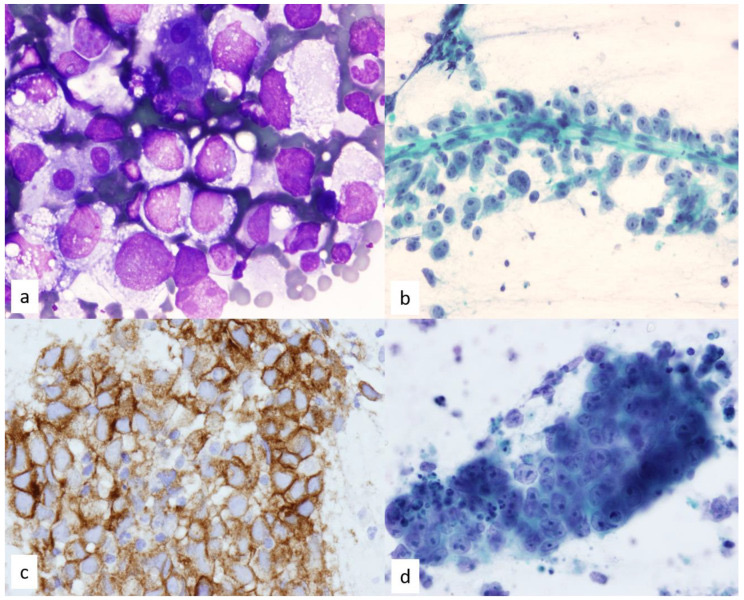
Mediastinal germ cell tumors on FNA. A, seminoma: poorly cohesive large cells with enlarged nuclei and clear to vacuolated cytoplasm on a frothy (tigroid) background, with a few lymphocytes (Diff-Quik, 60× objective). B, seminoma: cells with prominent nucleoli arranged around a vessel (Papanicolaou stain, 40× objective). C, seminoma cell block with membranous CD117 (c-KIT) staining (20× objective). D, embryonal carcinoma for contrast: a cohesive group of larger, more pleomorphic cells with prominent nucleoli (Papanicolaou stain).Image: Kundu Uma et al., PMC open access (Diagnostics (Basel). 2023 Jul 18;13(14):2400. doi: 10.3390/diagnostics13142400) Figure 17, CC BY.
Neurogenic Tumors of the Posterior Mediastinum and Retroperitoneal Mass Fine-Needle Aspiration
Definition. Neurogenic tumors of the paravertebral (posterior) mediastinum comprise peripheral neuroblastic tumors (neuroblastoma, ganglioneuroblastoma and ganglioneuroma), nerve sheath tumors (schwannoma and malignant peripheral nerve sheath tumor) and paraganglioma (Marx et al. 2022). They are the most frequent lesions of the paravertebral compartment (Roden et al. 2020). Peripheral neuroblastic tumors are classified by the International Neuroblastoma Pathology Classification (INPC), which combines the category, the grade of neuroblastic differentiation, the mitosis-karyorrhexis index (MKI) and age into a favorable or unfavorable histology group (Shimada et al. 1999; Conces, Shibui and Shimada 2026). Retroperitoneal masses include primary sarcomas, lymphomas, metastatic carcinomas, metastatic germ cell tumors and paragangliomas (Swallow et al. 2021). Fine-needle aspiration (FNA) is used widely for retroperitoneal lymph nodes and nonsarcomatous lesions (Gupta et al. 2017). For a suspected retroperitoneal sarcoma, image-guided core needle biopsy is the standard of care and FNA is to be avoided (Swallow et al. 2021).
Mesenchymal tumors of the thorax in the WHO 5th edition. The peripheral nerve sheath and neural tumors listed are extra-adrenal paraganglioma, granular cell tumor, schwannoma, malignant peripheral nerve sheath tumor, ganglioneuroma, ganglioneuroblastoma and neuroblastoma. Ganglioneuroma, ganglioneuroblastoma and neuroblastoma are among the mesenchymal tumors that most often occur in the mediastinum (Marx et al. 2022).
Change from the 4th edition. The WHO Thoracic Tumours 5th edition (2021) discusses only the most common mesenchymal tumors and those characteristic of the thorax. The previous thoracic edition covered a broader range. The full range of soft tissue tumors is covered in the WHO Soft Tissue and Bone Tumours volume, which was the 5th edition (2020) when the thoracic volume appeared and is now the 6th edition (2026) (Marx et al. 2022). The soft tissue facts on this page come from the WHO Thoracic Tumours 5th edition and primary studies.
Neuroblastic tumors in the thymus. Neuroblastoma and ganglioneuroma rarely arise in the thymus of adults, sometimes with the syndrome of inappropriate antidiuretic hormone secretion or a thymic cyst (Marx et al. 2022).
Horner syndrome. Horner syndrome is associated especially with a mediastinal primary neuroblastic tumor (Conces, Shibui and Shimada 2026).
INPC categories of peripheral neuroblastic tumors
INPC category
Schwannian stroma
Defining features
INPC prognostic group
Neuroblastoma
Stroma-poor
Neuroblastic cells in sheets or nests separated by thin fibrovascular septa; subtyped as undifferentiated, poorly differentiated or differentiating
Favorable or unfavorable by subtype, MKI and age
Ganglioneuroblastoma, intermixed
Stroma-rich
More than 50% ganglioneuromatous tissue with well-defined microscopic nests of neuroblastic cells in various stages of differentiation
Always favorable
Ganglioneuroma
Stroma-dominant
Mature or maturing ganglion cells in Schwannian stroma; mature ganglion cells are covered by satellite cells
Always favorable
Ganglioneuroblastoma, nodular
Composite
Grossly visible, often hemorrhagic neuroblastoma nodule in intermixed ganglioneuroblastoma or ganglioneuroma
Favorable or unfavorable subset by the neuroblastoma nodule
Sources: Shimada et al. 1999; Peuchmaur et al. 2003; Conces, Shibui and Shimada 2026.
Nodular ganglioneuroblastoma subsets. The 2003 revision of the INPC divided nodular ganglioneuroblastoma into a favorable and an unfavorable subset using the age-linked criteria applied to neuroblastoma. The 8-year event-free survival was 86.1% in the favorable subset and 32.2% in the unfavorable subset (Peuchmaur et al. 2003). In the original 1999 INPC, all nodular ganglioneuroblastomas were assigned to the unfavorable histology group (Peuchmaur et al. 2003).
Timing of classification. The favorable or unfavorable group is assigned only on tumor obtained before chemotherapy or irradiation and is not changed during the clinical course (Conces, Shibui and Shimada 2026).
Adult tumors. The INPC is designed for pediatric tumors and does not apply to adult neuroblastic tumors. Adult neuroblastomas are clinically aggressive, often carry ATRX alterations, usually lack MYCN amplification and are frequently PHOX2B negative (Conces, Shibui and Shimada 2026).
Age-linked INPC criteria for neuroblastoma
Feature
Favorable histology
Unfavorable histology
Undifferentiated subtype
Never favorable
Any age
Poorly differentiated subtype
Diagnosed before 18 months (547 days)
Diagnosed at 18 months (548 days) or later
Differentiating subtype
Diagnosed before 5 years (1,824 days)
Diagnosed at 5 years (1,825 days) or later without maturation to intermixed ganglioneuroblastoma or ganglioneuroma
Low MKI (fewer than 100 per 5,000 cells)
Before 5 years
5 years or later
Intermediate MKI (100–200 per 5,000 cells)
Before 18 months
18 months or later
High MKI (200 or more per 5,000 cells)
Never favorable
Any age
Source: Conces, Shibui and Shimada 2026.
Differentiating neuroblasts. More than 5% of tumor cells must be differentiating neuroblasts for the differentiating subtype. These cells show synchronous nuclear and cytoplasmic enlargement, a cytoplasmic diameter at least twice the nuclear diameter and an eccentric vesicular nucleus with one prominent nucleolus (Conces, Shibui and Shimada 2026).
Poorly differentiated subtype. Neuroblasts produce neurites, with or without Homer Wright rosettes, recognizable on hematoxylin and eosin staining (Conces, Shibui and Shimada 2026).
Undifferentiated subtype. Cells lack recognizable neurites, and nuclear PHOX2B staining is critical for diagnosis (Conces, Shibui and Shimada 2026).
Histology of MYCN-amplified tumors.MYCN-amplified neuroblastoma shows the undifferentiated subtype or the poorly differentiated subtype with high MKI (Conces, Shibui and Shimada 2026).
Cytology of peripheral neuroblastic tumors
Series of 90 aspirates. Aspirates from 83 neuroblastomas, 6 ganglioneuroblastomas and 1 ganglioneuroma, in patients aged 12 days to 12 years, showed distinctive cytomorphology for each type. INPC subtyping on smears classified 61 neuroblastomas as poorly differentiated, 14 as undifferentiated and 8 as differentiating. MKI was high in 63, low in 22 and intermediate in 2 (Koshy et al. 2019).
Immunocytochemistry. Cell blocks of neuroblastoma were positive for at least two neuronal markers (Koshy et al. 2019).
MYCN FISH on smears. Fluorescence in situ hybridization (FISH) for MYCN was feasible on smears and showed amplification in 6 of 11 cases (Koshy et al. 2019).
Ganglioneuroma. Two retroperitoneal ganglioneuromas sampled by EUS-FNA showed large scattered ganglion cells with abundant cytoplasm and large nuclei against a background of wavy spindle cells (Fang et al. 2022). S100 is present in the Schwann cell component of ganglioneuroma and absent in pure neuroblastic tumors (Weiss, Langloss and Enzinger 1983).
Core needle biopsy. Multiple cores obtained by image-guided percutaneous biopsy are generally adequate for diagnosis, INPC classification and molecular testing. Extensive necrosis that prevents MKI determination, sparse cellularity and crush artifact cause inadequate samples (Conces, Shibui and Shimada 2026).
Sampling of composite tumors. Needle samples can miss the neuroblastoma nodule of a nodular ganglioneuroblastoma. When a needle biopsy shows only intermixed ganglioneuroblastoma or ganglioneuroma, the report states that the favorable classification applies only to the sampled tissue (Conces, Shibui and Shimada 2026).
Immunohistochemistry of neuroblastic tumors
Marker
Type of marker
Finding
Source
PHOX2B
Neural crest lineage, nuclear
Detects neuroblastoma as well as synaptophysin does; negative in rhabdomyosarcoma, Ewing sarcoma and Wilms tumor; stable after decalcification of marrow
Hata et al. 2015; Conces, Shibui and Shimada 2026
Synaptophysin, chromogranin, CD56, PGP9.5
Neuronal phenotype
Positive in neuroblastoma; cytoplasmic
Conces, Shibui and Shimada 2026
CD57
Neuronal
Similar to synaptophysin in detecting neuroblastoma, including after treatment
Hata et al. 2015
WT1
Nephrogenic
No nuclear staining in 15 neuroblastomas; nuclear blastemal staining in 44% of Wilms tumors
Chen et al. 2004
Cytokeratin
Epithelial
Negative in neuroblastoma
Reagans and Li 2026
PHOX2B in the diagnostic panel. PHOX2B is recommended with neuronal markers. A negative PHOX2B stain in a small round cell tumor favors a diagnosis other than a peripheral neuroblastic tumor (Conces, Shibui and Shimada 2026).
Molecular and risk assessment of neuroblastic tumors
MYCN amplification. Amplification is defined by FISH as a more than 4-fold increase of MYCN signals over a chromosome 2 reference probe. It is a strong adverse prognostic factor and correlates with higher stage and unfavorable histology (Conces, Shibui and Shimada 2026).
DNA ploidy and segmental aberrations. Hyperdiploid tumors with whole-chromosome gains have an excellent prognosis. Segmental chromosomal aberrations such as 1p loss, 11q loss and 17q gain indicate a poor outcome, and about 70% of MYCN-amplified tumors have 1p36 deletion (Conces, Shibui and Shimada 2026).
WHO desirable testing. The WHO 5th edition lists testing for MYCN, ALK, TERT and ATRX status, DNA index and segmental chromosomal aberrations as desirable for predicting behavior (Marx et al. 2022).
ALK on FNA material. In 54 advanced neuroblastomas diagnosed by FNA, ALK protein was expressed in 65% on cell blocks. Two tumors with the ALK F1174L mutation had high ALK expression, and both patients died of disease (Bhardwaj et al. 2023).
Familial tumors. Germline ALK variants account for about 75–80% of familial neuroblastoma, and germline PHOX2B variants also occur (Conces, Shibui and Shimada 2026).
Staging. The International Neuroblastoma Risk Group Staging System is a presurgical system with stages L1, L2, M and MS. Stage MS applies to children younger than 18 months with metastases limited to skin, liver and/or bone marrow (Conces, Shibui and Shimada 2026).
Schwannoma of the paravertebral mediastinum on FNA
Cytologic features. In 116 resected schwannomas, diagnostic smears showed spindle cells with wavy nuclei in a fibrillary, collagenous or myxoid matrix and Antoni A and Antoni B fragments. Nuclear palisading was present in 41 smears and Verocay bodies in 10 (Domanski et al. 2006).
Degenerative atypia. Markedly pleomorphic nuclei occurred in 8 ancient and 6 conventional schwannomas, and slight to moderate pleomorphism in 38 more (Domanski et al. 2006).
Diagnostic yield. FNA gave an unequivocally benign diagnosis in 80 of 116 cases. There were 23 insufficient smears, 7 inconclusive smears and 6 false-positive malignant diagnoses (Domanski et al. 2006).
Sources of error. Aspirates with predominantly Antoni A tissue, nuclear pleomorphism or myxoid change are confused with other benign or malignant soft tissue tumors (Domanski et al. 2006).
Immunostains. Schwannoma shows diffuse SOX10 (Nonaka, Chiriboga and Rubin 2008). Virtually all benign nerve sheath tumors contain numerous S100-positive cells, against 18 of 36 malignant schwannomas (Weiss, Langloss and Enzinger 1983). The nerve sheath immunopanel is described in Benign Soft Tissue Tumors on FNA.
Malignant peripheral nerve sheath tumor on FNA
Cytologic diagnosis. In 55 FNA samples of malignant peripheral nerve sheath tumor (MPNST) from 52 patients, 26 of whom had neurofibromatosis type 1, a definitive or consistent diagnosis was made in 30% of primary, 93% of locally recurrent and 70% of metastatic tumors (Wakely, Ali and Bishop 2012).
SOX10 and S100. SOX10 was expressed in 49% and S100 protein in 30% of MPNSTs (Nonaka, Chiriboga and Rubin 2008).
H3K27me3 loss in resections. Complete H3K27me3 loss occurred in 51% of 100 resected MPNSTs and was absent in cellular schwannomas and atypical neurofibromas (Schaefer, Fletcher and Hornick 2016). Its use as a marker is described in Molecular and Ancillary Testing of Soft Tissue FNA Material.
H3K27me3 on FNA and needle biopsy. Complete loss was seen in 13 of 24 MPNSTs (54%) and in only 2 of 156 mimics, a specificity of 98.7%. Partial loss occurred in both MPNSTs and other tumors (Mito et al. 2017).
Differential diagnosis of spindle cell aspirates from the paravertebral mediastinum
Diagnosis
Smear findings
Key ancillary findings
Source
Schwannoma
Wavy spindle cells, fibrillary matrix, Antoni A fragments, Verocay bodies; degenerative pleomorphism
Diffuse SOX10 and S100; H3K27me3 retained
Domanski et al. 2006; Nonaka, Chiriboga and Rubin 2008; Weiss, Langloss and Enzinger 1983; Schaefer, Fletcher and Hornick 2016
Neurofibroma
Spindle cell aspirate
Diffuse SOX10; H3K27me3 retained
Nonaka, Chiriboga and Rubin 2008; Prieto-Granada et al. 2016
Ganglioneuroma
Large ganglion cells among wavy spindle cells
S100 in the Schwann cell component
Fang et al. 2022; Weiss, Langloss and Enzinger 1983
Malignant peripheral nerve sheath tumor
Malignant spindle cells
Complete H3K27me3 loss in 54% on needle samples and in 61% of 68 resected tumors, with partial loss in a further 7%; SOX10 in 49%
Mito et al. 2017; Prieto-Granada et al. 2016; Nonaka, Chiriboga and Rubin 2008
Solitary fibrous tumor
Spindle cells in a patternless arrangement
Nuclear STAT6 in 59 of 60; CD34; NAB2::STAT6 fusion
Doyle et al. 2014; Marx et al. 2022
Synovial sarcoma
Uniform spindle cells with scant cytoplasm
Nuclear TLE1 in 91 of 94; SS18::SSX fusion or SS18-SSX antibody
Terry et al. 2007; Marx et al. 2022
Retroperitoneal mass FNA: spectrum of lesions
Series of 389 aspirates. Of 389 retroperitoneal aspirates, 15.7% were inadequate. Sites were pancreas (29.0%), retroperitoneal lymph nodes (24.9%), kidneys (17.9%), adrenals (11.5%), retroperitoneal soft tissue (10.5%) and gut (5.9%). Malignant neoplasms outnumbered benign neoplasms by 10.8 to 1. Cytologic and histologic diagnoses agreed in 27 of 33 cases with histology (81.8%) (Gupta et al. 2017).
Series of 338 patients. Of 338 retroperitoneal masses, 28 were inadequate. Of the 310 adequate cases, 274 (88.4%) were malignant, 8 (2.6%) benign and 28 (9.0%) nonneoplastic. Renal origin (106, 34.2%) and retroperitoneal soft tissue (96, 31%) were most common (Das et al. 2019).
Common diagnoses by age. Tubercular lymphadenitis was the most common nonneoplastic lesion and paraganglioma the most common benign lesion. Wilms tumor, neuroblastoma and germ cell tumor predominated in infancy and early childhood, renal cell carcinoma and pancreatic adenocarcinoma in middle age, and metastatic carcinoma in the elderly (Das et al. 2019).
Accuracy. Image-guided FNA had a sensitivity of 98.02%, a specificity of 72.22% and a diagnostic accuracy of 94.12% (Das et al. 2019).
Biopsy of a suspected retroperitoneal sarcoma
The Transatlantic Australasian Retroperitoneal Sarcoma Working Group consensus (Swallow et al. 2021) gives the following recommendations.
Recommendation
Detail
Differential diagnosis of an undiagnosed retroperitoneal mass
Metastatic adenocarcinoma, lymphoma, metastatic germ cell tumor and paraganglioma
Preferred sampling
Image-guided percutaneous coaxial core needle biopsy, 14–18 gauge, as the standard of care, with multiple cores for subtyping, grading and biobanking
Omission of biopsy
Acceptable only when an expert radiologist in a multidisciplinary tumor board judges the imaging pathognomonic, such as heterogeneous well-differentiated and dedifferentiated liposarcoma, and no preoperative treatment is planned
Target within the tumor
The solid, well-perfused or most fluorodeoxyglucose-avid area, to avoid undergrading
FNA
Rarely diagnostic; delays treatment; to be avoided
Open or laparoscopic biopsy
To be avoided because of peritoneal contamination
Immunohistochemistry for liposarcoma
MDM2 staining is the minimum standard for well-differentiated and dedifferentiated liposarcoma; CDK4 and MDM2 amplification testing at the pathologist's discretion
Needle tract seeding
Rare after core needle biopsy; lowest with a coaxial sheathed needle
Main subtypes. Four histologic subtypes account for about 90% of retroperitoneal sarcomas. Well-differentiated and dedifferentiated (low- and intermediate-grade) liposarcoma forms about one-half of retroperitoneal sarcomas (Swallow et al. 2021).
WHO Thoracic Tumours 5th edition criteria for liposarcoma. MDM2 and CDK4 nuclear expression or MDM2 amplification is a desirable diagnostic criterion for well-differentiated and dedifferentiated liposarcoma in the WHO 5th edition (Marx et al. 2022).
Differential diagnosis of retroperitoneal aspirates
Diagnosis
Smear findings
Ancillary test
Source
Lymphoma
Dispersed lymphoid cells
Flow cytometry on a fresh needle rinse
Swallow et al. 2021; Reagans and Li 2026
Metastatic germ cell tumor
Seminoma or nonseminoma pattern
OCT4, SALL4, glypican-3
Bokemeyer et al. 2002; Cao et al. 2009
Paraganglioma
Round to oval cells with abundant finely granular cytoplasm
Reagans and Li 2026; Nonaka, Chiriboga and Rubin 2008
Well-differentiated or dedifferentiated liposarcoma
Adipocytic or pleomorphic spindle cells
MDM2 and CDK4; MDM2 amplification
Swallow et al. 2021; Marx et al. 2022
Schwannoma
Wavy spindle cells, Verocay bodies
Diffuse SOX10
Domanski et al. 2006; Nonaka, Chiriboga and Rubin 2008
Neuroblastoma
Small round cells, neuropil, rosettes
PHOX2B
Conces, Shibui and Shimada 2026; Hata et al. 2015
Metastatic carcinoma
Cohesive epithelial groups
Keratin and site-specific markers
Das et al. 2019
Extra-adrenal paraganglioma beside the duodenum on EUS-guided fine-needle biopsy. A, nests of polygonal cells with granular cytoplasm (H&E, 200×). B and C, strong chromogranin and synaptophysin staining (200×). D, nuclear GATA3 (200×). E, keratin negative (100×). F, CD34 outlines the rich capillary network around the nests (400×).Image: Nikas Ilias P. et al., PMC open access (Diagnostics (Basel). 2022 Jul 28;12(8):1819. doi: 10.3390/diagnostics12081819) Figure 2, CC BY.
References
Ali SZ, Erozan YS. Thymoma: cytopathologic features and differential diagnosis on fine needle aspiration. Acta Cytol. 1998;42(4):845–854. doi:10.1159/000331958. PMID 9684567
Angirekula M, Chang SY, Jenkins SM, et al. CD117, BAP1, MTAP, and TdT is a useful immunohistochemical panel to distinguish thymoma from thymic carcinoma. Cancers (Basel). 2022;14(9):2299. doi:10.3390/cancers14092299. PMID 35565429
Annema JT, van Meerbeeck JP, Rintoul RC, et al. Mediastinoscopy vs endosonography for mediastinal nodal staging of lung cancer: a randomized trial. JAMA. 2010;304(20):2245–2252. doi:10.1001/jama.2010.1705. PMID 21098770
Aravena C, Patel J, Goyal A, et al. Role of endobronchial ultrasound-guided transbronchial needle aspiration in the diagnosis and management of mediastinal cyst. J Bronchology Interv Pulmonol. 2020;27(2):142–146. doi:10.1097/LBR.0000000000000640. PMID 31855882
Bauer DE, Mitchell CM, Strait KM, et al. Clinicopathologic features and long-term outcomes of NUT midline carcinoma. Clin Cancer Res. 2012;18(20):5773–5779. doi:10.1158/1078-0432.CCR-12-1153. PMID 22896655
Bernard C, Frih H, Pasquet F, et al. Thymoma associated with autoimmune diseases: 85 cases and literature review. Autoimmun Rev. 2016;15(1):82–92. doi:10.1016/j.autrev.2015.09.005. PMID 26408958
Bhardwaj N, Rohilla M, Gautam U, et al. ALK gene mutation and ALK protein expression in advanced neuroblastoma and the potential value in risk stratification in fine-needle aspiration biopsy samples. Am J Clin Pathol. 2023;159(4):407–415. doi:10.1093/ajcp/aqac172. PMID 36812383
Bishop JA, French CA, Ali SZ. Cytopathologic features of NUT midline carcinoma: a series of 26 specimens from 13 patients. Cancer Cytopathol. 2016;124(12):901–908. doi:10.1002/cncy.21761. PMID 27400194
Bokemeyer C, Nichols CR, Droz JP, et al. Extragonadal germ cell tumors of the mediastinum and retroperitoneum: results from an international analysis. J Clin Oncol. 2002;20(7):1864–1873. doi:10.1200/JCO.2002.07.062. PMID 11919246
Calaminici M, Piper K, Lee AM, Norton AJ. CD23 expression in mediastinal large B-cell lymphomas. Histopathology. 2004;45(6):619–624. doi:10.1111/j.1365-2559.2004.01969.x. PMID 15569053
Cao D, Li J, Guo CC, Allan RW, Humphrey PA. SALL4 is a novel diagnostic marker for testicular germ cell tumors. Am J Surg Pathol. 2009;33(7):1065–1077. doi:10.1097/PAS.0b013e3181a13eef. PMID 19390421
Cao F, Zhang S, Dai Z, et al. Diagnosis of mediastinal cysts: the role and safety of EUS-FNA with 19-gauge needle: a retrospective cohort study. J Thorac Dis. 2022;14(9):3544–3551. doi:10.21037/jtd-22-1105. PMID 36245595
Carter BW, Tomiyama N, Bhora FY, et al. A modern definition of mediastinal compartments. J Thorac Oncol. 2014;9(9 Suppl 2):S97–S101. doi:10.1097/JTO.0000000000000292. PMID 25396318
Chen BF, Tzen CY, Liang DC, Liu HC, Huang YW, Fan CC. Immunohistochemical expression of Wilms' tumor 1 protein in nephroblastoma. J Chin Med Assoc. 2004;67(10):506–510. PMID 15648285
Conces MR, Shibui Y, Shimada H. Pathology of peripheral neuroblastic tumors. Diagn Pathol. 2026;21(1):22. doi:10.1186/s13000-026-01758-y. PMID 41612478
Copie-Bergman C, Gaulard P, Maouche-Chrétien L, et al. The MAL gene is expressed in primary mediastinal large B-cell lymphoma. Blood. 1999;94(10):3567–3575. PMID 10552968
Cozzolino I, Calaminici M, Ronchi A, et al. The World Health Organization Reporting System for Lymph Node, Spleen, and Thymus Cytopathology: part 2 – spleen and thymus. Acta Cytol. 2026;70(2):201–210. doi:10.1159/000548853. PMID 41086121
Das A, Gahine R, Patre V, Hussain N. Retroperitoneal tumor: a silent trespasser. Role of image-guided fine-needle aspiration cytology with histopathological correlation in early diagnosis. Acta Cytol. 2019;63(3):189–197. doi:10.1159/000497077. PMID 30893686
Domanski HA, Akerman M, Engellau J, et al. Fine-needle aspiration of neurilemoma (schwannoma): a clinicocytopathologic study of 116 patients. Diagn Cytopathol. 2006;34(6):403–412. doi:10.1002/dc.20449. PMID 16680779
Doyle LA, Vivero M, Fletcher CD, Mertens F, Hornick JL. Nuclear expression of STAT6 distinguishes solitary fibrous tumor from histologic mimics. Mod Pathol. 2014;27(3):390–395. doi:10.1038/modpathol.2013.164. PMID 24030747
Eom DW, Kang GH, Kim JW, Ryu DS. Unusual bronchopulmonary foregut malformation associated with pericardial defect: bronchogenic cyst communicating with tubular esophageal duplication. J Korean Med Sci. 2007;22(3):564–567. doi:10.3346/jkms.2007.22.3.564. PMID 17596673
Fang C, Pizzillo I, Shi Y, Sun W, Brandler TC. Ganglioneuroma on fine needle aspiration cytology: case series and review of the literature. Diagn Cytopathol. 2022;50(6):E146–E150. doi:10.1002/dc.24931. PMID 34985204
Feigin DS, Fenoglio JJ, McAllister HA, Madewell JE. Pericardial cysts: a radiologic-pathologic correlation and review. Radiology. 1977;125(1):15–20. doi:10.1148/125.1.15. PMID 897162
Foss HD, Reusch R, Demel G, et al. Frequent expression of the B-cell-specific activator protein in Reed-Sternberg cells of classical Hodgkin's disease provides further evidence for its B-cell origin. Blood. 1999;94(9):3108–3113. PMID 10556196
Gardić N, Tomić S, Miljković D, et al. A case series: can a cytological smear indicate the histological type of thymoma and distinguish thymoma from thymic carcinoma? Diagn Cytopathol. 2025;53(9):E165–E169. doi:10.1002/dc.25493. PMID 40464409
Gorczyca W, Tugulea S, Liu Z, et al. Flow cytometry in the diagnosis of mediastinal tumors with emphasis on differentiating thymocytes from precursor T-lymphoblastic lymphoma/leukemia. Leuk Lymphoma. 2004;45(3):529–538. doi:10.1080/10428190310001598008. PMID 15160915
Gu P, Zhao YZ, Jiang LY, et al. Endobronchial ultrasound-guided transbronchial needle aspiration for staging of lung cancer: a systematic review and meta-analysis. Eur J Cancer. 2009;45(8):1389–1396. doi:10.1016/j.ejca.2008.11.043. PMID 19124238
Gupta P, Rajwanshi A, Nijhawan R, et al. Fine needle aspiration in retroperitoneal lesions. APMIS. 2017;125(1):16–23. doi:10.1111/apm.12627. PMID 27807894
Gupta R, Mathur SR, Arora VK, Sharma SG. Cytologic features of extragonadal germ cell tumors: a study of 88 cases with aspiration cytology. Cancer. 2008;114(6):504–511. doi:10.1002/cncr.23983. PMID 18980289
Haack H, Johnson LA, Fry CJ, et al. Diagnosis of NUT midline carcinoma using a NUT-specific monoclonal antibody. Am J Surg Pathol. 2009;33(7):984–991. doi:10.1097/PAS.0b013e318198d666. PMID 19363441
Hartmann JT, Nichols CR, Droz JP, et al. Hematologic disorders associated with primary mediastinal nonseminomatous germ cell tumors. J Natl Cancer Inst. 2000;92(1):54–61. doi:10.1093/jnci/92.1.54. PMID 10620634
Hata JL, Correa H, Krishnan C, et al. Diagnostic utility of PHOX2B in primary and treated neuroblastoma and in neuroblastoma metastatic to the bone marrow. Arch Pathol Lab Med. 2015;139(4):543–546. doi:10.5858/arpa.2014-0255-OA. PMID 25822764
Hittmair A, Rogatsch H, Hobisch A, Mikuz G, Feichtinger H. CD30 expression in seminoma. Hum Pathol. 1996;27(11):1166–1171. doi:10.1016/s0046-8177(96)90310-x. PMID 8912826
Hoeller S, Zihler D, Zlobec I, et al. BOB.1, CD79a and cyclin E are the most appropriate markers to discriminate classical Hodgkin's lymphoma from primary mediastinal large B-cell lymphoma. Histopathology. 2010;56(2):217–228. doi:10.1111/j.1365-2559.2009.03462.x. PMID 20102401
Hsu SM, Jaffe ES. Leu M1 and peanut agglutinin stain the neoplastic cells of Hodgkin's disease. Am J Clin Pathol. 1984;82(1):29–32. doi:10.1093/ajcp/82.1.29. PMID 6741873
Jacobsen GK, Jacobsen M. Alpha-fetoprotein (AFP) and human chorionic gonadotropin (HCG) in testicular germ cell tumours: a prospective immunohistochemical study. Acta Pathol Microbiol Immunol Scand A. 1983;91(3):165–176. doi:10.1111/j.1699-0463.1983.tb02742.x. PMID 6190350
Jiménez-Heffernan JA, Rodríguez-García AM, Cima L, et al. A comprehensive review of the "tigroid" background cytological concept: what, when, where and why? Pathologica. 2022;114(2):121–127. doi:10.32074/1591-951X-287. PMID 35481562
Jones TD, Ulbright TM, Eble JN, Baldridge LA, Cheng L. OCT4 staining in testicular tumors: a sensitive and specific marker for seminoma and embryonal carcinoma. Am J Surg Pathol. 2004;28(7):935–940. doi:10.1097/00000478-200407000-00014. PMID 15223965
Kondo K, Monden Y. Thymoma and myasthenia gravis: a clinical study of 1,089 patients from Japan. Ann Thorac Surg. 2005;79(1):219–224. doi:10.1016/j.athoracsur.2004.06.090. PMID 15620947
Koshy A, Jain R, Srinivasan R, et al. Cytopathological spectrum of peripheral neuroblastic tumours in fine needle aspiration cytology and categorisation as per International Neuroblastoma Pathology Classification. Cytopathology. 2019;30(6):634–643. doi:10.1111/cyt.12747. PMID 31231909
Labarca G, Sierra-Ruiz M, Kheir F, et al. Diagnostic accuracy of endobronchial ultrasound transbronchial needle aspiration in lymphoma: a systematic review and meta-analysis. Ann Am Thorac Soc. 2019;16(11):1432–1439. doi:10.1513/AnnalsATS.201902-175OC. PMID 31291126
Marcus A, Narula N, Kamel MK, et al. Sensitivity and specificity of fine needle aspiration for the diagnosis of mediastinal lesions. Ann Diagn Pathol. 2019;39:69–73. doi:10.1016/j.anndiagpath.2019.02.011. PMID 30797131
Marx A, Chan JKC, Chalabreysse L, et al. The 2021 WHO classification of tumors of the thymus and mediastinum: what is new in thymic epithelial, germ cell, and mesenchymal tumors? J Thorac Oncol. 2022;17(2):200–213. doi:10.1016/j.jtho.2021.10.010. PMID 34695605
Marx A, Chan JKC, Coindre JM, et al. The 2015 World Health Organization classification of tumors of the thymus: continuity and changes. J Thorac Oncol. 2015;10(10):1383–1395. doi:10.1097/JTO.0000000000000654. PMID 26295375
Marx A, Willcox N, Leite MI, et al. Thymoma and paraneoplastic myasthenia gravis. Autoimmunity. 2010;43(5–6):413–427. doi:10.3109/08916930903555935. PMID 20380583
Maturu VN, Dhooria S, Agarwal R. Efficacy and safety of transbronchial needle aspiration in diagnosis and treatment of mediastinal bronchogenic cysts: systematic review of case reports. J Bronchology Interv Pulmonol. 2015;22(3):195–203. doi:10.1097/LBR.0000000000000174. PMID 26165889
Menter T, Tzankov A. Genetic alterations of 9p24 in lymphomas and their impact for cancer (immuno-)therapy. Virchows Arch. 2019;474(4):497–509. doi:10.1007/s00428-018-2438-6. PMID 30132131
Micames CG, McCrory DC, Pavey DA, Jowell PS, Gress FG. Endoscopic ultrasound-guided fine-needle aspiration for non-small cell lung cancer staging: a systematic review and metaanalysis. Chest. 2007;131(2):539–548. doi:10.1378/chest.06-1437. PMID 17296659
Mito JK, Qian X, Doyle LA, Hornick JL, Jo VY. Role of histone H3K27 trimethylation loss as a marker for malignant peripheral nerve sheath tumor in fine-needle aspiration and small biopsy specimens. Am J Clin Pathol. 2017;148(2):179–189. doi:10.1093/ajcp/aqx060. PMID 28898989
Moch H, Amin MB, Berney DM, et al. The 2022 World Health Organization classification of tumours of the urinary system and male genital organs, part A: renal, penile, and testicular tumours. Eur Urol. 2022;82(5):458–468. doi:10.1016/j.eururo.2022.06.016. PMID 35853783
Moran CA, Suster S. Mediastinal seminomas with prominent cystic changes: a clinicopathologic study of 10 cases. Am J Surg Pathol. 1995;19(9):1047–1053. doi:10.1097/00000478-199509000-00008. PMID 7661278
Nakagawa K, Matsuno Y, Kunitoh H, et al. Immunohistochemical KIT (CD117) expression in thymic epithelial tumors. Chest. 2005;128(1):140–144. doi:10.1378/chest.128.1.140. PMID 16002927
Nonaka D, Chiriboga L, Rubin BP. Sox10: a pan-schwannian and melanocytic marker. Am J Surg Pathol. 2008;32(9):1291–1298. doi:10.1097/PAS.0b013e3181658c14. PMID 18636017
Nonaka D, Henley JD, Chiriboga L, Yee H. Diagnostic utility of thymic epithelial markers CD205 (DEC205) and Foxn1 in thymic epithelial neoplasms. Am J Surg Pathol. 2007;31(7):1038–1044. doi:10.1097/PAS.0b013e31802b4917. PMID 17592270
Nonaka D. Differential expression of SOX2 and SOX17 in testicular germ cell tumors. Am J Clin Pathol. 2009;131(5):731–736. doi:10.1309/AJCP7MNCNBCRN8NO. PMID 19369635
Ordóñez NG. Value of GATA3 immunostaining in the diagnosis of parathyroid tumors. Appl Immunohistochem Mol Morphol. 2014;22(10):756–761. doi:10.1097/PAI.0000000000000007. PMID 25046229
Pallesen G, Hamilton-Dutoit SJ. Ki-1 (CD30) antigen is regularly expressed by tumor cells of embryonal carcinoma. Am J Pathol. 1988;133(3):446–450. PMID 2849300
Peuchmaur M, d'Amore ES, Joshi VV, et al. Revision of the International Neuroblastoma Pathology Classification: confirmation of favorable and unfavorable prognostic subsets in ganglioneuroblastoma, nodular. Cancer. 2003;98(10):2274–2281. doi:10.1002/cncr.11773. PMID 14601099
Posligua L, Ylagan L. Fine-needle aspiration cytology of thymic basaloid carcinoma: case studies and review of the literature. Diagn Cytopathol. 2006;34(5):358–366. doi:10.1002/dc.20433. PMID 16604555
Powers CN, Silverman JF, Geisinger KR, Frable WJ. Fine-needle aspiration biopsy of the mediastinum: a multi-institutional analysis. Am J Clin Pathol. 1996;105(2):168–173. doi:10.1093/ajcp/105.2.168. PMID 8607440
Prieto-Granada CN, Wiesner T, Messina JL, et al. Loss of H3K27me3 expression is a highly sensitive marker for sporadic and radiation-induced MPNST. Am J Surg Pathol. 2016;40(4):479–489. doi:10.1097/PAS.0000000000000564. PMID 26645727
Reagans R, Li Z. Approach to mediastinal cytopathology: a diagnostic overview. Mediastinum. 2026;10:21. doi:10.21037/med-2025-1-58. PMID 42427473
Roden AC, Ahmad U, Cardillo G, et al. Thymic carcinomas: a concise multidisciplinary update on recent developments from the Thymic Carcinoma Working Group of the International Thymic Malignancy Interest Group. J Thorac Oncol. 2022;17(5):637–650. doi:10.1016/j.jtho.2022.01.021. PMID 35227908
Roden AC, Fang W, Shen Y, et al. Distribution of mediastinal lesions across multi-institutional, international, radiology databases. J Thorac Oncol. 2020;15(4):568–579. doi:10.1016/j.jtho.2019.12.108. PMID 31870881
Roden AC, Judge M, den Bakker MA, et al. Dataset for reporting of thymic epithelial tumours: recommendations from the International Collaboration on Cancer Reporting (ICCR). Histopathology. 2023;83(6):967–980. doi:10.1111/his.15047. PMID 37722860
Ruffini E, Huang J, Cilento V, et al. The International Association for the Study of Lung Cancer Thymic Epithelial Tumors Staging Project: proposal for a stage classification for the forthcoming (ninth) edition of the TNM classification of malignant tumors. J Thorac Oncol. 2023;18(12):1655–1671. doi:10.1016/j.jtho.2023.09.002. PMID 37689391
Ryu HS, Koh JS, Park S, et al. Classification of thymoma by fine needle aspiration biopsy according to WHO classification: a cytological algorithm for stepwise analysis in the classification of thymoma. Acta Cytol. 2012;56(5):487–494. doi:10.1159/000339001. PMID 23075888
Schaefer IM, Fletcher CD, Hornick JL. Loss of H3K27 trimethylation distinguishes malignant peripheral nerve sheath tumors from histologic mimics. Mod Pathol. 2016;29(1):4–13. doi:10.1038/modpathol.2015.134. PMID 26585554
Shimada H, Ambros IM, Dehner LP, et al. Terminology and morphologic criteria of neuroblastic tumors: recommendations by the International Neuroblastoma Pathology Committee. Cancer. 1999;86(2):349–363. PMID 10421272
Shimada H, Ambros IM, Dehner LP, et al. The International Neuroblastoma Pathology Classification (the Shimada system). Cancer. 1999;86(2):364–372. PMID 10421273
Stein H, Mason DY, Gerdes J, et al. The expression of the Hodgkin's disease associated antigen Ki-1 in reactive and neoplastic lymphoid tissue: evidence that Reed-Sternberg cells and histiocytic malignancies are derived from activated lymphoid cells. Blood. 1985;66(4):848–858. PMID 3876124
Suster S, Rosai J. Multilocular thymic cyst: an acquired reactive process. Study of 18 cases. Am J Surg Pathol. 1991;15(4):388–398. PMID 2006719
Swallow CJ, Strauss DC, Bonvalot S, et al. Management of primary retroperitoneal sarcoma (RPS) in the adult: an updated consensus approach from the Transatlantic Australasian RPS Working Group. Ann Surg Oncol. 2021;28(12):7873–7888. doi:10.1245/s10434-021-09654-z. PMID 33852100
Takata S, Horie M, Miyabe K, et al. Thymic cyst: a clinicopathological, immunohistochemical, and molecular study of 36 cases focusing on squamous- and ciliated-type epithelium. Hum Pathol. 2026;172:106109. doi:10.1016/j.humpath.2026.106109. PMID 41850630
Takeda S, Miyoshi S, Minami M, et al. Clinical spectrum of mediastinal cysts. Chest. 2003;124(1):125–132. doi:10.1378/chest.124.1.125. PMID 12853514
Terry J, Saito T, Subramanian S, et al. TLE1 as a diagnostic immunohistochemical marker for synovial sarcoma emerging from gene expression profiling studies. Am J Surg Pathol. 2007;31(2):240–246. doi:10.1097/01.pas.0000213330.71745.39. PMID 17255769
Thomas-de-Montpréville V, Dulmet E. Cysts of the posterior mediastinum showing müllerian differentiation (Hattori's cysts). Ann Diagn Pathol. 2007;11(6):417–420. doi:10.1016/j.anndiagpath.2006.12.011. PMID 18022126
Ulbright TM. Concerning the seminal study of anterior mediastinal teratomas by H. G. Schlumberger: an update and modern contextualization. Arch Pathol Lab Med. 2025;150(1):27–36. doi:10.5858/arpa.2025-0234-RA. PMID 41429179
Wakely PE, Ali SZ, Bishop JA. The cytopathology of malignant peripheral nerve sheath tumor: a report of 55 fine-needle aspiration cases. Cancer Cytopathol. 2012;120(5):334–341. doi:10.1002/cncy.21195. PMID 22434579
Wakely PE. Fine needle aspiration in the diagnosis of thymic epithelial neoplasms. Hematol Oncol Clin North Am. 2008;22(3):433–442. doi:10.1016/j.hoc.2008.03.003. PMID 18514125
Weiss SW, Langloss JM, Enzinger FM. Value of S-100 protein in the diagnosis of soft tissue tumors with particular reference to benign and malignant Schwann cell tumors. Lab Invest. 1983;49(3):299–308. PMID 6310227
Wildi SM, Hoda RS, Fickling W, et al. Diagnosis of benign cysts of the mediastinum: the role and risks of EUS and FNA. Gastrointest Endosc. 2003;58(3):362–368. PMID 14528209
Willner J, Zhou F, Moreira AL. Diagnostic challenges in the cytology of thymic epithelial neoplasms. Cancers (Basel). 2022;14(8):2013. doi:10.3390/cancers14082013. PMID 35454918
Zakowski MF, Huang J, Bramlage MP. The role of fine needle aspiration cytology in the diagnosis and management of thymic neoplasia. J Thorac Oncol. 2010;5(10 Suppl 4):S281–S285. doi:10.1097/JTO.0b013e3181f23e19. PMID 20859120
Zhang Y, Yu L, Ke J. Pathological features and prognosis of thymoma with or without myasthenia gravis. Front Surg. 2022;9:726673. doi:10.3389/fsurg.2022.726673. PMID 35252321
Zynger DL, Dimov ND, Luan C, Teh BT, Yang XJ. Glypican 3: a novel marker in testicular germ cell tumors. Am J Surg Pathol. 2006;30(12):1570–1575. doi:10.1097/01.pas.0000213322.89670.48. PMID 17122513
Extra-adrenal paraganglioma beside the duodenum on EUS-guided fine-needle biopsy. A, nests of polygonal cells with granular cytoplasm (H&E, 200×). B and C, strong chromogranin and synaptophysin staining (200×). D, nuclear GATA3 (200×). E, keratin negative (100×). F, CD34 outlines the rich capillary network around the nests (400×). Image: Nikas Ilias P. et al., PMC open access (Diagnostics (Basel). 2022 Jul 28;12(8):1819. doi: 10.3390/diagnostics12081819) Figure 2, CC BY.