Last reviewed 2026-11-10 by a board-certified pathologist. 238 references.
Colorectal Adenomas, Serrated Polyps and Other Colorectal Polyps
Definition. Colorectal polyps are mucosal lesions that protrude into the lumen or form flat mucosal elevations. The neoplastic precursors of colorectal carcinoma are conventional adenomas and serrated polyps (Nagtegaal et al. 2020; Arends et al. 2026). Non-neoplastic polyps include prolapse-related inflammatory polyps, inflammatory myoglandular polyps, post-inflammatory polyps, juvenile polyps and benign lymphoid polyps. The WHO Classification of Digestive System Tumours, 6th edition (2026), divides colorectal serrated polyps into three sections: hyperplastic polyp, sessile serrated lesion (SSL) and traditional serrated adenoma (TSA). It adds TSA with conventional dysplasia (Arends et al. 2026).
Fibromuscular obliteration of the lamina propria; fibrin or granulation tissue cap
Reactive lesion attributed to mucosal prolapse
Chetty et al. 1993; Ng et al. 2004
Post-inflammatory polyp
Regenerative mucosa and granulation tissue after ulceration in colitis; no dysplasia
Reactive; associated with severe, extensive inflammation
ICCR polypectomy 2020; Mahmoud et al. 2019
Benign lymphoid polyp
Hyperplastic lymphoid follicles with germinal centers in the mucosa and submucosa
Reactive lymphoid hyperplasia
Nishimura et al. 2024; Hong et al. 2021
Juvenile polyp
Abundant lamina propria with cystically dilated glands and no smooth muscle core
Germline SMAD4 or BMPR1A variants in juvenile polyposis syndrome
Syngal et al. 2015; Arends et al. 2026
Conventional adenomas of the colorectum
Morphology and grading of conventional adenomas
Definition. Conventional adenomas are neoplastic polyps of dysplastic intestinal epithelium. Architecture is recorded as tubular, tubulovillous or villous (Gupta et al. 2020).
Villous component. Tubular adenomas have a villous component of <25% of the polyp and villous adenomas >75%. All other adenomas are tubulovillous. An adenoma with a villous component of >25% is an advanced lesion (ICCR polypectomy 2020).
Criteria for high-grade dysplasia. High-grade dysplasia shows marked architectural changes at low magnification: complex crowding, irregular glands, cribriform architecture and intraluminal necrosis. These accompany cytologic features: loss of cell polarity, enlarged nuclei with prominent nucleoli and often atypical mitotic figures (ICCR polypectomy 2020).
Two-tier grading. The 5th edition used a two-tier system of low-grade and high-grade dysplasia for precursor lesions at all digestive sites. The term "carcinoma in situ" is discouraged and is included in high-grade dysplasia (Nagtegaal et al. 2020). The 6th edition retains two-tier (low-grade and high-grade) grading across precursor lesions (Arends et al. 2026). The same two-tier model applies to Barrett dysplasia (Barrett Esophagus and Dysplasia).
Reproducibility. Five gastrointestinal pathologists reviewing 107 adenomas smaller than 1 cm showed poor agreement for villous component and high-grade dysplasia. Agreement remained poor after consensus criteria were applied (Mahajan et al. 2013).
Risk factors for high-grade dysplasia. In 3371 adenomas from the National Polyp Study, size and villous component were the major independent risk factors for high-grade dysplasia. The adjusted odds ratio was 7.7 for large adenomas compared with small adenomas, and 8.1 for the most villous categories compared with tubular adenomas. Age of 60 years or older was an independent patient factor (O'Brien et al. 1990).
Adenoma–carcinoma sequence
Molecular steps.RAS mutations occurred in 58% of adenomas larger than 1 cm and in 9% of adenomas smaller than 1 cm. Chromosome 18 deletions were present in 73% of carcinomas and 47% of advanced adenomas. Chromosome 17p loss was largely confined to carcinomas (75%) (Vogelstein et al. 1988).
Pathway share. About 60% of colorectal carcinomas arise from conventional adenomas through the chromosomal instability pathway. About 35% arise through the serrated pathway, and about 5% arise in Lynch syndrome (Snover 2011).
Effect of polypectomy. Colonoscopic polypectomy in the National Polyp Study reduced the expected incidence of colorectal carcinoma by 76–90% (Winawer et al. 1993). Long-term follow-up of the same cohort showed a 53% reduction in colorectal cancer mortality (Zauber et al. 2012).
Incomplete resection. In the CARE study, 10.1% of neoplastic polyps of 5–20 mm were incompletely resected. The rate was 31.0% for SSLs and 7.2% for conventional adenomas (Pohl et al. 2013).
Epithelial misplacement in colorectal adenomas
Mechanism. Epithelial misplacement (pseudoinvasion) is the passage of benign adenomatous epithelium through the muscularis mucosae into the submucosa. Repeated twisting of the stalk of a polyp with hemorrhage is the proposed mechanism (Muto et al. 1973).
Site. Diagnostic difficulty is concentrated in sigmoid adenomas detected by bowel cancer screening (Shepherd and Griggs 2015).
Histologic discriminators. Eight gastrointestinal pathologists showed substantial agreement for desmoplasia (κ=0.70) and invasive adenocarcinoma (κ=0.63). Agreement was moderate for stromal hemosiderin (κ=0.53) and low for lamina propria around glands (κ=0.12) (Lee et al. 2022).
Lymphoglandular complexes. Adenomatous glands within submucosal lymphoglandular complexes are invested by lamina propria, are continuous with the surface adenoma and are contained within the lymphoid tissue. Invasive carcinomas show at least one of single cells or small clusters, fused or irregular glands, solid nests, desmoplasia, intraluminal necrosis or lymphovascular invasion (Lee et al. 2018).
Unresolved cases. When misplacement cannot be separated from invasion, ICCR records the additional value "Suspicious for adenocarcinoma". The elements for adenocarcinoma are not completed unless the diagnosis is definite (ICCR polypectomy 2020).
Feature
Epithelial misplacement
Invasive adenocarcinoma in an adenoma
Source
Glands in the submucosa
Rounded, low-grade glands like the surface adenoma
Irregular, fused or angulated glands, single cells, solid nests
Lee et al. 2018; Lee et al. 2022
Stroma around glands
Lamina propria; hemosiderin from hemorrhage
Desmoplastic stroma
Muto et al. 1973; Lee et al. 2022
Lymphoid tissue
Glands confined within a lymphoglandular complex in one pattern
Tumor extends beyond the lymphoid aggregate
Lee et al. 2018
Lymphovascular invasion
Absent
May be present
Lee et al. 2018
Usual site
Sigmoid colon, pedunculated polyps
Any site
Shepherd and Griggs 2015
Serrated polyps of the colorectum
Classification of serrated polyps by edition
WHO 5th edition. Serrated lesions of the colon, rectum and appendix were described together. The term sessile serrated lesion replaced sessile serrated polyp/adenoma because these lesions may be flat. BRAF or KRAS mutation was used to separate two neoplastic pathways (Nagtegaal et al. 2020).
Carcinomas arising in serrated lesions. These carcinomas are usually proximal and are overrepresented among cancers found after colonoscopy (Rex et al. 2012).
Hyperplastic polyps
Architecture. Straight crypts extend symmetrically from the surface to the muscularis mucosae. Serration is most pronounced in the upper half and at the surface. Crypts do not branch horizontally (Rex et al. 2012).
Microvesicular type. Most cells contain small-droplet mucin, with scattered goblet cells. Microvesicular hyperplastic polyps are predominantly left-sided, and multiple polyps are common in the rectum (Rex et al. 2012).
Goblet cell-rich type. The epithelium consists almost entirely of goblet cells, with few or no luminal serrations. About 90% occur in the left colon and most are smaller than 0.5 cm (Rex et al. 2012). KRAS mutations are associated with goblet cell hyperplastic polyps (Spring et al. 2006).
Mucin-poor type. This type is rare, and its molecular features are poorly characterized (Rex et al. 2012).
Sessile serrated lesion
Diagnostic architecture. Crypt bases are dilated or branched, often horizontally along the muscularis mucosae, forming boot-shaped, L-shaped or anchor-shaped crypts. Serration extends into the basal half of the crypts (Rex et al. 2012).
Diagnostic threshold. A consensus panel recommended that a single unequivocal architecturally distorted, dilated or horizontally branched crypt is sufficient for the diagnosis (Rex et al. 2012). In 6340 polyps, serrated polyps with any SSL-type crypt behaved clinically like SSLs. Applying the one-crypt threshold raised the SSL rate from 12.1% to 14.7% of polyps (Bettington et al. 2014).
Proliferation pattern. Ki-67 is irregular and asymmetric in SSLs. Hyperplastic polyps show regular, symmetric basal proliferation (Torlakovic et al. 2008).
Prevalence. A systematic review of 74 colonoscopy studies found a pooled SSL prevalence of 4.6% in the United States and Europe. Of SSLs, 71.5% were proximal (Meester et al. 2020). In a prospective chromoendoscopy series, SSLs formed 9% of polyps and 75% were proximal (Spring et al. 2006).
Molecular features.BRAF mutation was present in 75% of SSLs and 0% of conventional adenomas (Kambara et al. 2004). BRAF mutation occurred in 78% of SSLs and in 1 of 248 adenomas (Spring et al. 2006). The use of BRAF mutation to separate sporadic MSI-high carcinoma from Lynch syndrome is covered in Hereditary Colorectal Cancer, Polyposis Syndromes and Mismatch Repair Testing.
Sessile serrated lesion with dysplasia
Frequency. Among 2139 patients with SSLs, 12% had low-grade dysplasia, 2% high-grade dysplasia and 1% adenocarcinoma. Median age rose from 61 years without dysplasia to 76 years with carcinoma (Lash et al. 2010). In a Dutch nationwide study of 186,427 SSLs, 9.4% had dysplasia (Angerilli et al. 2026).
Molecular profile. Of 137 SSLs with dysplasia or carcinoma, 92.7% were BRAF mutated and 74.5% had lost MLH1. Most were smaller than 10 mm and proximal (Bettington et al. 2017).
Dysplasia patterns. In 266 SSLs with dysplasia, the patterns were minimal deviation (19%), serrated (12%), adenomatous (8%) and not otherwise specified (79%). Forty-six lesions (17%) showed more than one pattern. MLH1 was lost in 91% of minimal deviation dysplasia, 13% of serrated dysplasia, 5% of adenomatous dysplasia and 83% of dysplasia not otherwise specified (Liu et al. 2017).
Minimal deviation dysplasia. This pattern shows mild crypt crowding, reduced serration and hypermucinous cells with mild nuclear hyperchromasia. Loss of MLH1 expression by immunohistochemistry usually supports the diagnosis (Liu et al. 2017).
MLH1 immunohistochemistry. MLH1 was the only useful biomarker for detecting dysplasia in SSLs among MLH1, p16, p53, β-catenin and c-myc (Angerilli et al. 2026).
Collision lesions. BRAF V600E immunostaining was negative in the dysplastic component of 83% of BRAF-mutant SSLs with adenomatous dysplasia. A subset of these lesions represents a collision of an SSL with a conventional adenoma (Bettington et al. 2019).
Traditional serrated adenoma
Morphology. TSAs are exophytic villiform polyps of eosinophilic cells with flat-topped slit-like serrations and numerous ectopic crypt foci. Variants include flat, filiform and mucin-rich TSA (Chetty 2016).
Ectopic crypt formation. Ectopic crypts have bases that do not reach the muscularis mucosae. They were present in all TSAs and were nearly exclusive to TSA among serrated polyps (Torlakovic et al. 2008).
Clinical and molecular features. In 200 TSAs, 71% were distal, 67% were BRAF mutant and 22% were KRAS mutant. BRAF-mutant TSAs were more often proximal and were associated with a precursor polyp. MLH1 was retained in 97% of advanced TSAs (Bettington et al. 2015).
Progression. Progression occurs through TP53 mutation and Wnt pathway activation (Bettington et al. 2015). In 60 TSAs with dysplasia or carcinoma, mismatch repair proteins were retained in all lesions (Tsai et al. 2014).
Dysplasia in TSA. Serrated dysplasia and conventional adenomatous dysplasia both occur in TSA. About 20–25% of TSAs contain adenomatous dysplasia (Chetty 2016).
WHO 6th edition: TSA with conventional dysplasia. An adenomatous polyp with any amount of TSA component is classified as TSA with low-grade or high-grade dysplasia, graded by nuclear cytology (Arends et al. 2026).
Reporting rule under the 5th edition. Dysplasia in an SSL is not graded, because biologically advanced lesions may show only mild morphologic change. TSA is regarded as a low-grade dysplastic lesion and is recorded as "TSA, high grade" only when high-grade dysplasia is present (ICCR polypectomy 2020).
Previous rule. The 5th edition recognized SSL with conventional dysplasia but had no TSA with conventional dysplasia category. Large polyps combining conventional dysplasia with a TSA or SSL component might have been diagnosed as mixed polyps (Arends et al. 2026).
Feature
Hyperplastic polyp
Sessile serrated lesion
Traditional serrated adenoma
Source
Usual site
Left colon and rectum
Proximal colon (71.5%)
Distal colon (71%)
Rex et al. 2012; Meester et al. 2020; Bettington et al. 2015
Crypt architecture
Straight, symmetric
Dilated, boot- or L-shaped bases
Ectopic crypts
Rex et al. 2012; Torlakovic et al. 2008
Cytology
Microvesicular or goblet cells
No cytologic atypia without dysplasia
Eosinophilic cells with pencillate nuclei
Rex et al. 2012; Liu et al. 2017
Ki-67 distribution
Regular, symmetric, basal
Irregular, asymmetric
Low, limited to ectopic crypts
Torlakovic et al. 2008
Driver mutation
BRAF (microvesicular) or KRAS (goblet cell)
BRAF
BRAF or KRAS
O'Brien et al. 2006; Bettington et al. 2015
MLH1 status in advanced lesions
Not reported
MLH1 loss in 74.5% with dysplasia
MLH1 retained in 97%
Bettington et al. 2017; Bettington et al. 2015
Non-neoplastic and hamartomatous colorectal polyps
Inflammatory myoglandular polyp. These are solitary, red, pedunculated polyps, usually in the sigmoid colon. Inflamed granulation tissue, smooth muscle proliferation and hyperplastic glands with occasional cystic dilatation are characteristic. Abundant smooth muscle separates them from juvenile and inflammatory polyps. They lack the fibrin cap of inflammatory cap polyps (Nakamura et al. 1992).
Prolapse-induced inflammatory polyps. Fibrin caps, fibromuscular obliteration of the lamina propria, goblet cell hypertrophy and serrated tubules are consistent findings. Inflammatory cloacogenic polyps, cap polyps, polypoid mucosal folds of diverticular disease and inflammatory myoglandular polyps are all attributed to mucosal prolapse (Chetty et al. 1993).
Mucosal prolapse syndrome. Solitary rectal ulcer syndrome and rectal mucosal prolapse share histologic features and a prolapse mechanism (du Boulay et al. 1983). Prolapsing mucosal polyps of the sigmoid colon are usually associated with diverticular disease. They show crypt abnormalities, fibromuscular obliteration of the lamina propria and a thickened, splayed muscularis mucosae (Tendler et al. 2002).
Cap polyposis. Inflammatory polyps carry a cap of granulation tissue. In 11 patients (median age 20 years), rectal bleeding (82%) and mucous diarrhea (46%) were the most common symptoms. Chronic straining was present in 64%, and the lower rectum was involved in 82% (Ng et al. 2004).
Juvenile polyp. In juvenile polyposis, juvenile polyps show abundant lamina propria with elongated, cystically dilated glands and no smooth muscle core (Syngal et al. 2015). In a pediatric series of 120 patients, 77.5% of polyps were juvenile and 76.6% of patients had a solitary polyp (Rathi et al. 2015). Solitary juvenile polyps contain abundant neutrophils and eosinophils (Zysset et al. 2021). The 6th edition describes juvenile polyps in the colorectal chapter. The 5th edition described them only as part of juvenile polyposis syndrome (Arends et al. 2026).
Post-inflammatory polyp. Inflammatory polyps do not show dysplasia, and dysplasia in an inflammatory polyp raises the possibility of colitis-associated dysplasia (ICCR polypectomy 2020). Post-inflammatory polyps were present in 29.2% of 1582 patients with inflammatory bowel disease. They were associated with more severe and extensive inflammation and with colectomy, and were not associated with colorectal neoplasia (Mahmoud et al. 2019).
Benign lymphoid polyp. Benign lymphoid polyps of the rectum (rectal tonsil) are submucosal tumor-like lesions of hyperplastic lymphoid follicles with germinal centers. Endoscopically they resemble neuroendocrine tumors and lymphomas (Nishimura et al. 2024). In 65 patients with rectal localized lymphoid hyperplasia, 81.5% of lesions resolved over a median of 31 months. Ulcerative colitis (5 patients) or MALT lymphoma (1 patient) was later diagnosed in 9.2% (Hong et al. 2021).
Mesenchymal polyps. Polypectomies may contain perineurioma, Schwann cell hamartoma, schwannoma, neurofibroma, ganglioneuroma, lipoma, granular cell tumor, inflammatory fibroid polyp or gastrointestinal stromal tumor (ICCR polypectomy 2020). Mucosal neural lesions are covered in Anal Canal Neoplasia, Colorectal Neuroendocrine Tumors and Other Lower GI Tumors.
The US Multi-Society Task Force intervals assume a high-quality colonoscopy with complete resection. They do not apply to hereditary syndromes, inflammatory bowel disease or serrated polyposis syndrome (Gupta et al. 2020). The guideline uses the term sessile serrated polyp (SSP) for SSL.
Baseline finding
Interval
Source
Normal colonoscopy
10 years
Gupta et al. 2020
1–2 tubular adenomas <10 mm
7–10 years
Gupta et al. 2020
3–4 tubular adenomas <10 mm
3–5 years
Gupta et al. 2020
5–10 tubular adenomas <10 mm
3 years
Gupta et al. 2020
Adenoma ≥10 mm, tubulovillous or villous histology, or high-grade dysplasia
3 years
Gupta et al. 2020
>10 adenomas at one examination
1 year; genetic testing considered
Gupta et al. 2020
Piecemeal resection of adenoma or SSP ≥20 mm
6 months
Gupta et al. 2020
≤20 hyperplastic polyps <10 mm in the rectum or sigmoid colon
10 years
Gupta et al. 2020
≤20 hyperplastic polyps <10 mm proximal to the sigmoid colon
10 years
Gupta et al. 2020
1–2 SSPs <10 mm
5–10 years
Gupta et al. 2020
3–4 SSPs <10 mm
3–5 years
Gupta et al. 2020
5–10 SSPs <10 mm
3 years
Gupta et al. 2020
SSP ≥10 mm, SSP with dysplasia, or TSA
3 years
Gupta et al. 2020
Hyperplastic polyp ≥10 mm
3–5 years
Gupta et al. 2020
Advanced adenoma. An advanced adenoma is an adenoma ≥10 mm, with tubulovillous or villous histology, or with high-grade dysplasia (Gupta et al. 2020).
Hyperplastic polyp ≥10 mm. A 3-year interval is favored when the local distinction of SSP from hyperplastic polyp is inconsistent (Gupta et al. 2020).
Previous rule. The 2012 Task Force recommended 5–10 years after 1–2 small tubular adenomas (Gupta et al. 2020).
Pathology input. The intervals depend on the number and size of adenomas, villous histology, high-grade dysplasia and the serrated polyp type. The poor reproducibility of villous component and high-grade dysplasia in small adenomas therefore affects surveillance (Mahajan et al. 2013).
Differential diagnosis of colorectal polyps
Entity
Main mimic
Distinguishing finding
Source
Sessile serrated lesion
Microvesicular hyperplastic polyp
One unequivocal distorted, dilated or horizontally branched crypt base
Rex et al. 2012; Bettington et al. 2014
Traditional serrated adenoma
Sessile serrated lesion
Ectopic crypts and eosinophilic villiform epithelium
Torlakovic et al. 2008
SSL with minimal deviation dysplasia
SSL without dysplasia
Crypt crowding, hypermucinous cells and MLH1 loss
Liu et al. 2017
SSL with adenomatous dysplasia
Collision of SSL and conventional adenoma
BRAF V600E immunostaining negative in the adenomatous component
Bettington et al. 2019
Epithelial misplacement
Invasive adenocarcinoma
Lamina propria and hemosiderin around rounded glands; no desmoplasia
Lee et al. 2022
Adenoma in a lymphoglandular complex
Invasive adenocarcinoma
Glands confined within lymphoid tissue, invested by lamina propria
Lee et al. 2018
Inflammatory myoglandular polyp
Juvenile polyp
Abundant smooth muscle in inflamed lamina propria
Nakamura et al. 1992
Benign lymphoid polyp
Neuroendocrine tumor or MALT lymphoma
Hyperplastic follicles with germinal centers
Nishimura et al. 2024; Hong et al. 2021
Prolapsing mucosal polyp
Adenoma or carcinoma on endoscopy
Fibromuscular obliteration of lamina propria; splayed muscularis mucosae
Tendler et al. 2002
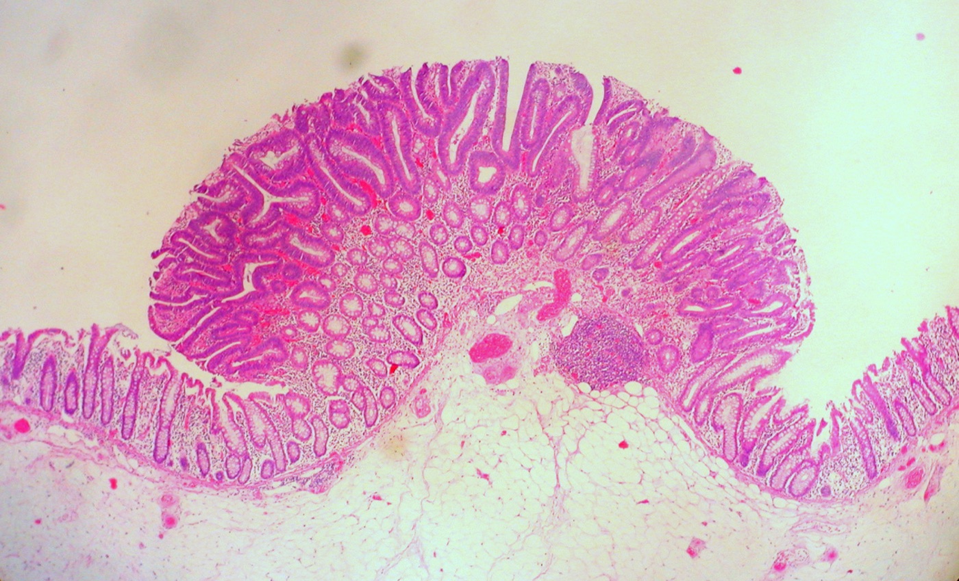
Tubular adenoma of the colon: a small polyp whose crypts are lined by crowded, elongated, hyperchromatic (pencillate) nuclei with mucin depletion, sharply contrasting with the normal flat mucosa on either side; a lymphoid aggregate sits in the stalk at center and submucosal fat lies below. H&E, low power (magnification not stated).Image: Ed Uthman, Wikimedia Commons, CC BY 2.0.
Hereditary Colorectal Cancer, Polyposis Syndromes and Mismatch Repair Testing
Definition. Hereditary colorectal cancer syndromes are germline conditions that raise the risk of colorectal carcinoma and, in most, of other cancers. They comprise Lynch syndrome, the adenomatous polyposes (familial adenomatous polyposis, MUTYH-associated polyposis and rarer recessive and dominant forms) and the hamartomatous polyposes (juvenile polyposis, Peutz–Jeghers syndrome and PTEN hamartoma tumor syndrome) (Syngal et al. 2015). Lynch syndrome is the most common cause of inherited colorectal carcinoma (Syngal et al. 2015). The WHO Classification of Digestive System Tumours, 6th edition (2026), classifies genetic tumour syndromes by mechanism, pathway and gene, in line with the WHO Classification of Genetic Tumour Syndromes (Arends et al. 2026).
Few (usually <10) early-onset adenomas, often proximal
46% (MLH1), 35% (MSH2), 20% (MSH6) and 10% (PMS2) by age 70 years under surveillance
Syngal et al. 2015; Møller et al. 2017
Familial adenomatous polyposis
APC
Autosomal dominant
≥100 adenomas
100% without colectomy
Syngal et al. 2015
Attenuated FAP
APC (5′ end, 3′ end, parts of exon 9)
Autosomal dominant
10–99 adenomas, proximal predominance
69% by age 80 years
Syngal et al. 2015
MUTYH-associated polyposis
MUTYH (biallelic)
Autosomal recessive
Usually 20–99 adenomas; serrated polyps in some
43–100%
Syngal et al. 2015; Boparai et al. 2008
Polymerase proofreading-associated polyposis
POLE, POLD1
Autosomal dominant, high penetrance
Multiple adenomas, early-onset carcinoma
High penetrance; not quantified
Palles et al. 2013
NTHL1-associated polyposis
NTHL1 (biallelic)
Autosomal recessive
Adenomatous polyposis
Carcinoma in at least one member of each family
Weren et al. 2015
Juvenile polyposis syndrome
SMAD4, BMPR1A
Autosomal dominant
Juvenile polyps, mostly colorectal
38–68%
Syngal et al. 2015
Peutz–Jeghers syndrome
STK11
Autosomal dominant
Hamartomatous polyps with arborizing smooth muscle
39%
Syngal et al. 2015
PTEN hamartoma tumor syndrome
PTEN
Autosomal dominant
Mixed polyps; hyperplastic polyps most frequent
9–16%
Syngal et al. 2015; Heald et al. 2010
Serrated polyposis syndrome
Mostly nongermline; RNF43 in about 2%
Mostly nongermline
Multiple serrated polyps, mostly proximal
>50% estimated, probably an overestimate
Syngal et al. 2015; Arends et al. 2026
Lynch syndrome
Genetics and cancer risks in Lynch syndrome
Definition. Lynch syndrome is an autosomal dominant condition caused by a germline pathogenic variant in a DNA mismatch repair (MMR) gene or in EPCAM. It was formerly called hereditary nonpolyposis colorectal cancer (Syngal et al. 2015).
EPCAM deletions. Germline deletions of the 3′ exons of EPCAM (TACSTD1), the gene upstream of MSH2, cause transcriptional read-through into MSH2. The MSH2 promoter in cis is methylated in Ep-CAM-positive tissues, and the tumors are MSH2-deficient (Ligtenberg et al. 2009).
Frequency. In 1066 unselected patients with colorectal adenocarcinoma, 19.5% had MSI and 2.2% had Lynch syndrome. Five of the 23 probands did not meet the Amsterdam criteria or the Bethesda guidelines (Hampel et al. 2005).
Gene-specific risks. In the Prospective Lynch Syndrome Database, cumulative endometrial cancer incidence at 70 years was 34%, 51%, 49% and 24% for MLH1, MSH2, MSH6 and PMS2 carriers (Møller et al. 2017).
Extended cohort. In 6350 carriers, MSH6 variants caused a modest colorectal risk, and no significantly increased cancer risk was shown for PMS2 carriers (Dominguez-Valentin et al. 2020).
Lynch syndrome-related cancers. These include colorectal, endometrial, gastric, ovarian, pancreatic, ureter and renal pelvis, biliary tract, brain (usually glioblastoma) and small intestinal cancers, sebaceous gland adenomas and keratoacanthomas (Syngal et al. 2015).
Accelerated progression. The estimated polyp-to-cancer dwell time is 35 months in Lynch syndrome and 10–15 years in sporadic cancer (Syngal et al. 2015).
Lynch syndrome adenomas. The precursor adenoma is often proximal and occasionally flat. It is likely to show villous features, high-grade dysplasia and many tumor-infiltrating lymphocytes (Syngal et al. 2015).
Turcot syndrome. Turcot syndrome, brain tumors with multiple colorectal adenomas, has two germline causes. APC mutations underlie the medulloblastoma families, and MMR gene defects underlie the glioblastoma families (Hamilton et al. 1995).
Clinical criteria for Lynch syndrome
Amsterdam II criteria. At least three relatives have a Lynch syndrome-associated cancer (colorectal, endometrial, small bowel, ureter or renal pelvis). One is a first-degree relative of the other two. At least two successive generations are affected, and at least one cancer is diagnosed before age 50 years. Familial adenomatous polyposis is excluded, and tumors are verified pathologically (Vasen et al. 1999; Syngal et al. 2015).
Revised Bethesda guidelines. Tumor testing for MSI is indicated for colorectal carcinoma before age 50 years and for synchronous or metachronous Lynch syndrome-related tumors at any age. It is also indicated for carcinoma with MSI-high histology before age 60 years, and for specified family histories (Umar et al. 2004; Syngal et al. 2015).
MSI-high histology. The Bethesda guidelines list tumor-infiltrating lymphocytes, a Crohn-like lymphocytic reaction, mucinous or signet-ring differentiation and a medullary growth pattern (Syngal et al. 2015).
Histologic predictors of MSI. In 528 consecutive carcinomas, more than 2 tumor-infiltrating lymphocytes per high-power field was the best single predictor of MSI. Absence of dirty necrosis, a Crohn-like reaction, right-sided location, well or poor differentiation and any mucinous differentiation were independent predictors (Greenson et al. 2003).
Universal tumor testing. The EGAPP Working Group found sufficient evidence to recommend offering Lynch syndrome testing to all individuals with newly diagnosed colorectal cancer, to reduce morbidity and mortality in relatives (EGAPP Working Group 2009). The ASCP/CAP/AMP/ASCO guideline recommends MMR status testing of colorectal cancers to identify patients at high risk for Lynch syndrome and for prognostic stratification (Sepulveda et al. 2017).
Mismatch repair immunohistochemistry and MSI testing
Principles of MMR testing
Heterodimers. MLH1 pairs with PMS2, and MSH2 pairs with MSH6. Loss of MLH1 or MSH2 causes degradation of PMS2 or MSH6. Loss of PMS2 or MSH6 does not cause loss of its partner (Hall et al. 2010).
MSI definition. The NCI reference panel uses five microsatellites. A tumor is MSI-high when two or more markers are unstable and MSI-low when one is unstable (Boland et al. 1998).
Test performance. MSI testing has a sensitivity of 80–91% for MLH1 or MSH2 carriers and 55–77% for MSH6 or PMS2 carriers, with 90% specificity. MMR immunohistochemistry has 83% sensitivity and 89% specificity (Syngal et al. 2015).
Sporadic mechanism. MMR deficiency occurs in 15–20% of colorectal cancers, and about three-fourths of these are due to MLH1 epigenetic silencing. BRAF p.V600 mutations rarely occur with germline MMR deficiency and are reported in up to three-fourths of tumors with epigenetic silencing (Sepulveda et al. 2017). MLH1 promoter methylation was present in sporadic tumors that lacked MLH1 expression and absent in adjacent normal tissue (Kane et al. 1997).
BRAF in sporadic and hereditary tumors.BRAF mutations were present in 87% of colorectal cancers and cell lines with methylated MLH1 and in 4% of those without methylation. No BRAF mutation was found in 20 MSI cancers from Lynch syndrome patients (Deng et al. 2004).
Guideline statement.BRAF p.V600 testing is performed in MMR-deficient tumors with MLH1 loss. A BRAF mutation strongly favors sporadic pathogenesis, and its absence does not exclude Lynch syndrome. MLH1 promoter methylation testing further refines the risk before germline testing (Sepulveda et al. 2017).
ACG algorithm. Patients with MMR-deficient tumors without BRAF mutation or MLH1 hypermethylation undergo genetic evaluation (Syngal et al. 2015).
Immunohistochemical result
Most likely underlying defect
Next step
Source
Loss of MLH1 and PMS2
Somatic MLH1 promoter hypermethylation or germline MLH1 variant
BRAF V600E or MLH1 promoter methylation testing
Hall et al. 2010; Sepulveda et al. 2017
Loss of MSH2 and MSH6
Germline MSH2 variant or EPCAM 3′ deletion
Germline MSH2 and EPCAM testing
Ligtenberg et al. 2009; Syngal et al. 2015
Isolated loss of MSH6
MSH6 defect
Germline MSH6 testing
Hall et al. 2010; Syngal et al. 2015
Isolated loss of PMS2
PMS2 defect
Germline PMS2 testing
Hall et al. 2010; Syngal et al. 2015
MMR loss without methylation or germline variant
Double somatic MMR mutations in most
Tumor sequencing of MMR genes
Haraldsdottir et al. 2014; Mensenkamp et al. 2014
Loss in tumor and in non-neoplastic cells
Constitutional MMR deficiency
Germline MMR gene testing
Colas et al. 2024
Sources of error in mismatch repair immunohistochemistry
MSH6 loss after neoadjuvant therapy. In 51 colorectal carcinomas resected after neoadjuvant therapy, MLH1, PMS2 and MSH2 staining was preserved in all, and MSH6 staining was decreased in 10 (20%). Nine of the 10 had preserved MSH6 in the pretreatment sample, and all 10 were microsatellite stable. The authors recommend immunohistochemistry on the pretreatment biopsy when MSH6 staining is diminished in a treated tumor, before germline testing for Lynch syndrome (Bao et al. 2010).
MSH6 reduction linked to Ki-67. Reduced MSH6 expression was found in 12 of 50 microsatellite-stable carcinomas after neoadjuvant therapy (loss in 20–100% of tumor cells) and in 8 of 64 untreated microsatellite-stable carcinomas (loss in 10–50%). MSH6 and Ki-67 loss coincided in the same tumor areas. Three carcinomas from carriers of a germline MSH6 mutation showed complete MSH6 loss with positive Ki-67 staining (Kuan et al. 2017).
Heterogeneous (subclonal) MSH6 loss. Heterogeneous MSH6 staining was defined as discrete tumor areas with abrupt loss of staining next to areas with retained staining. It was found in 9 of 1754 colorectal cancers, and none of the 9 patients had a germline MSH6 mutation. All 9 tumors had an MLH1 or PMS2 abnormality: MLH1 methylation in 6, a germline PMS2 mutation in 2 and a germline MLH1 mutation in 1. The authors recommend reporting the pattern as MSH6 staining present, with a comment that it usually indicates a defect in another MMR gene (Chen et al. 2020).
Heterogeneous loss with double-loss areas. In 200 colorectal cancers, 9 (4.5%) showed heterogeneous MMR protein expression. Six of the 9 also had loss of another MMR protein, so that some areas showed loss of two proteins. Both components had a high tumor mutation burden (Berrino et al. 2023).
Lynch-like syndrome and double somatic mutations
Somatic explanation. In 32 patients with MMR-deficient tumors without MLH1 hypermethylation or a germline variant, 22 (69%) had two somatic MMR mutations matching the lost protein. Six (19%) had false-positive screening results (Haraldsdottir et al. 2014).
Second cohort. Two somatic hits explained MMR deficiency in more than 50% of MSI tumors without germline variants or promoter methylation (Mensenkamp et al. 2014).
Histology. Carcinomas with double somatic MMR mutations are histologically indistinguishable from Lynch syndrome carcinomas (Hemminger et al. 2018).
Somatic POLE mutations. Five of six tumors with more than 200 mutations per megabase had somatic POLE mutations (Haraldsdottir et al. 2014).
Constitutional mismatch repair deficiency
Definition. Constitutional mismatch repair deficiency (CMMRD) results from biallelic germline variants in MLH1, MSH2, MSH6 or PMS2. It is a childhood cancer predisposition syndrome with mainly hematologic, brain and intestinal tract tumors (Wimmer et al. 2014).
Immunohistochemical confirmation. Loss of one or more MMR proteins in neoplastic and non-neoplastic cells, including tumor-infiltrating leukocytes or endothelial cells, confirms CMMRD when a positive control stains (Colas et al. 2024).
Adenomatous polyposis syndromes
Familial adenomatous polyposis and attenuated FAP
Gene. Truncating germline mutations of APC cause adenomatous polyposis coli (Groden et al. 1991). The APC locus is on chromosome 5q (Spirio et al. 1993).
Definition. Familial adenomatous polyposis (FAP) is defined by ≥100 synchronous colorectal adenomas with autosomal dominant inheritance. Its prevalence is 1 in 6850 to 1 in 31,250 live births (Syngal et al. 2015).
Attenuated FAP. Patients with 10–99 adenomas have attenuated FAP or oligopolyposis. Adenomas average about 25 and are proximally predominant. Cancer is delayed by 10–20 years compared with classic FAP (Syngal et al. 2015).
Genotype. Attenuated alleles cluster near the 5′ end of APC (Spirio et al. 1993). Mutations at the far 3′ end and in parts of exon 9 also give an attenuated phenotype (Syngal et al. 2015).
Extracolonic manifestations. These include duodenal and ampullary adenomas, desmoid tumors, papillary thyroid cancer and congenital hypertrophy of the retinal pigment epithelium (Syngal et al. 2015). Duodenal adenomas occur in more than half of patients, and the lifetime risk of duodenal cancer is 3–5%. Gastric polyps, mostly fundic gland polyps, occur in 23–100% (Syngal et al. 2015).
GAPPS. Gastric adenocarcinoma and proximal polyposis of the stomach is recognized as an FAP variant with a unique phenotype (Nagtegaal et al. 2020).
APC promoter 1B in GAPPS. GAPPS is an autosomal dominant syndrome with a significant risk of gastric adenocarcinoma and no significant risk of colorectal adenocarcinoma. Point mutations in APC promoter 1B co-segregated with disease in all six families studied. The mutations reduced binding of the YY1 transcription factor and impaired promoter 1B activity. Promoter 1A is methylated in normal stomach and in fundic gland polyps, which suggests that gastric mucosa relies on 1B transcripts and that colonic cells are usually protected by the 1A isoform. Similar promoter 1B mutations occur in rare FAP families (Li et al. 2016).
Referral threshold. A personal history of more than 10 cumulative colorectal adenomas prompts genetic evaluation for the adenomatous polyposis syndromes (Syngal et al. 2015).
MUTYH-associated polyposis
Mechanism. Biallelic MUTYH (MYH) variants impair base excision repair of 8-oxoguanine mispairs. Tumors show an excess of somatic G:C→T:A transversions in APC (Al-Tassan et al. 2002).
Phenotype. About one-third of patients with more than 15 adenomas had biallelic MUTYH variants. No patient with biallelic variants had more than 1000 adenomas (Sieber et al. 2003). Biallelic variants occur in 16–40% of patients with 15–99 adenomas without FAP (Syngal et al. 2015).
Serrated polyps. Hyperplastic polyps or SSLs were present in 47% of patients with MUTYH-associated polyposis, and 18% met criteria for hyperplastic polyposis. KRAS mutations in these polyps were G:C→T:A transversions in 94% (Boparai et al. 2008).
Polymerase proofreading-associated and NTHL1-associated polyposis
POLE and POLD1. Heterozygous germline POLE p.Leu424Val and POLD1 p.Ser478Asn variants affect the proofreading (exonuclease) domains of DNA polymerases ε and δ. They predispose with high penetrance to multiple adenomas and colorectal carcinoma. Tumors are microsatellite stable with excess base substitutions, and POLD1 variants also predispose to endometrial cancer (Palles et al. 2013).
NTHL1. A homozygous germline nonsense variant in the base excision repair gene NTHL1 caused recessive adenomatous polyposis and colorectal carcinoma in three families. Tumors showed a non-hypermutated profile enriched for C>T transitions. All three affected women developed endometrial malignancy or premalignancy (Weren et al. 2015).
Hamartomatous polyposis syndromes
Juvenile polyposis syndrome
Clinical criteria. At least five juvenile polyps in the colorectum, juvenile polyps elsewhere in the gastrointestinal tract, or any number of juvenile polyps with a family history of juvenile polyposis (Syngal et al. 2015). Jass and colleagues' 1988 working definition required more than five colorectal juvenile polyps (Jass et al. 1988).
Genes. Germline SMAD4 (Howe et al. 1998) and BMPR1A (Howe et al. 2001) variants cause juvenile polyposis. Up to 60% of clinically defined patients carry a variant in one of these genes (Syngal et al. 2015).
Dysplasia by polyp shape. Of 169 multilobulated or villous juvenile polyps, 46.7% contained dysplasia. Only 9.0% of typical spherical juvenile polyps were dysplastic (Jass et al. 1988).
Cancer risk. Colorectal cancer risk is 17–22% by age 35 years and approaches 68% by age 60 years. Gastric cancer risk is 30% with SMAD4 variants (Syngal et al. 2015).
SMAD4 and hereditary hemorrhagic telangiectasia. All patients with advanced upper gastrointestinal disease, and all genotyped patients with telangiectasia, had SMAD4 variants (Latchford et al. 2012). Evaluation for hereditary hemorrhagic telangiectasia is recommended in SMAD4 carriers (Syngal et al. 2015).
Hereditary mixed polyposis syndrome. A 40-kb duplication upstream of GREM1 causes ectopic epithelial GREM1 expression and reduced bone morphogenetic protein signaling (Jaeger et al. 2012). Affected patients have mixed juvenile–adenomatous, adenomatous, hyperplastic and serrated polyps (Syngal et al. 2015).
Peutz–Jeghers syndrome
Gene. Germline STK11 (LKB1) mutations on chromosome 19p13.3 cause Peutz–Jeghers syndrome (Hemminki et al. 1998; Jenne et al. 1998).
Clinical features. Mucocutaneous melanin pigmentation is present in more than 95% of patients, most often perioral and buccal. Gastrointestinal polyps occur in 88–100% (Syngal et al. 2015).
Polyp histology. Peutz–Jeghers polyps are nondysplastic and covered by the normal epithelium of their segment. Branching fronds contain extensions of the muscularis mucosae in an arborizing pattern (Syngal et al. 2015). The 6th edition describes these polyps in the jejunum and ileum chapter (Arends et al. 2026).
Cancer risk. In 1644 patients, the lifetime risk of any cancer was 37–93%. Colorectal cancer was the most common, followed by breast, small bowel, gastric and pancreatic cancer (van Lier et al. 2010). Early endoscopy mainly removes polyps that could cause intussusception or obstruction (Beggs et al. 2010).
PTEN hamartoma tumor syndrome
Gene. Germline PTEN mutations on chromosome 10q23 cause Cowden disease (Liaw et al. 1997).
Gastrointestinal phenotype. Of 69 PTEN carriers who had endoscopy, 93% had polyps, and half of these had hyperplastic polyps. Nine (13%) had colorectal cancer before age 50 years (Heald et al. 2010).
Polyp types. Several polyp types usually occur in one patient. Diffuse esophageal glycogenic acanthosis with colonic polyposis has been suggested as pathognomonic (Syngal et al. 2015).
Revised criteria. Colon cancer and esophageal glycogenic acanthosis were added to the diagnostic criteria (Pilarski et al. 2013).
Cronkhite–Canada syndrome
Definition. Cronkhite–Canada syndrome is a noninherited gastrointestinal polyposis sparing the esophagus, with ectodermal abnormalities and an often fatal course (Slavik and Montgomery 2014).
Features. In 14 patients, all had upper and lower gastrointestinal polyps, and most had alopecia, hyperpigmentation, onychodystrophy and protein-losing enteropathy. Adenomatous polyps occurred in 71% and colorectal cancer in 14% (Sweetser et al. 2012).
IgG4. IgG4-positive plasma cells (>5 per high-power field) were present in 52% of Cronkhite–Canada polyps and 12% of juvenile polyposis polyps. Corticosteroids induced remission in 91% (Sweetser et al. 2012).
Upper gastrointestinal mucosa. Gastric biopsies showed edema and eosinophil-rich inflammation, and duodenal biopsies showed villous atrophy (Bettington et al. 2014).
Serrated polyposis syndrome
Classification by edition. The 6th edition describes serrated polyposis in the colorectal chapter (Arends et al. 2026). The 5th edition placed it in the genetic tumour syndromes chapter (Arends et al. 2026).
Genetics. Germline RNF43 variants occur in about 2% of patients, and no other high-penetrance gene has been found (Quintana et al. 2018; Arends et al. 2026). Serrated polyposis is now viewed as a largely sporadic syndrome (Nagtegaal et al. 2026). Nonsense RNF43 variants were found in 2 of 20 patients with multiple SSLs (Gala et al. 2014). Not all carriers of likely pathogenic RNF43 variants meet the criteria (Brinch et al. 2024).
Testing yield. In 139 patients who had multigene panel testing, 2.9% had a pathogenic germline variant. Three had Lynch syndrome and one had an RNF43 variant (Upadhye et al. 2025).
Overlap. Serrated polyposis or multiple serrated polyps can occur within PTEN hamartoma tumor syndrome and hereditary mixed polyposis syndrome (Arends et al. 2026).
Carcinomas in serrated polyposis. About half of the carcinomas have a BRAF mutation, 40% are MLH1 deficient and fewer than 5% have a KRAS mutation (Nagtegaal et al. 2026).
Diagnostic criteria. The WHO 2019 criteria replaced the 2010 criteria (Dekker et al. 2020). The 6th-edition summary of changes does not restate them (Arends et al. 2026). The 2019 criteria apply to the "cumulative lifetime count" of serrated polyps: "(i) ≥5 serrated lesions/polyps proximal to the rectum all being ≥5 mm in size, with ≥2 being ≥10 mm in size or (ii) >20 serrated lesions/polyps of any size distributed throughout the large bowel, with ≥5 being proximal to the rectum" (Upadhye et al. 2025).
Criteria set
Criterion 1
Criterion 2
Criterion 3
Source
WHO 2019 (5th edition)
≥5 serrated lesions or polyps proximal to the rectum, all ≥5 mm, with ≥2 ≥10 mm
>20 serrated lesions or polyps of any size throughout the large bowel, with ≥5 proximal to the rectum
None (counts are cumulative over a lifetime)
Upadhye et al. 2025; Dekker et al. 2020
WHO 2010 (previous)
≥5 serrated polyps proximal to the sigmoid colon, with ≥2 >10 mm
Any serrated polyps proximal to the sigmoid colon in a person with a first-degree relative with serrated polyposis
>20 serrated polyps of any size throughout the colon
Syngal et al. 2015
Differential diagnosis of hereditary colorectal syndromes
Presentation
Main differential
Distinguishing finding
Source
MLH1 and PMS2 loss
Lynch syndrome versus sporadic MSI carcinoma
BRAF V600E or MLH1 promoter methylation favors sporadic origin
Sepulveda et al. 2017; Deng et al. 2004
MMR loss, no germline variant, no methylation
Lynch syndrome versus double somatic mutations
Two somatic MMR mutations on tumor sequencing
Haraldsdottir et al. 2014
15–99 adenomas, recessive pattern
MUTYH-associated polyposis versus attenuated FAP
Biallelic MUTYH variants; G:C→T:A transversions
Sieber et al. 2003; Al-Tassan et al. 2002
Multiple adenomas, dominant pattern, MSS tumors
Polymerase proofreading-associated polyposis
POLE or POLD1 exonuclease-domain variants
Palles et al. 2013
Recessive polyposis with endometrial neoplasia
NTHL1-associated polyposis
Homozygous NTHL1 variant; C>T transitions
Weren et al. 2015
Juvenile-type polyps with alopecia and nail changes
Cronkhite–Canada syndrome versus juvenile polyposis
Ectodermal changes; IgG4-positive plasma cells
Sweetser et al. 2012
Mixed hamartomatous and hyperplastic polyps
PTEN hamartoma tumor syndrome
Glycogenic acanthosis; PTEN variant
Heald et al. 2010; Syngal et al. 2015
hereditary colorectal cancer syndrome lookup
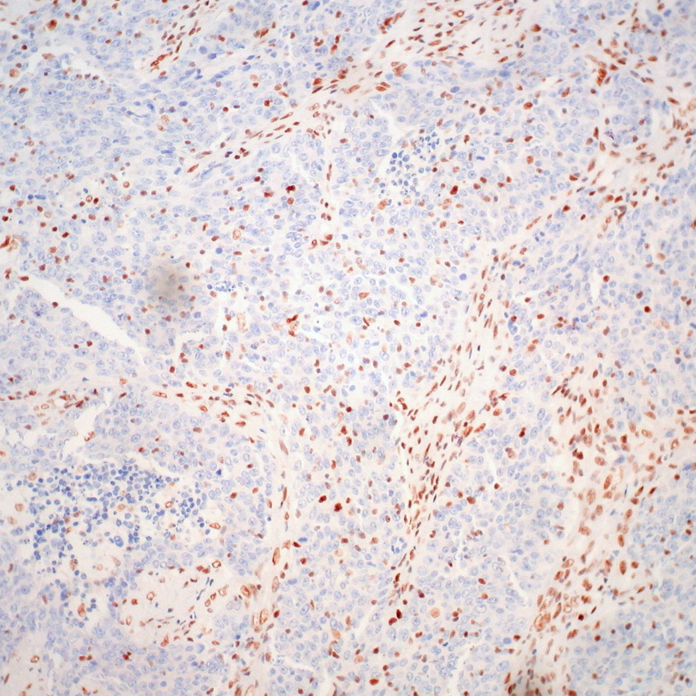
Loss of MLH1 in a medullary carcinoma of the colon: the sheets of tumor cells have no nuclear staining, while stromal cells and lymphocytes between the sheets keep strong nuclear MLH1, which serves as the internal positive control. MLH1 immunostain, medium power (magnification not stated).Image: Ed Uthman, Wikimedia Commons, CC BY 2.0.
Colorectal Adenocarcinoma: Histologic Subtypes, Grade, Tumor Budding and the Malignant Polyp
Definition. Colorectal adenocarcinoma is a malignant glandular epithelial tumor of the colon and rectum. Early invasive carcinoma extends through the muscularis mucosae into the submucosa (Kikuchi et al. 1995). The WHO Classification of Digestive System Tumours, 6th edition (2026), adds colorectal intramucosal carcinoma, low-grade tubuloglandular adenocarcinoma and lymphoglandular complex-like adenocarcinoma. It redefines high-grade carcinoma by gland formation and renames carcinoma with sarcomatoid components as carcinoma with mesenchymal differentiation (Arends et al. 2026).
Lymphoglandular complexes with desmoplasia and necrosis
Screening-detected; dMMR in 22% of one series
Low stage; nodal metastasis in 3 of 8 colectomies
Arends et al. 2026; Yilmaz et al. 2024
Mucinous and signet-ring cell carcinoma
Mucinous adenocarcinoma. Mucinous adenocarcinoma requires more than 50% of the tumor to consist of pools of extracellular mucin containing malignant glands or single tumor cells. Tumors with less than 50% are described as having a mucinous component (ICCR polypectomy 2020). Greenson and colleagues used the same >50% category (Greenson et al. 2003). A meta-analysis of 222,256 patients found mucinous carcinoma more often proximal and less frequent in men. Mucinous differentiation increased the hazard of death by 2–8% after correction for stage (Verhulst et al. 2012).
Signet-ring cell carcinoma. Signet-ring cell carcinoma requires more than 50% signet-ring cells. Tumors with less than 50% are described as having a signet-ring cell component (ICCR polypectomy 2020). CAP protocol 4.4.0.1 lists poorly cohesive carcinoma and signet-ring cell carcinoma as separate types (CAP 4.4.0.1). In 196,757 Dutch patients, signet-ring cell carcinoma formed 1.0% of colorectal carcinomas. It presented at stage III or IV in 75.2%. Five-year relative survival was 30.8% in the colon and 19.5% in the rectum. Adjuvant chemotherapy benefit was comparable to that in conventional adenocarcinoma (Hugen et al. 2015).
Metastatic pattern. In 5817 autopsies, conventional adenocarcinoma spread predominantly to the liver. Mucinous and signet-ring cell carcinomas more often spread to the peritoneum and to multiple sites (Hugen et al. 2014).
Medullary carcinoma
Criteria. In the 6th edition, more than 50% of the tumor consists of solid sheets of cells with vesicular nuclei, prominent nucleoli and abundant eosinophilic cytoplasm, with prominent infiltration by lymphocytes and neutrophils (Nagtegaal et al. 2026). Lanza and colleagues used solid growth in at least 70% of the tumor with little nuclear pleomorphism as a study criterion in 1265 carcinomas (Lanza et al. 1999).
Clinical and molecular features. Medullary carcinomas were proximal, large and expansile with a conspicuous peritumoral lymphocytic infiltrate and Crohn-like reaction. Of those tested, 90% showed widespread MSI and 84% were diploid. None of 33 patients with resectable stage II or III tumors had recurrence (Lanza et al. 1999).
Immunophenotype. Compared with poorly differentiated carcinomas, medullary carcinomas were less often CDX2 positive (19% versus 55%) and MLH1 positive (21% versus 60%). Calretinin was positive in 73% of medullary carcinomas and 12% of poorly differentiated carcinomas (Winn et al. 2009).
Micropapillary, serrated, adenoma-like and adenosquamous carcinoma
Micropapillary adenocarcinoma. Small clusters of tumor cells lie in stromal spaces that mimic vascular channels, in at least 5% of the tumor (ICCR polypectomy 2020; Nagtegaal et al. 2026). Of 178 colorectal carcinomas, 19.1% had a micropapillary component of 5–60%. Nodal metastasis was present in 73.5% with the component and 42.4% without it. The component was an independent predictor of nodal metastasis (Haupt et al. 2007).
Serrated adenocarcinoma. Serrated adenocarcinoma forms about 7.5% of colorectal carcinomas and up to 17.5% of the most proximal carcinomas. BRAF mutation and CpG island methylation are early events. MSI-high occurs in 16% and MSI-low in 29% (Mäkinen 2007).
Serrated adenocarcinoma cohort. Serrated adenocarcinomas formed 9.1% of 927 carcinomas, and 51.7% had residual serrated adenoma. Five-year survival was less favorable than in matched conventional carcinomas (García-Solano et al. 2010).
Adenoma-like adenocarcinoma. At least 50% of the invasive tumor has an adenoma-like appearance with villous architecture, low-grade cytology, a pushing growth pattern and minimal desmoplasia (ICCR polypectomy 2020; 5th edition). The 6th-edition appraisal gives >50% of the invasive areas (Nagtegaal et al. 2026). Adenoma-like areas show villiform architecture, rare foci of high-grade atypia and absent or limited desmoplasia. On average they formed about 50% of the tumor. Of 35 tumors, 43% had been diagnosed as adenoma on biopsy, 20% had nodal metastases and 58% had KRAS mutations. Survival was better than in conventional well-differentiated carcinoma (Gonzalez et al. 2016).
Adenosquamous carcinoma. Adenosquamous carcinoma formed 0.09% of 111,263 California registry cases. It was more often metastatic at surgery (36.56% versus 13.92%) and poorly differentiated (65.96% versus 19.74%), with shorter median survival (Masoomi et al. 2012).
New colorectal entities in the WHO 6th edition
Intramucosal carcinoma. Intramucosal adenocarcinoma shows unequivocal infiltration restricted to the lamina propria. It is diagnosed only in polypectomy and resection specimens, and only with overtly poor differentiation, poorly differentiated clusters, signet-ring cells or tumor budding. It forms less than 0.1% of colorectal carcinomas, correlates strongly with hereditary syndromes and carries negligible nodal risk (Arends et al. 2026).
Previous rule for intramucosal carcinoma. The 5th edition included carcinoma in situ within high-grade dysplasia (Nagtegaal et al. 2020). Intramucosal carcinoma was grouped with high-grade adenomas because of its very low nodal risk (Vink-Börger et al. 2025).
Staging of intramucosal carcinoma. In AJCC/CAP staging, pTis includes intramucosal carcinoma: tumor involving the lamina propria or muscularis mucosae without extension through it. A synoptic report is required only for invasive tumors (CAP 4.4.0.1).
Evidence for intramucosal carcinoma. In 14 strictly defined cases, the estimated incidence in screening was 0.01%. Seven (50%) were MMR deficient, and six of these were associated with Lynch syndrome (Vink-Börger et al. 2025). In a Japanese consensus study, stromal infiltration by single cells or small clusters was the best objective criterion of lamina propria invasion. Concordance for invasion was poor to fair (Kojima et al. 2017).
Low-grade tubuloglandular adenocarcinoma. This rare subtype is typically seen in inflammatory bowel disease. It may arise from low-grade conventional dysplasia or non-conventional colitis-associated dysplasia. CK7 is sometimes expressed and somatic IDH1 mutations are common (Arends et al. 2026).
Tubuloglandular evidence.IDH1 R132C mutations were found in 13% of inflammatory bowel disease-associated adenocarcinomas and in none of the sporadic ones. All IDH1-mutated tumors had tubuloglandular histology (Hartman et al. 2014). Of these carcinomas, 88% were flat or invisible, and crypt cell dysplasia was the most common associated non-conventional dysplasia (Akarca et al. 2023).
Lymphoglandular complex-like adenocarcinoma. The tumor formerly called dome carcinoma or gut-associated lymphoid tissue carcinoma is now lymphoglandular complex-like adenocarcinoma. It usually presents at low stage and is more often found in screening programs (Arends et al. 2026).
Lymphoglandular evidence. In 20 cases, all showed haphazard gland distribution with angulation, fusion or solid nests. Desmoplasia was present in 50%. Most arose with a surface tubular adenoma, and no patient had recurrence (Yilmaz et al. 2024). Adenomas involving submucosal lymphoglandular complexes are the main mimic; their features are given in Colorectal Adenomas, Serrated Polyps and Other Colorectal Polyps (Lee et al. 2018).
Undifferentiated carcinoma and carcinoma with mesenchymal differentiation
WHO 6th edition. Tumors without definite squamous, glandular or neuroendocrine differentiation are undifferentiated carcinomas. The 5th-edition term carcinoma with sarcomatoid components is replaced by carcinoma with mesenchymal differentiation. A SWI/SNF-deficient subset with alterations of ARID1A, SMARCA2, SMARCA4 or SMARCB1 is added (Arends et al. 2026). CAP protocol 4.4.0.1 still lists carcinoma with sarcomatoid component (CAP 4.4.0.1).
SWI/SNF deficiency. In 13 undifferentiated gastrointestinal carcinomas with variable rhabdoid features, 92% lost at least one SWI/SNF subunit (Agaimy et al. 2016). In 30 cases, 96.7% lost at least one subunit, 58.6% had negative or decreased pancytokeratin, and SMARCA4 loss predicted the worst survival (Chang et al. 2022).
Histologic grade of colorectal adenocarcinoma
WHO 6th-edition rule. Grade is based on gland formation in the least differentiated component. A tumor is high grade when at least one high-power field lacks gland formation, and low grade otherwise (Arends et al. 2026).
Exclusions from grading. Tumor budding and poorly differentiated clusters at the invasive front are not used for grading and are reported separately. Cribriform architecture counts as glandular differentiation (Arends et al. 2026).
Prognostic value. The 6th-edition system offers the most prognostic value, especially in microsatellite-stable tumors (Arends et al. 2026).
Previous rule (5th edition). Grading was two-tiered and based on gland formation: low grade (formerly well to moderately differentiated) and high grade (formerly poorly differentiated). It used the least differentiated component and excluded tumor buds and poorly differentiated clusters. Only adenocarcinoma NOS and mucinous adenocarcinoma were graded (ICCR polypectomy 2020).
CAP reporting. CAP protocol 4.4.0.1 uses four tiers: G1 well differentiated (>95% gland formation), G2 moderately differentiated (50–95%), G3 poorly differentiated (<50%) and G4 undifferentiated (no gland formation or mucin, and no squamous or neuroendocrine differentiation). The scheme applies to adenocarcinoma NOS and not to histologic variants. Medullary carcinoma behaves as a low-grade tumor even when it appears poorly differentiated (CAP 4.4.0.1).
Evidence on differentiation. Poor differentiation predicted nodal metastasis in pT1 carcinoma (Beaton et al. 2013; Bosch et al. 2013).
Poorly differentiated clusters. Clusters of ≥5 cancer cells without a gland-like structure were counted in the field with most clusters under a ×20 objective. Fewer than 5, 5–9 and ≥10 clusters defined G1, G2 and G3, with 5-year disease-free survival of 96%, 85% and 59% (Ueno et al. 2012). Interobserver agreement was higher than for conventional grading (weighted κ 0.66 versus 0.52) (Ueno et al. 2012).
Tumor budding in colorectal carcinoma (ITBCC 2016)
Definition. A tumor bud is a single tumor cell or a cluster of four or fewer tumor cells (Lugli et al. 2017). Poorly differentiated clusters contain ≥5 cells, which separates them from buds (ICCR polypectomy 2020).
Method. Budding is counted on H&E in one hotspot at the invasive front, in a field of 0.785 mm². A three-tier system is reported with the bud count. Tumor budding and tumor grade are separate parameters (Lugli et al. 2017).
Hotspot selection. The hotspot is chosen after review of all slides with invasive tumor. Keratin immunohistochemistry may be used when inflammation obscures the front, but scoring is done on H&E (CAP 4.4.0.1).
Applicability. CAP requires a budding score for conventional adenocarcinoma (well or moderately differentiated). Budding is not applicable to micropapillary, signet-ring cell or mucinous carcinoma, or to poorly differentiated or undifferentiated carcinoma, unless a well or moderately differentiated conventional component is present (CAP 4.4.0.1). The CAP polypectomy case summary, which includes a budding score, is recommended for clinical care and is not required for accreditation (CAP Bx 4.3.0.0).
Prognostic role. Budding independently predicts nodal metastasis in pT1 carcinoma and survival in stage II carcinoma (Lugli et al. 2017). In 43 studies of 17,831 patients, high-grade budding independently predicted worse disease-free, overall and cancer-specific survival in stage II and III disease (Shu et al. 2026).
Validation. In 379 resected stage I–IV carcinomas, BD3 was associated with higher pT and pN categories and poorer overall survival. BD3 predicted poorer disease-free survival in stage II (Dawson et al. 2019).
Stain choice. Pan-cytokeratin staining detected three to four times more buds than H&E and gave better interobserver agreement (Koelzer et al. 2015).
ITBCC budding grade
Buds per hotspot (0.785 mm²)
Source
BD1 (low)
0–4
Lugli et al. 2017; CAP 4.4.0.1
BD2 (intermediate)
5–9
Lugli et al. 2017; CAP 4.4.0.1
BD3 (high)
≥10
Lugli et al. 2017; CAP 4.4.0.1
BD0 versus BD1. In T1 and T2 carcinomas, nodal metastasis did not differ between tumors with no buds and tumors with 1–4 buds (Lee et al. 2025).
Immunohistochemistry of colorectal adenocarcinoma
Marker
Result in colorectal carcinoma
Limitation
Source
CDX2
Uniform expression in all but one colorectal and duodenal tumor
Positive in mucinous ovarian (64%) and urinary bladder (100%) adenocarcinomas
Werling et al. 2003
CDX2 (gastrointestinal comparison)
Positive in 97% of colorectal carcinomas
Positive in 61% of gastric and 16% of pancreatic carcinomas
Bayrak et al. 2012
CK20
Positive in 81.1%
Less often positive in high-grade (47.6%) and proximal tumors
Bayrak et al. 2011
CK7
Positive in 17.3%
More often positive in node-positive tumors (25.3%)
Bayrak et al. 2011
CK7−/CK20+
64–65.8% of colorectal carcinomas
Specificity 96.7% against gastric and pancreatic carcinomas
Bayrak et al. 2011; Bayrak et al. 2012
SATB2
Positive in 85%; SATB2 or CK20 positive in 97%
Restricted to lower gastrointestinal glandular epithelium among epithelia
Magnusson et al. 2011
SATB2 with CK7 and CK20
Sensitivity 93% and specificity 77% for SATB2 alone
Specificity 100% in combination with CK7 and CK20
Dragomir et al. 2014
Calretinin
Positive in 73% of medullary carcinomas
Positive in 12% of poorly differentiated carcinomas
Carcinoma of unknown primary. The colon-like subtype, defined as CDX2 and CK20 positive and CK7 negative or with a colorectal molecular profile, is the only favorable gastrointestinal-like subtype (Arends et al. 2026).
The malignant polyp (pT1 colorectal carcinoma)
Levels of invasion in malignant polyps
System
Level
Definition
Source
Haggitt (pedunculated)
0
Carcinoma confined to the mucosa
Haggitt et al. 1985
Haggitt
1
Invasion of the submucosa of the head
Haggitt et al. 1985
Haggitt
2
Invasion to the neck
Haggitt et al. 1985
Haggitt
3
Invasion of the stalk
Haggitt et al. 1985
Haggitt
4
Invasion of the submucosa of the underlying bowel wall
Haggitt et al. 1985
Kikuchi (sessile)
sm1
Slight invasion below the muscularis mucosae (200–300 µm)
Kikuchi et al. 1995
Kikuchi
sm2
Intermediate submucosal invasion
Kikuchi et al. 1995
Kikuchi
sm3
Invasion to the inner surface of the muscularis propria
Kikuchi et al. 1995
Haggitt outcome data. None of 65 patients with carcinoma confined to the mucosa had an adverse outcome. Level 4 invasion and rectal location were the only significant adverse factors, and 7 of 28 level 4 lesions had an adverse outcome (Haggitt et al. 1985).
Sessile polyps in the Haggitt system. In a sessile polyp, any invasion of the submucosa is Haggitt level 4 (Kuo et al. 2020).
Kikuchi outcome data. None of the 17 patients with local recurrence or nodal metastasis had sm1 invasion (Kikuchi et al. 1995).
Depth in micrometers. In 865 submucosal carcinomas, nodal metastasis was 0% for nonpedunculated tumors with depth <1000 µm. For pedunculated tumors, it was 0% for head invasion and for stalk invasion <3000 µm without lymphatic invasion. Depth in pedunculated tumors was measured from the Haggitt level 2 line (Kitajima et al. 2004).
High-risk features of a malignant polyp
Meta-analyses. Lymphatic invasion (relative risk 5.2), submucosal invasion ≥1 mm (5.2), budding (5.1) and poor differentiation (4.8) were the strongest predictors of nodal metastasis in pT1 carcinoma (Bosch et al. 2013). A second meta-analysis found nodal metastasis in up to 17% of pT1 carcinomas, with tumor budding showing an odds ratio of 7.74 (Beaton et al. 2013).
Combined risk factors. Unfavorable grade, definite vascular invasion and budding together discriminated nodal risk. Nodal involvement was 0.7% with no risk factor, 20.7% with one and 36.4% with several (Ueno et al. 2004).
Resection margin. Unfavorable histology comprised tumor at or within 1.0 mm of the margin, grade III carcinoma, or lymphatic or venous invasion. Adverse outcome occurred in 19.7% with unfavorable histology and 0% without it (Cooper et al. 1995).
JSCCR 2019 criteria. Surgical resection is recommended for a positive vertical margin. Intestinal resection with node dissection is recommended for submucosal invasion ≥1000 µm, lymphovascular invasion, poorly differentiated, signet-ring cell or mucinous carcinoma, or BD2/BD3 budding at the deepest invasion (Hashiguchi et al. 2020).
Depth alone. The nodal metastasis rate with submucosal invasion ≥1000 µm was 12.5%. About 90% of patients with depth ≥1000 µm had no nodal metastasis, so depth is weighed with the other risk factors (Hashiguchi et al. 2020).
CAP reporting of malignant polyps. A malignant polyp is an adenoma with invasive adenocarcinoma extending through the muscularis mucosae into the submucosa. High-grade dysplasia and intramucosal carcinoma are excluded. Histologic type and grade, margin status, lymphatic or venous invasion, tumor budding and depth of invasion guide management. High-grade carcinoma, tumor ≤1 mm from the margin, lymphatic or venous invasion, high-grade budding and deep submucosal invasion raise the risk. Micropapillary and signet-ring cell types count as high grade (CAP Bx 4.3.0.0).
CAP depth categories. Depth is recorded as <1 mm, ≥1 mm and <2 mm, >2 mm or the exact depth. Actual measurement is the recommended method, because Haggitt and Kikuchi levels apply to only a small subset of malignant polyps (CAP Bx 4.3.0.0). The case summary also records polyp configuration (pedunculated with stalk length, or sessile) and the type of precursor polyp (CAP Bx 4.3.0.0).
Measurement method. Depth is measured from the muscularis mucosae, or from the polyp surface when the carcinoma is ulcerated or has overgrown the precursor. An invasive width of ≥4 mm is associated with nodal metastasis. Most studies regard a deep-margin clearance of <1 mm as positive (ICCR polypectomy 2020).
Carcinoma ≤1 mm from the margin; positive vertical margin
Cooper et al. 1995; Hashiguchi et al. 2020
Grade
Well or moderately differentiated
Poorly differentiated, signet-ring, micropapillary or mucinous
Bosch et al. 2013; Hashiguchi et al. 2020; CAP Bx 4.3.0.0
Lymphovascular invasion
Absent
Present
Bosch et al. 2013; Cooper et al. 1995
Tumor budding
BD1
BD2 or BD3
Hashiguchi et al. 2020
Depth, nonpedunculated
<1000 µm
≥1000 µm
Kitajima et al. 2004; Hashiguchi et al. 2020
Level, pedunculated
Haggitt 1–3
Haggitt 4
Haggitt et al. 1985
Differential diagnosis of colorectal adenocarcinoma subtypes
Entity
Main mimic
Distinguishing finding
Source
Medullary carcinoma
Poorly differentiated adenocarcinoma
Calretinin positive, CDX2 and MLH1 often lost, lymphocytic infiltrate
Winn et al. 2009; Lanza et al. 1999
Adenoma-like adenocarcinoma
Villous adenoma on biopsy
Invasion of the wall on resection; villiform glands with limited desmoplasia
Gonzalez et al. 2016
Lymphoglandular complex-like adenocarcinoma
Adenoma in a lymphoglandular complex
Angulated or fused glands, solid nests, desmoplasia
Yilmaz et al. 2024; Lee et al. 2018
Intramucosal carcinoma
High-grade dysplasia
Single cells or small clusters infiltrating lamina propria
Kojima et al. 2017; Arends et al. 2026
Micropapillary adenocarcinoma
Lymphovascular invasion
Micropapillary clusters within the tumor; component independently predicts nodal metastasis
Haupt et al. 2007
Undifferentiated SWI/SNF-deficient carcinoma
Poorly differentiated adenocarcinoma
Rhabdoid cells, weak keratin, loss of SMARCA4, SMARCA2, SMARCB1 or ARID1A
Agaimy et al. 2016; Chang et al. 2022
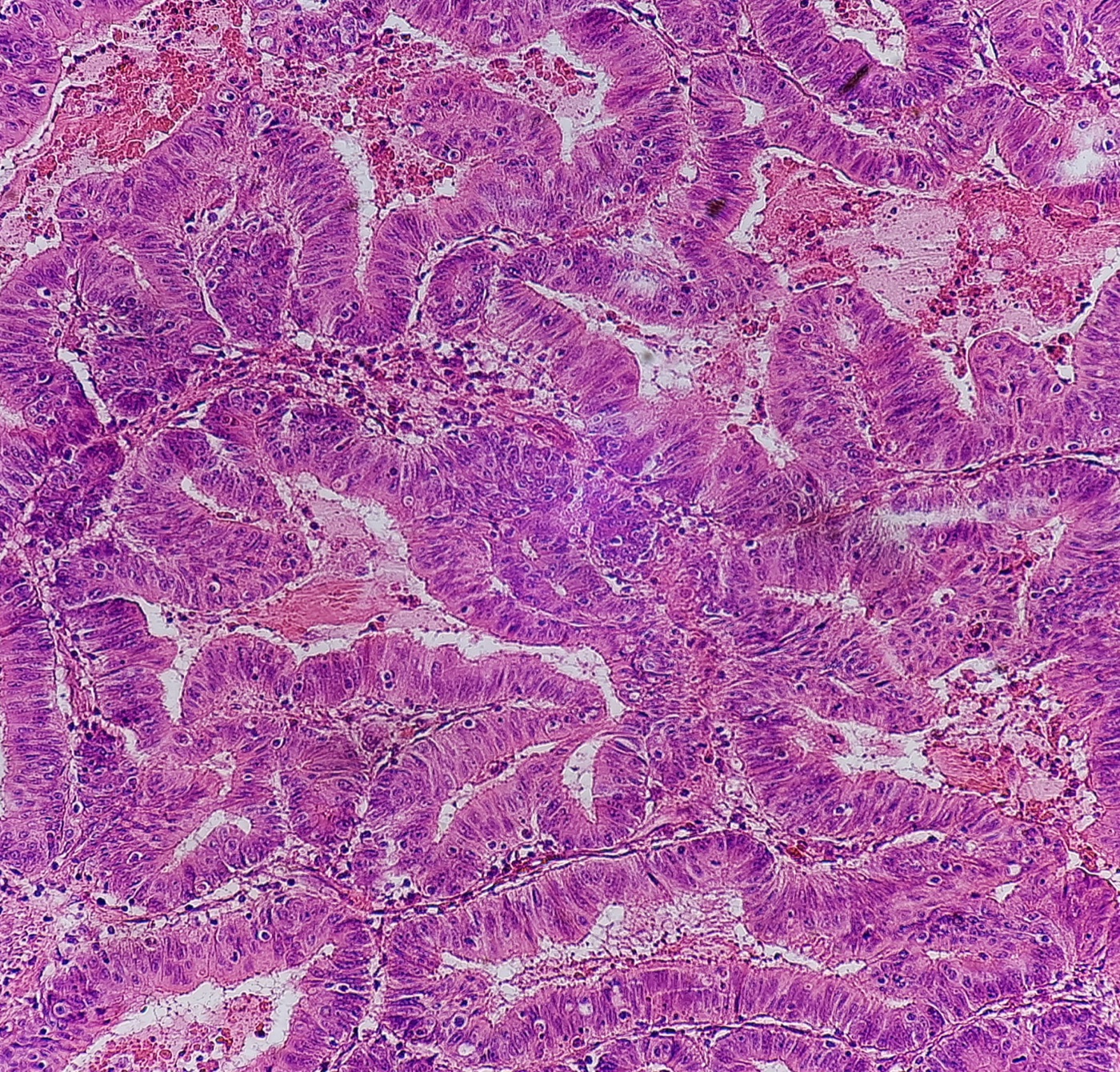
Colorectal adenocarcinoma, not otherwise specified: cribriform and complex glands lined by tall columnar cells with elongated, stratified, hyperchromatic nuclei; several lumina contain eosinophilic necrotic debris with nuclear dust ('dirty necrosis'), best seen at upper right and lower right. H&E, medium power (magnification not stated).Image: Mikael Häggström, M.D., Wikimedia Commons, CC0.
Colorectal Cancer Staging, CAP Reporting, Biomarkers and Neoadjuvant Response
Definition. Pathologic staging of colorectal carcinoma assigns the primary tumor (pT), regional lymph node (pN) and distant metastasis (pM) categories of the American Joint Committee on Cancer (AJCC) 8th edition from the resection specimen, using the College of American Pathologists (CAP) colon and rectum protocol (CAP 2025). The same report records margins, lymphovascular and perineural invasion, tumor budding, tumor deposits and, after neoadjuvant therapy, tumor regression. Predictive biomarkers are reported in a separate CAP biomarker template (CAP 2021).
Standards that govern colorectal cancer staging and reporting in 2026
Standard
Current version
Content relevant to the pathology report
Source
AJCC TNM staging, colon and rectum
8th edition
pT, pN (including N1c) and pM categories
CAP 2025; AJCC 2026
CAP colon and rectum resection protocol
4.4.0.1 (September 2025; required March 2026)
Synoptic core and conditional elements
CAP 2025
CAP colon and rectum biomarker template
1.3.0.0 (June 2021; not required for accreditation)
MMR, MSI, KRAS, NRAS, BRAF, HER2 results and methods
CAP 2021
WHO Classification of Digestive System Tumours
6th edition (2026)
Histologic type and two-tier grade
Arends et al. 2026
ITBCC tumor budding consensus
2016
Hotspot count and three-tier budding score
Lugli et al. 2017
ASCP/CAP/AMP/ASCO molecular biomarker guideline
2017
Extended RAS, BRAF V600 and MMR testing
Sepulveda et al. 2017
CAP/AMP MMR and MSI guideline for immune checkpoint inhibitors
2022
Choice of assay for MMR deficiency
Bartley et al. 2022
AJCC version 9 for colon and rectum. The AJCC 2026 staging chart lists colon and rectum under the 8th edition and stages appendix carcinoma and anus with version 9 (AJCC 2026). Version 9 colon and rectum protocols are expected in the fourth quarter of 2026 and will apply to cases diagnosed from 1 January 2027 (ACS 2026).
Primary tumor (pT) categories of colorectal carcinoma
pT category
Definition (AJCC 8th edition, as given in the CAP protocol)
pTis
Intramucosal carcinoma: invasion of the lamina propria or muscularis mucosae without extension through it
pT1
Invasion through the muscularis mucosae into the submucosa
pT2
Invasion of the muscularis propria
pT3
Invasion through the muscularis propria into pericolorectal tissues
pT4a
Invasion through the visceral peritoneum, including gross perforation through tumor and tumor continuous with the serosal surface through inflammation
pT4b
Direct invasion of, or adherence to, adjacent organs or structures
Source: CAP 2025.
Intramucosal carcinoma. Carcinoma confined to the lamina propria or muscularis mucosae is staged pTis. A synoptic report is required for invasive tumors only (CAP 2025).
Serosal involvement (pT4a). Tumor at the serosal surface, free tumor cells on the serosa with mesothelial erosion, hyperplasia or inflammation, and perforation with tumor continuous with the surface through inflammation all indicate pT4a. Elastic stains are not standard practice for this purpose. The pT4a category does not apply to nonperitonealized surfaces such as the posterior ascending and descending colon and the lower rectum (CAP 2025).
Evidence for peritoneal involvement. In a prospective series of 412 colonic resections, local peritoneal involvement was the most powerful independent prognostic factor after curative surgery and predicted intraperitoneal recurrence (Shepherd et al. 1997). Serosal scrape cytology was positive in 46% of pT3 cancers lying 1 mm or less from a serosal fibroinflammatory reaction, a rate comparable to pT4a tumors (55%) (Panarelli et al. 2013). The CAP protocol recommends additional levels or sections for tumors less than 1 mm from the serosa with a serosal reaction, and assigns pT3 if involvement is not found (CAP 2025).
Adhesions. A tumor grossly adherent to another organ is classified cT4b. If no tumor is found in the adhesion microscopically, the pT category follows the depth of wall invasion (CAP 2025).
Rectal sphincter and pelvic structures. Invasion of the external sphincter or levator ani is pT4b. Intramural extension into an adjacent segment, the ileum or the anal canal does not change pT (CAP 2025).
Regional lymph node (pN) categories, tumor deposits and the 12-node minimum
pN category
Definition (AJCC 8th edition)
pN0
No regional lymph node metastasis
pN1a
One regional lymph node positive (tumor ≥0.2 mm)
pN1b
Two or three regional lymph nodes positive
pN1c
No positive nodes, with tumor deposits in the subserosa, mesentery or nonperitonealized pericolic or perirectal or mesorectal tissue
pN2a
Four to six regional lymph nodes positive
pN2b
Seven or more regional lymph nodes positive
Source: CAP 2025.
pM category
Definition (AJCC 8th edition)
pM1a
Metastasis to one site or organ without peritoneal metastasis
pM1b
Metastasis to two or more sites or organs without peritoneal metastasis
pM1c
Metastasis to the peritoneal surface, alone or with other site or organ metastases
Source: CAP 2025.
AJCC 8th stage group
T
N
M
0
Tis
N0
M0
I
T1, T2
N0
M0
IIA
T3
N0
M0
IIB
T4a
N0
M0
IIC
T4b
N0
M0
IIIA
T1–T2
N1/N1c
M0
IIIA
T1
N2a
M0
IIIB
T3–T4a
N1/N1c
M0
IIIB
T2–T3
N2a
M0
IIIB
T1–T2
N2b
M0
IIIC
T4a
N2a
M0
IIIC
T3–T4a
N2b
M0
IIIC
T4b
N1–N2
M0
IVA
Any T
Any N
M1a
IVB
Any T
Any N
M1b
IVC
Any T
Any N
M1c
Source: NCI PDQ 2025, reproducing the AJCC 8th edition.
Classification of nodules in pericolorectal fat
Tumor deposit. A tumor focus in pericolic or perirectal fat or adjacent mesentery, within the lymph drainage area of the primary tumor, without identifiable lymph node tissue or vascular structure. Size and shape do not affect the classification (CAP 2025).
Venous invasion. A nodule with an identifiable vessel wall or remnant on H&E, elastic or another stain is classified as venous invasion (CAP 2025).
Perineural invasion. A tumor focus in or around a large nerve is classified as perineural invasion (CAP 2025).
Effect on N category. Tumor deposits without nodal metastasis are N1c regardless of T. When positive nodes are present, the N category follows the number of positive nodes, and the number of deposits is still recorded. After neoadjuvant therapy, a deposit may represent residual primary tumor (CAP 2025).
Evidence. In a meta-analysis of 10,106 patients, 22% had tumor deposits. Deposits combined with nodal metastasis were the strongest predictor of liver, lung and peritoneal metastasis, and the authors proposed adding the number of deposits to the number of positive nodes (Nagtegaal et al. 2017). In 228 advanced colon cancers, pericolonic deposits retained independent prognostic value in multivariate analysis (Puppa et al. 2007).
Lymph node harvest in colorectal resections
Minimum target. The minimum target is 12 lymph nodes, and all nodes that can be found are examined. Grossly negative or equivocal nodes are submitted entirely. If fewer than 12 nodes are found, the specimen is re-examined (CAP 2025).
Evidence for the target. In a systematic review of 17 studies and 61,371 patients, 16 studies found better survival in stage II colon cancer as more nodes were evaluated (Chang et al. 2007). In the INT-0089 trial (3,411 patients), survival increased with the number of nodes analyzed after control for the number involved, including node-negative patients (Le Voyer et al. 2003).
Small nodes and neoadjuvant therapy. Metastases are often found in nodes smaller than 5 mm. Nodes are harder to find in obese and elderly patients and after neoadjuvant therapy (CAP 2025).
Isolated tumor cells. Single cells or clusters smaller than 0.2 mm are classified N0, and the number of nodes with isolated tumor cells is stated in a comment. Nodal metastases of 0.2 to 2.0 mm are counted as positive. Levels and immunohistochemistry are not recommended for routine node examination (CAP 2025). In a meta-analysis, micrometastases in stage I or II colorectal cancer increased recurrence (odds ratio 5.63), and isolated tumor cells did not (odds ratio 1.00) (Sloothaak et al. 2014).
Nonregional nodes. Metastasis to nonregional nodes is M1. For rectal cancer, external iliac and common iliac nodes are distant (CAP 2025).
Lymphovascular invasion, perineural invasion and other prognostic elements in the CAP report
Element
CAP definition or rule
Evidence
Source
Small vessel invasion
Endothelium-lined space without smooth muscle or elastic lamina; reported separately from venous invasion
Associated with nodal metastasis
CAP 2025
Venous (large vessel) invasion
Endothelium-lined space with smooth muscle or elastic lamina; intramural or extramural
Extramural invasion is an independent adverse factor and a risk factor for liver metastasis
CAP 2025; Betge et al. 2012
Perineural invasion
Present or not identified
5-year disease-free survival 65% without and 16% with perineural invasion
Independent predictor of nodal metastasis in pT1 and survival in stage II
CAP 2025; Lugli et al. 2017
Macroscopic perforation
Present or not identified
Rates 2.6% to 9%; poor outcome
CAP 2025
Detection of venous invasion. The orphan artery and protruding tongue signs raise suspicion for venous invasion (CAP 2025). An elastin (Movat) stain raised the venous invasion detection rate from 19.6% to 46.4% and improved interobserver agreement for extramural venous invasion (kappa 0.23 to 0.41) (Kirsch et al. 2013). Detection also rises with the number of tissue blocks examined (Betge et al. 2012).
Underreporting of perineural invasion. Perineural invasion appeared in less than 0.5% of original reports and in 22% of tumors on review. It was an independent prognostic factor in that series (Liebig et al. 2009).
Margins of colorectal resections and the circumferential resection margin
Margins reported. Proximal, distal and radial or mesenteric margins are reported for all resections. Deep and mucosal margins are reported for transanal disk excisions. The serosal surface is not a surgical margin (CAP 2025).
Radial margin. The radial margin is the adventitial soft tissue margin closest to the deepest tumor. It is assessed in all rectal carcinomas and in colonic segments with nonperitonealized surfaces. Below the anterior peritoneal reflection the margin is circumferential (CAP 2025).
Positive circumferential resection margin (CRM). The CRM is recorded as positive when tumor lies 1 mm or less from the inked nonperitonealized surface. Data on intranodal or intravascular tumor within 1 mm of the margin are limited (CAP 2025).
Distal margin. For low anterior resections, a 2-cm distal margin is considered adequate, and 1 cm may be sufficient for T1 and T2 tumors (CAP 2025).
Study
Population
Finding
Source
Leeds whole-mount study
52 rectal adenocarcinomas
Lateral margin involvement in 27%; 12 of 14 developed pelvic recurrence
Quirke et al. 1986
Leeds surgeon audit
586 rectal cancers
Local recurrence 38.2% with CRM involvement and 10.0% without
Birbeck et al. 2002
Dutch TME trial, nonirradiated arm
656 patients
Local recurrence 16% with margin ≤2 mm and 5.8% with wider margins
Nagtegaal et al. 2002b
MRC CR07 and NCIC-CTG CO16
1,156 patients
3-year local recurrence 6% CRM-negative and 17% CRM-positive
Quirke et al. 2009
Total mesorectal excision and grading of the mesorectum
Basis of total mesorectal excision (TME). Foci of adenocarcinoma were found in the mesorectum several centimeters distal to rectal cancers, and total mesorectal excision was introduced to remove this tissue (Heald et al. 1982). In the Dutch TME trial of 1,861 patients, 2-year local recurrence was 2.4% with preoperative radiotherapy plus TME and 8.2% with TME alone (Kapiteijn et al. 2001).
Required element. Macroscopic evaluation of the mesorectum is required for rectal cancers resected by low anterior or abdominoperineal resection. The nonperitonealized surface of the fresh specimen is examined circumferentially, and the specimen is scored by its worst area (CAP 2025).
Mesorectal grade
Macroscopic criteria (CAP)
Complete
Intact bulky mesorectum with a smooth surface; only minor irregularities; no defect deeper than 5 mm; no coning toward the distal margin; smooth circumferential margin on transverse slices
Nearly complete
Moderate bulk; irregular surface with defects deeper than 5 mm, none reaching the muscularis propria; muscularis propria not visible except at the levator insertion
Incomplete
Little bulk; defects down to the muscularis propria; very irregular circumferential margin on transverse slices
Source: CAP 2025.
Prognostic value. In the Dutch trial, 24% of 180 mesorectums were incomplete, and recurrence was 36.1% with an incomplete mesorectum and 20.3% with a complete one (Nagtegaal et al. 2002a). In the CR07 trial, the plane of surgery was mesorectal in 52%, intramesorectal in 34% and muscularis propria in 13%, with 3-year local recurrence of 4%, 7% and 13% (Quirke et al. 2009).
Rectal cancer after neoadjuvant therapy: ypTNM and tumor regression
The y prefix. The ypTNM categories describe the extent of tumor present at examination after neoadjuvant therapy (CAP 2025).
Sampling. Specimens after chemoradiation are thoroughly sectioned with careful examination of the tumor site. Regression is assessed in the primary tumor only, and lymph node metastases are excluded from the score (CAP 2025).
Acellular mucin. Acellular mucin pools after neoadjuvant therapy represent completely eradicated tumor. They are not used to assign pT and are not counted as positive nodes (CAP 2025).
Modified Ryan score
Description
0
No viable cancer cells (complete response)
1
Single cells or rare small groups of cancer cells (near-complete response)
2
Residual cancer with evident regression, more than single cells or rare small groups (partial response)
3
Extensive residual cancer with no evident regression (poor or no response)
Source: CAP 2025.
Regression system
Tiers
Basis
Source
Mandard
5 (TRG 1 complete regression to TRG 5 no regressive change)
Proportion of residual cancer and fibrosis; developed in esophageal carcinoma
Mandard et al. 1994
Ryan 3-point
3
Kappa 0.84 for the 3-point grade and 0.64 for the 5-point grade
Ryan et al. 2005
Modified Ryan (CAP and AJCC)
4 (0 to 3)
Suggested by CAP for rectal and anal carcinoma
CAP 2025
Dworak grading. Dworak et al. proposed a regression grading system for rectal cancer from 17 resections after preoperative radiochemotherapy (Dworak et al. 1997).
Comparison of systems. In 563 patients with long-course chemoradiation and TME, the four-tier AJCC system predicted recurrence more accurately than the three-tier versions of the Mandard and Dworak/Rödel systems used in that study (concordance index 0.694) (Trakarnsanga et al. 2014).
Pathologic complete response. In a pooled analysis of 3,105 patients, 484 had a pathologic complete response. Five-year disease-free survival was 83.3% with a complete response and 65.6% without it (Maas et al. 2010).
Organ preservation in dMMR rectal cancer. All 12 patients with mismatch repair-deficient, locally advanced rectal cancer treated with dostarlimab had a clinical complete response, and none had chemoradiotherapy or surgery at the time of the report (Cercek et al. 2022).
Predictive and prognostic biomarkers in colorectal carcinoma
Biomarker
Frequency or context
Clinical use
Source
KRAS and NRAS (extended RAS)
KRAS exon 2 mutation in 42.3% to 43%; other RAS mutations in 17% of exon 2 wild-type tumors
Mutation predicts no benefit from anti-EGFR antibodies
Karapetis et al. 2008; Amado et al. 2008; Douillard et al. 2013
BRAF V600E
Up to 70% of tumors with MLH1 promoter hypermethylation
Prognostic; favors sporadic dMMR; target of encorafenib-based therapy
CAP 2021; Sepulveda et al. 2017; Kopetz et al. 2019
dMMR or MSI-H
About 15% of CRC, most of them sporadic (MLH1 promoter hypermethylation); Lynch syndrome 2% to 3% of CRC
About 2% of CRC; 5% of KRAS exon 2 wild-type tumors in HERACLES screening
Dual anti-HER2 therapy or trastuzumab deruxtecan
CAP 2021; Sartore-Bianchi et al. 2016; Siena et al. 2021
KRAS G12C
Metastatic chemorefractory CRC
Sotorasib plus panitumumab; median progression-free survival 5.6 versus 2.2 months
Fakih et al. 2023
POLE proofreading mutation
1.0% of 6,517 CRC; mutually exclusive with dMMR
Excellent prognosis; immunogenic
Domingo et al. 2016
NTRK fusion
Rare; tumor-agnostic target
Larotrectinib response rate 75% across tumor types
Drilon et al. 2018
RAS and BRAF testing in colorectal carcinoma
Extended RAS testing. Patients considered for anti-EGFR therapy must receive RAS mutational testing that covers KRAS and NRAS codons 12 and 13 (exon 2), 59 and 61 (exon 3), and 117 and 146 (exon 4) (Sepulveda et al. 2017).
Trial evidence. Cetuximab improved overall survival only in KRAS wild-type tumors (median 9.5 versus 4.8 months) (Karapetis et al. 2008). Panitumumab efficacy was confined to KRAS wild-type tumors (Amado et al. 2008). In PRIME, RAS mutations outside KRAS exon 2 also predicted lack of benefit from panitumumab-FOLFOX4 (Douillard et al. 2013).
BRAF V600 testing.BRAF p.V600 analysis is recommended for prognostic stratification and in dMMR tumors with MLH1 loss, where a mutation strongly favors sporadic origin and its absence does not exclude Lynch syndrome. Evidence was insufficient to recommend BRAF as a predictor of anti-EGFR response (Sepulveda et al. 2017).
BRAF V600E-directed therapy. In BEACON, encorafenib, binimetinib and cetuximab gave a median overall survival of 9.0 months against 5.4 months with control therapy, with a response rate of 26% (Kopetz et al. 2019).
Markers without a testing recommendation. Evidence was insufficient to recommend PIK3CA mutation analysis or PTEN analysis for therapy selection outside a trial (Sepulveda et al. 2017).
Specimen requirements. Metastatic or recurrent tumor is preferred for predictive testing, and primary tumor is acceptable. Formalin-fixed paraffin-embedded tissue is acceptable, and cytology specimens require additional validation. Methods detect mutations at a mutant allele frequency of at least 5%, and the pathologist documents the tumor cell fraction (Sepulveda et al. 2017).
Mismatch repair deficiency as a predictive biomarker in colorectal carcinoma
Assays for immunotherapy selection. For colorectal cancer, the guideline recommends MMR immunohistochemistry, MSI by PCR or both. A validated MSI by next-generation sequencing assay may be used if it shows equivalence. Tumor mutational burden is not a surrogate for MMR deficiency. An MMR deficiency consistent with Lynch syndrome is communicated to the treating physician (Bartley et al. 2022).
Evidence for checkpoint blockade. In the first pembrolizumab study, the immune-related response rate was 40% in dMMR colorectal cancer and 0% in MMR-proficient colorectal cancer, and dMMR tumors carried a mean of 1,782 somatic mutations against 73 (Le et al. 2015). In KEYNOTE-177, first-line pembrolizumab gave a median progression-free survival of 16.5 months against 8.2 months with chemotherapy (André et al. 2020).
HER2, KRAS G12C and NTRK as therapeutic targets in colorectal carcinoma
HER2 scoring criteria. HERACLES required 3+ immunohistochemistry in at least 50% of tumor cells, or amplification in at least 50% of cells in 2+ tumors. Other trials used more permissive criteria adapted from the gastroesophageal guideline (CAP 2021).
Anti-HER2 trial results. Trastuzumab and lapatinib produced objective responses in 30% of 27 refractory patients (Sartore-Bianchi et al. 2016). Trastuzumab deruxtecan produced responses in 45.3% of HER2-positive patients, with interstitial lung disease in 6% (Siena et al. 2021). Tucatinib plus trastuzumab produced a confirmed response rate of 38.1% in RAS wild-type disease (Strickler et al. 2023).
Molecular classes of colorectal carcinoma
Hypermutated tumors. In The Cancer Genome Atlas, 16% of colorectal carcinomas were hypermutated. Three-quarters had MSI, usually with MLH1 silencing, and one-quarter had somatic MMR gene and POLE mutations (Cancer Genome Atlas Network 2012).
Consensus molecular subtypes. Expression profiling defines CMS1 (MSI immune, 14%), CMS2 (canonical, 37%), CMS3 (metabolic, 13%) and CMS4 (mesenchymal, 23%), and 13% of samples have mixed features (Guinney et al. 2015). CMS classification is not part of the CAP report.
Differential diagnosis of borderline staging findings in colorectal resections
Finding
Classification
Distinguishing rule
Source
Nodule in fat without nodal tissue, vessel or nerve
Tumor deposit (N1c if nodes negative)
Size and shape irrelevant
CAP 2025
Nodule with vessel wall or elastic lamina
Venous invasion
Elastic stain shows the wall
CAP 2025; Kirsch et al. 2013
Tumor at serosa with mesothelial reaction
pT4a
Additional levels when within 1 mm of a serosal reaction
Definition. Appendiceal epithelial neoplasms comprise serrated lesions, adenomas, appendiceal mucinous neoplasms (AMNs), adenocarcinoma and goblet cell adenocarcinoma. Well-differentiated neuroendocrine tumors (NETs) of the appendix are classified with the digestive neuroendocrine neoplasms. Mucinous neoplasms are the most frequent cause of pseudomyxoma peritonei (PMP) (Arends et al. 2026). The current CAP protocols are Appendix version 5.1.0.0 (posted December 2022) and Appendix NET version 5.0.0.0 (posted December 2023). Both stage with AJCC version 9 (CAP 2022; CAP 2023), which is the current AJCC system for appendix carcinoma and appendiceal NETs (AJCC 2026).
Classification of appendiceal epithelial neoplasms: WHO 6th edition and the previous rules
Category (WHO 6th edition)
Entities
Defining feature
Source
Benign
Serrated lesions
Serrated architecture confined to the mucosa
Arends et al. 2026
Benign
Conventional appendiceal adenoma
Resembles colorectal adenoma
Arends et al. 2026
Benign
Mucinous adenoma
Circumferential mucinous neoplastic epithelium confined to the mucosa and completely surrounded by an intact muscularis mucosae; cured by appendectomy
Arends et al. 2026
Malignant
Low-grade appendiceal mucinous neoplasm (LAMN)
Pushing growth through the wall, low-grade cytology
Arends et al. 2026; CAP 2022
Malignant
High-grade appendiceal mucinous neoplasm (HAMN)
Pushing growth, high-grade cytology
Arends et al. 2026; Carr et al. 2016
Malignant
Adenocarcinoma
Infiltrative (destructive) invasion
Arends et al. 2026; Carr et al. 2016
Malignant
Goblet cell adenocarcinoma
Amphicrine-like carcinoma of the appendix
Arends et al. 2026
Changes from the 5th edition in appendiceal classification
Benign and malignant division. The 6th edition divides appendiceal epithelial neoplasms into benign and malignant groups (Arends et al. 2026).
AMNs as malignant. LAMN and HAMN are placed with adenocarcinoma and goblet cell adenocarcinoma among malignant neoplasms because some spread to the peritoneum (Arends et al. 2026). Before the 6th edition, the CAP protocol already regarded LAMN as a low-grade carcinoma and staged it (CAP 2022).
Mucinous adenoma. The term is reinstated in the 6th edition. In the 5th edition, these mucosa-confined lesions were not clearly defined and were grouped with AMNs (Arends et al. 2026). The 2016 PSOGI consensus had recommended that "cystadenoma" no longer be used (Carr et al. 2016).
Pseudomyxoma peritonei. The 6th edition moves PMP and mucinous carcinoma peritonei (MCP) to a new peritoneal metastases section, subdivided into metastatic nonappendiceal tumors and metastatic AMNs.
Neuroendocrine marker rule. The 6th-edition marker rule for digestive neuroendocrine neoplasms is given in Gastrointestinal neuroendocrine neoplasms on cytology. The 2023 CAP appendix NET protocol lists chromogranin A, synaptophysin, INSM1 and CD56 as usable markers, recommends chromogranin A and synaptophysin, and states that immunohistochemistry is generally not required to diagnose a well-differentiated NET (CAP 2023).
Serrated lesions and adenomas of the appendix
Terminology. PSOGI preferred "serrated polyp with or without dysplasia" for serrated tumors confined to the mucosa with an intact muscularis mucosae (Carr et al. 2016). The CAP protocol classifies an adenomatous proliferation with an intact muscularis mucosae as an appendiceal adenoma (CAP 2022). A PSOGI review applies the classification to routine reporting of LAMN, HAMN, serrated polyps, adenomas and adenocarcinomas (Carr et al. 2017).
Molecular findings. In 56 serrated appendiceal polyps, 59% lacked dysplasia. Decreased MLH1 and MGMT expression and BRAF mutations were more common in serrated polyps than in mucinous neoplasms termed cystadenomas in that study, and none of 28 polyps with decreased MLH1 showed high-frequency microsatellite instability (Yantiss et al. 2007).
Relation to carcinoma. Serrated-pathway features were present with similar frequency in dysplastic and nondysplastic serrated polyps and were not highly prevalent in adjacent carcinomas (Yantiss et al. 2007).
Appendiceal mucinous neoplasms: LAMN and HAMN
Histologic definitions of appendiceal mucinous neoplasms
LAMN. LAMN shows obliteration of the muscularis mucosae, with neoplastic epithelium resting on fibrous tissue, or nondestructive mural or peritoneal involvement (CAP 2022). In 107 cases, LAMN showed a villous or flat proliferation of mucinous epithelium with low-grade atypia, and peritoneal tumor consisted of mucin pools with low-grade epithelium dissecting in hyalinized stroma (Misdraji et al. 2003).
HAMN. PSOGI introduced HAMN for lesions without infiltrative invasion that show high-grade cytologic atypia (Carr et al. 2016). HAMN is staged like mucinous adenocarcinoma (CAP 2022). In 34 HAMNs treated by right colectomy, HAMN without perforation had a prognosis similar to LAMN, and the authors proposed staging both by the same system (Dartigues et al. 2025).
Mucinous adenocarcinoma. PSOGI reserved this term for lesions with infiltrative invasion (Carr et al. 2016). Destructive invasion with desmoplasia defines invasive adenocarcinoma (CAP 2022).
Reporting the primary. If the appendiceal primary qualifies for LAMN, LAMN is selected as the histologic type even when the peritoneum is involved (CAP 2022).
Molecular findings in appendiceal mucinous neoplasms
GNAS and KRAS. Activating GNAS mutations were found in 16 of 32 LAMNs and in none of 3 mucinous adenocarcinomas. KRAS mutations were present in 30 LAMNs (Nishikawa et al. 2013).
Pseudomyxoma peritonei. Across 46 studies and 2,181 samples, the most frequent mutations in PMP were KRAS (38% to 100%), GNAS (17% to 100%) and TP53 (5% to 23%) (Murage et al. 2023).
MMR testing. A minority of appendiceal carcinomas are MSI-high, and MSI or MMR testing is considered appropriate for all invasive appendiceal carcinomas (CAP 2022).
Behavior of appendiceal mucinous neoplasms by extent of spread
Setting
Outcome
Source
AMN confined to the appendix (337 patients; 194 with ≥6 months follow-up)
No recurrence, PMP or disease-specific death; all 910 lymph nodes examined negative
Polydorides and Wen 2022
LAMN confined to the appendix
No recurrence at a median of 6 years
Misdraji et al. 2003
LAMN with extra-appendiceal spread
3-, 5- and 10-year survival 100%, 86% and 45%
Misdraji et al. 2003
Acellular periappendiceal mucin (localized)
2 of about 50 patients (4%) developed diffuse disease; neither appendix was submitted entirely
Yantiss et al. 2009
Cellular periappendiceal mucin (localized)
5 of 15 (33%) developed mucinous ascites
Yantiss et al. 2009
LAMN or adenoma at the proximal margin (16 patients)
No residual neoplasia in 6 cecal resections; no recurrence at a mean of 4.7 years
Arnason et al. 2015
HAMN treated by right colectomy (34 patients)
No lymph node metastases; without perforation, prognosis similar to LAMN
Dartigues et al. 2025
Gross examination. Microscopic examination of the entire appendix is necessary when extra-appendiceal mucin is present, because extra-appendiceal tumor cells predict progression (Yantiss et al. 2009). For specimens with acellular mucin, additional tissue is examined to evaluate cellularity (CAP 2022).
Margins. The proximal margin of an appendectomy is taken en face. Acellular mucin at a margin is not a positive margin and is recorded in a comment. LAMN or HAMN at the margin is recorded (CAP 2022).
Serosal mucin and handling artifact. Acellular mucin on the serosa without stromal reaction is likely carryover from handling. Serosal mucin with mesothelial reaction, fibrosis or neovascularization represents involvement and is relevant for staging (CAP 2022).
Mimics. Ruptured appendiceal diverticula can show extra-appendiceal mucin with or without epithelium and are distinguished from LAMN. Diverticula may also be a route of mucin egress in LAMN (CAP 2022).
Pseudomyxoma peritonei and the PSOGI peritoneal categories
PSOGI defines PMP as intraperitoneal accumulation of mucus due to mucinous neoplasia characterized by the redistribution phenomenon (Carr et al. 2016).
PSOGI category
Synonym
Defining features
Source
Acellular mucin
None
Mucin without neoplastic epithelium; classified separately
Carr et al. 2016
Low-grade mucinous carcinoma peritonei
Disseminated peritoneal adenomucinosis (DPAM)
Low-grade mucinous epithelium in mucin pools
Carr et al. 2016; Bradley et al. 2006
High-grade mucinous carcinoma peritonei
Peritoneal mucinous carcinomatosis (PMCA)
High-grade features
Carr et al. 2016
High-grade mucinous carcinoma peritonei with signet ring cells
None
Signet ring cell component
Carr et al. 2016
Survival by category. Five-year overall survival was 100% with acellular mucin, 74.4% with low-grade PMP, 44.4% with high-grade PMP and 20% with signet ring cells. The appendiceal and peritoneal grades differed in 13.3% of cases (Rauwerdink et al. 2024).
Historical categories. DPAM had 5- and 10-year survival of 75% and 68%, against 14% and 3% for PMCA. The authors proposed PMP as a clinical descriptor for the DPAM pattern (Ronnett et al. 2001). In 101 patients, survival did not differ between DPAM and the intermediate PMCA-I group, and both were grouped as low-grade MCP (Bradley et al. 2006).
Treatment outcome. In 2,298 patients treated with cytoreductive surgery, median survival was 16.3 years. The PMCA subtype, prior chemotherapy and incomplete cytoreduction predicted poorer survival (Chua et al. 2012).
Ovarian involvement. Ovarian involvement was present in 66.2% of female patients with PMP (Rauwerdink et al. 2024).
Three-tier AJCC grade for mucinous appendiceal tumors
Grade
Criteria
Usual morphologic correlate
Source
G1, well differentiated
Low-grade cytology, no signet ring cells; peritoneal tumor acellular or low cellularity (typically <20%) without infiltrative invasion
LAMN with or without peritoneal spread
CAP 2022
G2, moderately differentiated
Mixed or diffuse high-grade cytology; no signet ring cells
HAMN or mucinous adenocarcinoma with destructive invasion
CAP 2022
G3, poorly differentiated
High-grade cytology, usually with a signet ring cell component
Signet ring cell carcinoma or high-grade goblet cell adenocarcinoma
CAP 2022
Basis of the scheme. In 219 disseminated mucinous neoplasms, destructive invasion, high cytologic grade, high cellularity, angiolymphatic and perineural invasion and signet ring cells predicted worse survival. G2 and G3 had worse prognosis than G1 (Davison et al. 2014). Valasek and Pai (2018) proposed specific criteria for the three-tier AJCC grade.
Discordant grades. When appendiceal and peritoneal grades differ, both are recorded, and the peritoneal grade determines the grade for staging (CAP 2022).
Pseudosignet ring cells. Degenerate cells floating in mucin do not qualify as G3 (CAP 2022).
AJCC version 9 staging of appendiceal carcinoma and mucinous neoplasms
Category
Definition (AJCC version 9, CAP appendix protocol)
pTis
Intramucosal carcinoma
pTis (LAMN)
LAMN confined to the muscularis propria; acellular mucin or mucinous epithelium may invade the muscularis propria
pT1
Invasion of the submucosa
pT2
Invasion of the muscularis propria
pT3
Invasion through the muscularis propria into the subserosa or mesoappendix
pT4a
Invasion through the visceral peritoneum, including acellular mucin or mucinous epithelium on the serosa of the appendix or mesoappendix
pT4b
Direct invasion of, or adherence to, adjacent organs or structures
pN1a / pN1b / pN1c
One node / two or three nodes / tumor deposits with negative nodes
pN2
Four or more regional nodes
pM1a
Intraperitoneal acellular mucin without tumor cells
pM1b
Intraperitoneal metastasis only, including mucinous deposits with tumor cells
pM1c
Metastasis to sites other than the peritoneum
Source: CAP 2022.
LAMN T categories. T1 and T2 are not applicable to LAMN. LAMN in the submucosa or muscularis propria remains Tis (LAMN), and extension into the subserosa or serosa is T3 or T4a (CAP 2022).
Serosal acellular mucin. Acellular mucin involving the serosal surface is T4a because of a small risk of peritoneal recurrence (CAP 2022).
Lymph nodes. Regional nodes are the anterior cecal, posterior cecal, ileocolic and right colic nodes. Acellular mucin in a node is N0. Tumor deposits are not relevant for LAMN or HAMN (CAP 2022).
Peritoneal implants. Implants on the serosa of bowel and on the ovary, spleen or liver surface are M1b, even with infiltration of the underlying organ (CAP 2022).
Grade in stage IV (CAP wording). Peritoneal mucinous deposits containing tumor cells are staged M1b and are grouped as stage IVA (mucinous G1 tumors) or stage IVB (nonmucinous G1 and all G2/G3/G4 tumors) (CAP 2022).
Grade in stage IV (version 9 table). The published version 9 stage table assigns M1a tumors of any grade to stage IVA, M1b G1 tumors to stage IVA, M1b G2, G3 or GX tumors to stage IVB and M1c tumors to stage IVC. The table has no mucinous qualifier (Janczewski et al. 2023).
Change from the 8th edition. The 8th-edition stage definitions were maintained in version 9 after survival analysis of 11,799 National Cancer Database patients (Janczewski et al. 2023).
Cecal or appendiceal origin. Tumors at the appendiceal base are distinguished from cecal carcinomas by the location of the tumor bulk and any precursor lesion (CAP 2022).
Appendiceal adenocarcinoma
Types. The CAP appendix protocol lists adenocarcinoma, mucinous adenocarcinoma, signet-ring cell carcinoma, goblet cell adenocarcinoma, neuroendocrine carcinoma and other types (CAP 2022).
Grade of nonmucinous tumors. Nonmucinous tumors are graded by gland formation: G1 >95%, G2 50% to 95% and G3 <50% (CAP 2022).
Population data. In SEER data, mucinous tumors were the most common appendiceal malignancy. Five-year survival was highest for neuroendocrine tumors (reported as carcinoid tumors; 83%) and lowest for signet ring cell carcinoma (18%) (McGory et al. 2005).
Mucinous and nonmucinous histology. In 11,871 patients in the National Cancer Data Base, 50.3% had mucinous, 40.5% nonmucinous and 9.2% signet ring cell neoplasms. In stage IV well-differentiated disease, median survival was 6.4 years for mucinous and 2.3 years for nonmucinous tumors (Asare et al. 2016).
Lymph node metastasis. Nodal metastases were present in 17.8% of 163 appendiceal adenocarcinomas treated by right colectomy (Dartigues et al. 2025).
Goblet cell adenocarcinoma
Nomenclature. Goblet cell adenocarcinoma replaced the terms goblet cell carcinoid and mixed goblet cell carcinoid-adenocarcinoma in the 5th edition, because the neuroendocrine component is minor (Nagtegaal et al. 2020; CAP 2022).
6th-edition position. Goblet cell adenocarcinoma is the most common amphicrine-like carcinoma of the appendix (Gastrointestinal neuroendocrine neoplasms on cytology). It is classified and staged as a carcinoma. Its prognosis is worse than that of NET and slightly better than that of conventional appendiceal adenocarcinoma (Arends et al. 2026).
Staging errors. In a review of 58 tumors, NET staging was used incorrectly for 25% of goblet cell carcinoids, and 43% of surveyed pathologists believed NET staging applied (Wen et al. 2017).
Immunoprofile. Typical tumors showed focal neuroendocrine marker staining, MUC2 expression without MUC1, and normal membranous β-catenin and E-cadherin. Poorly differentiated adenocarcinoma ex goblet cell carcinoid showed abnormal p53 and β-catenin staining (Tang et al. 2008).
High-grade patterns. In 77 high-grade tumors, 74% occurred in female patients, 77% had peritoneal spread, 58% involved the gynecologic tract and 65% were stage IV. Patterns included poorly cohesive goblet cells, poorly cohesive nonmucinous cells, microglandular units and solid sheets (Reid et al. 2016).
Grading of goblet cell adenocarcinoma (5th-edition rule)
Basis. Grading by the proportion of tumor with tubular or clustered growth separated median overall survival of 204, 86 and 29 months for low-, intermediate- and high-grade tumors, independent of stage (Yozu et al. 2018). The WHO 5th edition grades by the proportion of low-grade areas: grade 1, more than 75%; grade 2, 50% to 75%; grade 3, less than 50% (Marins et al. 2026).
Patterns. The five low-grade features are tubules of goblet-like mucinous cells, small cohesive groups of goblet-like cells, mild architectural disarray or tubular fusion, mild nuclear atypia and infrequent mitoses. The ten high-grade features include single mucinous or nonmucinous cells, complex anastomosing tubules, cribriform masses, sheets, large aggregates of goblet-like or signet ring-like cells, conventional adenocarcinoma, desmoplasia, high-grade cytology, numerous or atypical mitoses and necrosis. Extracellular mucin is not used in grading (Marins et al. 2026).
Reproducibility. Among seven pathologists grading 20 tumors by WHO 5th-edition criteria, all agreed on the grade in 20% of cases, with a Fleiss kappa of 0.29 (Marins et al. 2026).
Edition status. The three-tier grade is the 5th-edition rule. Arends et al. (2026) do not describe 6th-edition grading criteria for goblet cell adenocarcinoma.
Earlier classification. Tang et al. divided tumors into typical goblet cell carcinoid and adenocarcinoma ex goblet cell carcinoid of signet ring cell or poorly differentiated type, with stage IV-matched 5-year survival of 100%, 38% and 0% (Tang et al. 2008).
Appendiceal neuroendocrine tumors
pT category
Definition (AJCC version 9)
pT1
≤2 cm
pT2
>2 cm to ≤4 cm
pT3
>4 cm, or subserosal invasion, or involvement of the mesoappendix
pT4
Perforation of the peritoneum, or direct invasion of adjacent organs or structures (excluding direct mural extension to adjacent subserosa of adjacent bowel)
pN1
Regional lymph node metastasis
pM1a / pM1b / pM1c
Hepatic only / at least one extrahepatic site / both
Source: CAP 2023.
Change from the 8th edition. Version 9 adds T1NXM0 to stage I for appendiceal NETs, follows the WHO separation of G3 NETs from neuroendocrine carcinomas, and advises against serum chromogranin A for diagnosis and monitoring (Chauhan et al. 2024).
Grade (well-differentiated NET)
Mitotic count per 2 mm²
Ki-67 index
Source
G1
<2
<3%
CAP 2023; Arends et al. 2026
G2
2–20
3%–20%
CAP 2023; Arends et al. 2026
G3
>20
>20%
CAP 2023; Arends et al. 2026
Grading method. Mitoses are counted in at least 10 mm² of the most active area and expressed per 2 mm². The Ki-67 index is counted in a hotspot, with at least 500 cells. When the two disagree, the higher grade is assigned (CAP 2023). The 6th edition endorses validated digital Ki-67 analysis and leaves the NET grade thresholds unchanged (Arends et al. 2026). The recording of tumor necrosis beside the grade is covered in Gastrointestinal neuroendocrine neoplasms on cytology. The common IARC-WHO framework separates well-differentiated NETs from poorly differentiated neuroendocrine carcinomas across organ systems (Rindi et al. 2018).
Size. Tumors smaller than 1.0 cm do not recur or metastasize, and tumors of 1.0 to 2.0 cm rarely do (CAP 2023). In 150 patients, none of 127 tumors smaller than 2.0 cm metastasized, against 3 of 14 tumors of 2.0 to 2.9 cm and 4 of 9 tumors of 3.0 cm or larger (Moertel et al. 1987).
Tumors of 1 to 2 cm. In 278 patients followed for a median of 13 years, nodal metastases were found in 20% of right hemicolectomies, and overall survival did not differ between appendectomy and hemicolectomy. The authors concluded that right hemicolectomy is not indicated after complete appendectomy (Nesti et al. 2023).
Predictors of nodal metastasis. In the series of Galanopoulos et al., grade, vascular invasion and lymphatic invasion predicted nodal involvement, while size and mesoappendiceal invasion did not reach significance (Galanopoulos et al. 2019).
Incidental tumors. When a NET is first identified on histology, the entire appendix and margin are submitted, and size is estimated from the number of involved blocks and slice thickness, typically 3 mm (CAP 2023).
Tubular (L-cell) NET. Tear-drop tubules in fibrous stroma are negative for chromogranin A and express enteroglucagon, peptide YY and pancreatic polypeptide. These tumors are small, behave benignly and are distinguished from adenocarcinoma (CAP 2023).
Markers. INSM1 had a pooled sensitivity of 0.99 and specificity of 0.96 for gastroenteropancreatic NENs (Zhang et al. 2021).
Demographics. In SEER data, appendiceal NETs (reported as carcinoid tumors) presented at a mean age of 41 years, and 71% occurred in women (McGory et al. 2005).
Differential diagnosis of appendiceal neoplasms
Lesion
Key features
Distinguishing finding
Source
Mucinous adenoma
Mucinous epithelium confined to mucosa
Intact muscularis mucosae around the whole circumference
Arends et al. 2026
LAMN
Low-grade epithelium, pushing growth
Loss of muscularis mucosae; no destructive invasion
CAP 2022; Carr et al. 2016
HAMN
High-grade epithelium, pushing growth
No infiltrative invasion
Carr et al. 2016
Mucinous adenocarcinoma
Infiltrative invasion
Desmoplasia
Carr et al. 2016; CAP 2022
Ruptured diverticulum
Extra-appendiceal mucin
Diverticulum with or without epithelium; no neoplastic mucosa
Staged by size and mesoappendix or subserosal involvement
CAP 2023
Tubular NET
Small tubules in stroma
Chromogranin A negative; benign
CAP 2023
Cecal adenocarcinoma involving appendix
Bulk in cecum
Precursor lesion location
CAP 2022
Anal Canal Neoplasia, Colorectal Neuroendocrine Tumors and Other Lower GI Tumors
Definition. Neoplasms of the anal canal and perianal region include squamous intraepithelial lesions (SILs), squamous cell carcinoma (SCC), perianal Paget disease, anal gland adenocarcinoma and anorectal melanoma. The colon and rectum also give rise to well-differentiated neuroendocrine tumors (NETs) and to mucosal neural and smooth muscle lesions. The current CAP protocols are Anus resection version 5.0.0.0 (posted June 2022), Anus excision version 5.0.0.0 (posted September 2023) and Colon and Rectum NET version 5.0.0.0 (posted December 2023). All three stage with AJCC version 9 (CAP 2022; CAP 2023a; CAP 2023b), which is the current AJCC system for the anus and for colorectal NETs (AJCC 2026).
Anatomy of the anal canal and perianal region for staging
Anal canal. The anal canal begins where the rectum enters the puborectalis sling at the apex of the sphincter complex, about 1 to 2 cm proximal to the dentate line. It ends where the squamous mucosa joins the perianal skin (CAP 2022).
Mucosal zones. From proximal to distal, the canal contains colorectal-type glandular mucosa, a narrow transitional zone near the dentate line and nonkeratinizing squamous epithelium without skin appendages. Anal glands lie beneath the mucosa near the dentate line and often penetrate the internal sphincter (CAP 2022).
Anal and perianal cancers. A tumor that cannot be seen entirely with gentle traction on the buttocks is an anal cancer. A tumor at or distal to the mucocutaneous junction, entirely visible with traction and within 5 cm of the anus, is perianal. The CAP anus protocol does not apply to tumors more than 5 cm from the anus (CAP 2022).
Lymphatic drainage. Anal canal tumors usually spread first to mesorectal nodes, and anal margin tumors spread to superficial inguinal nodes (CAP 2022).
Anal squamous intraepithelial lesions: WHO 6th edition terminology
Changes from the 5th edition in anal precursor terminology
LAST terminology. The 6th edition aligns anal canal neoplasia with the Lower Anogenital Squamous Terminology (LAST) project (Darragh et al. 2012) and adopts the two-tier LSIL and HSIL system for HPV-related lesions (Arends et al. 2026). Before the 6th edition, the CAP anus protocols already recommended the LAST two-tier LSIL and HSIL terms for anal canal lesions (CAP 2023a). The 5th-edition summary listed the precursor as anal squamous dysplasia, with p16 and HPV testing recommended (Nagtegaal et al. 2020).
HPV-independent neoplasia. The 6th edition recognizes for the first time that subsets of anal intraepithelial neoplasia arise independently of HPV, as TP53-mutant or TP53-wild-type lesions (Arends et al. 2026).
p16 staining patterns in anal intraepithelial neoplasia
LAST biomarker rules. LAST recommends p16 as the biomarker for HPV-associated squamous lesions. Positivity is strong and block-like, and routine use is not advocated. p16 is used to separate HSIL from mimics such as squamous metaplasia, atrophy and reparative change, and to classify a lesion with -IN2 morphology as LSIL or HSIL, because -IN2 mixes low- and high-grade lesions that H&E cannot separate. The -IN term may follow the SIL term as a qualifier, as in "HSIL-CIN3" (Darragh et al. 2012; Gadkari et al. 2022). In the anus, HSIL is typically diffusely block-positive for p16 (Nagtegaal et al. 2026).
Staining patterns. A band-like pattern, with more than 90% of contiguous cells positive, was seen in 21.4% of AIN I, 80% of AIN II and 87.5% of AIN III, and in none of the condylomas. Ki-67 positivity in more than 50% of nuclei was also associated with high-grade AIN (Walts et al. 2006).
Condylomas. Anal condylomas are sampled thoroughly, because LSIL and HSIL may coexist when several HPV types are present (Arends et al. 2026).
HPV-independent anal squamous precursors
Differentiated (TP53-associated) lesions. In 86 anal SCCs, 15% had differentiated squamous intraepithelial neoplasia with basal and parabasal atypia, acanthosis and a cobblestone appearance. These epithelia were p16-negative with strong basal p53 and Ki-67 staining, and the associated carcinomas often required radical resection (Wasserman et al. 2016).
TP53-wild-type lesions. Five HPV-negative, p53-wild-type anal lesions with verrucous or acanthotic growth, hyperkeratosis and hypergranulosis were described, and 3 had concurrent invasive SCC. HPV underlies about 90% of anal and perianal SCCs (Pinto et al. 2025).
Vulvar counterpart. HPV-independent, p53-wild-type vulvar precursors show acanthosis, verruciform growth and altered maturation, with recurrent PIK3CA and HRAS alterations (Parra-Herran et al. 2022).
Screening and treatment. Treatment of HSIL and anal cancer screening, including the IANS screening ages, are covered in Anal Cancer Screening with Anal Cytology (Palefsky et al. 2022; Stier et al. 2024).
Anal squamous cell carcinoma
Histologic types of anal carcinoma
Squamous cell carcinoma. Most anal carcinomas are SCCs. The WHO recommends the generic term "squamous cell carcinoma" for all anal squamous malignancies, because the earlier subtypes (large cell keratinizing, large cell nonkeratinizing and basaloid) were poorly reproducible and mostly lacked prognostic differences. Descriptive comments on cell size, basaloid features and keratinization are encouraged (CAP 2022).
Basaloid features. Prominent basaloid features and small cell size relate to high-risk HPV. The term cloacogenic carcinoma is obsolete. Basaloid SCC is distinguished from basal cell carcinoma, which is rare, often perianal and less aggressive (CAP 2022).
Verrucous carcinoma. Verrucous carcinoma is an exophytic, hyperkeratotic tumor with bulbous pushing rete pegs and no infiltrative invasion. It is rarely HPV-associated and is regarded as distinct from giant condyloma (Buschke-Lowenstein tumor), which is associated with HPV 6 and 11. p16 does not separate them because both are negative or focal (CAP 2022). In 27 verrucous lesions, all 3 giant condylomas contained HPV 6 or 11 with active transcription (del Pino et al. 2012).
SCC with mucinous microcysts. Cystic spaces containing Alcian blue- or PAS-positive mucin define this variant, which has a worse prognosis than ordinary SCC (CAP 2022).
Aggressive rare types. Undifferentiated carcinoma and small cell carcinoma have an unfavorable prognosis (CAP 2022).
Grade, immunohistochemistry and HPV status of anal SCC
Grade. SCC is graded G1 to G3, and the highest grade present is recorded (CAP 2022).
Immunohistochemistry. Anal SCC expresses CK7, CK5/6, p53 and p63 and is negative for CK20 (CAP 2022).
HPV and p16 as prognostic markers. In a meta-analysis of 17 cohorts (1,635 patients) treated with chemoradiotherapy, HPV-positive tumors had fewer locoregional recurrences (hazard ratio 0.27) and better overall survival (hazard ratio 0.26). p16-positive tumors had better overall survival (hazard ratio 0.44) (Parwaiz et al. 2019).
Treatment context of anal SCC
Primary chemoradiation. Most anal canal carcinomas are treated without surgery, and resection specimens are mainly small anal margin lesions or treatment failures (CAP 2022).
ACT I. At 12 years, chemoradiation produced 25.3 fewer locoregional relapses and 12.5 fewer anal cancer deaths per 100 patients than radiotherapy alone (Northover et al. 2010).
ACT II. Complete response at 26 weeks was 90.5% with mitomycin and 89.6% with cisplatin, and maintenance chemotherapy did not improve 3-year progression-free survival (James et al. 2013).
Regression after therapy. The modified Ryan score (0 to 3) is suggested for resections after chemotherapy or radiation (CAP 2022).
AJCC version 9 staging of anal carcinoma
Category
Definition (AJCC version 9, CAP anus protocol)
pT1
≤2 cm
pT2
>2 cm to ≤5 cm
pT3
>5 cm
pT4
Any size, invading adjacent organs such as the vagina, urethra or bladder
pN1a
Inguinal, mesorectal, superior rectal, internal iliac or obturator nodes
pN1b
External iliac nodes
pN1c
External iliac nodes with any N1a nodes
pM1
Distant metastasis
Source: CAP 2022.
Scope. The system applies to carcinomas of the anal canal, including those arising in anorectal fistulas and anal glands. It excludes melanoma, well-differentiated NETs and sarcoma (CAP 2022).
Change from the 8th edition. Version 9 kept the T, N and M categories and revised the stage groups, because 8th-edition stage IIIA had better survival than stage IIB. Version 9 redefined stage IIB as T1–T2 N1 M0 and stage IIIA as T3 N0–N1 M0, and removed stage 0 (Janczewski et al. 2023).
AJCC 8th stage group
T
N
M
0
Tis
N0
M0
I
T1
N0
M0
IIA
T2
N0
M0
IIB
T3
N0
M0
IIIA
T1–T2
N1
M0
IIIB
T4
N0
M0
IIIC
T3–T4
N1
M0
IV
Any T
Any N
M1
Source: NCI PDQ 2025, reproducing the AJCC 8th edition.
Vessel invasion. Lymphatic or venous invasion does not change the T category (CAP 2022).
Rectal carcinoma involving the anal canal. Colorectal-type adenocarcinoma extending from the rectum into the anal canal is staged with the colorectal system (CAP 2022).
Excision specimens. Excisional biopsies and transanal disk excisions are reported with the anus excision protocol, which records the distance to the deep margin (CAP 2023a).
Perianal Paget disease
Primary and secondary disease. CK7 detects both primary and secondary Paget cells. CK20 expression identifies Paget disease likely to be associated with rectal adenocarcinoma, while CK20-negative, GCDFP-positive Paget cells indicate a primary cutaneous intraepithelial malignancy (CAP 2022).
Goldblum and Hart series. Five of 11 patients had rectal adenocarcinoma. Their Paget cells and invasive carcinoma were CK7+/CK20+/GCDFP−, and intraepithelial glands with dirty necrosis were present in four cases. Four patients without rectal carcinoma had CK7+/CK20−/GCDFP+ Paget cells (Goldblum and Hart 1998).
Nowak series. Three of 5 patients had concurrent rectal adenocarcinoma, with CK7 and CK20 expression and no GCDFP-15. The 2 cases without an underlying carcinoma were GCDFP-15 and CK7 positive and CK20 negative (Nowak et al. 1998).
Paget immunophenotype
Interpretation
Source
CK7+, CK20+, GCDFP-15−
Secondary; associated with anorectal adenocarcinoma
Goldblum and Hart 1998; Nowak et al. 1998
CK7+, CK20−, GCDFP-15+
Primary cutaneous (apocrine-type) Paget disease
Goldblum and Hart 1998; CAP 2022
Anal gland adenocarcinoma and other anal adenocarcinomas
Types. Anal adenocarcinomas include colorectal-type and anal gland-type tumors, plus IBD- or fistula-associated tumors and adnexal-type neoplasms. Colorectal-type tumors arise in the columnar mucosa of the canal and are managed like rectal adenocarcinoma (CAP 2022).
Immunophenotype. Anal gland carcinomas contain mucin, express CK7, CK20 or both, and are negative for CDX2, CK5/6 and p63 (CAP 2022). In two anal gland carcinomas, both tumors were diffusely CK7-positive and negative for CK5/6, p63, CDX2 and CK20. Normal anal glands expressed CK5/6 and p63 in basal layers, and no myoepithelial cells were found (Lisovsky et al. 2007).
Fistulas and Crohn disease. Adenocarcinoma and SCC have been reported in chronic anorectal fistulas in long-standing Crohn disease (CAP 2022).
Anorectal melanoma
Morphology. In 17 anorectal melanomas, patterns were epithelioid, spindle, lymphoma-like and pleomorphic. Pigment was present in 9 cases and junctional change in 6 (Chute et al. 2006).
Immunohistochemistry. S100 was positive in all cases, HMB-45 in 16 of 17, Melan-A in 14 of 15, and KIT in 12 of 16. Pankeratin was negative in all. KIT expression in spindle cell tumors may lead to confusion with GIST (Chute et al. 2006).
KIT alterations. Mutations or copy number increases of KIT were found in 39% of mucosal melanomas (Curtin et al. 2006).
Outcome. In 143 SEER patients, median survival was 16 months after abdominoperineal resection and 18 months after transanal excision, and 5-year survival was 16.8% and 19.3% (Iddings et al. 2010).
Staging. Anorectal melanoma is excluded from the anal carcinoma staging system (CAP 2022).
Colorectal neuroendocrine tumors
Site-specific behavior of colorectal NETs
Rectal NETs. Rectal NETs are about one-quarter of GI NETs. They are usually small, solitary and asymptomatic, and they arise 4 to 13 cm from the anal verge. Tumors smaller than 2.0 cm or without mitotic activity are almost always indolent (CAP 2023b).
Colonic NETs. NETs of the colon outside the ileocecal region are rare, large and often metastatic at presentation. Two-thirds arise in the cecum or right colon (CAP 2023b).
Registry data. In 345 colorectal NETs (reported as carcinoids), nodal metastasis was associated with size of 11 mm or more and lymphatic invasion, and distant metastasis with size of 21 mm or more and venous invasion. Tumors without these features had no metastasis (Konishi et al. 2007).
Management guidance. The most common rectal NETs are small G1 tumors that can be treated by adequate endoscopic resection (Rinke et al. 2023).
Immunophenotype and grade of rectal NETs
L-cell tumors. L-cell NETs represent 60% to 80% of rectal NETs. They can be chromogranin-negative, and additional endocrine markers are used when the diagnosis is suspected. Rectal NETs express prostatic acid phosphatase, a potential source of misclassification in male patients (CAP 2023b).
Prognosis by phenotype. In 208 rectal NETs, a non-L-cell phenotype combined with size greater than 1 cm was associated with higher grade, higher T category, lymphovascular and perineural invasion, metastasis and worse survival (Kim et al. 2015).
Neuroendocrine markers. The 6th-edition marker rule is given in Gastrointestinal neuroendocrine neoplasms on cytology. The CAP colorectal NET protocol (2023) lists chromogranin A, synaptophysin, INSM1 and CD56 as usable markers and recommends chromogranin A and synaptophysin (CAP 2023b).
Definition (AJCC version 9, CAP colorectal NET protocol)
pT1a
Mucosa or submucosa, ≤1 cm
pT1b
Mucosa or submucosa, >1 cm to ≤2 cm
pT2
Muscularis propria, or >2 cm with mucosal or submucosal invasion
pT3
Through the muscularis propria into subserosal tissue without serosal penetration
pT4
Visceral peritoneum, or other organs or adjacent structures
pN1
Regional lymph node metastasis
pM1a / pM1b / pM1c
Liver only / at least one extrahepatic site / both
Source: CAP 2023b.
Change from the 8th edition. Version 9 adds T1NXM0 to stage I for colorectal NETs, recognizing the role of endoscopic resection, and advises against serum chromogranin A for diagnosis and monitoring (Chauhan et al. 2024).
Multiple tumors. The largest tumor assigns T, with the number of tumors or the (m) suffix recorded (CAP 2023b).
Lymphovascular invasion. In 21 studies of 1,816 small rectal NETs treated endoscopically, the overall prevalence of lymphovascular invasion was 21.8%; it was detected in 13.2% on H&E alone and in 35.8% with immunohistochemistry. Size greater than 5 mm predicted it, and recurrence was 0.3% at 5 years (Kang et al. 2019).
Margins of local excisions. Margins are reliable only in unfragmented specimens. Most patients with positive margins neither recur nor progress (CAP 2023b).
Necrosis. Coagulative tumor necrosis may indicate more aggressive behavior and is reported (CAP 2023b).
Mucosal neural and smooth muscle lesions of the colorectum
Lesion
Histology
Immunohistochemistry
Syndrome association
Source
Mucosal Schwann cell hamartoma
Uniform bland spindle cells in lamina propria; no ganglion cells
Solitary polyp, multiple polyps or diffuse involvement by ganglioneuroma
Ganglion cells present
Polypoid form: none; diffuse form: MEN2B, NF1 and other tumors
Shekitka and Sobin 1994
Leiomyoma of muscularis mucosae
Eosinophilic smooth muscle merging with muscularis mucosae
SMA and desmin positive; CD34, CD117 and S100 negative
None
Miettinen et al. 2001
Schwann cell hamartoma. All 26 polyps were sessile, 1 to 6 mm, mostly distal, and no patient developed NF1 or another syndrome. NF1 neurofibromas were less uniformly cellular and contained neurofilament-positive axons (Gibson and Hornick 2009).
Perineurioma. Intramucosal perineuriomas were small sessile polyps of 0.2 to 0.6 cm, mostly rectosigmoid, composed of bland spindle cells entrapping crypts. They were negative for S100, KIT, smooth muscle markers and keratin (Hornick and Fletcher 2005). In 29 fibroblastic polyps, serrated crypts were present in all cases (Agaimy et al. 2010).
Ganglioneuroma. In 43 patients, 28 had polypoid ganglioneuroma, 7 ganglioneuromatous polyposis and 8 diffuse ganglioneuromatosis. Solitary polypoid ganglioneuroma was not followed by NF1 or multiple endocrine neoplasia (Shekitka and Sobin 1994).
Leiomyoma. Of 88 tumors of the muscularis mucosae, most measured 1 to 22 mm (median 4 mm), arose in the rectosigmoid and were removed by polypectomy. All were benign leiomyomas, and none was a GIST (Miettinen et al. 2001).
Primary colorectal lymphoma
Frequency. Primary colorectal lymphoma represents 0.2–0.6% of colorectal cancers (Hangge et al. 2019).
Subtypes and site. In 3860 SEER cases diagnosed from 1973 to 2012, 99.2% were non-Hodgkin lymphomas. Diffuse large B-cell lymphoma was the most frequent type (43.4%), followed by extranodal marginal zone lymphoma (14.2%), follicular lymphoma (8.2%), mantle cell lymphoma (7.4%) and Burkitt lymphoma (6.2%). The cecum was the most frequent site (34.2%) (Li et al. 2020).
Survival. In 2942 SEER patients, 5-year survival was 58.4%. Aggressive histology and stage III or IV presentation were associated with worse survival (Hangge et al. 2019).
Differential diagnosis of anal and lower GI tumors
Agaimy A, Daum O, Märkl B, et al. SWI/SNF complex-deficient undifferentiated/rhabdoid carcinomas of the gastrointestinal tract: a series of 13 cases highlighting mutually exclusive loss of SMARCA4 and SMARCA2 and frequent co-inactivation of SMARCB1 and SMARCA2. Am J Surg Pathol. 2016;40(4):544–553. doi:10.1097/PAS.0000000000000554. PMID 26551623
Agaimy A, Stoehr R, Vieth M, Hartmann A. Benign serrated colorectal fibroblastic polyps/intramucosal perineuriomas are true mixed epithelial-stromal polyps (hybrid hyperplastic polyp/mucosal perineurioma) with frequent BRAF mutations. Am J Surg Pathol. 2010;34(11):1663–1671. doi:10.1097/PAS.0b013e3181f4a458. PMID 20962618
Akarca FG, Yozu M, Alpert L, et al. Non-conventional dysplasia is frequently associated with low-grade tubuloglandular and mucinous adenocarcinomas in inflammatory bowel disease. Histopathology. 2023;83(2):276–285. doi:10.1111/his.14922. PMID 37055929
Al-Tassan N, Chmiel NH, Maynard J, et al. Inherited variants of MYH associated with somatic G:C→T:A mutations in colorectal tumors. Nat Genet. 2002;30(2):227–232. doi:10.1038/ng828. PMID 11818965
Amado RG, Wolf M, Peeters M, et al. Wild-type KRAS is required for panitumumab efficacy in patients with metastatic colorectal cancer. J Clin Oncol. 2008;26(10):1626–1634. doi:10.1200/JCO.2007.14.7116. PMID 18316791
André T, Shiu KK, Kim TW, et al. Pembrolizumab in microsatellite-instability-high advanced colorectal cancer. N Engl J Med. 2020;383(23):2207–2218. doi:10.1056/NEJMoa2017699. PMID 33264544
Angerilli V, Vink-Börger ME, van Roermund NS, et al. Dysplasia in sessile serrated lesions: frequency, interobserver variability and added value of immunohistochemistry. Histopathology. 2026;89(1):98–109. doi:10.1111/his.70135. PMID 41830338
Arends MJ, Esposito I, Gill AJ, et al. Changes in the 6th edition of the World Health Organization classification of tumours of the digestive system. Histopathology. 2026;88(7):1295–1314. doi:10.1111/his.70116. PMID 41724188
Arnason T, Kamionek M, Yang M, Yantiss RK, Misdraji J. Significance of proximal margin involvement in low-grade appendiceal mucinous neoplasms. Arch Pathol Lab Med. 2015;139(4):518–521. doi:10.5858/arpa.2014-0246-OA. PMID 24971927
Asare EA, Compton CC, Hanna NN, et al. The impact of stage, grade, and mucinous histology on the efficacy of systemic chemotherapy in adenocarcinomas of the appendix: analysis of the National Cancer Data Base. Cancer. 2016;122(2):213–221. doi:10.1002/cncr.29744. PMID 26506400
Bao F, Panarelli NC, Rennert H, Sherr DL, Yantiss RK. Neoadjuvant therapy induces loss of MSH6 expression in colorectal carcinoma. Am J Surg Pathol. 2010;34(12):1798–1804. doi:10.1097/PAS.0b013e3181f906cc. PMID 21107085
Bartley AN, Mills AM, Konnick E, et al. Mismatch repair and microsatellite instability testing for immune checkpoint inhibitor therapy: guideline from the College of American Pathologists in collaboration with the Association for Molecular Pathology and Fight Colorectal Cancer. Arch Pathol Lab Med. 2022;146(10):1194–1210. doi:10.5858/arpa.2021-0632-CP. PMID 35920830
Bayrak R, Haltas H, Yenidunya S. The value of CDX2 and cytokeratins 7 and 20 expression in differentiating colorectal adenocarcinomas from extraintestinal gastrointestinal adenocarcinomas: cytokeratin 7-/20+ phenotype is more specific than CDX2 antibody. Diagn Pathol. 2012;7:9. doi:10.1186/1746-1596-7-9. PMID 22268990
Bayrak R, Yenidünya S, Haltas H. Cytokeratin 7 and cytokeratin 20 expression in colorectal adenocarcinomas. Pathol Res Pract. 2011;207(3):156–160. doi:10.1016/j.prp.2010.12.005. PMID 21282015
Beaton C, Twine CP, Williams GL, Radcliffe AG. Systematic review and meta-analysis of histopathological factors influencing the risk of lymph node metastasis in early colorectal cancer. Colorectal Dis. 2013;15(7):788–797. doi:10.1111/codi.12129. PMID 23331927
Beggs AD, Latchford AR, Vasen HF, et al. Peutz-Jeghers syndrome: a systematic review and recommendations for management. Gut. 2010;59(7):975–986. doi:10.1136/gut.2009.198499. PMID 20581245
Berrino E, Aquilano MC, Valtorta E, et al. Unique patterns of heterogeneous mismatch repair protein expression in colorectal cancer unveil different degrees of tumor mutational burden and distinct tumor microenvironment features. Mod Pathol. 2023;36(2):100012. doi:10.1016/j.modpat.2022.100012. PMID 36853785
Betge J, Pollheimer MJ, Lindtner RA, et al. Intramural and extramural vascular invasion in colorectal cancer: prognostic significance and quality of pathology reporting. Cancer. 2012;118(3):628–638. doi:10.1002/cncr.26310. PMID 21751188
Bettington M, Brown IS, Kumarasinghe MP, et al. The challenging diagnosis of Cronkhite-Canada syndrome in the upper gastrointestinal tract: a series of 7 cases with clinical follow-up. Am J Surg Pathol. 2014;38(2):215–223. doi:10.1097/PAS.0000000000000098. PMID 24418855
Bettington M, Liu C, Gill A, et al. BRAF V600E immunohistochemistry demonstrates that some sessile serrated lesions with adenomatous dysplasia may represent collision lesions. Histopathology. 2019;75(1):81–87. doi:10.1111/his.13851. PMID 30825335
Bettington M, Walker N, Rosty C, et al. Clinicopathological and molecular features of sessile serrated adenomas with dysplasia or carcinoma. Gut. 2017;66(1):97–106. doi:10.1136/gutjnl-2015-310456. PMID 26475632
Bettington M, Walker N, Rosty C, et al. Critical appraisal of the diagnosis of the sessile serrated adenoma. Am J Surg Pathol. 2014;38(2):158–166. doi:10.1097/PAS.0000000000000103. PMID 24418851
Bettington ML, Walker NI, Rosty C, et al. A clinicopathological and molecular analysis of 200 traditional serrated adenomas. Mod Pathol. 2015;28(3):414–427. doi:10.1038/modpathol.2014.122. PMID 25216220
Birbeck KF, Macklin CP, Tiffin NJ, et al. Rates of circumferential resection margin involvement vary between surgeons and predict outcomes in rectal cancer surgery. Ann Surg. 2002;235(4):449–457. doi:10.1097/00000658-200204000-00001. PMID 11923599
Boland CR, Thibodeau SN, Hamilton SR, et al. A National Cancer Institute Workshop on Microsatellite Instability for cancer detection and familial predisposition: development of international criteria for the determination of microsatellite instability in colorectal cancer. Cancer Res. 1998;58(22):5248–5257. PMID 9823339
Boparai KS, Dekker E, Van Eeden S, et al. Hyperplastic polyps and sessile serrated adenomas as a phenotypic expression of MYH-associated polyposis. Gastroenterology. 2008;135(6):2014–2018. doi:10.1053/j.gastro.2008.09.020. PMID 19013464
Bosch SL, Teerenstra S, de Wilt JH, et al. Predicting lymph node metastasis in pT1 colorectal cancer: a systematic review of risk factors providing rationale for therapy decisions. Endoscopy. 2013;45(10):827–834. doi:10.1055/s-0033-1344238. PMID 23884793
Bradley RF, Stewart JH 4th, Russell GB, Levine EA, Geisinger KR. Pseudomyxoma peritonei of appendiceal origin: a clinicopathologic analysis of 101 patients uniformly treated at a single institution, with literature review. Am J Surg Pathol. 2006;30(5):551–559. doi:10.1097/01.pas.0000202039.74837.7d. PMID 16699309
Brinch HH, Byrjalsen A, Lohse Z, et al. Germline pathogenic variants in RNF43 in patients with and without serrated polyposis syndrome. Fam Cancer. 2024;24(1):3. doi:10.1007/s10689-024-00428-6. PMID 39546056
Cancer Genome Atlas Network. Comprehensive molecular characterization of human colon and rectal cancer. Nature. 2012;487(7407):330–337. doi:10.1038/nature11252. PMID 22810696
Carr NJ, Bibeau F, Bradley RF, et al. The histopathological classification, diagnosis and differential diagnosis of mucinous appendiceal neoplasms, appendiceal adenocarcinomas and pseudomyxoma peritonei. Histopathology. 2017;71(6):847–858. doi:10.1111/his.13324. PMID 28746986
Carr NJ, Cecil TD, Mohamed F, et al. A consensus for classification and pathologic reporting of pseudomyxoma peritonei and associated appendiceal neoplasia: the results of the Peritoneal Surface Oncology Group International (PSOGI) modified Delphi process. Am J Surg Pathol. 2016;40(1):14–26. doi:10.1097/PAS.0000000000000535. PMID 26492181
Cercek A, Lumish M, Sinopoli J, et al. PD-1 blockade in mismatch repair-deficient, locally advanced rectal cancer. N Engl J Med. 2022;386(25):2363–2376. doi:10.1056/NEJMoa2201445. PMID 35660797
Chang B, Sheng W, Wang L, et al. SWI/SNF complex-deficient undifferentiated carcinoma of the gastrointestinal tract: clinicopathologic study of 30 cases with an emphasis on variable morphology, immune features, and the prognostic significance of different SMARCA4 and SMARCA2 subunit deficiencies. Am J Surg Pathol. 2022;46(7):889–906. doi:10.1097/PAS.0000000000001836. PMID 34812766
Chang GJ, Rodriguez-Bigas MA, Skibber JM, Moyer VA. Lymph node evaluation and survival after curative resection of colon cancer: systematic review. J Natl Cancer Inst. 2007;99(6):433–441. doi:10.1093/jnci/djk092. PMID 17374833
Chauhan A, Chan K, Halfdanarson TR, et al. Critical updates in neuroendocrine tumors: version 9 American Joint Committee on Cancer staging system for gastroenteropancreatic neuroendocrine tumors. CA Cancer J Clin. 2024;74(4):359–367. doi:10.3322/caac.21840. PMID 38685134
Chen W, Pearlman R, Hampel H, et al. MSH6 immunohistochemical heterogeneity in colorectal cancer: comparative sequencing from different tumor areas. Hum Pathol. 2020;96:104–111. doi:10.1016/j.humpath.2019.11.003. PMID 31783044
Chetty R, Bhathal PS, Slavin JL. Prolapse-induced inflammatory polyps of the colorectum and anal transitional zone. Histopathology. 1993;23(1):63–67. doi:10.1111/j.1365-2559.1993.tb01184.x. PMID 8365715
Chetty R. Traditional serrated adenoma (TSA): morphological questions, queries and quandaries. J Clin Pathol. 2016;69(1):6–11. doi:10.1136/jclinpath-2015-203452. PMID 26553935
Chua TC, Moran BJ, Sugarbaker PH, et al. Early- and long-term outcome data of patients with pseudomyxoma peritonei from appendiceal origin treated by a strategy of cytoreductive surgery and hyperthermic intraperitoneal chemotherapy. J Clin Oncol. 2012;30(20):2449–2456. doi:10.1200/JCO.2011.39.7166. PMID 22614976
Cooper HS, Deppisch LM, Gourley WK, et al. Endoscopically removed malignant colorectal polyps: clinicopathologic correlations. Gastroenterology. 1995;108(6):1657–1665. doi:10.1016/0016-5085(95)90126-4. PMID 7768369
Curtin JA, Busam K, Pinkel D, Bastian BC. Somatic activation of KIT in distinct subtypes of melanoma. J Clin Oncol. 2006;24(26):4340–4346. doi:10.1200/JCO.2006.06.2984. PMID 16908931
Darragh TM, Colgan TJ, Cox JT, et al. The Lower Anogenital Squamous Terminology Standardization Project for HPV-associated lesions: background and consensus recommendations from the College of American Pathologists and the American Society for Colposcopy and Cervical Pathology. Arch Pathol Lab Med. 2012;136(10):1266–1297. doi:10.5858/arpa.LGT200570. PMID 22742517
Dartigues P, Kepenekian V, Illac-Vauquelin C, et al. Insights into the clinical prognosis of high-grade appendiceal mucinous neoplasms. Am J Surg Pathol. 2025;49(5):499–507. doi:10.1097/PAS.0000000000002373. PMID 39975985
Davison JM, Choudry HA, Pingpank JF, et al. Clinicopathologic and molecular analysis of disseminated appendiceal mucinous neoplasms: identification of factors predicting survival and proposed criteria for a three-tiered assessment of tumor grade. Mod Pathol. 2014;27(11):1521–1539. doi:10.1038/modpathol.2014.37. PMID 24633196
Dawson H, Galuppini F, Träger P, et al. Validation of the International Tumor Budding Consensus Conference 2016 recommendations on tumor budding in stage I-IV colorectal cancer. Hum Pathol. 2019;85:145–151. doi:10.1016/j.humpath.2018.10.023. PMID 30428391
Dekker E, Bleijenberg A, Balaguer F; Dutch-Spanish-British Serrated Polyposis Syndrome collaboration. Update on the World Health Organization criteria for diagnosis of serrated polyposis syndrome. Gastroenterology. 2020;158(6):1520–1523. doi:10.1053/j.gastro.2019.11.310. PMID 31982410
del Pino M, Bleeker MC, Quint WG, Snijders PJ, Meijer CJ, Steenbergen RD. Comprehensive analysis of human papillomavirus prevalence and the potential role of low-risk types in verrucous carcinoma. Mod Pathol. 2012;25(10):1354–1363. doi:10.1038/modpathol.2012.91. PMID 22684225
Deng G, Bell I, Crawley S, et al. BRAF mutation is frequently present in sporadic colorectal cancer with methylated hMLH1, but not in hereditary nonpolyposis colorectal cancer. Clin Cancer Res. 2004;10(1 Pt 1):191–195. doi:10.1158/1078-0432.ccr-1118-3. PMID 14734469
Domingo E, Freeman-Mills L, Rayner E, et al. Somatic POLE proofreading domain mutation, immune response, and prognosis in colorectal cancer: a retrospective, pooled biomarker study. Lancet Gastroenterol Hepatol. 2016;1(3):207–216. doi:10.1016/S2468-1253(16)30014-0. PMID 28404093
Dominguez-Valentin M, Sampson JR, Seppälä TT, et al. Cancer risks by gene, age, and gender in 6350 carriers of pathogenic mismatch repair variants: findings from the Prospective Lynch Syndrome Database. Genet Med. 2020;22(1):15–25. doi:10.1038/s41436-019-0596-9. PMID 31337882
Douillard JY, Oliner KS, Siena S, et al. Panitumumab-FOLFOX4 treatment and RAS mutations in colorectal cancer. N Engl J Med. 2013;369(11):1023–1034. doi:10.1056/NEJMoa1305275. PMID 24024839
Dragomir A, de Wit M, Johansson C, et al. The role of SATB2 as a diagnostic marker for tumors of colorectal origin: results of a pathology-based clinical prospective study. Am J Clin Pathol. 2014;141(5):630–638. doi:10.1309/AJCPWW2URZ9JKQJU. PMID 24713733
Drilon A, Laetsch TW, Kummar S, et al. Efficacy of larotrectinib in TRK fusion-positive cancers in adults and children. N Engl J Med. 2018;378(8):731–739. doi:10.1056/NEJMoa1714448. PMID 29466156
du Boulay CE, Fairbrother J, Isaacson PG. Mucosal prolapse syndrome: a unifying concept for solitary ulcer syndrome and related disorders. J Clin Pathol. 1983;36(11):1264–1268. doi:10.1136/jcp.36.11.1264. PMID 6630576
Dworak O, Keilholz L, Hoffmann A. Pathological features of rectal cancer after preoperative radiochemotherapy. Int J Colorectal Dis. 1997;12(1):19–23. doi:10.1007/s003840050072. PMID 9112145
Evaluation of Genomic Applications in Practice and Prevention (EGAPP) Working Group. Recommendations from the EGAPP Working Group: genetic testing strategies in newly diagnosed individuals with colorectal cancer aimed at reducing morbidity and mortality from Lynch syndrome in relatives. Genet Med. 2009;11(1):35–41. doi:10.1097/GIM.0b013e31818fa2ff. PMID 19125126
Fakih MG, Salvatore L, Esaki T, et al. Sotorasib plus panitumumab in refractory colorectal cancer with mutated KRAS G12C. N Engl J Med. 2023;389(23):2125–2139. doi:10.1056/NEJMoa2308795. PMID 37870968
Gadkari R, Ravi R, Bhatia JK. Cervical cancers: varieties and the Lower Anogenital Squamous Terminology. Cytojournal. 2022;19:39. doi:10.25259/CMAS_03_14_2021. PMID 35928526
Gala MK, Mizukami Y, Le LP, et al. Germline mutations in oncogene-induced senescence pathways are associated with multiple sessile serrated adenomas. Gastroenterology. 2014;146(2):520–529. doi:10.1053/j.gastro.2013.10.045. PMID 24512911
Galanopoulos M, McFadyen R, Drami I, et al. Challenging the current risk factors of appendiceal neuroendocrine neoplasms: can they accurately predict local lymph nodal invasion? Results from a large case series. Neuroendocrinology. 2019;109(2):179–186. doi:10.1159/000499381. PMID 31060039
García-Solano J, Pérez-Guillermo M, Conesa-Zamora P, et al. Clinicopathologic study of 85 colorectal serrated adenocarcinomas: further insights into the full recognition of a new subset of colorectal carcinoma. Hum Pathol. 2010;41(10):1359–1368. doi:10.1016/j.humpath.2010.04.002. PMID 20594582
Gibson JA, Hornick JL. Mucosal Schwann cell "hamartoma": clinicopathologic study of 26 neural colorectal polyps distinct from neurofibromas and mucosal neuromas. Am J Surg Pathol. 2009;33(5):781–787. doi:10.1097/PAS.0b013e31818dd6ca. PMID 19065103
Goldblum JR, Hart WR. Perianal Paget's disease: a histologic and immunohistochemical study of 11 cases with and without associated rectal adenocarcinoma. Am J Surg Pathol. 1998;22(2):170–179. doi:10.1097/00000478-199802000-00004. PMID 9500217
Gonzalez RS, Cates JM, Washington MK, et al. Adenoma-like adenocarcinoma: a subtype of colorectal carcinoma with good prognosis, deceptive appearance on biopsy and frequent KRAS mutation. Histopathology. 2016;68(2):183–190. doi:10.1111/his.12725. PMID 25913616
Greenson JK, Bonner JD, Ben-Yzhak O, et al. Phenotype of microsatellite unstable colorectal carcinomas: well-differentiated and focally mucinous tumors and the absence of dirty necrosis correlate with microsatellite instability. Am J Surg Pathol. 2003;27(5):563–570. doi:10.1097/00000478-200305000-00001. PMID 12717242
Groden J, Thliveris A, Samowitz W, et al. Identification and characterization of the familial adenomatous polyposis coli gene. Cell. 1991;66(3):589–600. doi:10.1016/0092-8674(81)90021-0. PMID 1651174
Guinney J, Dienstmann R, Wang X, et al. The consensus molecular subtypes of colorectal cancer. Nat Med. 2015;21(11):1350–1356. doi:10.1038/nm.3967. PMID 26457759
Gupta S, Lieberman D, Anderson JC, et al. Recommendations for follow-up after colonoscopy and polypectomy: a consensus update by the US Multi-Society Task Force on Colorectal Cancer. Gastroenterology. 2020;158(4):1131–1153.e5. doi:10.1053/j.gastro.2019.10.026. PMID 32044092
Haggitt RC, Glotzbach RE, Soffer EE, Wruble LD. Prognostic factors in colorectal carcinomas arising in adenomas: implications for lesions removed by endoscopic polypectomy. Gastroenterology. 1985;89(2):328–336. doi:10.1016/0016-5085(85)90333-6. PMID 4007423
Hall G, Clarkson A, Shi A, et al. Immunohistochemistry for PMS2 and MSH6 alone can replace a four antibody panel for mismatch repair deficiency screening in colorectal adenocarcinoma. Pathology. 2010;42(5):409–413. doi:10.3109/00313025.2010.493871. PMID 20632815
Hamilton SR, Liu B, Parsons RE, et al. The molecular basis of Turcot's syndrome. N Engl J Med. 1995;332(13):839–847. doi:10.1056/NEJM199503303321302. PMID 7661930
Hampel H, Frankel WL, Martin E, et al. Screening for the Lynch syndrome (hereditary nonpolyposis colorectal cancer). N Engl J Med. 2005;352(18):1851–1860. doi:10.1056/NEJMoa043146. PMID 15872200
Hangge PT, Calderon E, Habermann EB, Glasgow AE, Mishra N. Primary colorectal lymphoma: institutional experience and review of a national database. Dis Colon Rectum. 2019;62(10):1167–1176. doi:10.1097/DCR.0000000000001279. PMID 30489325
Haraldsdottir S, Hampel H, Tomsic J, et al. Colon and endometrial cancers with mismatch repair deficiency can arise from somatic, rather than germline, mutations. Gastroenterology. 2014;147(6):1308–1316.e1. doi:10.1053/j.gastro.2014.08.041. PMID 25194673
Hartman DJ, Binion D, Regueiro M, et al. Isocitrate dehydrogenase-1 is mutated in inflammatory bowel disease-associated intestinal adenocarcinoma with low-grade tubuloglandular histology but not in sporadic intestinal adenocarcinoma. Am J Surg Pathol. 2014;38(8):1147–1156. doi:10.1097/PAS.0000000000000239. PMID 25029120
Hashiguchi Y, Muro K, Saito Y, et al. Japanese Society for Cancer of the Colon and Rectum (JSCCR) guidelines 2019 for the treatment of colorectal cancer. Int J Clin Oncol. 2020;25(1):1–42. doi:10.1007/s10147-019-01485-z. PMID 31203527
Haupt B, Ro JY, Schwartz MR, Shen SS. Colorectal adenocarcinoma with micropapillary pattern and its association with lymph node metastasis. Mod Pathol. 2007;20(7):729–733. doi:10.1038/modpathol.3800790. PMID 17464318
Heald B, Mester J, Rybicki L, et al. Frequent gastrointestinal polyps and colorectal adenocarcinomas in a prospective series of PTEN mutation carriers. Gastroenterology. 2010;139(6):1927–1933. doi:10.1053/j.gastro.2010.06.061. PMID 20600018
Heald RJ, Husband EM, Ryall RD. The mesorectum in rectal cancer surgery: the clue to pelvic recurrence? Br J Surg. 1982;69(10):613–616. doi:10.1002/bjs.1800691019. PMID 6751457
Hemminger JA, Pearlman R, Haraldsdottir S, et al. Histology of colorectal adenocarcinoma with double somatic mismatch-repair mutations is indistinguishable from those caused by Lynch syndrome. Hum Pathol. 2018;78:125–130. doi:10.1016/j.humpath.2018.04.017. PMID 29723603
Hemminki A, Markie D, Tomlinson I, et al. A serine/threonine kinase gene defective in Peutz-Jeghers syndrome. Nature. 1998;391(6663):184–187. doi:10.1038/34432. PMID 9428765
Hong JT, Kim ER, Park SW, et al. Long-term prognosis of localized lymphoid hyperplasia of the rectum. Gut Liver. 2021;15(1):77–84. doi:10.5009/gnl19288. PMID 32062874
Hornick JL, Fletcher CD. Intestinal perineuriomas: clinicopathologic definition of a new anatomic subset in a series of 10 cases. Am J Surg Pathol. 2005;29(7):859–865. doi:10.1097/01.pas.0000154130.87219.2c. PMID 15958849
Howe JR, Bair JL, Sayed MG, et al. Germline mutations of the gene encoding bone morphogenetic protein receptor 1A in juvenile polyposis. Nat Genet. 2001;28(2):184–187. doi:10.1038/88919. PMID 11381269
Howe JR, Roth S, Ringold JC, et al. Mutations in the SMAD4/DPC4 gene in juvenile polyposis. Science. 1998;280(5366):1086–1088. doi:10.1126/science.280.5366.1086. PMID 9582123
Hugen N, van de Velde CJH, de Wilt JHW, Nagtegaal ID. Metastatic pattern in colorectal cancer is strongly influenced by histological subtype. Ann Oncol. 2014;25(3):651–657. doi:10.1093/annonc/mdt591. PMID 24504447
Hugen N, Verhoeven RH, Lemmens VE, et al. Colorectal signet-ring cell carcinoma: benefit from adjuvant chemotherapy but a poor prognostic factor. Int J Cancer. 2015;136(2):333–339. doi:10.1002/ijc.28981. PMID 24841868
Iddings DM, Fleisig AJ, Chen SL, Faries MB, Morton DL. Practice patterns and outcomes for anorectal melanoma in the USA, reviewing three decades of treatment: is more extensive surgical resection beneficial in all patients? Ann Surg Oncol. 2010;17(1):40–44. doi:10.1245/s10434-009-0705-0. PMID 19774417
Jaeger E, Leedham S, Lewis A, et al. Hereditary mixed polyposis syndrome is caused by a 40-kb upstream duplication that leads to increased and ectopic expression of the BMP antagonist GREM1. Nat Genet. 2012;44(6):699–703. doi:10.1038/ng.2263. PMID 22561515
James RD, Glynne-Jones R, Meadows HM, et al. Mitomycin or cisplatin chemoradiation with or without maintenance chemotherapy for treatment of squamous-cell carcinoma of the anus (ACT II): a randomised, phase 3, open-label, 2 × 2 factorial trial. Lancet Oncol. 2013;14(6):516–524. doi:10.1016/S1470-2045(13)70086-X. PMID 23578724
Janczewski LM, Browner AE, Cotler JH, et al. Survival outcomes used to validate version 9 of the American Joint Committee on Cancer staging system for appendiceal cancer. CA Cancer J Clin. 2023;73(6):590–596. doi:10.3322/caac.21806. PMID 37358310
Janczewski LM, Faski J, Nelson H, et al. Survival outcomes used to generate version 9 American Joint Committee on Cancer staging system for anal cancer. CA Cancer J Clin. 2023;73(5):516–523. doi:10.3322/caac.21780. PMID 37114458
Jass JR, Williams CB, Bussey HJ, Morson BC. Juvenile polyposis: a precancerous condition. Histopathology. 1988;13(6):619–630. doi:10.1111/j.1365-2559.1988.tb02093.x. PMID 2853131
Jenne DE, Reimann H, Nezu J, et al. Peutz-Jeghers syndrome is caused by mutations in a novel serine threonine kinase. Nat Genet. 1998;18(1):38–43. doi:10.1038/ng0198-38. PMID 9425897
Kambara T, Simms LA, Whitehall VL, et al. BRAF mutation is associated with DNA methylation in serrated polyps and cancers of the colorectum. Gut. 2004;53(8):1137–1144. doi:10.1136/gut.2003.037671. PMID 15247181
Kane MF, Loda M, Gaida GM, et al. Methylation of the hMLH1 promoter correlates with lack of expression of hMLH1 in sporadic colon tumors and mismatch repair-defective human tumor cell lines. Cancer Res. 1997;57(5):808–811. PMID 9041175
Kang HS, Kwon MJ, Kim TH, Han J, Ju YS. Lymphovascular invasion as a prognostic value in small rectal neuroendocrine tumor treated by local excision: a systematic review and meta-analysis. Pathol Res Pract. 2019;215(11):152642. doi:10.1016/j.prp.2019.152642. PMID 31585816
Kapiteijn E, Marijnen CA, Nagtegaal ID, et al. Preoperative radiotherapy combined with total mesorectal excision for resectable rectal cancer. N Engl J Med. 2001;345(9):638–646. doi:10.1056/NEJMoa010580. PMID 11547717
Karapetis CS, Khambata-Ford S, Jonker DJ, et al. K-ras mutations and benefit from cetuximab in advanced colorectal cancer. N Engl J Med. 2008;359(17):1757–1765. doi:10.1056/NEJMoa0804385. PMID 18946061
Kikuchi R, Takano M, Takagi K, et al. Management of early invasive colorectal cancer. Risk of recurrence and clinical guidelines. Dis Colon Rectum. 1995;38(12):1286–1295. doi:10.1007/BF02049154. PMID 7497841
Kim JY, Kim KS, Kim KJ, et al. Non-L-cell immunophenotype and large tumor size in rectal neuroendocrine tumors are associated with aggressive clinical behavior and worse prognosis. Am J Surg Pathol. 2015;39(5):632–643. doi:10.1097/PAS.0000000000000400. PMID 25724002
Kirsch R, Messenger DE, Riddell RH, et al. Venous invasion in colorectal cancer: impact of an elastin stain on detection and interobserver agreement among gastrointestinal and nongastrointestinal pathologists. Am J Surg Pathol. 2013;37(2):200–210. doi:10.1097/PAS.0b013e31826a92cd. PMID 23108018
Kitajima K, Fujimori T, Fujii S, et al. Correlations between lymph node metastasis and depth of submucosal invasion in submucosal invasive colorectal carcinoma: a Japanese collaborative study. J Gastroenterol. 2004;39(6):534–543. doi:10.1007/s00535-004-1339-4. PMID 15235870
Koelzer VH, Zlobec I, Berger MD, et al. Tumor budding in colorectal cancer revisited: results of a multicenter interobserver study. Virchows Arch. 2015;466(5):485–493. doi:10.1007/s00428-015-1740-9. PMID 25701480
Kojima M, Shimazaki H, Iwaya K, et al. Intramucosal colorectal carcinoma with invasion of the lamina propria: a study by the Japanese Society for Cancer of the Colon and Rectum. Hum Pathol. 2017;66:230–237. doi:10.1016/j.humpath.2017.04.031. PMID 28711649
Konishi T, Watanabe T, Kishimoto J, et al. Prognosis and risk factors of metastasis in colorectal carcinoids: results of a nationwide registry over 15 years. Gut. 2007;56(6):863–868. doi:10.1136/gut.2006.109157. PMID 17213340
Kopetz S, Grothey A, Yaeger R, et al. Encorafenib, binimetinib, and cetuximab in BRAF V600E-mutated colorectal cancer. N Engl J Med. 2019;381(17):1632–1643. doi:10.1056/NEJMoa1908075. PMID 31566309
Kuan SF, Ren B, Brand R, Dudley B, Pai RK. Neoadjuvant therapy in microsatellite-stable colorectal carcinoma induces concomitant loss of MSH6 and Ki-67 expression. Hum Pathol. 2017;63:33–39. doi:10.1016/j.humpath.2017.02.003. PMID 28232158
Kuo E, Wang K, Liu X. A focused review on advances in risk stratification of malignant polyps. Gastroenterology Res. 2020;13(5):163–183. doi:10.14740/gr1329. PMID 33224364
Lanza G, Gafà R, Matteuzzi M, et al. Medullary-type poorly differentiated adenocarcinoma of the large bowel: a distinct clinicopathologic entity characterized by microsatellite instability and improved survival. J Clin Oncol. 1999;17(8):2429–2438. doi:10.1200/JCO.1999.17.8.2429. PMID 10561306
Lash RH, Genta RM, Schuler CM. Sessile serrated adenomas: prevalence of dysplasia and carcinoma in 2139 patients. J Clin Pathol. 2010;63(8):681–686. doi:10.1136/jcp.2010.075507. PMID 20547691
Latchford AR, Neale K, Phillips RK, Clark SK. Juvenile polyposis syndrome: a study of genotype, phenotype, and long-term outcome. Dis Colon Rectum. 2012;55(10):1038–1043. doi:10.1097/DCR.0b013e31826278b3. PMID 22965402
Le DT, Uram JN, Wang H, et al. PD-1 blockade in tumors with mismatch-repair deficiency. N Engl J Med. 2015;372(26):2509–2520. doi:10.1056/NEJMoa1500596. PMID 26028255
Le Voyer TE, Sigurdson ER, Hanlon AL, et al. Colon cancer survival is associated with increasing number of lymph nodes analyzed: a secondary survey of intergroup trial INT-0089. J Clin Oncol. 2003;21(15):2912–2919. doi:10.1200/JCO.2003.05.062. PMID 12885809
Lee HE, Wu TT, Chandan VS, et al. Colonic adenomatous polyps involving submucosal lymphoglandular complexes: a diagnostic pitfall. Am J Surg Pathol. 2018;42(8):1083–1089. doi:10.1097/PAS.0000000000001081. PMID 29738362
Lee JY, Roh MS, Lee JH, et al. Comparison of the prognosis and lymph node metastasis between no tumor budding and low-grade tumor budding in T1 and T2 colorectal cancer. Sci Rep. 2025;15(1):212. doi:10.1038/s41598-024-84035-6. PMID 39747554
Lee M, Kudose S, Del Portillo A, et al. Invasive carcinoma versus pseudoinvasion: interobserver variability in the assessment of left-sided colorectal polypectomies. J Clin Pathol. 2022;75(9):593–597. doi:10.1136/jclinpath-2021-207406. PMID 33846218
Li J, Woods SL, Healey S, et al. Point mutations in exon 1B of APC reveal gastric adenocarcinoma and proximal polyposis of the stomach as a familial adenomatous polyposis variant. Am J Hum Genet. 2016;98(5):830–842. doi:10.1016/j.ajhg.2016.03.001. PMID 27087319
Li Q, Mo S, Dai W, et al. Changes in incidence and survival by decade of patients with primary colorectal lymphoma: a SEER analysis. Front Public Health. 2020;8:486401. doi:10.3389/fpubh.2020.486401. PMID 33178655
Liaw D, Marsh DJ, Li J, et al. Germline mutations of the PTEN gene in Cowden disease, an inherited breast and thyroid cancer syndrome. Nat Genet. 1997;16(1):64–67. doi:10.1038/ng0597-64. PMID 9140396
Liebig C, Ayala G, Wilks J, et al. Perineural invasion is an independent predictor of outcome in colorectal cancer. J Clin Oncol. 2009;27(31):5131–5137. doi:10.1200/JCO.2009.22.4949. PMID 19738119
Ligtenberg MJ, Kuiper RP, Chan TL, et al. Heritable somatic methylation and inactivation of MSH2 in families with Lynch syndrome due to deletion of the 3′ exons of TACSTD1. Nat Genet. 2009;41(1):112–117. doi:10.1038/ng.283. PMID 19098912
Lisovsky M, Patel K, Cymes K, Chase D, Bhuiya T, Morgenstern N. Immunophenotypic characterization of anal gland carcinoma: loss of p63 and cytokeratin 5/6. Arch Pathol Lab Med. 2007;131(8):1304–1311. doi:10.5858/2007-131-1304-ICOAGC. PMID 17683193
Liu C, Walker NI, Leggett BA, et al. Sessile serrated adenomas with dysplasia: morphological patterns and correlations with MLH1 immunohistochemistry. Mod Pathol. 2017;30(12):1728–1738. doi:10.1038/modpathol.2017.92. PMID 28752838
Lugli A, Kirsch R, Ajioka Y, et al. Recommendations for reporting tumor budding in colorectal cancer based on the International Tumor Budding Consensus Conference (ITBCC) 2016. Mod Pathol. 2017;30(9):1299–1311. doi:10.1038/modpathol.2017.46. PMID 28548122
Maas M, Nelemans PJ, Valentini V, et al. Long-term outcome in patients with a pathological complete response after chemoradiation for rectal cancer: a pooled analysis of individual patient data. Lancet Oncol. 2010;11(9):835–844. doi:10.1016/S1470-2045(10)70172-8. PMID 20692872
Magnusson K, de Wit M, Brennan DJ, et al. SATB2 in combination with cytokeratin 20 identifies over 95% of all colorectal carcinomas. Am J Surg Pathol. 2011;35(7):937–948. doi:10.1097/PAS.0b013e31821c3dae. PMID 21677534
Mahajan D, Downs-Kelly E, Liu X, et al. Reproducibility of the villous component and high-grade dysplasia in colorectal adenomas <1 cm: implications for endoscopic surveillance. Am J Surg Pathol. 2013;37(3):427–433. doi:10.1097/PAS.0b013e31826cf50f. PMID 23348206
Mahmoud R, Shah SC, Ten Hove JR, et al. No association between pseudopolyps and colorectal neoplasia in patients with inflammatory bowel diseases. Gastroenterology. 2019;156(5):1333–1344.e3. doi:10.1053/j.gastro.2018.11.067. PMID 30529584
Marins LV, Murden R, Misdraji J, et al. High interobserver variability exists in grading appendiceal goblet cell adenocarcinoma using World Health Organization 5th edition criteria. Histopathology. 2026;89(4):658–669. doi:10.1111/his.70205. PMID 42322148
Masoomi H, Ziogas A, Lin BS, et al. Population-based evaluation of adenosquamous carcinoma of the colon and rectum. Dis Colon Rectum. 2012;55(5):509–514. doi:10.1097/DCR.0b013e3182420953. PMID 22513428
McGory ML, Maggard MA, Kang H, O'Connell JB, Ko CY. Malignancies of the appendix: beyond case series reports. Dis Colon Rectum. 2005;48(12):2264–2271. doi:10.1007/s10350-005-0196-4. PMID 16258711
Meester RGS, van Herk MMAGC, Lansdorp-Vogelaar I, Ladabaum U. Prevalence and clinical features of sessile serrated polyps: a systematic review. Gastroenterology. 2020;159(1):105–118.e25. doi:10.1053/j.gastro.2020.03.025. PMID 32199884
Mensenkamp AR, Vogelaar IP, van Zelst-Stams WA, et al. Somatic mutations in MLH1 and MSH2 are a frequent cause of mismatch-repair deficiency in Lynch syndrome-like tumors. Gastroenterology. 2014;146(3):643–646.e8. doi:10.1053/j.gastro.2013.12.002. PMID 24333619
Miettinen M, Sarlomo-Rikala M, Sobin LH. Mesenchymal tumors of muscularis mucosae of colon and rectum are benign leiomyomas that should be separated from gastrointestinal stromal tumors: a clinicopathologic and immunohistochemical study of eighty-eight cases. Mod Pathol. 2001;14(10):950–956. doi:10.1038/modpathol.3880417. PMID 11598163
Misdraji J, Yantiss RK, Graeme-Cook FM, Balis UJ, Young RH. Appendiceal mucinous neoplasms: a clinicopathologic analysis of 107 cases. Am J Surg Pathol. 2003;27(8):1089–1103. doi:10.1097/00000478-200308000-00006. PMID 12883241
Moertel CG, Weiland LH, Nagorney DM, Dockerty MB. Carcinoid tumor of the appendix: treatment and prognosis. N Engl J Med. 1987;317(27):1699–1701. doi:10.1056/NEJM198712313172704. PMID 3696178
Møller P, Seppälä T, Bernstein I, et al. Cancer incidence and survival in Lynch syndrome patients receiving colonoscopic and gynaecological surveillance: first report from the prospective Lynch syndrome database. Gut. 2017;66(3):464–472. doi:10.1136/gutjnl-2015-309675. PMID 26657901
Murage NW, Ahmed NM, Underwood TJ, Walters ZS, Breininger SP. The genetic profile and molecular subtypes of human pseudomyxoma peritonei and appendiceal mucinous neoplasms: a systematic review. Cancer Metastasis Rev. 2023;42(1):335–359. doi:10.1007/s10555-023-10088-0. PMID 36723696
Muto T, Bussey HJ, Morson BC. Pseudo-carcinomatous invasion in adenomatous polyps of the colon and rectum. J Clin Pathol. 1973;26(1):25–31. doi:10.1136/jcp.26.1.25. PMID 4540378
Nagtegaal ID, Knijn N, Hugen N, et al. Tumor deposits in colorectal cancer: improving the value of modern staging—a systematic review and meta-analysis. J Clin Oncol. 2017;35(10):1119–1127. doi:10.1200/JCO.2016.68.9091. PMID 28029327
Nagtegaal ID, Marijnen CA, Kranenbarg EK, van de Velde CJ, van Krieken JH. Circumferential margin involvement is still an important predictor of local recurrence in rectal carcinoma: not one millimeter but two millimeters is the limit. Am J Surg Pathol. 2002;26(3):350–357. doi:10.1097/00000478-200203000-00009. PMID 11859207 (cited as Nagtegaal et al. 2002b)
Nagtegaal ID, Montgomery EA, Polydorides AD, et al. Knowledge gaps in tubular gut tumours: a critical appraisal of the 6th edition of the World Health Organization classification of tumours. J Pathol. 2026;270(1):4–22. doi:10.1002/path.70089. PMID 42411933
Nagtegaal ID, Odze RD, Klimstra D, et al. The 2019 WHO classification of tumours of the digestive system. Histopathology. 2020;76(2):182–188. doi:10.1111/his.13975. PMID 31433515
Nagtegaal ID, van de Velde CJ, van der Worp E, et al. Macroscopic evaluation of rectal cancer resection specimen: clinical significance of the pathologist in quality control. J Clin Oncol. 2002;20(7):1729–1734. doi:10.1200/JCO.2002.07.010. PMID 11919228 (cited as Nagtegaal et al. 2002a)
Nakamura S, Kino I, Akagi T. Inflammatory myoglandular polyps of the colon and rectum. A clinicopathological study of 32 pedunculated polyps, distinct from other types of polyps. Am J Surg Pathol. 1992;16(8):772–779. doi:10.1097/00000478-199208000-00005. PMID 1309176
Nesti C, Bräutigam K, Benavent M, et al. Hemicolectomy versus appendectomy for patients with appendiceal neuroendocrine tumours 1–2 cm in size: a retrospective, Europe-wide, pooled cohort study. Lancet Oncol. 2023;24(2):187–194. doi:10.1016/S1470-2045(22)00750-1. PMID 36640790
Ng KH, Mathur P, Kumarasinghe MP, et al. Cap polyposis: further experience and review. Dis Colon Rectum. 2004;47(7):1208–1215. doi:10.1007/s10350-004-0561-8. PMID 15164251
Nishimura T, Tanaka T, Ishimaru S, et al. Benign lymphoid polyp of the rectum diagnosed and treated with endoscopic submucosal resection with a ligation device. Cureus. 2024;16(3):e55921. doi:10.7759/cureus.55921. PMID 38469365
Northover J, Glynne-Jones R, Sebag-Montefiore D, et al. Chemoradiation for the treatment of epidermoid anal cancer: 13-year follow-up of the first randomised UKCCCR Anal Cancer Trial (ACT I). Br J Cancer. 2010;102(7):1123–1128. doi:10.1038/sj.bjc.6605605. PMID 20354531
Nowak MA, Guerriere-Kovach P, Pathan A, Campbell TE, Deppisch LM. Perianal Paget's disease: distinguishing primary and secondary lesions using immunohistochemical studies including gross cystic disease fluid protein-15 and cytokeratin 20 expression. Arch Pathol Lab Med. 1998;122(12):1077–1081. PMID 9870855
O'Brien MJ, Winawer SJ, Zauber AG, et al. The National Polyp Study. Patient and polyp characteristics associated with high-grade dysplasia in colorectal adenomas. Gastroenterology. 1990;98(2):371–379. PMID 2403953
O'Brien MJ, Yang S, Mack C, et al. Comparison of microsatellite instability, CpG island methylation phenotype, BRAF and KRAS status in serrated polyps and traditional adenomas indicates separate pathways to distinct colorectal carcinoma end points. Am J Surg Pathol. 2006;30(12):1491–1501. doi:10.1097/01.pas.0000213313.36306.85. PMID 17122504
Palefsky JM, Lee JY, Jay N, et al. Treatment of anal high-grade squamous intraepithelial lesions to prevent anal cancer. N Engl J Med. 2022;386(24):2273–2282. doi:10.1056/NEJMoa2201048. PMID 35704479
Palles C, Cazier JB, Howarth KM, et al. Germline mutations affecting the proofreading domains of POLE and POLD1 predispose to colorectal adenomas and carcinomas. Nat Genet. 2013;45(2):136–144. doi:10.1038/ng.2503. PMID 23263490
Panarelli NC, Schreiner AM, Brandt SM, Shepherd NA, Yantiss RK. Histologic features and cytologic techniques that aid pathologic stage assessment of colonic adenocarcinoma. Am J Surg Pathol. 2013;37(8):1252–1258. doi:10.1097/PAS.0b013e3182960e7c. PMID 23774176
Parra-Herran C, Nucci MR, Singh N, et al. HPV-independent, p53-wild-type vulvar intraepithelial neoplasia: a review of nomenclature and the journey to characterize verruciform and acanthotic precursor lesions of the vulva. Mod Pathol. 2022;35(10):1317–1326. doi:10.1038/s41379-022-01079-7. PMID 35437330
Parwaiz I, MacCabe TA, Thomas MG, Messenger DE. A systematic review and meta-analysis of prognostic biomarkers in anal squamous cell carcinoma treated with primary chemoradiotherapy. Clin Oncol (R Coll Radiol). 2019;31(12):e1–e13. doi:10.1016/j.clon.2019.06.013. PMID 31301958
Pilarski R, Burt R, Kohlman W, et al. Cowden syndrome and the PTEN hamartoma tumor syndrome: systematic review and revised diagnostic criteria. J Natl Cancer Inst. 2013;105(21):1607–1616. doi:10.1093/jnci/djt277. PMID 24136893
Pinto A, Singhi AD, Voltaggio L, et al. TP53 wild-type, human papillomavirus-independent anal growth/(intra)epithelial lesion (ANGEL): a potential precursor of anal squamous cell carcinoma. Mod Pathol. 2025;38(5):100721. doi:10.1016/j.modpat.2025.100721. PMID 39863109
Pohl H, Srivastava A, Bensen SP, et al. Incomplete polyp resection during colonoscopy: results of the complete adenoma resection (CARE) study. Gastroenterology. 2013;144(1):74–80.e1. doi:10.1053/j.gastro.2012.09.043. PMID 23022496
Polydorides AD, Wen X. Clinicopathologic parameters and outcomes of mucinous neoplasms confined to the appendix: a benign entity with excellent prognosis. Mod Pathol. 2022;35(11):1732–1739. doi:10.1038/s41379-022-01114-7. PMID 35676331
Puppa G, Maisonneuve P, Sonzogni A, et al. Pathological assessment of pericolonic tumor deposits in advanced colonic carcinoma: relevance to prognosis and tumor staging. Mod Pathol. 2007;20(8):843–855. doi:10.1038/modpathol.3800791. PMID 17491597
Quintana I, Mejías-Luque R, Terradas M, et al. Evidence suggests that germline RNF43 mutations are a rare cause of serrated polyposis. Gut. 2018;67(12):2230–2232. doi:10.1136/gutjnl-2017-315733. PMID 29330307
Quirke P, Durdey P, Dixon MF, Williams NS. Local recurrence of rectal adenocarcinoma due to inadequate surgical resection: histopathological study of lateral tumour spread and surgical excision. Lancet. 1986;2(8514):996–999. doi:10.1016/s0140-6736(86)92612-7. PMID 2430152
Quirke P, Steele R, Monson J, et al. Effect of the plane of surgery achieved on local recurrence in patients with operable rectal cancer: a prospective study using data from the MRC CR07 and NCIC-CTG CO16 randomised clinical trial. Lancet. 2009;373(9666):821–828. doi:10.1016/S0140-6736(09)60485-2. PMID 19269520
Rathi C, Ingle M, Pandav N, et al. Clinical, endoscopic, and pathologic characteristics of colorectal polyps in Indian children and adolescents. Indian J Gastroenterol. 2015;34(6):453–457. doi:10.1007/s12664-015-0612-1. PMID 26573842
Rauwerdink P, Al-Toma D, Wassenaar ECE, et al. Reclassification of appendiceal mucinous neoplasms and associated pseudomyxoma peritonei according to the Peritoneal Surface Oncology Group International consensus: clinicopathological reflections of a two-center cohort study. Ann Surg Oncol. 2024;31(13):8572–8584. doi:10.1245/s10434-024-16254-0. PMID 39327362
Reid MD, Basturk O, Shaib WL, et al. Adenocarcinoma ex-goblet cell carcinoid (appendiceal-type crypt cell adenocarcinoma) is a morphologically distinct entity with highly aggressive behavior and frequent association with peritoneal/intra-abdominal dissemination: an analysis of 77 cases. Mod Pathol. 2016;29(10):1243–1253. doi:10.1038/modpathol.2016.105. PMID 27338636
Rex DK, Ahnen DJ, Baron JA, et al. Serrated lesions of the colorectum: review and recommendations from an expert panel. Am J Gastroenterol. 2012;107(9):1315–1329. doi:10.1038/ajg.2012.161. PMID 22710576
Rindi G, Klimstra DS, Abedi-Ardekani B, et al. A common classification framework for neuroendocrine neoplasms: an International Agency for Research on Cancer (IARC) and World Health Organization (WHO) expert consensus proposal. Mod Pathol. 2018;31(12):1770–1786. doi:10.1038/s41379-018-0110-y. PMID 30140036
Rinke A, Ambrosini V, Dromain C, et al. European Neuroendocrine Tumor Society (ENETS) 2023 guidance paper for colorectal neuroendocrine tumours. J Neuroendocrinol. 2023;35(6):e13309. doi:10.1111/jne.13309. PMID 37345509
Ronnett BM, Yan H, Kurman RJ, Shmookler BM, Wu L, Sugarbaker PH. Patients with pseudomyxoma peritonei associated with disseminated peritoneal adenomucinosis have a significantly more favorable prognosis than patients with peritoneal mucinous carcinomatosis. Cancer. 2001;92(1):85–91. doi:10.1002/1097-0142(20010701)92:1<85::aid-cncr1295>3.0.co;2-r. PMID 11443613
Ryan R, Gibbons D, Hyland JM, et al. Pathological response following long-course neoadjuvant chemoradiotherapy for locally advanced rectal cancer. Histopathology. 2005;47(2):141–146. doi:10.1111/j.1365-2559.2005.02176.x. PMID 16045774
Sartore-Bianchi A, Trusolino L, Martino C, et al. Dual-targeted therapy with trastuzumab and lapatinib in treatment-refractory, KRAS codon 12/13 wild-type, HER2-positive metastatic colorectal cancer (HERACLES): a proof-of-concept, multicentre, open-label, phase 2 trial. Lancet Oncol. 2016;17(6):738–746. doi:10.1016/S1470-2045(16)00150-9. PMID 27108243
Sepulveda AR, Hamilton SR, Allegra CJ, et al. Molecular biomarkers for the evaluation of colorectal cancer: guideline from the American Society for Clinical Pathology, College of American Pathologists, Association for Molecular Pathology, and American Society of Clinical Oncology. J Mol Diagn. 2017;19(2):187–225. doi:10.1016/j.jmoldx.2016.11.001. PMID 28185757
Sepulveda AR, Hamilton SR, Allegra CJ, et al. Molecular biomarkers for the evaluation of colorectal cancer: guideline from the American Society for Clinical Pathology, College of American Pathologists, Association for Molecular Pathology, and the American Society of Clinical Oncology. Am J Clin Pathol. 2017;147(3):221–260. doi:10.1093/ajcp/aqw209. PMID 28165529
Shekitka KM, Sobin LH. Ganglioneuromas of the gastrointestinal tract: relation to von Recklinghausen disease and other multiple tumor syndromes. Am J Surg Pathol. 1994;18(3):250–257. PMID 7906923
Shepherd NA, Baxter KJ, Love SB. The prognostic importance of peritoneal involvement in colonic cancer: a prospective evaluation. Gastroenterology. 1997;112(4):1096–1102. doi:10.1016/s0016-5085(97)70119-7. PMID 9097991
Shepherd NA, Griggs RK. Bowel cancer screening-generated diagnostic conundrum of the century: pseudoinvasion in sigmoid colonic polyps. Mod Pathol. 2015;28(Suppl 1):S88–S94. doi:10.1038/modpathol.2014.138. PMID 25560603
Shu Y, Zhang H, Lugli A, et al. The prognostic value of tumour budding for survival in stage II and III colorectal cancer: a systematic review and meta-analysis. Histopathology. 2026;89(6):927–940. doi:10.1111/his.70225. PMID 42322147
Sieber OM, Lipton L, Crabtree M, et al. Multiple colorectal adenomas, classic adenomatous polyposis, and germ-line mutations in MYH. N Engl J Med. 2003;348(9):791–799. doi:10.1056/NEJMoa025283. PMID 12606733
Siena S, Di Bartolomeo M, Raghav K, et al. Trastuzumab deruxtecan (DS-8201) in patients with HER2-expressing metastatic colorectal cancer (DESTINY-CRC01): a multicentre, open-label, phase 2 trial. Lancet Oncol. 2021;22(6):779–789. doi:10.1016/S1470-2045(21)00086-3. PMID 33961795
Slavik T, Montgomery EA. Cronkhite–Canada syndrome six decades on: the many faces of an enigmatic disease. J Clin Pathol. 2014;67(10):891–897. doi:10.1136/jclinpath-2014-202488. PMID 25004941
Sloothaak DA, Sahami S, van der Zaag-Loonen HJ, et al. The prognostic value of micrometastases and isolated tumour cells in histologically negative lymph nodes of patients with colorectal cancer: a systematic review and meta-analysis. Eur J Surg Oncol. 2014;40(3):263–269. doi:10.1016/j.ejso.2013.12.002. PMID 24368050
Spirio L, Olschwang S, Groden J, et al. Alleles of the APC gene: an attenuated form of familial polyposis. Cell. 1993;75(5):951–957. doi:10.1016/0092-8674(93)90538-2. PMID 8252630
Spring KJ, Zhao ZZ, Karamatic R, et al. High prevalence of sessile serrated adenomas with BRAF mutations: a prospective study of patients undergoing colonoscopy. Gastroenterology. 2006;131(5):1400–1407. doi:10.1053/j.gastro.2006.08.038. PMID 17101316
Stier EA, Clarke MA, Deshmukh AA, et al. International Anal Neoplasia Society's consensus guidelines for anal cancer screening. Int J Cancer. 2024;154(10):1694–1702. doi:10.1002/ijc.34850. PMID 38297406
Strickler JH, Cercek A, Siena S, et al. Tucatinib plus trastuzumab for chemotherapy-refractory, HER2-positive, RAS wild-type unresectable or metastatic colorectal cancer (MOUNTAINEER): a multicentre, open-label, phase 2 study. Lancet Oncol. 2023;24(5):496–508. doi:10.1016/S1470-2045(23)00150-X. PMID 37142372
Sweetser S, Ahlquist DA, Osborn NK, et al. Clinicopathologic features and treatment outcomes in Cronkhite-Canada syndrome: support for autoimmunity. Dig Dis Sci. 2012;57(2):496–502. doi:10.1007/s10620-011-1874-9. PMID 21881972
Syngal S, Brand RE, Church JM, et al. ACG clinical guideline: genetic testing and management of hereditary gastrointestinal cancer syndromes. Am J Gastroenterol. 2015;110(2):223–262. doi:10.1038/ajg.2014.435. PMID 25645574
Tang LH, Shia J, Soslow RA, et al. Pathologic classification and clinical behavior of the spectrum of goblet cell carcinoid tumors of the appendix. Am J Surg Pathol. 2008;32(10):1429–1443. doi:10.1097/PAS.0b013e31817f1816. PMID 18685490
Tendler DA, Aboudola S, Zacks JF, et al. Prolapsing mucosal polyps: an underrecognized form of colonic polyp. A clinicopathological study of 15 cases. Am J Gastroenterol. 2002;97(2):370–376. doi:10.1111/j.1572-0241.2002.05472.x. PMID 11866275
Torlakovic EE, Gomez JD, Driman DK, et al. Sessile serrated adenoma (SSA) vs. traditional serrated adenoma (TSA). Am J Surg Pathol. 2008;32(1):21–29. doi:10.1097/PAS.0b013e318157f002. PMID 18162766
Trakarnsanga A, Gönen M, Shia J, et al. Comparison of tumor regression grade systems for locally advanced rectal cancer after multimodality treatment. J Natl Cancer Inst. 2014;106(10):dju248. doi:10.1093/jnci/dju248. PMID 25249540
Tsai JH, Liau JY, Lin YL, et al. Traditional serrated adenoma has two pathways of neoplastic progression that are distinct from the sessile serrated pathway of colorectal carcinogenesis. Mod Pathol. 2014;27(10):1375–1385. doi:10.1038/modpathol.2014.35. PMID 24603588
Ueno H, Kajiwara Y, Shimazaki H, et al. New criteria for histologic grading of colorectal cancer. Am J Surg Pathol. 2012;36(2):193–201. doi:10.1097/PAS.0b013e318235edee. PMID 22251938
Ueno H, Mochizuki H, Hashiguchi Y, et al. Risk factors for an adverse outcome in early invasive colorectal carcinoma. Gastroenterology. 2004;127(2):385–394. doi:10.1053/j.gastro.2004.04.022. PMID 15300569
Umar A, Boland CR, Terdiman JP, et al. Revised Bethesda Guidelines for hereditary nonpolyposis colorectal cancer (Lynch syndrome) and microsatellite instability. J Natl Cancer Inst. 2004;96(4):261–268. doi:10.1093/jnci/djh034. PMID 14970275
Upadhye I, Al Maliki H, Cuthill V, et al. Low yield of genetic testing in serrated polyposis syndrome. Clin Transl Gastroenterol. 2025;16(11):e00923. doi:10.14309/ctg.0000000000000923. PMID 41002021
Valasek MA, Pai RK. An update on the diagnosis, grading, and staging of appendiceal mucinous neoplasms. Adv Anat Pathol. 2018;25(1):38–60. doi:10.1097/PAP.0000000000000178. PMID 29016471
van Lier MG, Wagner A, Mathus-Vliegen EM, et al. High cancer risk in Peutz-Jeghers syndrome: a systematic review and surveillance recommendations. Am J Gastroenterol. 2010;105(6):1258–1264. doi:10.1038/ajg.2009.725. PMID 20051941
Vasen HF, Watson P, Mecklin JP, Lynch HT. New clinical criteria for hereditary nonpolyposis colorectal cancer (HNPCC, Lynch syndrome) proposed by the International Collaborative group on HNPCC. Gastroenterology. 1999;116(6):1453–1456. doi:10.1016/s0016-5085(99)70510-x. PMID 10348829
Verhulst J, Ferdinande L, Demetter P, Ceelen W. Mucinous subtype as prognostic factor in colorectal cancer: a systematic review and meta-analysis. J Clin Pathol. 2012;65(5):381–388. doi:10.1136/jclinpath-2011-200340. PMID 22259177
Vink-Börger E, Knijn N, de Bruine A, et al. Is it time to acknowledge intramucosal colorectal carcinoma? Histopathology. 2025;86(5):805–812. doi:10.1111/his.15389. PMID 39648952
Vogelstein B, Fearon ER, Hamilton SR, et al. Genetic alterations during colorectal-tumor development. N Engl J Med. 1988;319(9):525–532. doi:10.1056/NEJM198809013190901. PMID 2841597
Walts AE, Lechago J, Bose S. P16 and Ki67 immunostaining is a useful adjunct in the assessment of biopsies for HPV-associated anal intraepithelial neoplasia. Am J Surg Pathol. 2006;30(7):795–801. doi:10.1097/01.pas.0000208283.14044.a9. PMID 16819320
Wasserman JK, Bateman J, Mai KT. Differentiated squamous intraepithelial neoplasia associated with squamous cell carcinoma of the anal canal. Histopathology. 2016;68(6):834–842. doi:10.1111/his.12874. PMID 26402383
Wen KW, Hale G, Shafizadeh N, Hosseini M, Huang A, Kakar S. Appendiceal goblet cell carcinoid: common errors in staging and clinical interpretation with a proposal for an improved terminology. Hum Pathol. 2017;65:187–193. doi:10.1016/j.humpath.2017.05.012. PMID 28551326
Weren RD, Ligtenberg MJ, Kets CM, et al. A germline homozygous mutation in the base-excision repair gene NTHL1 causes adenomatous polyposis and colorectal cancer. Nat Genet. 2015;47(6):668–671. doi:10.1038/ng.3287. PMID 25938944
Werling RW, Yaziji H, Bacchi CE, Gown AM. CDX2, a highly sensitive and specific marker of adenocarcinomas of intestinal origin: an immunohistochemical survey of 476 primary and metastatic carcinomas. Am J Surg Pathol. 2003;27(3):303–310. doi:10.1097/00000478-200303000-00003. PMID 12604886
Wimmer K, Kratz CP, Vasen HF, et al. Diagnostic criteria for constitutional mismatch repair deficiency syndrome: suggestions of the European consortium 'care for CMMRD' (C4CMMRD). J Med Genet. 2014;51(6):355–365. doi:10.1136/jmedgenet-2014-102284. PMID 24737826
Winawer SJ, Zauber AG, Ho MN, et al. Prevention of colorectal cancer by colonoscopic polypectomy. The National Polyp Study Workgroup. N Engl J Med. 1993;329(27):1977–1981. doi:10.1056/NEJM199312303292701. PMID 8247072
Winn B, Tavares R, Fanion J, et al. Differentiating the undifferentiated: immunohistochemical profile of medullary carcinoma of the colon with an emphasis on intestinal differentiation. Hum Pathol. 2009;40(3):398–404. doi:10.1016/j.humpath.2008.08.014. PMID 18992917
Yantiss RK, Panczykowski A, Misdraji J, et al. A comprehensive study of nondysplastic and dysplastic serrated polyps of the vermiform appendix. Am J Surg Pathol. 2007;31(11):1742–1753. doi:10.1097/PAS.0b013e31806bee6d. PMID 18059232
Yantiss RK, Shia J, Klimstra DS, et al. Prognostic significance of localized extra-appendiceal mucin deposition in appendiceal mucinous neoplasms. Am J Surg Pathol. 2009;33(2):248–255. doi:10.1097/PAS.0b013e31817ec31e. PMID 18852679
Yilmaz O, Westerhoff M, Panarelli N, et al. Lymphoglandular complex-like colorectal carcinoma: a series of 20 colorectal cases, including newly reported features of malignant behavior. Am J Surg Pathol. 2024;48(1):70–79. doi:10.1097/PAS.0000000000002141. PMID 38054635
Yozu M, Johncilla ME, Srivastava A, et al. Histologic and outcome study supports reclassifying appendiceal goblet cell carcinoids as goblet cell adenocarcinomas, and grading and staging similarly to colonic adenocarcinomas. Am J Surg Pathol. 2018;42(7):898–910. doi:10.1097/PAS.0000000000001056. PMID 29579011
Zauber AG, Winawer SJ, O'Brien MJ, et al. Colonoscopic polypectomy and long-term prevention of colorectal-cancer deaths. N Engl J Med. 2012;366(8):687–696. doi:10.1056/NEJMoa1100370. PMID 22356322
Zhang Q, Huang J, He Y, Cao R, Shu J. Insulinoma-associated protein 1 (INSM1) is a superior marker for the diagnosis of gastroenteropancreatic neuroendocrine neoplasms: a meta-analysis. Endocrine. 2021;74(1):61–71. doi:10.1007/s12020-021-02754-6. PMID 34021851
Zysset D, Montani M, Spalinger J, et al. Molecular and histological profiling reveals an innate-shaped immune microenvironment in solitary juvenile polyps. Clin Transl Gastroenterol. 2021;12(6):e00361. doi:10.14309/ctg.0000000000000361. PMID 34060497
Image credits
Tubular adenoma of the colon. Image: Ed Uthman, Wikimedia Commons, CC BY 2.0.
Loss of MLH1 in a medullary carcinoma of the colon. Image: Ed Uthman, Wikimedia Commons, CC BY 2.0.
Colorectal adenocarcinoma, not otherwise specified. Image: Mikael Häggström, M.D., Wikimedia Commons, CC0.