Library › Serous effusion cytology and the International System
Serous effusion cytology and the International System
Last reviewed 2026-11-04 by a board-certified pathologist. 146 references.
The International System for Reporting Serous Fluid Cytopathology (ISRSFC)
Definition. The International System for Reporting Serous Fluid Cytopathology (ISRSFC) is a five-category system for reporting pleural, peritoneal and pericardial fluid cytology. Each category has an implied risk of malignancy (ROM). It was developed jointly by the International Academy of Cytology and the American Society of Cytopathology and published as a monograph in 2020 (Chandra et al.). The monograph uses the abbreviation TIS. This site uses ISRSFC throughout. A 2nd edition is in preparation. A 2024 survey of users was published in 2025, and the planned edition will add cerebrospinal fluid (Kurtycz et al. 2025; Chandra et al. 2026).
Clinical use of the International System for Reporting Serous Fluid Cytopathology
- Indications: effusion cytology is performed to find the cause of a new effusion, to stage a known cancer and to detect recurrence.
- Diagnostic value: a malignant effusion cytology result is a definitive diagnosis, comparable to a histologic diagnosis (Pinto et al. 2020).
- Staging value: a malignant pleural or pericardial effusion indicates M1a disease in lung carcinoma (AJCC 8th edition). A malignant peritoneal fluid changes the stage of gynecologic and gastrointestinal cancers.
- Sensitivity: a meta-analysis of 80 studies (34,941 samples) estimated accuracy from 73 of those studies. Serous effusion cytology showed a sensitivity of 73.1% and a specificity of 99.9% (Farahani and Baloch 2019). A negative result therefore does not exclude malignancy.
Specimen handling and adequacy
- Fresh fluid is preferred. Fluid that cannot be processed promptly is refrigerated at 2–8 °C and is not frozen (Shidham 2022). In one study, effusions stored at 4 °C remained interpretable for 14 days, with stable immunostaining and amplifiable DNA (Manosca et al. 2007).
- Anticoagulant: heparin at 3 units per mL of fluid prevents clotting and does not alter cytologic detail. It can cause background staining on Romanowsky-stained smears (Shidham 2022).
- Preparations: cytocentrifuge or liquid-based slides, direct smears and a cell block. The cell block supports immunocytochemistry and molecular testing. Formalin fixation of the cell block matches the fixation used to validate most immunostains and predictive biomarker assays.
- Volume: in a study of 2540 pleural fluids, the malignancy fraction rose with volume up to 75 mL and was independent of volume at or above 75 mL (Rooper et al. 2014). A volume of at least 75–100 mL is recommended for optimal detection of malignancy. Smaller volumes are not rejected, because they can contain adequate cellularity (Wang et al. 2023).
- No evidence-based minimum cell count: the nondiagnostic category is used for acellular, degenerated or blood-only specimens, or for specimens with only rare benign cells. Mesothelial cells are not required for adequacy. A specimen of any volume that contains malignant cells or significant atypia is adequate (Wang et al. 2023).
- Low volume alone does not make a specimen nondiagnostic. A well-preserved benign specimen of low volume is reported as negative for malignancy. A comment on cellularity is stated for specimens with low cellularity (Wang et al. 2023).
Reporting categories
| Category | Abbreviation | Criteria | Mean ROM | Frequency in pooled data |
|---|---|---|---|---|
| Nondiagnostic | ND | No cells, or only rare benign-appearing cells (fewer than 10), such as lymphocytes, macrophages or red blood cells; degenerated or poorly preserved cells; used only after an adequate and representative amount of fluid has been processed | 17% | 0.2% |
| Negative for malignancy | NFM | Benign cells only: mesothelial cells (including reactive forms), macrophages, inflammatory cells and benign elements | 21% | 72.7% |
| Atypia of undetermined significance | AUS | Atypical cells that are probably benign, in which malignancy cannot be excluded | 66% | 0.6% |
| Suspicious for malignancy | SFM | Features that strongly suggest malignancy, insufficient in quantity or quality for a definitive diagnosis | 82% | 2.3% |
| Malignant | MAL | Unequivocal malignant cells, reported as primary or secondary | 99% | 21.3% |
- ROM and frequency values are the means from the Farahani and Baloch meta-analysis (2019), which reclassified published effusion series into the five categories. The ISRSFC uses the same ROM values: ND 17%, NFM 21%, AUS 66%, SFM 82% and MAL 99% (Wang et al. 2023).
- The ROM of NFM (21%) is high because a false-negative result is common in effusions from known cancers. The value reflects the sampling limits of the method.
- Later meta-analysis: pooled data from 16 studies (19,128 cases) gave a sensitivity of 70% and a specificity of 99% when only the malignant category was counted as positive (Ahuja et al. 2024).
- Blood-only specimens are mostly ND. Degenerated or poorly preserved cells are reported as ND and are excluded from AUS (Wang et al. 2023).
Malignant category: primary and secondary
- Malignant, primary: diffuse mesothelioma and primary effusion lymphoma. Mesothelioma accounts for 1–6% of malignant effusions. Primary effusion lymphoma is strongly associated with HHV8 and HIV and is usually EBV positive (Wang et al. 2023).
- Malignant, secondary: metastatic carcinoma (most common), lymphoma and leukemia involving the cavity, melanoma, sarcoma and germ cell tumor.
- The report names the tumor type and, where ancillary studies allow, the probable primary site.
Indeterminate categories
- AUS and SFM are categories of last resort. The ISRSFC recommends ancillary studies on the cell block to resolve atypia before an indeterminate report is issued (Pinto et al. 2020).
- Preliminary reports may be issued while ancillary results are pending. The final report gives the definitive category.
- AUS is a diagnosis of exclusion. Its rate is kept as low as possible (Wang et al. 2023).
Cytomorphology by category
- ND: acellular fluid, fluid with only degenerated cells, or preparations obscured by blood.
- NFM:
- benign and reactive mesothelial cells, including binucleated and multinucleated forms
- macrophages, lymphocytes, neutrophils and eosinophils
- lymphocyte-rich fluids, such as tuberculous, chylous or post-surgical effusions
- AUS: atypical mesothelial or epithelial cells in which benign and malignant processes cannot be separated, for example scattered enlarged cells after chemotherapy.
- SFM: few cells with malignant features, or features of mesothelioma without confirmatory ancillary results.
- MAL:
- metastatic adenocarcinoma: a second, foreign cell population, often in three-dimensional clusters with vacuolated cytoplasm
- mesothelioma: abundant atypical mesothelial cells, supported by BAP1 or MTAP loss or CDKN2A homozygous deletion
- lymphoma: a monotonous population of atypical lymphoid cells, confirmed by flow cytometry or immunocytochemistry
Ancillary testing within the International System for Reporting Serous Fluid Cytopathology
- Lineage panel: at least two mesothelial and two carcinoma markers, each with a sensitivity or specificity above 80% (Husain et al. 2018). Related page: Mesothelial vs Carcinoma Immunohistochemistry in Effusions.
- Benign versus malignant mesothelial cells: BAP1 and MTAP immunostains and CDKN2A FISH (Husain et al. 2024). Related page: Diffuse Mesothelioma in Effusions.
- Primary site: TTF-1, GATA3, PAX8, CDX2 and other site markers.
- Lymphoid populations: flow cytometry.
- Predictive testing: ER, PR and HER2 in breast carcinoma; molecular testing in lung adenocarcinoma. Cell blocks are suitable when the assay has been validated on them.
Common misclassifications in the International System for Reporting Serous Fluid Cytopathology
- Reactive mesothelial cells versus adenocarcinoma: reactive cells form a single population with a morphologic range from normal to atypical. Adenocarcinoma forms a second, foreign population. Related page: Benign and Reactive Mesothelial Cells.
- Reactive mesothelial cells versus mesothelioma: morphology overlaps. BAP1 or MTAP loss, or CDKN2A homozygous deletion, supports mesothelioma.
- Histiocytes versus signet-ring or lobular carcinoma cells: CD163 and keratin immunostains separate them.
International System for Reporting Serous Fluid Cytopathology category lookup
Benign and Reactive Mesothelial Cells
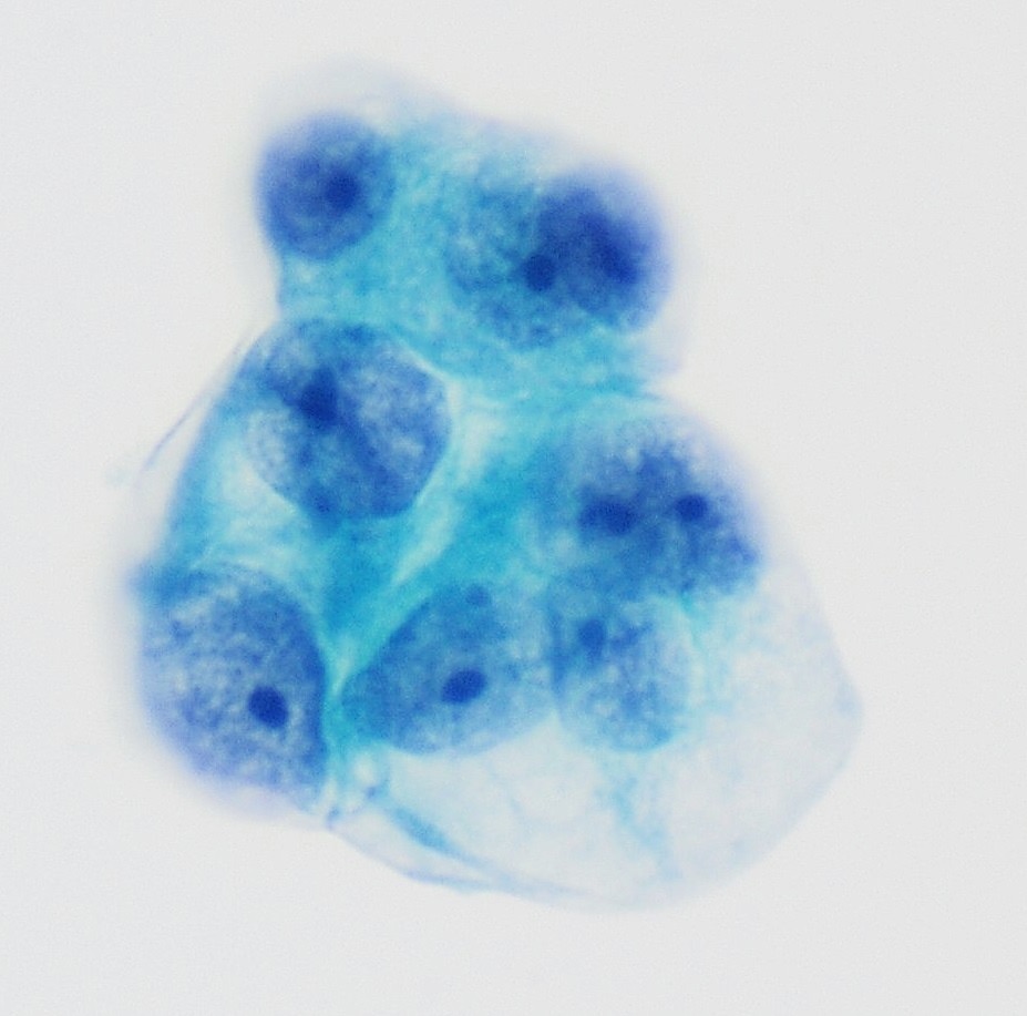
Definition. Mesothelial cells line the pleural, peritoneal and pericardial cavities. They are the normal resident cells of effusion specimens. Injury, inflammation and fluid accumulation make them proliferate and enlarge, a change called reactive mesothelial hyperplasia. Benign and reactive mesothelial cells are reported as negative for malignancy in the International System for Reporting Serous Fluid Cytopathology (ISRSFC).
Clinical features of benign and reactive mesothelial cells
- Common causes of reactive change: congestive heart failure, cirrhosis, renal failure, pneumonia, pulmonary infarction, pancreatitis, connective tissue disease, surgery, radiation and chemotherapy (Jhala et al. 2022).
- Diagnostic problem: reactive mesothelial cells can be numerous and atypical. They mimic metastatic adenocarcinoma and mesothelioma, and they are a major source of false-positive effusion diagnoses.
- Clinical correlation with the cause of the effusion and with imaging is part of the interpretation.
Normal and reactive cytomorphology
- Arrangement: single cells, pairs and small flat groups. Reactive fluids can contain larger groups and small papillary clusters, especially in peritoneal fluid from cirrhosis.
- Intercellular "windows": narrow clear spaces between adjacent cells. They result from surface microvilli.
- Cell borders: knobby or scalloped ("blebs"), from microvilli and cytoplasmic projections.
- Two-tone cytoplasm: dense perinuclear cytoplasm (endoplasm) and a pale, lacy outer rim (ectoplasm). The pattern is clearest on Diff-Quik preparations.
- Nucleus: central, round to oval, with smooth membranes, fine evenly distributed chromatin and one small nucleolus.
- Binucleation and multinucleation are common.
- Cell-in-cell arrangements ("clasping"): one mesothelial cell partly wraps around another.
Reactive atypia
- Nuclear enlargement with a preserved, moderate N/C ratio.
- Prominent nucleoli, sometimes large.
- Mitotic figures, which do not by themselves indicate malignancy.
- A continuous morphologic range: reactive cells form one population that grades from normal to atypical. A second, distinct population favors metastatic carcinoma.
- Psammoma bodies occur occasionally in benign effusions and do not by themselves establish malignancy.
Ancillary testing in benign and reactive mesothelial cells
- Mesothelial markers: calretinin (nuclear and cytoplasmic), WT1 (nuclear), D2-40 (podoplanin; membranous) and CK5/6.
- Carcinoma markers are negative: claudin-4, MOC-31 and Ber-EP4. Focal MOC-31 or Ber-EP4 staining can occur in reactive mesothelial cells (Wang et al. 2023).
- Keratin: mesothelial cells are keratin positive. Histiocytes are keratin negative.
- Benign versus malignant: BAP1 nuclear staining and MTAP cytoplasmic staining are retained in reactive mesothelial cells, and CDKN2A is not homozygously deleted. Retained staining does not exclude mesothelioma (Husain et al. 2024).
- Desmin stains reactive mesothelial cells more often than mesothelioma, and EMA stains mesothelioma more often (Husain et al. 2018). These markers lack adequate specificity for diagnostic use. IMIG authors no longer recommend desmin, EMA, GLUT-1, p53 or IMP3 for routine diagnostic use (Chapel et al. 2020).
- PAX8 stained benign mesothelium in 8 of 25 cases (32%), more often in females, and the PAX8-positive mesothelium was ER negative (Chapel et al. 2017). PAX8 positivity alone therefore does not establish a Müllerian carcinoma in peritoneal fluid.
Differential diagnosis of benign and reactive mesothelial cells
| Mimic | Distinguishing features |
|---|---|
| Histiocytes (macrophages) | Eccentric bean-shaped nuclei, foamy or finely vacuolated cytoplasm, no windows; CD68 and CD163 positive, keratin and calretinin negative |
| Metastatic adenocarcinoma | A second, foreign population; three-dimensional clusters with smooth "community" borders; claudin-4, MOC-31 and Ber-EP4 positive |
| Diffuse mesothelioma | Many large atypical cells and large clusters; BAP1 or MTAP loss, or CDKN2A homozygous deletion |
| Signet-ring and lobular carcinoma cells | Small single cells with intracytoplasmic mucin vacuoles; keratin positive, CD163 negative; GATA3 positive in lobular carcinoma |
| Degenerated mesothelial cells | Vacuolated cytoplasm and pyknotic nuclei that mimic signet-ring cells; same mesothelial immunoprofile |
Reporting of benign and reactive mesothelial cells
- ISRSFC category: benign and reactive mesothelial cells are reported as negative for malignancy (NFM).
- Atypia of undetermined significance (AUS) is used when atypical mesothelial cells cannot be classified as benign after ancillary studies on the cell block.
- Mesothelial cells are not required for adequacy. A lymphocyte-only effusion, for example in tuberculosis or a chylous effusion, is interpretable and is reported as NFM (Wang et al. 2023). Related page: The International System for Reporting Serous Fluid Cytopathology.
Mesothelial vs Carcinoma Immunohistochemistry in Effusions
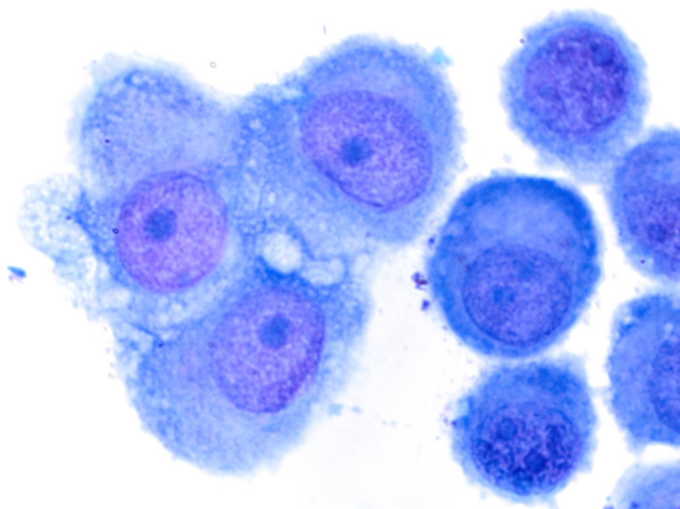
Definition. Immunohistochemistry on effusion cell blocks answers two separate questions. The first is lineage: mesothelial cells or carcinoma. The second applies only to a mesothelial population: benign or malignant. Different marker sets answer each question (Husain et al. 2018; Husain et al. 2024).
Clinical features of mesothelial versus carcinoma immunohistochemistry in effusions
- Indication: an effusion with atypical cells or a second cell population, or an effusion in a patient with a pleural or peritoneal mass.
- Cell block preferred: formalin-fixed, paraffin-embedded cell blocks allow the same validated protocols used for tissue.
- Panel rules (IMIG):
- panels contain both positive and negative markers for mesothelial differentiation and for the lesions in the differential
- each marker has a sensitivity or specificity above 80% for the lesion in question
- staining location is assessed (nuclear, cytoplasmic or membranous)
- staining in more than 10% of cells is the suggested threshold for a positive cytoplasmic or membranous marker (Husain et al. 2018)
- Common practice: a panel of two to three mesothelial and two carcinoma markers, adjusted to the clinical differential (Chapel et al. 2020).
Step 1: lineage
Mesothelial markers
| Marker | Pattern | Pitfalls |
|---|---|---|
| Calretinin | Nuclear and cytoplasmic | Cytoplasmic staining alone is nonspecific. Positive in 35–40% of lung squamous cell carcinomas (Chapel et al. 2020) |
| WT1 | Nuclear | Positive in tubo-ovarian serous carcinoma |
| D2-40 (podoplanin) | Membranous | Positive in 13–65% of serous ovarian carcinomas and about half of squamous cell carcinomas (Chapel et al. 2020) |
| CK5/6 | Cytoplasmic | Positive in squamous cell and urothelial carcinoma |
Carcinoma (epithelial) markers
| Marker | Pattern | Notes |
|---|---|---|
| Claudin-4 | Membranous | Positive in 99% of adenocarcinomas and negative in all 64 evaluable mesotheliomas in an effusion series; the single negative adenocarcinoma was a serous carcinoma (Jo et al. 2014) |
| MOC-31 (EpCAM) | Membranous | Staining in 0–10% of epithelioid mesotheliomas, and up to 30% in occasional studies (Chapel et al. 2020) |
| Ber-EP4 (EpCAM) | Membranous | Focal MOC-31 or Ber-EP4 staining occurs in reactive mesothelial cells and in up to 35% of mesotheliomas (Wang et al. 2023) |
| CEA (monoclonal), B72.3 | Cytoplasmic or membranous | Lower sensitivity; useful in a panel |
Site-specific markers
| Marker | Primary site | Pitfalls |
|---|---|---|
| TTF-1, napsin A | Lung adenocarcinoma (also thyroid for TTF-1) | Negative in mesothelioma |
| GATA3 | Breast, urothelial | Positive in many mesotheliomas, strongly and diffusely in sarcomatoid mesothelioma (Berg and Churg 2017) |
| TRPS1 | Breast | More specific than GATA3 against mesothelioma; positive in a subset of tubo-ovarian serous carcinomas |
| PAX8 | Müllerian, renal, thyroid | Positive in 93% of serous carcinomas and 0 of 40 peritoneal mesotheliomas (Ordóñez 2013). A later series found PAX8 in 5 of 27 (18%) peritoneal mesotheliomas and 8 of 25 (32%) benign mesothelium samples (Chapel et al. 2017) |
| SOX17 | Müllerian | Highly specific for Müllerian origin and negative in mesothelial lesions (Issin et al. 2026) |
| p40 | Squamous | Negative in mesothelioma |
Peritoneal fluid in a female patient
- Serous carcinoma and mesothelioma share WT1 and can share D2-40 and calretinin.
- Claudin-4 and SOX17 are the most specific discriminators. Claudin-4 was positive in 98% of serous carcinomas and in no peritoneal mesothelioma (Ordóñez 2013). SOX17 is negative in mesothelial lesions (Issin et al. 2026).
- PAX8 can stain mesothelium. It is positive in a subset of peritoneal mesotheliomas and benign mesothelium, more often in females (Chapel et al. 2017).
Step 2: benign or malignant mesothelial cells
These tests are applied once mesothelial lineage is established.
| Test | Malignant result | Notes |
|---|---|---|
| BAP1 | Loss of nuclear staining | Internal control in inflammatory and stromal cells |
| MTAP | Loss of cytoplasmic staining | Surrogate for CDKN2A homozygous deletion in pleural mesothelioma. In peritoneal mesothelioma, MTAP loss is less frequent (11 of 53 versus 35 of 60 pleural; Davidson 2024) and agrees poorly with CDKN2A FISH (κ=0.091; Quaranta 2025), so retained MTAP does not exclude malignancy |
| CDKN2A FISH | Homozygous 9p21 deletion | Detects some deletions with retained MTAP |
| Merlin | Loss of staining | Reflects NF2 loss; adds sensitivity |
| p53 | Diffuse (mutant-pattern) staining | Rare in mesothelioma (7%); null pattern is nonspecific (Chapel et al. 2022) |
- Sensitivity of panels: BAP1, MTAP and Merlin losses were each 100% specific and 54%, 46% and 52% sensitive for pleural mesothelioma. BAP1 plus MTAP was 79% sensitive, and BAP1 plus MTAP plus Merlin was 90% sensitive (Chapel et al. 2022).
- These markers do not define lineage. BAP1 loss also occurs in other cancers, for example uveal melanoma, renal cell carcinoma and cholangiocarcinoma, and MTAP loss occurs in many carcinomas.
- Retained staining does not exclude mesothelioma.
Historic markers
- Desmin stains reactive mesothelial cells more often than mesothelioma.
- EMA shows thick membranous staining more often in mesothelioma.
- GLUT-1 and IMP3 were also proposed.
- These markers overlap between benign and malignant proliferations. IMIG authors no longer recommend desmin, EMA, GLUT-1, IMP3 or p53 for routine diagnostic use (Chapel et al. 2020). A later validation study found mutant-pattern p53 in only 7% of pleural mesotheliomas (Chapel et al. 2022).
Differential diagnosis of mesothelial versus carcinoma immunohistochemistry in effusions
- Reactive mesothelial cells versus metastatic adenocarcinoma: lineage panel (Step 1). Related page: Benign and Reactive Mesothelial Cells.
- Reactive mesothelial cells versus mesothelioma: malignancy panel (Step 2). Related page: Diffuse Mesothelioma in Effusions.
- Mesothelioma versus serous carcinoma (peritoneum): claudin-4 and SOX17. PAX8 is interpreted with caution.
- Mesothelioma versus metastatic breast carcinoma: TRPS1 is more specific than GATA3. Related page: Metastatic Breast Carcinoma in Effusions.
- Mesothelioma versus squamous cell carcinoma: both can be CK5/6 and D2-40 positive. p40 and claudin-4 favor squamous cell carcinoma.
Reporting of mesothelial versus carcinoma immunohistochemistry in effusions
- Report content: the immunostains performed, the staining pattern of each, and the conclusion on lineage and, in a mesothelial population, on malignancy.
- ISRSFC category: BAP1 or MTAP loss, or CDKN2A homozygous deletion, in a mesothelial population supports malignant (primary). A lineage panel that shows carcinoma supports malignant (secondary). Related page: The International System for Reporting Serous Fluid Cytopathology.
Diffuse Mesothelioma in Effusions
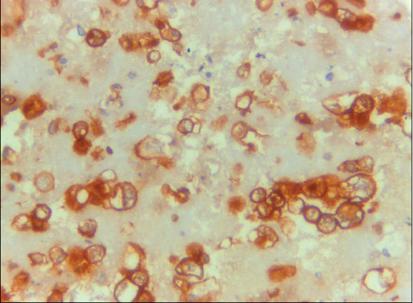
Definition. Diffuse mesothelioma (formerly malignant mesothelioma) is a malignant neoplasm of mesothelial cells that spreads diffusely over a serosal surface. The WHO Classification of Thoracic Tumours (5th edition, 2021) dropped the prefix "malignant" from localized and diffuse mesothelioma. The three histologic subtypes are epithelioid, biphasic and sarcomatoid. Effusion cytology can establish the diagnosis when morphology is supported by ancillary tests (Hjerpe et al. 2015; Husain et al. 2024).
WHO 2021 terminology (pleura and pericardium)
- Diffuse mesothelioma: epithelioid, biphasic or sarcomatoid.
- Localized mesothelioma: a circumscribed mass with the histology of diffuse mesothelioma and no diffuse serosal spread.
- Mesothelioma in situ (MIS): a single layer of surface mesothelial cells with loss of BAP1 or MTAP, or CDKN2A homozygous deletion, without invasion. It is a new entity in 2021.
- Well-differentiated papillary mesothelial tumor (WDPMT): the new name for well-differentiated papillary mesothelioma, reflecting its indolent behavior.
- Epithelioid diffuse mesothelioma is graded. Nuclear atypia and mitotic count are each scored from 1 to 3, and the summed score gives a nuclear grade of I to III. Low grade is nuclear grade I, or nuclear grade II without necrosis. High grade is nuclear grade II with necrosis, or nuclear grade III. The grade is based on the highest-grade features and applies only to epithelioid tumors (Sauter et al. 2022; Mastromarino et al. 2022).
- Biphasic mesothelioma can be diagnosed in a small biopsy with both components, even when one component is less than 10% (Sauter et al. 2022).
Clinical features of diffuse mesothelioma in effusions
- Frequency: mesothelioma accounts for 1–6% of malignant effusions (Wang et al. 2023).
- Risk factor: asbestos exposure, with a latency of decades. Germline BAP1 pathogenic variants (BAP1 tumor predisposition syndrome) account for a minority of cases.
- Presentation: a recurrent unilateral pleural effusion is the usual first sign of pleural mesothelioma. Peritoneal mesothelioma presents with ascites.
- Cytology yield: epithelioid mesothelioma sheds cells into effusions. Sarcomatoid mesothelioma rarely sheds diagnostic cells, so a negative effusion does not exclude it.
- Imaging correlation: cytology cannot separate MIS from invasive mesothelioma. Imaging and thoracoscopic findings establish that distinction (Churg et al. 2022).
Cytomorphology (epithelioid diffuse mesothelioma)
- High cellularity: more and larger mesothelial cells, in more and larger clusters, than in reactive effusions (Hjerpe et al. 2015).
- Large three-dimensional clusters (morulae) with knobby, scalloped outer borders. Papillary clusters may contain collagen cores.
- Mesothelial features are preserved: windows, two-tone cytoplasm, central nuclei.
- Nuclear atypia ranges from mild to marked. Many mesotheliomas show only mild atypia. Ancillary tests are therefore needed.
- Other findings: cell-in-cell arrangements, multinucleation, prominent nucleoli and intracytoplasmic vacuoles.
- A single population of mesothelial cells, without a second foreign population.
Ancillary testing in diffuse mesothelioma in effusions
Lineage is established first with mesothelial and carcinoma markers (related page: Mesothelial vs Carcinoma Immunohistochemistry in Effusions). Malignancy is then established with the tests below.
| Test | Abnormal result | Notes |
|---|---|---|
| BAP1 immunostain | Loss of nuclear staining in tumor cells | Inflammatory and stromal cells serve as the internal positive control. Loss in 10 of 15 (67%) epithelioid mesotheliomas in matched cytology and biopsy specimens; BAP1 loss, p16 deletion or both in 11 of 11 evaluable pairs (Hwang et al. 2016). Loss in 54% of a pleural series of all subtypes (Chapel et al. 2022) |
| MTAP immunostain | Loss of cytoplasmic staining | Surrogate for CDKN2A homozygous deletion; 100% specific for mesothelioma vs reactive hyperplasia in a pleural series (Hida et al. 2017) |
| CDKN2A (p16) FISH | Homozygous deletion of 9p21 | 73% of evaluable effusion cell blocks (Hwang et al. 2016). Present in about one-third of peritoneal mesotheliomas (9 of 26), with MTAP co-deleted in every case (Krasinskas et al. 2010) |
| Merlin immunostain | Loss of staining | Reflects NF2 inactivation. Loss in 52% of pleural mesotheliomas with 100% specificity; BAP1, MTAP and Merlin together were 90% sensitive (Chapel et al. 2022) |
- Combined panels increase sensitivity: BAP1 plus MTAP immunostains detected 76.5% of pleural mesotheliomas with 100% specificity (Hida et al. 2017) and 79% in a later series (Chapel et al. 2022).
- A retained result does not exclude mesothelioma. CDKN2A FISH can detect deletions that MTAP immunostaining misses.
- These tests separate benign from malignant mesothelial cells. They do not establish lineage, because BAP1 and MTAP loss also occur in other cancers.
- EZH2 immunohistochemistry is also listed, with BAP1 and MTAP, as useful for separating benign from malignant mesothelial proliferations (Sauter et al. 2022).
- Desmin and EMA are historic markers and are not used alone. IMIG authors no longer recommend desmin, EMA, GLUT-1, p53 or IMP3 for routine diagnostic use (Chapel et al. 2020).
- Molecular profile: the most frequently altered genes in diffuse pleural mesothelioma are BAP1, CDKN2A, NF2, TP53, SETD2 and SETDB1 (Sauter et al. 2022).
Reporting in effusions (ISRSFC)
- Malignant, primary (diffuse mesothelioma): atypical mesothelial cells with BAP1 or MTAP loss, or CDKN2A homozygous deletion, in the appropriate clinical and imaging setting.
- Suspicious for malignancy: cytology that suggests mesothelioma without confirmatory ancillary results, for example abundant, markedly atypical mesothelial cells with retained BAP1 and MTAP.
- Atypia of undetermined significance: atypical mesothelial cells that cannot be classified as benign, with retained BAP1 and MTAP and a lower degree of cellularity or atypia.
- The choice between AUS and SFM depends on the degree of atypia and cellularity. Related page: The International System for Reporting Serous Fluid Cytopathology.
- A comment may state that cytology cannot separate MIS from invasive mesothelioma.
Differential diagnosis of diffuse mesothelioma in effusions
- Reactive mesothelial hyperplasia (related page: Benign and Reactive Mesothelial Cells): fewer and smaller clusters, a clinical cause, retained BAP1 and MTAP, no CDKN2A deletion.
- Metastatic adenocarcinoma: a second population; claudin-4, MOC-31 and Ber-EP4 positive; calretinin negative.
- Serous carcinoma (peritoneum): WT1 positive, like mesothelioma; PAX8 and claudin-4 positive.
- WDPMT: papillary clusters of bland cells, usually with retained BAP1 and MTAP.
Metastatic Breast Carcinoma in Effusions
Definition. Metastatic breast carcinoma in an effusion is the presence of breast carcinoma cells in pleural, peritoneal or pericardial fluid. The International System for Reporting Serous Fluid Cytopathology (ISRSFC) reports it in the malignant (MAL) category as a secondary malignancy. Its two cytologic patterns follow the histologic type: invasive carcinoma of no special type (NST, formerly ductal) and invasive lobular carcinoma.
Clinical features of metastatic breast carcinoma in effusions
- Frequency: metastatic carcinoma in pleural effusions comes most commonly from the lung and the breast (Wang et al. 2023).
- Timing: the effusion may appear years after treatment of the primary tumor. A history of breast carcinoma is sometimes absent from the requisition.
- Lobular carcinoma spreads to the peritoneum, gastrointestinal tract and ovaries more often than NST carcinoma.
- Predictive testing: an effusion may be the only accessible metastatic specimen. ER, PR and HER2 status on the cell block guides endocrine, HER2-directed and antibody-drug conjugate therapy.
Cytomorphology of metastatic breast carcinoma in effusions
Invasive carcinoma of no special type
- Three-dimensional spherical clusters ("cannonballs," proliferation spheres) with smooth, sharply defined "community" borders.
- Cells: enlarged nuclei with irregular membranes and nucleoli; vacuolated cytoplasm.
- A second, foreign population distinct from the background mesothelial cells.
- Other patterns: papillary clusters, acinar groups and single cells.
Invasive lobular carcinoma
- Small, uniform single cells and linear single-file chains.
- Intracytoplasmic vacuoles, some with a central dot of mucin (targetoid vacuoles). Signet-ring forms occur.
- Mild atypia: the cells are often smaller and blander than reactive mesothelial cells. They mimic histiocytes, lymphocytes and mesothelial cells, and they are easily missed at low magnification.
- E-cadherin loss (absent membranous staining) on the cell block supports the lobular type, with cytoplasmic p120.
Immunohistochemistry
- Carcinoma lineage: claudin-4, MOC-31 and Ber-EP4 positive; calretinin negative. Related page: Mesothelial vs Carcinoma Immunohistochemistry in Effusions.
- GATA3: nuclear; sensitive for breast carcinoma. It is also positive in urothelial carcinoma and other tumors (Miettinen et al. 2014). In one series of 40 pleural specimens (36 ThinPrep fluids and 4 fine-needle aspirates; 21 breast carcinomas and 19 mesotheliomas), it stained all breast carcinomas and 84% of mesotheliomas (Baban et al. 2023).
- TRPS1: nuclear.
- sensitive for all breast carcinoma subtypes, including triple-negative carcinoma: positive in 86% of metaplastic and nonmetaplastic triple-negative carcinomas (Ai et al. 2021)
- positive in 92% of breast carcinomas in pleural effusion cell blocks (Wang et al. 2022)
- low positivity in 5% of mesotheliomas, compared with GATA3 positivity in 84% (Baban et al. 2023)
- positive in a subset of tubo-ovarian serous carcinomas: 4 of 6 Müllerian carcinomas (Wang et al. 2022) and 48% of tubo-ovarian serous carcinomas (Issin et al. 2026). Ai et al. (2021) found little TRPS1 in ovarian carcinoma, so the reported frequency varies between series.
- positive predictive value for breast origin in malignant effusions of only 55%, with a negative predictive value of 97% (Issin et al. 2026)
- ER and PR: positive in most NST and lobular carcinomas. ER is also positive in Müllerian carcinomas. PAX8 and SOX17 identify the Müllerian tumors.
- Mammaglobin and GCDFP-15: specific but less sensitive.
- SOX10: positive in a subset of triple-negative carcinomas.
Predictive biomarkers on effusion specimens
- ER and PR (ASCO/CAP 2020): ≥1% of tumor nuclei staining is positive. 1–10% is reported as ER low positive, with a recommended comment. The status of the controls is reported for cases with 0–10% staining (Allison et al. 2020).
- HER2 IHC (ASCO/CAP 2018, affirmed 2023):
| Score | Staining | Interpretation |
|---|---|---|
| 0 | No staining, or faint incomplete membrane staining in ≤10% of tumor cells | Negative |
| 1+ | Faint, barely perceptible, incomplete membrane staining in >10% of tumor cells | Negative |
| 2+ | Weak to moderate complete membrane staining in >10% of tumor cells | Equivocal; reflex in situ hybridization (ISH) |
| 3+ | Circumferential, complete, intense membrane staining in >10% of tumor cells | Positive |
- HER2-low is a therapy-selection term. It denotes IHC 1+, or IHC 2+ without amplification by ISH, the eligibility criteria for trastuzumab deruxtecan. It is not an ASCO/CAP reporting category. The 2023 update affirms the 2018 recommendations, states that new result categories such as HER2-low are premature, and adds best practices for the distinction between IHC 0 and 1+ (Wolff et al. 2023).
- Cell-block fixation: the guidelines call for fixation in 10% neutral buffered formalin for 6 to 72 hours.
- Receptor conversion: receptor and HER2 status can differ between the primary tumor and a distant metastasis. In a meta-analysis, ER changed from positive to negative in 22.5% of cases and HER2 from positive to negative in 21.3% (Schrijver et al. 2018).
- Tumor cell content: scant cell blocks limit scoring. The report states when the number of tumor cells is low.
Differential diagnosis of metastatic breast carcinoma in effusions
| Entity | Distinguishing features |
|---|---|
| Reactive mesothelial cells | Windows, two-tone cytoplasm, one continuous population; calretinin and WT1 positive, claudin-4 negative |
| Diffuse mesothelioma | Knobby clusters with mesothelial features; calretinin positive, claudin-4 negative; often GATA3 positive, rarely TRPS1 positive |
| Histiocytes (lobular mimic) | Bean-shaped nuclei, foamy cytoplasm; CD163 positive, keratin negative |
| Tubo-ovarian serous carcinoma | Papillary clusters, psammoma bodies; PAX8, WT1 and SOX17 positive; may be ER and TRPS1 positive |
| Lung adenocarcinoma | TTF-1 and napsin A positive; GATA3 usually negative |
| Gastric poorly cohesive carcinoma | Signet-ring cells with E-cadherin loss; GATA3, ER and TRPS1 usually negative; CDX2 may be positive |
| Plasmacytoid urothelial carcinoma | Single plasmacytoid cells with E-cadherin loss; GATA3 positive; ER and TRPS1 negative |
Reporting of metastatic breast carcinoma in effusions
- ISRSFC category: malignant (MAL), secondary: metastatic carcinoma, consistent with breast primary, with the histologic type (no special type or lobular) when the morphology and immunoprofile support one. Related page: The International System for Reporting Serous Fluid Cytopathology.
- Report content: the immunostains used, the ER, PR and HER2 results, and a statement when the number of tumor cells limits scoring.
Metastatic Lung Adenocarcinoma in Serous Effusions
Definition. Metastatic lung adenocarcinoma in a serous effusion is the presence of adenocarcinoma cells of pulmonary origin in pleural, pericardial or peritoneal fluid. The International System for Reporting Serous Fluid Cytopathology (ISRSFC, 2020) reports it in the malignant (MAL) category as a secondary malignancy. The cell block also serves as the specimen for predictive biomarker testing.
Clinical features of metastatic lung adenocarcinoma in serous effusions
- Frequency: lung carcinoma is the most common cause of malignant pleural effusion overall. Adenocarcinoma is the lung carcinoma type most frequently found in effusions (ISRSFC 2020).
- Staging: a malignant pleural or pericardial effusion is classified as M1a in the AJCC 8th edition (IASLC 8th edition TNM). The IASLC proposals for the 9th edition keep M1a unchanged and divide M1c into M1c1 and M1c2 (Fong et al. 2024).
- Clinical setting: the effusion is often the first or the only accessible specimen in a patient with advanced disease. It can establish the diagnosis, the stage and the treatment target in a single procedure.
- Treatment: first-line therapy for advanced nonsquamous non-small cell lung carcinoma depends on driver alterations and PD-L1 expression. Biomarker results from the effusion cell block therefore determine treatment.
Specimen handling
- Volume: ISRSFC recommends at least 75 mL of pleural fluid for diagnosis. Larger volumes increase the cellularity of the cell block.
- Preparations: direct or cytospin smears, liquid-based preparations and a formalin-fixed, paraffin-embedded cell block.
- Cell block priority: the CAP-led guideline for thoracic small biopsy and cytology specimens supports the use of cytology specimens, including cell blocks, for immunohistochemistry and molecular testing (Roy-Chowdhuri et al. 2020).
- Tissue conservation: a limited immunostain panel and early allocation of unstained sections for molecular testing preserve tumor cells.
- Non-formalin fixatives: in 33 matched pleural effusion cell blocks, PreservCyt and CytoLyt showed only moderate PD-L1 concordance with formalin at the ≥1% cutoff (κ 0.46–0.59). SP263 with CytoLyt showed fair concordance (κ 0.38). All alcohol-based fixatives gave significantly weaker staining intensity than formalin (Mansour et al. 2026). Alcohol-based fixation therefore risks false-negative PD-L1 results, and laboratories validate each assay on the cell-block fixation protocol in use.
Cytomorphology of metastatic lung adenocarcinoma in serous effusions
- Cell populations: a second population of cells distinct from the background mesothelial cells ("two-cell population").
- Architecture:
- three-dimensional clusters, papillae, acinar groups and morulae (proliferation spheres)
- smooth "community borders" around clusters
- single cells in some cases
- Cytoplasm: finely vacuolated or foamy, with distinct cell borders. Large mucin vacuoles in mucinous types.
- Nuclei: enlarged, eccentric and round to oval, with vesicular chromatin and prominent nucleoli. The N/C ratio is increased.
- Mesothelial cells for comparison: knobby cell borders, two-tone cytoplasm, windows between adjacent cells and central nuclei.
- Invasive mucinous adenocarcinoma: columnar cells with abundant mucin and bland nuclei. These cells resemble metastatic gastrointestinal or pancreaticobiliary carcinoma.
Ancillary testing in metastatic lung adenocarcinoma in serous effusions
Carcinoma versus mesothelial cells
- Carcinoma markers: claudin-4, MOC-31, Ber-EP4 and BG8 are positive in adenocarcinoma. Claudin-4 is the most specific of these for carcinoma versus mesothelial cells. In 159 malignant effusions, claudin-4 stained 99% of adenocarcinomas (83 of 84) and none of 64 mesotheliomas (Jo et al. 2014).
- Mesothelial markers: calretinin, WT1 and D2-40 are positive in mesothelial cells and negative in lung adenocarcinoma.
Site of origin
| Marker | Lung adenocarcinoma | Main other positive sites |
|---|---|---|
| TTF-1 (nuclear) | Positive in most nonmucinous adenocarcinomas | Thyroid carcinoma, small cell carcinoma and other neuroendocrine carcinomas |
| Napsin A (granular cytoplasmic) | Positive in most nonmucinous adenocarcinomas | Clear cell carcinoma of the ovary and endometrium, papillary renal cell carcinoma |
| CK7 / CK20 | CK7+ / CK20− | Many CK7+ carcinomas, including breast and Müllerian carcinomas |
| GATA3, PAX8, CDX2, SATB2 | Negative in nonmucinous types | Breast, Müllerian, gastrointestinal origin |
- TTF-1 clone: the SPT24 and SP141 clones are more sensitive and less specific than the 8G7G3/1 clone. Lung adenocarcinomas were positive in 89% (8G7G3/1) and 93% (SPT24 and SP141). Colorectal metastases to the lung were positive in 2%, 7% and 8%, respectively (Vidarsdottir et al. 2018).
- Napsin A pitfall: macrophages show granular napsin A staining. Napsin A positivity therefore requires interpretation in clearly identified tumor cells.
- Invasive mucinous adenocarcinoma: TTF-1 and napsin A are usually negative. CK7 is positive, and CK20 and HNF4α can be positive (WHO Thoracic Tumours 2021). Clinical and radiologic correlation separates it from a gastrointestinal primary.
Predictive biomarkers on the cell block
- Molecular testing (CAP/IASLC/AMP 2018):
- EGFR, ALK and ROS1 testing is required for all advanced adenocarcinomas.
- BRAF, MET, RET, ERBB2 and KRAS are included when a larger panel is performed.
- Cytology specimens, including cell blocks and other cytologic preparations, are suitable for molecular testing.
- Next-generation sequencing (NGS) panels are preferred over sequential single-gene tests.
- EGFR: detected by DNA-based mutation analysis (PCR or NGS). EGFR immunohistochemistry is not recommended for treatment selection (CAP/IASLC/AMP 2018). Exon 19 deletions and L858R are the common sensitizing mutations.
- ALK: ALK immunohistochemistry (D5F3 clone) is an acceptable alternative to FISH. Positive staining is strong granular cytoplasmic staining. EML4::ALK is the most common fusion.
- ROS1: ROS1 immunohistochemistry can be used as a screening test. A positive result is confirmed by FISH, RNA-based NGS or another molecular method (CAP/IASLC/AMP 2018).
- Other targets: KRAS G12C, BRAF V600E, MET exon 14 skipping, RET fusions, NTRK fusions and ERBB2 mutations each have FDA-approved targeted therapy (Zhang et al. 2024).
- Tumor content: the pathologist assesses the tumor cell content of each specimen. Each laboratory establishes the minimum proportion and number of tumor cells for mutation detection during assay validation (Lindeman et al. 2013, reaffirmed in 2018).
- Cell-free DNA: plasma cell-free DNA testing can "rule in" a targetable mutation when tissue is limited or hard to obtain (CAP/IASLC/AMP 2018).
- PD-L1 (22C3):
- Reported as the tumor proportion score (TPS): the percentage of viable tumor cells with partial or complete membranous staining at any intensity.
- Cytoplasmic-only staining and staining of macrophages and lymphocytes are excluded.
- At least 100 viable tumor cells are required for an evaluable result (22C3 pharmDx label).
- TPS ≥50% supported first-line pembrolizumab monotherapy (Reck et al. 2016). TPS ≥1% was the threshold in KEYNOTE-042 (Mok et al. 2019).
- Cell blocks for PD-L1: the CAP-led 2020 guideline states that thoracic small biopsy and cytology specimens can be processed for immunohistochemical and molecular biomarkers (Roy-Chowdhuri et al. 2020). In a small series using the SP263 assay, PD-L1 categories on effusion cell blocks matched histology in 10 of 11 paired cases (91%). A CD163 immunostain identified macrophages and excluded them from the score (Mahajan et al. 2022).
Differential diagnosis of metastatic lung adenocarcinoma in serous effusions
| Entity | Distinguishing features |
|---|---|
| Reactive mesothelial cells | Windows, knobby borders, two-tone cytoplasm; calretinin and WT1 positive; claudin-4 negative |
| Diffuse mesothelioma, epithelioid | Large mesothelial groups with windows; calretinin and WT1 positive; BAP1 or MTAP loss |
| Metastatic breast carcinoma | Cannonball clusters or single files; GATA3 positive; ER often positive; TTF-1 negative |
| Metastatic Müllerian carcinoma | Papillae and psammoma bodies; PAX8 and WT1 positive (serous); napsin A positive in clear cell carcinoma, with PAX8 positivity and TTF-1 negativity |
| Metastatic gastrointestinal carcinoma | Columnar cells, dirty necrosis or signet-ring cells; CDX2 and SATB2 positive in colorectal carcinoma |
| Metastatic thyroid carcinoma | TTF-1 and PAX8 positive; thyroglobulin positive; napsin A negative |
| Small cell carcinoma | Nuclear molding, scant cytoplasm; TTF-1 positive; INSM1, synaptophysin and chromogranin positive |
Reporting of metastatic lung adenocarcinoma in serous effusions
- ISRSFC category: malignant (MAL), secondary: metastatic adenocarcinoma, consistent with lung primary.
- Report content: the immunostains used, a statement on the adequacy of the cell block for biomarker testing, and the tests sent or pending.
- Atypia of undetermined significance (AUS) and suspicious for malignancy (SFM) are used when the atypical cells are too few or too poorly preserved for a definite diagnosis. Ancillary tests on the cell block resolve many of these cases.
Metastatic Gastrointestinal and Pancreaticobiliary Carcinoma in Serous Effusions
Definition. Metastatic gastrointestinal (GI) and pancreaticobiliary carcinoma in a serous effusion is the presence of carcinoma cells from the stomach, small or large intestine, appendix, pancreas, biliary tract or liver in peritoneal, pleural or pericardial fluid. The International System for Reporting Serous Fluid Cytopathology (ISRSFC, 2020) reports it in the malignant (MAL) category as a secondary malignancy.
Clinical features of metastatic gastrointestinal and pancreaticobiliary carcinoma in serous effusions
- Site of effusion: GI and pancreaticobiliary carcinomas most often involve the peritoneum. In a series of 209 patients with malignant ascites, ovarian carcinoma was the most common cause and GI carcinomas were second (Ayantunde and Parsons 2007).
- Gastric poorly cohesive carcinoma (signet-ring cell type) spreads diffusely over the peritoneum and can involve both ovaries (Krukenberg tumor).
- Pancreatic carcinoma: peritoneal seeding or positive peritoneal cytology is classified as M1 in the AJCC 8th edition (CAP pancreas protocol, version 4.2.0.0, 2021).
- Gastric adenocarcinoma: positive peritoneal cytology is an adverse prognostic finding. In 291 patients with positive cytology, median disease-specific survival was 1.3 years without visible metastases and 0.8 years with visible metastases (Mezhir et al. 2010).
- Pseudomyxoma peritonei (PMP) is a clinical syndrome of mucinous ascites and mucinous peritoneal implants. Most cases arise from an appendiceal mucinous neoplasm (Carr et al. 2016).
Cytomorphology of metastatic gastrointestinal and pancreaticobiliary carcinoma in serous effusions
Colorectal adenocarcinoma
- Tall columnar cells in palisaded groups, acini and three-dimensional clusters.
- Elongated, cigar-shaped, hyperchromatic nuclei with pseudostratification.
- "Dirty" necrosis (granular necrotic debris) in the background.
- In fluid, clusters can round up, so the columnar shape is less evident.
Gastric and other poorly cohesive carcinomas
- Single cells and small loose groups.
- Signet-ring cells: an intracytoplasmic mucin vacuole indents the nucleus into a crescent.
- Nuclei can be small and bland. This feature causes underdiagnosis.
- Mucin is shown by mucicarmine, PAS with diastase or Alcian blue stains.
Pancreaticobiliary adenocarcinoma
- Sheets and three-dimensional clusters of cuboidal to columnar cells.
- Nuclear enlargement, irregular nuclear membranes and anisonucleosis (variation of nuclear size of 4:1 or more within one group, the "drunken honeycomb" pattern).
- Cytoplasmic mucin vacuoles in some cells.
Pseudomyxoma peritonei
- Abundant thick mucin, with histiocytes and mesothelial cells within the mucin.
- Low-grade disease: few strips of bland columnar mucinous cells.
- High-grade disease: more cells, cytologic atypia, and signet-ring cells in some cases.
- PSOGI terms for the peritoneal disease: acellular mucin, low-grade mucinous carcinoma peritonei (formerly disseminated peritoneal adenomucinosis, DPAM), high-grade mucinous carcinoma peritonei (formerly peritoneal mucinous carcinomatosis, PMCA), and high-grade mucinous carcinoma peritonei with signet-ring cells (Carr et al. 2016).
Hepatocellular carcinoma
- Rare in effusions. Polygonal cells with central nuclei, prominent nucleoli and granular cytoplasm; bile pigment in some cases.
- HepPar-1 and arginase-1 positive.
Ancillary testing in metastatic gastrointestinal and pancreaticobiliary carcinoma in serous effusions
Carcinoma versus mesothelial cells and macrophages
- Carcinoma: claudin-4, MOC-31 and Ber-EP4 positive; calretinin and WT1 negative.
- Vacuolated macrophages: CD68 and CD163 positive; keratin negative; mucin stains negative.
- Vacuolated mesothelial cells: calretinin, WT1 and D2-40 positive; claudin-4 negative.
Site of origin
| Primary | CK7 / CK20 | CDX2 | SATB2 | Other |
|---|---|---|---|---|
| Colorectum | − / + | + | + | CK7 positive in a minority |
| Appendix (mucinous neoplasm, PMP) | − or focal / + | + | + | MUC2 positive |
| Stomach | + or − / + or − | Variable | Usually − | Variable profile; HER2 testing in advanced disease |
| Pancreas and biliary tract | + / + or − | Variable | − | SMAD4 (DPC4) inactivated in about 55% of pancreatic adenocarcinomas (McCarthy et al. 2003) |
| Liver (hepatocellular) | − / − | − | − | HepPar-1, arginase-1, glypican-3 |
- CK7/CK20 pattern: CK7−/CK20+ indicates colorectal origin in most cases (Chu et al. 2000).
- CDX2 is a sensitive marker of intestinal differentiation. It is also positive in gastric, pancreaticobiliary and mucinous ovarian carcinomas and in invasive mucinous adenocarcinoma of the lung in some cases (Werling et al. 2003).
- SATB2 combined with CK20 identifies more than 95% of colorectal carcinomas (Magnusson et al. 2011). SATB2 is usually negative in gastric and pancreaticobiliary carcinomas.
- Signet-ring cells of breast origin (lobular carcinoma) are GATA3 and ER positive, with loss of E-cadherin. GI signet-ring cells are GATA3 negative.
Predictive and molecular tests on the cell block
- Colorectal carcinoma: KRAS, NRAS and BRAF mutation analysis, and mismatch repair (MMR) or microsatellite instability (MSI) testing.
- Gastric and gastroesophageal carcinoma: HER2, PD-L1 (combined positive score), MMR, and claudin 18.2.
- HER2 is scored by the gastroesophageal (Rüschoff/Hofmann) criteria. Complete membranous staining is not required, and strong basolateral or lateral membranous staining in ≥10% of tumor cells of a resection specimen is 3+ (Bartley et al. 2017).
- Claudin 18.2: positive is moderate to strong (2+ or 3+) membranous staining in ≥75% of tumor cells (Rogers et al. 2026). The FDA approved zolbetuximab on 18 October 2024 for first-line treatment of HER2-negative, claudin 18.2-positive advanced gastric or gastroesophageal junction adenocarcinoma, with the VENTANA CLDN18 (43-14A) assay as companion diagnostic.
- Pancreatic carcinoma: ASCO recommends assessment of hereditary risk for all patients. Germline testing "may be discussed" with every patient, even when the family history is unremarkable (Stoffel et al. 2019).
Differential diagnosis of metastatic gastrointestinal and pancreaticobiliary carcinoma in serous effusions
| Entity | Distinguishing features |
|---|---|
| Vacuolated macrophages | Kidney-shaped nuclei, foamy cytoplasm; CD68 and CD163 positive; keratin and mucin stains negative |
| Reactive mesothelial cells with vacuoles | Windows, knobby borders; calretinin and WT1 positive; mucin stains negative |
| Metastatic breast lobular carcinoma | Single cells and single files, targetoid mucin; GATA3 and ER positive; E-cadherin loss |
| Metastatic Müllerian carcinoma | Papillae and psammoma bodies; PAX8 and WT1 positive (serous); CK20 negative |
| Primary ovarian mucinous carcinoma | Large unilateral ovarian mass; CK7 positive; SATB2 negative; immunoprofile overlaps with gastric carcinoma; PMP rarely arises from it |
| Metastatic lung adenocarcinoma, invasive mucinous type | Lung consolidation or mass; CK7 positive; TTF-1 usually negative; CDX2 positive in some cases |
Reporting of metastatic gastrointestinal and pancreaticobiliary carcinoma in serous effusions
- ISRSFC category: malignant (MAL), secondary. The report states the favored primary site and the immunostains used.
- Atypia of undetermined significance (AUS) applies to scant, bland mucinous cells when neoplasia cannot be confirmed. Suspicious for malignancy (SFM) applies when the atypia is strong and the cells are few.
Metastatic Müllerian Carcinoma in Serous Effusions
Definition. Metastatic Müllerian carcinoma in a serous effusion is the presence of carcinoma cells of tubo-ovarian, primary peritoneal or endometrial origin in peritoneal, pleural or pericardial fluid. The main types are high-grade serous, low-grade serous, endometrioid and clear cell carcinoma, classified by the WHO Female Genital Tumours, 5th edition (2020). The International System for Reporting Serous Fluid Cytopathology (ISRSFC, 2020) reports these tumors in the malignant (MAL) category as secondary malignancies.
Clinical features of metastatic Müllerian carcinoma in serous effusions
- Frequency: Müllerian carcinoma, mainly tubo-ovarian high-grade serous carcinoma (HGSC), is the most common source of malignant ascites in females. In a series of 209 patients with malignant ascites, ovarian carcinoma was the most common cause overall (Ayantunde and Parsons 2007).
- Presentation: HGSC presents at an advanced stage with ascites, omental disease and, in some patients, a malignant pleural effusion.
- FIGO staging (2014, ovary, fallopian tube and peritoneum):
- malignant cells in ascites or peritoneal washings with tumor confined to the ovaries or tubes: IC3
- malignant pleural effusion: IVA
- Diagnosis before neoadjuvant chemotherapy (SGO/ASCO 2016): the guideline recommends histologic confirmation of an invasive ovarian, tubal or peritoneal carcinoma in every patient before neoadjuvant chemotherapy, preferably by core biopsy. Cytology is acceptable only in exceptional cases when a biopsy cannot be performed. It is then combined with a serum CA-125:CEA ratio >25 to exclude a nongynecologic primary (Wright et al. 2016).
- Endometrial carcinoma: in FIGO 2023, positive peritoneal washing cytology is not considered for staging purposes (Berek et al. 2023).
- Germline and tumor testing (ASCO 2020): every patient with epithelial ovarian cancer should have germline testing for BRCA1, BRCA2 and other ovarian cancer susceptibility genes. Somatic tumor BRCA1/2 testing follows a negative germline result. Clear cell, endometrioid and mucinous carcinomas are tested for mismatch repair deficiency (Konstantinopoulos et al. 2020).
- PARP inhibitor maintenance: germline or somatic BRCA1/2 status and tumor genomic instability (homologous recombination deficiency) guide the choice of maintenance therapy (Tew et al. 2020).
Cytomorphology of metastatic Müllerian carcinoma in serous effusions
High-grade serous carcinoma
- Large three-dimensional clusters, papillae and spheres with scalloped borders.
- Single cells in some cases.
- Marked nuclear pleomorphism (variation of 3:1 or more), coarse chromatin and macronucleoli. The N/C ratio is high.
- Cytoplasmic vacuoles; mitoses, including atypical forms.
- Psammoma bodies in some cases.
Low-grade serous carcinoma and serous borderline tumor
- Papillary clusters of small, uniform cells with mild nuclear atypia and small nucleoli.
- Numerous psammoma bodies.
- Low-grade serous carcinoma and implants of serous borderline tumor cannot be separated on cytology.
Endometrioid carcinoma
- Glandular and acinar groups of columnar cells with oval nuclei.
- Squamous morules in some cases.
- Low-grade nuclei in most cases.
Clear cell carcinoma
- Clusters and papillae of cells with abundant clear or granular eosinophilic cytoplasm.
- Hobnail cells; round nuclei with prominent nucleoli.
- Hyaline globules (basement membrane material) within clusters.
Psammoma bodies
- Psammoma bodies occur in benign and malignant settings: endosalpingiosis, mesothelial hyperplasia, serous borderline tumor, low-grade and high-grade serous carcinoma, mesothelioma and papillary thyroid carcinoma.
Ancillary testing in metastatic Müllerian carcinoma in serous effusions
Carcinoma versus mesothelial cells
- WT1 is positive in both mesothelial cells and serous carcinoma. WT1 therefore does not separate them.
- Carcinoma: PAX8, claudin-4, MOC-31, Ber-EP4 and ER positive; calretinin and D2-40 negative or focal.
- Mesothelial cells: calretinin, D2-40 and WT1 positive; claudin-4 negative.
- PAX8 in mesothelium: PAX8 stained 8 of 25 benign mesothelium samples (32%), significantly more often in women, and 5 of 27 peritoneal mesotheliomas (18%). The PAX8-positive mesothelium was ER negative (Chapel et al. 2017). PAX8 is therefore interpreted with claudin-4, ER and calretinin.
- SOX17: in tissue, SOX17 was 87% sensitive and 100% specific for Müllerian tumors, with no staining of mesothelial lesions that coexpressed PAX8 and WT1. The specificity held in effusion cell blocks (Issin et al. 2026).
Müllerian carcinoma types
| Type | PAX8 | WT1 | p53 | ER | Other |
|---|---|---|---|---|---|
| Tubo-ovarian HGSC | + | + | Aberrant (overexpression, complete absence or cytoplasmic) | Usually + | p16 diffuse; TP53 mutation in nearly all |
| Endometrial serous carcinoma | + | Usually − or weak | Aberrant | Variable | p16 diffuse |
| Low-grade serous carcinoma | + | + | Wild-type | + | KRAS, BRAF or NRAS mutation |
| Endometrioid carcinoma | + | − | Wild-type in most | + | PR+; nuclear β-catenin (CTNNB1); MMR deficiency in a subset |
| Clear cell carcinoma | + | − | Wild-type in most | − | HNF1β+, napsin A+; ARID1A loss in a subset |
| Mesonephric-like adenocarcinoma | + | − | Wild-type | − | GATA3+, TTF-1+; KRAS mutation |
- p53 patterns (Köbel and Kang 2021):
- wild-type: variable nuclear staining of different intensity
- overexpression: strong nuclear staining in >80% of tumor cell nuclei (Köbel et al. 2016)
- complete absence (null): no staining in tumor cells, with internal control staining in stromal or inflammatory cells
- cytoplasmic: cytoplasmic staining with or without nuclear staining
- A complete absence of p53 staining is an aberrant (mutant) pattern. It is a recognized pitfall, because it can be misread as negative.
- Napsin A in clear cell carcinoma: clear cell carcinoma is napsin A positive and TTF-1 negative. PAX8 positivity separates it from lung adenocarcinoma.
- PAX8 outside the Müllerian tract: renal cell carcinoma and thyroid carcinoma are also PAX8 positive.
Differential diagnosis of metastatic Müllerian carcinoma in serous effusions
| Entity | Distinguishing features |
|---|---|
| Reactive mesothelial cells | Windows, knobby borders; calretinin and WT1 positive; claudin-4, ER and SOX17 negative; PAX8 positive in about one-third (mainly in women) |
| Diffuse mesothelioma, epithelioid | Calretinin and D2-40 positive; PAX8 positive in about 18% of peritoneal cases; BAP1 or MTAP loss |
| Endosalpingiosis | Ciliated cells, bland nuclei, psammoma bodies; few cells |
| Metastatic breast carcinoma | GATA3 positive; PAX8 and WT1 negative |
| Metastatic GI carcinoma | CK20, CDX2 and SATB2 positive in colorectal and appendiceal tumors; PAX8 negative |
| Metastatic lung adenocarcinoma | TTF-1 and napsin A positive; PAX8 negative |
Reporting of metastatic Müllerian carcinoma in serous effusions
- ISRSFC category: malignant (MAL), secondary: metastatic carcinoma, consistent with Müllerian origin, with a favored type when the morphology and immunoprofile support one.
- Low-grade serous neoplasms: the report states that a serous borderline tumor and low-grade serous carcinoma cannot be separated on cytology.
Lymphoma and Primary Effusion Lymphoma in Serous Effusions
Definition. Lymphoid neoplasms reach a serous cavity in two ways. Systemic lymphoma or leukemia involves the pleura, peritoneum or pericardium secondarily. A small group of large B-cell lymphomas arises in the body cavity and grows as an effusion without a solid mass. The International System for Reporting Serous Fluid Cytopathology (ISRSFC) reports primary effusion lymphoma (PEL) as a primary malignancy and other lymphomas and leukemias as secondary malignancies (Chandra 2020). WHO-HAEM5 recognizes PEL, with its extracavitary subtype, within the KSHV/HHV8-associated B-cell lymphoid proliferations, and adds fluid overload-associated large B-cell lymphoma (FO-LBCL) as a new HHV8-negative entity (Alaggio 2022). The ICC 2022 term for the HHV8-negative entity is HHV8- and EBV-negative primary effusion-based lymphoma (Ferry 2024). Related topics: The International System for Reporting Serous Fluid Cytopathology, Benign and Reactive Mesothelial Cells and Lymph Node FNA Triage for Flow Cytometry, Cultures and Molecular Testing.
Clinical features of lymphoma in serous effusions
- Frequency of pleural effusion: pleural effusion occurs in 20–30% of patients with non-Hodgkin lymphoma and Hodgkin lymphoma. Peritoneal and pericardial involvement is uncommon (Das 2006). Effusions are most frequent when the mediastinum is involved. Acute and chronic leukemias and myelodysplastic syndromes are rarely accompanied by pleural involvement (Alexandrakis 2004).
- Share of malignant effusions: in 3011 pleural and peritoneal specimens, lymphoreticular neoplasms accounted for 16% of malignant pleural effusions and 7% of malignant peritoneal effusions (Sears 1987).
- Mechanisms: thoracic duct obstruction and impaired lymphatic drainage are the main mechanism in Hodgkin lymphoma. Direct pleural infiltration predominates in non-Hodgkin lymphoma (Das 2006).
- Subtypes: T-cell neoplasms, especially lymphoblastic lymphoma, involve serous fluids more often than other subtypes (Das 2006). Because B-cell lymphomas are more common overall, they are the lymphomas most often found in fluid specimens (Michelow 2026).
- Children: hematolymphoid neoplasms are the most common cause of malignant serous effusion in children (Michelow 2026). Related topic: Special Non-Neoplastic Effusions and Pediatric and Pericardial Fluid Cytology.
- Nonlymphomatous causes: a pleural effusion in a patient with a hematologic malignancy can also result from drug toxicity, infection, a second malignancy or, rarely, an autoimmune process (Alexandrakis 2004).
- Positive cytology rate: published rates of positive cytology for non-Hodgkin lymphoma in pleural fluid range from 22.2% to 94.1% (Das 2006).
Cytomorphology of lymphoma in serous effusions
- Triage by cell size: lymphocyte-predominant effusions are first sorted into small-to-medium and large lymphoid cell populations. The main differential diagnosis of a small-to-medium population is reactive lymphocytosis. The main differential diagnosis of a large-cell population is metastatic malignancy and atypical or malignant mesothelial cells (Michelow 2026).
- Small-cell lymphomas: chronic lymphocytic leukemia/small lymphocytic lymphoma (CLL/SLL), follicular lymphoma and mantle cell lymphoma produce a population of small to medium lymphoid cells. Reactive lymphocytosis is their main mimic, and flow cytometry separates them (Michelow 2026; Czader 2003).
- Lymphoblastic lymphoma: in 15 T-lymphoblastic lymphomas sampled by aspiration or effusion, the blasts had a high nuclear-to-cytoplasmic ratio, scant basophilic cytoplasm and nuclear notches, clefts and indentations. Chromatin was condensed in small and intermediate blasts and dispersed in larger blasts. Nucleoli were present only in the larger blasts (Bhaker 2015).
- Primary effusion lymphoma: large cells with moderate to abundant, deeply basophilic cytoplasm on Romanowsky stains, sometimes with vacuoles. The nuclei are round or irregular and multilobated, often with large nucleoli (Ehinger 2026). The morphology is intermediate between large-cell immunoblastic lymphoma and anaplastic large cell lymphoma (Nador 1996). A perinuclear hof and coarse chromatin were described in HIV-negative PEL (Chu 2025).
- Hodgkin lymphoma: rare in effusion specimens. The differential diagnosis of Reed-Sternberg-like cells in a fluid is broad and includes reactive and other neoplastic large cells (Das 2006).
Flow cytometry in serous effusions with lymphoid cells
- Effect on diagnosis: in 115 consecutive serous effusions with concurrent flow cytometry, the provisional cytologic diagnosis was modified in 18 cases (16%). Eight atypical or suspicious cases were changed to benign. Ten benign or atypical cases were changed to malignant, including follicular lymphoma (3), mantle cell lymphoma (2), chronic lymphocytic leukemia, angioimmunoblastic lymphoma, large cell lymphoma, NK-cell leukemia and plasma cell myeloma (Czader 2003).
- Limits: flow cytometry did not contribute in Hodgkin lymphoma or in nonlymphoid malignancies (Czader 2003).
- Concordance: in 51 effusions with flow cytometry or immunocytochemistry, the initial cytologic diagnosis agreed with the final diagnosis in 94.1%. The final diagnoses were T-lymphoblastic lymphoma (18), mature B-cell non-Hodgkin lymphoma (10), Burkitt lymphoma (7), diffuse large B-cell lymphoma (4), and smaller numbers of follicular lymphoma, T-cell non-Hodgkin lymphoma, chronic lymphocytic leukemia, hairy cell leukemia, plasmablastic lymphoma and Hodgkin lymphoma (Gupta 2021).
- Combined methods: a review of published series reported that combined morphology and flow cytometric immunophenotyping reached a sensitivity and specificity of 100% (Das 2006).
- T-lymphoblastic lymphoma: flow cytometry showed CD2 in 15 of 15 cases, cytoplasmic CD3 in 15 of 15, surface CD3 in 14 of 15, CD5 in 13 of 15, CD10 in 7 of 15 and TdT in 8 of 15. All cases showed a tight cluster of CD4 and CD8 double-positive cells. In the 7 cases with TdT negative by flow cytometry, cell block immunocytochemistry was TdT positive (Bhaker 2015).
- Primary effusion lymphoma: the cells are CD45 dim, CD38 positive and CD138 positive. Surface and cytoplasmic light chains are absent, and B-cell antigens are often absent (Ehinger 2026). Light chain restriction therefore cannot establish clonality in PEL.
Primary effusion lymphoma
- Discovery: KSHV DNA sequences were found in 8 of 193 lymphomas. All 8 were AIDS-related body-cavity-based lymphomas, and all 8 also contained the EBV genome (Cesarman 1995).
- Definition as an entity: in 19 lymphomatous effusions without a contiguous mass, 15 were KSHV positive. The 4 KSHV-negative effusions had Burkitt or Burkitt-like morphology and MYC rearrangement (Nador 1996).
- Patients: in the same series, all 15 KSHV-positive patients were men and 13 of 15 had HIV infection. Fourteen of 15 tumors contained EBV, and 13 of 13 had clonal immunoglobulin gene rearrangement (Nador 1996). In a later cohort of 70 patients, 67 were men, the median age was 46 years, 80% had HIV infection and 27% had Kaposi sarcoma (Hu 2021).
- HIV-negative PEL: occurs in older patients. In a series of 7 HIV-negative men with a mean age of 76 years, 6 died of disease, with a mean survival of 5.4 months (Chu 2025).
- Dual infection: KSHV and EBV can infect and transform the same B cell, a finding frequent in KSHV-positive, EBV-positive PEL and extracavitary PEL (Cesarman 2022).
- Immunophenotype: CD45 positive and T-cell antigen negative; CD30, EMA and CD138 positive; CD10, CD15 and BCL6 negative (Chadburn 2004). CD138 was expressed in 5 of 7 PEL samples and was absent in 13 other lymphomatous effusions (Gaidano 1997). In HIV-negative PEL, HHV8 latent nuclear antigen was positive in 7 of 7 cases, CD138 in 6 of 7, and CD3 and CD20 were negative in all (Chu 2025).
- HHV8 detection: KSHV/HHV8 infection is required for the diagnosis (Ehinger 2026). A monoclonal antibody to latent nuclear antigen-1 (LANA-1) of HHV8 identifies latently infected cells in PEL, Kaposi sarcoma and multicentric Castleman disease (Dupin 1999).
- Extracavitary PEL: KSHV-positive solid lymphomas share the morphology, immunophenotype and genotype of PEL and are designated extracavitary PEL (Chadburn 2004). Compared with effusion-only PEL, extracavitary-only PEL was less often positive for CD45 (69.2% versus 96.2%) and EMA (26.7% versus 100%) (Hu 2021).
- Genetics of PEL: all 13 tested KSHV-positive effusion lymphomas had clonal immunoglobulin gene rearrangement despite absent immunoglobulin expression, all 12 studied lacked MYC rearrangement, and 13 of 15 lacked BCL2, BCL6, RAS and TP53 alterations (Nador 1996). Complex karyotypes were present in 3 of 5 HIV-negative cases studied (Chu 2025).
- Outcome: PEL accounted for 11 of 277 (4%) HIV-associated systemic non-Hodgkin lymphomas in one institution, with a median survival of 6 months (Simonelli 2003). In the 70-patient cohort, the median overall survival of the 52 patients with follow-up was 42.5 months, and 6.8 months in HIV-negative patients. The authors nonetheless concluded that PEL is aggressive with a poor prognosis regardless of HIV status (Hu 2021).
Fluid overload-associated large B-cell lymphoma
- WHO-HAEM5 entity: patients often have an underlying cause of fluid overload, such as chronic heart failure, renal failure, protein-losing enteropathy or liver failure and cirrhosis. The neoplastic cells have a mature B-cell immunophenotype. KSHV/HHV8 is negative, and EBV is positive in 13–30% of cases. The genomic profile differs from that of PEL, and the prognosis is more favorable (Alaggio 2022).
- Japanese cohort: of 69 effusion-based lymphomas tested for HHV8, 64 were negative. The median age of the HHV8-negative patients was 77 years, and all 58 tested were HIV negative. The pleural cavity was involved in 77%. All expressed at least one pan-B-cell antigen, 30 of 38 were non-germinal center type by the Hans algorithm, and EBER was positive in 6 of 45. Fifty-six of 64 patients received systemic therapy within 3 months, mostly CHOP-like regimens, and the 2-year overall survival after this treatment was 84.7% (Kaji 2020).
- Multi-institutional series: in 55 HHV8-negative effusion-based large B-cell lymphomas, the median age was 80 years. The pleural cavity was involved in 73% and the pericardial cavity in 31%. CD20 was positive in 98%, PAX5 in 100% and EBV in 6%. Forty-four percent of patients were managed with drainage alone, and some patients achieve complete remission after drainage alone. The median overall survival was 14.9 months, and the authors judged the prognosis worse than previously reported. Japanese residence, pericardial effusion and absence of MYC rearrangement were favorable factors (Gisriel 2022; Ferry 2024).
- Genetics: FO-LBCL showed recurrent CD79B, PIM1 and MYD88 alterations. All PEL cases in the same series lacked CD20 (Jeon 2026). A review of 54 HHV8-unrelated cases recommended MYC or 8q24 testing for subclassification (Wu 2013).
| Feature | Primary effusion lymphoma | Fluid overload-associated large B-cell lymphoma |
|---|---|---|
| WHO-HAEM5 family | KSHV/HHV8-associated B-cell lymphoid proliferations and lymphomas | Large B-cell lymphomas |
| Typical patient | Younger or middle-aged man; HIV infection in most | Elderly; HIV negative; fluid overload state |
| HHV8 (LANA) | Positive in all cases (defining) | Negative |
| EBV | Positive in many cases; 14 of 15 in the defining series | Positive in a minority |
| CD20 and PAX5 | Usually negative | Positive |
| Phenotype | Plasmablastic: CD138, CD30 and EMA positive | Mature B cell |
| Hans algorithm | Not applicable | Mostly non-germinal center type |
| Outcome | Aggressive | More favorable; some remit after drainage |
Sources: Alaggio 2022; Chadburn 2004; Gisriel 2022; Kaji 2020; Hu 2021; Nador 1996; Ferry 2024.
Immunocytochemistry panel for lymphoid cells in serous effusions
| Question | Markers on the cell block | Interpretation |
|---|---|---|
| Lymphoid versus nonlymphoid | CD45, keratin, calretinin, SOX10 | CD45 positive and keratin negative in lymphoma; PEL is CD45 dim |
| B-cell or T-cell lineage | CD20, PAX5, CD3 | PEL lacks CD20, PAX5 and CD3 in most cases |
| Precursor lymphoid neoplasm | TdT, CD34, CD1a, CD99 | TdT positive in lymphoblastic lymphoma |
| Plasmablastic differentiation | CD138, MUM1, CD38 | Positive in PEL and plasma cell neoplasms |
| Viral association | HHV8 LANA immunostain, EBER in situ hybridization | LANA defines PEL; EBER positive in many PEL |
| Small B-cell lymphoma type | CD5, CD10, CD23, cyclin D1 | Applied with flow cytometry results |
Sources: Bhaker 2015; Chadburn 2004; Chu 2025; Ehinger 2026; Nador 1996.
Differential diagnosis of lymphoma in serous effusions
| Entity | Distinguishing features |
|---|---|
| Reactive lymphocytosis (tuberculous, chylous, post-surgical effusions) | Polymorphous small lymphocytes; polytypic B cells and phenotypically normal T cells by flow cytometry |
| CLL/SLL, follicular lymphoma, mantle cell lymphoma | Monotonous small cells; light chain-restricted B cells with the characteristic CD5, CD10 and CD23 pattern |
| Diffuse large B-cell lymphoma (secondary) | Large cells; mature B-cell phenotype; known nodal or extranodal disease |
| Burkitt lymphoma | Medium cells with vacuolated basophilic cytoplasm; HHV8 negative; MYC rearrangement (Nador 1996) |
| T-lymphoblastic lymphoma | Blasts with convoluted nuclei; cytoplasmic CD3 and TdT; CD4 and CD8 double positive (Bhaker 2015) |
| Primary effusion lymphoma | Plasmablastic or anaplastic large cells; CD45 dim, CD138 positive; HHV8 positive |
| Fluid overload-associated large B-cell lymphoma | Elderly patient with fluid overload; CD20 and PAX5 positive; HHV8 negative |
| EBV-positive DLBCL associated with chronic inflammation (pyothorax-associated lymphoma) | Mass in a closed space after prolonged chronic suppurative inflammation, such as long-standing pyothorax; EBV positive; HHV8 negative (Ferry 2024) |
| Plasma cell myeloma involving the effusion | Plasma cells with light chain restriction by flow cytometry (Czader 2003) |
| Metastatic carcinoma, melanoma or mesothelioma | Cohesive groups or large single cells; keratin, SOX10 or mesothelial markers (Michelow 2026) |
Reporting of lymphoma in serous effusions
- ISRSFC category: malignant (MAL). PEL is reported as a primary malignancy. Secondary involvement by lymphoma or leukemia is reported as a secondary malignancy with the subtype when ancillary studies support it (Chandra 2020).
- Known lymphoma: a positive fluid with flow cytometric confirmation establishes recurrence or progression and can change the stage and management (Czader 2003).
- Material for ancillary studies: fresh fluid is sent for flow cytometry, and a cell block is prepared for immunocytochemistry, EBER and HHV8 studies and FISH. Handling details are given in The International System for Reporting Serous Fluid Cytopathology.
Uncommon Nonlymphoid Malignancies in Serous Effusions
Definition. Most malignant effusions contain adenocarcinoma. A minority contain squamous cell carcinoma, small cell carcinoma, melanoma, sarcoma or germ cell tumor. The International System for Reporting Serous Fluid Cytopathology (ISRSFC) reports these tumors in the malignant (MAL) category as secondary malignancies (Chandra 2020). They are uncommon and often lose the features of the primary tumor in fluid. They mimic adenocarcinoma, mesothelioma or reactive mesothelial cells. A cell block for immunocytochemistry is the basis of the workup. Related topics: The International System for Reporting Serous Fluid Cytopathology, Mesothelial vs Carcinoma Immunohistochemistry in Effusions, Lymphoma and Primary Effusion Lymphoma in Serous Effusions and Small Cell Lung Carcinoma (Cytology).
Clinical features of uncommon malignancies in serous effusions
- Overall distribution: in 584 malignant pleural effusions from 472 patients, adenocarcinoma accounted for 47.4% of specimens. Large cell undifferentiated carcinoma (14.3%) and lymphoma or leukemia (15.0%) were the next most common groups. Melanoma accounted for 1.4% of malignant pleural effusions in men (Johnston 1985).
- Squamous cell carcinoma: a search of 9297 serous fluids from 7389 patients during an aggregate of 33 years found squamous cell carcinoma cells in the fluids of only 46 patients. The primary tumors were mostly in the lung (16), female genital tract (8) and larynx (6) (Smith-Purslow 1989).
- Sarcoma: sarcomas account for less than 6% of malignant effusions. The diagnosis is usually made in a patient with a known primary tumor (Abadi 1998).
- Melanoma: of 123 effusion specimens from patients with a history of melanoma, 59% were negative, 19% contained metastatic melanoma, 16% contained another malignancy and 6% were atypical. Pleural fluids were twice as likely as peritoneal fluids to contain melanoma (Shield 2023).
- Germ cell tumors: malignant effusions from germ cell tumors are rare. In 48 resected malignant ovarian germ cell tumors, 9 (19%) had peritoneal cytology suspicious for or diagnostic of metastasis, and 3 of these were first misclassified as carcinoma (Sood 2026).
- Site of origin by sex: in men, lung carcinoma (49.1%) and lymphoma or leukemia (21.1%) were the most common sources of malignant pleural effusion. In women, breast carcinoma (37.4%) and female genital tract carcinoma (20.3%) were the most common sources (Johnston 1985).
- Unknown primary: 15% of patients in a large series presented with a malignant effusion from an unknown primary site (Sears 1987). The primary site was never determined in 10.2% of patients with malignant pleural effusion in another series (Johnston 1985).
Squamous cell carcinoma in serous effusions
- Cytomorphology: every recognized type of squamous cell carcinoma cell occurs in serous fluids. These cells are mistaken for adenocarcinoma and mesothelioma cells when the squamous features are not recognized (Smith-Purslow 1989).
- Immunoprofile: p40 was positive in 96.8% of pulmonary squamous cell carcinomas and in 1 of 41 mesotheliomas (2.4%), so it is rarely positive in mesothelioma (Tatsumori 2014). In 30 nonkeratinizing lung squamous cell carcinomas, calretinin was positive in 40%, podoplanin (D2-40) in 15%, CK5/6 in 100% and WT1 in 0% (Ordóñez 2006). Claudin-4 also favors squamous cell carcinoma. Calretinin, D2-40 and CK5/6 are expressed in a subset of squamous cell carcinomas, so a mesothelial panel alone does not exclude it. The marker data are given in Mesothelial vs Carcinoma Immunohistochemistry in Effusions.
Small cell carcinoma in serous effusions
- Cytomorphology in fluids: effusion specimens of small cell carcinoma show cleaner backgrounds, fewer naked nuclei and more prominent nucleoli than aspirates, and crush artifact and necrosis are absent (Ng 2024). Nuclear molding was present in 95% of small cell carcinoma specimens of all types in that series.
- Neuroendocrine markers: INSM1 was positive in 94.9% of small cell lung carcinomas, 3.3% of adenocarcinomas and 4.2% of squamous cell carcinomas (Rooper 2017).
- Fixation pitfall: all neuroendocrine tumors in one series were INSM1 positive in formalin-fixed cell blocks. In CytoLyt-fixed cell blocks from the same cases, INSM1 was negative in 65% and equivocal in 30% (Garlin-Politis 2024). A negative INSM1 result on a CytoLyt-fixed cell block does not exclude a neuroendocrine carcinoma.
- Differential diagnosis: lymphoma, lymphoblastic lymphoma and the small round cell tumors of childhood. CD45 and TdT separate the lymphoid neoplasms. Details of small cell carcinoma are given in Small Cell Lung Carcinoma (Cytology).
Melanoma in serous effusions
- Cytomorphology (32 effusions): lack of cohesion in 100%, large eccentric nuclei with prominent nucleoli in 100%, multinucleation in 84%, cytoplasmic vacuolization in 75%, pigment in 72% and cell-in-cell engulfment in 47% (Beaty 1997).
- Patterns (44 effusions): the epithelioid pattern was the most common: 88% of cases contained mainly dispersed plasmacytoid cells, and 61% also contained loose groups. Spindle cells, bizarre giant cells, small lymphoid-like cells and cells with large hard-edged vacuoles occurred in rare cases (Shield 2023).
- Mimic of reactive mesothelial cells: melanoma composed of plasmacytoid cells resembles reactive mesothelial cells, with binucleation, round nuclei, nucleoli and loose groups. Large nucleoli (95%), intranuclear cytoplasmic inclusions (41%), binucleate "bug-eyed demon" cells and small punctate vacuoles on air-dried preparations were more common in melanoma. Pigment was present in 36% (Shield 2023).
- Immunoprofile: sensitivity was 84% for S100, 100% for a pan-melanoma cocktail, 92% for HMB45, 92% for Melan-A and 91% for SOX10. Calretinin, AE1/AE3, EMA and Ber-EP4 were negative in all tested cases (Shield 2023). In an earlier series, 3 cases showed cytokeratin in melanoma cells (Beaty 1997).
- BAP1: BAP1 loss occurs in uveal melanoma and in mesothelioma. BAP1 loss therefore does not establish mesothelioma in a patient with uveal melanoma (Mesothelial vs Carcinoma Immunohistochemistry in Effusions).
Sarcoma in serous effusions
- Shared features (24 fluids): single-cell arrangement (95.8%), indistinct cell borders (75.0%), nuclear pleomorphism (75.0%), multinucleation (54.2%) and a proteinaceous background with lysed blood (70.8%). All 24 specimens were diagnosed as malignant (Abadi 1998).
- Loss of primary morphology: in 28 positive fluids, 18 resembled the primary tumor. Four small round blue cell tumors and four spindle cell tumors appeared epithelioid in the fluid. Nine fluids contained large cohesive clusters that mimicked carcinoma (Chen 2019).
- Outcome: of 28 patients with sarcoma in body cavity fluid, 24 died between 2 days and 2 years after the positive fluid, with a median of 19 days (Chen 2019).
- Angiosarcoma: angiosarcoma in an effusion overlaps with metastatic adenocarcinoma in morphology and has a poor prognosis. Immunocytochemistry for endothelial markers on the cell block is required (Lam 2025).
- Approach: the cell block panel is chosen by the known primary tumor. A first panel separates sarcoma from carcinoma and mesothelioma (Chen 2019).
Germ cell tumors in serous effusions
- SALL4: all seminomas, embryonal carcinomas and yolk sac tumors showed strong SALL4 staining in more than 90% of tumor cells. Only 11 of 275 nontesticular tumors (10 carcinomas and 1 sarcoma) showed focal staining in fewer than 25% of cells (Cao 2009).
- Yolk sac tumor markers: all 31 yolk sac tumors stained for glypican-3, although 45% stained in fewer than 30% of cells. Alpha-fetoprotein was present in 29 of 31 but was often focal and patchy (Cao 2009).
- OCT4: strong in seminoma and embryonal carcinoma, negative in yolk sac tumor, choriocarcinoma and teratoma (Cao 2009).
- Ovarian germ cell tumors: SALL4 on the cell block reclassified an indeterminate peritoneal fluid as suspicious or malignant. Cytologic detection of metastasis correlated with intra-abdominal spread, omental involvement and advanced stage (Sood 2026).
Immunocytochemistry for uncommon malignancies in serous effusions
| Tumor | Positive markers | Pitfall |
|---|---|---|
| Squamous cell carcinoma | p40, CK5/6, claudin-4 | Calretinin, D2-40 and CK5/6 also stain mesothelial cells |
| Small cell carcinoma | INSM1, synaptophysin, chromogranin, TTF-1 | INSM1 negative after CytoLyt fixation |
| Melanoma | SOX10, S100, Melan-A, HMB45 | Keratin positive in a minority; BAP1 loss in uveal melanoma |
| Angiosarcoma | Endothelial markers | Epithelioid cells mimic adenocarcinoma |
| Other sarcomas | Type-specific markers chosen by the known primary | Cohesive clusters mimic carcinoma |
| Seminoma, embryonal carcinoma | SALL4, OCT4 | Misclassified as carcinoma |
| Yolk sac tumor | SALL4, glypican-3, alpha-fetoprotein (focal); OCT4 negative | Misclassified as carcinoma |
Sources: Beaty 1997; Cao 2009; Chen 2019; Garlin-Politis 2024; Lam 2025; Ordóñez 2006; Rooper 2017; Shield 2023; Tatsumori 2014.
Differential diagnosis of uncommon malignancies in serous effusions
| Finding in the fluid | Main considerations | Separating features |
|---|---|---|
| Rounded groups of cells with dense cytoplasm | Squamous cell carcinoma, adenocarcinoma, mesothelioma | p40 and claudin-4 in squamous cell carcinoma; calretinin and WT1 in mesothelial cells |
| Small cells with molding | Small cell carcinoma, lymphoma, small round cell tumors | INSM1 and keratin in small cell carcinoma; CD45 in lymphoma |
| Dispersed plasmacytoid cells with macronucleoli | Melanoma, reactive mesothelial cells, plasma cell neoplasm | SOX10 and S100 in melanoma; calretinin in mesothelial cells; CD138 and light chains in plasma cells |
| Pleomorphic single cells and multinucleation | Sarcoma, pleomorphic carcinoma, melanoma | Keratin, SOX10 and lineage markers on the cell block |
| Large cells with clear cytoplasm in a young patient | Germ cell tumor, carcinoma | SALL4 and OCT4 |
Diagnostic yield of effusion cytology preparations
In 60 pleural fluids, the cell block increased cellularity and the yield of malignancy by 15% compared with conventional smears and preserved architectural patterns (Shivakumarswamy 2012). A conclusive diagnosis of cancer was made on the first fluid specimen in 90.5% of patients with malignant pleural effusion (Johnston 1985).
Management and prognosis of malignant pleural effusion
- Survival: the average time to death after diagnosis of a malignant effusion was 5 months or less, except in carcinoma of the breast and ovary (Sears 1987).
- LENT score: pleural fluid lactate dehydrogenase, ECOG performance status, the neutrophil-to-lymphocyte ratio and tumor type predict survival. Median survival was 319 days in the low-risk group, 130 days in the moderate-risk group and 44 days in the high-risk group (Clive 2014).
- ATS/STS/STR guideline: ultrasound guidance for pleural interventions; no pleural intervention in asymptomatic patients; an indwelling pleural catheter or chemical pleurodesis in symptomatic patients with expandable lung; and an indwelling pleural catheter for nonexpandable lung or failed pleurodesis (Feller-Kopman 2018).
- Indwelling catheter versus talc: in the TIME2 trial, dyspnea improved equally in the first 42 days. Initial hospitalization was shorter with an indwelling pleural catheter, and fewer patients needed further pleural procedures (6% versus 22%) (Davies 2012).
Reporting of uncommon malignancies in serous effusions
- ISRSFC category: malignant (MAL), secondary. The report names the tumor type and the likely primary site when the morphology and immunoprofile support it (Chandra 2020).
- Suspicious for malignancy: used when the cells are few or the immunoprofile is incomplete. Ancillary studies on the cell block are applied before an indeterminate category is issued (The International System for Reporting Serous Fluid Cytopathology).
Special Non-Neoplastic Effusions and Pediatric and Pericardial Fluid Cytology
Definition. Several benign effusions have a specific cytologic picture or a specific clinical setting. They include rheumatoid pleuritis, lupus pleuritis with lupus erythematosus (LE) cells, eosinophilic pleural effusion and peritoneal dialysis effluent. Pediatric and pericardial fluids differ from adult pleural and peritoneal fluids in their causes and in their malignant tumors. These specimens are reported as negative for malignancy (NFM) in the International System for Reporting Serous Fluid Cytopathology (ISRSFC) unless malignant cells are present (Chandra 2020). Related topics: The International System for Reporting Serous Fluid Cytopathology, Benign and Reactive Mesothelial Cells and Lymphoma and Primary Effusion Lymphoma in Serous Effusions.
Classification of pleural effusions as transudates or exudates
- Chylous and pseudochylous effusions: a pleural fluid triglyceride >110 mg/dL is highly suggestive of a chylous effusion, and values of 50–110 mg/dL require lipoprotein analysis for chylomicrons. The gross appearance was a poor indicator, described as chylous in less than 50% of chylous effusions (Staats 1980). Pleural fluid triglycerides separate chylothorax from pseudochylothorax (Arrigo 2023).
- Light's criteria: an effusion is an exudate when at least one criterion is met: a pleural fluid-to-serum protein ratio >0.5, a pleural fluid-to-serum lactate dehydrogenase (LDH) ratio >0.6, or a pleural fluid LDH more than two-thirds of the upper limit of normal serum LDH (Light 1972).
- Status of the criteria: Light's criteria remain the standard for separating exudates from transudates. They require serum sampling and lack specificity (Porcel 2026).
- Pleural fluid-only alternative: in 7280 thoracenteses, a triple combination of protein >3 g/dL, LDH >250 IU/L or cholesterol >55 mg/dL matched the accuracy of Light's criteria. It correctly reclassified 19–20% of transudates misclassified by Light's criteria, while the reverse misclassification occurred in 11–14% (Porcel 2026).
- Role of cytology: effusion cytology is sent to find the cause of a new effusion, to stage a known cancer and to detect recurrence. In a meta-analysis of 36 studies with 6057 patients, the pooled sensitivity of pleural fluid cytology for malignant pleural effusion was 58.2% (range 20.5–86.0%). Sensitivity was highest in lung adenocarcinoma (83.6%) and ovarian cancer (85.2%) and lowest in lung squamous cell carcinoma (24.2%) and mesothelioma (28.9%) (Kassirian 2023).
Fluid biomarkers in the evaluation of serous effusions
| Biomarker | Use | Pooled performance |
|---|---|---|
| Carcinoembryonic antigen (CEA), pleural fluid | Malignant pleural effusion | Sensitivity 0.54, specificity 0.94 (45 studies) |
| CEA above cutoff | Carcinoma versus mesothelioma | Sensitivity 0.97, specificity 0.60 for excluding mesothelioma (11 studies) |
| Soluble mesothelin-related peptides, pleural fluid | Pleural mesothelioma | Sensitivity 0.79, specificity 0.85 |
| Adenosine deaminase (ADA), pleural fluid | Tuberculous pleurisy | Sensitivity 0.92, specificity 0.90 (63 studies) |
Sources: Shi 2008; Cui 2014; Liang 2008.
- Interpretation: a negative mesothelin result does not exclude mesothelioma, and a positive result is an indication for further invasive investigation (Cui 2014). CEA and ADA results are interpreted with the clinical findings and conventional tests (Shi 2008; Liang 2008).
Next steps after negative effusion cytology
- Repeat specimens: among 55 patients with malignant fluid cytology on at least one of several specimens, the first positive result came from the initial specimen in 65%, the second in 27%, the third in 5% and the fifth in 2%. Specimens after the third provided 2% of first positive results. When two direct smears and two cytospin preparations were examined, the initial specimen was diagnostic in 89% (Garcia 1994).
- Pleural biopsy: in 348 patients with exudative effusions that remained undiagnosed after thoracentesis, the procedural yield was 93.2% for medical thoracoscopy and 84.5% for closed-blind pleural biopsy. Ultrasound guidance raised the thoracoscopic yield to 98.7% (Maturu 2015).
- Choice of biopsy method: no clinical or radiologic finding predicted the histologic diagnosis of an undiagnosed exudate with reasonable certainty. Thoracoscopy had a mortality of 0.37% and a complication rate of 5.6%; closed-blind biopsy had a complication rate of 8.3% (Maturu 2015).
- Mesothelioma: sarcomatoid mesothelioma rarely sheds diagnostic cells, so a negative fluid does not exclude it (Diffuse Mesothelioma in Effusions).
Rheumatoid pleuritis in effusion cytology
Patients with rheumatoid arthritis of any severity or duration can develop necrotizing granulomatous pleuritis, which replicates the inflammation of rheumatoid synovitis and rheumatoid nodules. The cytologic triad consists of slender, elongated multinucleated macrophages; round, giant multinucleated macrophages; and granular necrotic background material. In a review of 24 cases and the literature, this picture was pathognomonic of rheumatoid disease, including in patients who had not yet developed arthritis (Naylor 1990). The effusion is an exudate with low pH and glucose and high LDH and cell count. Neutrophils tend to predominate at diagnosis and resemble empyema, and lymphocytes replace them 7–11 days later. About 95% of patients have high titers of rheumatoid factor (Avnon 2007).
Lupus erythematosus cells in serous effusions
LE cells are neutrophils or macrophages that contain a phagocytosed, homogenized nuclear body. In 33 patients with systemic lupus erythematosus, the effusions of 9 contained LE cells, identified in Papanicolaou-stained smears, toluidine blue wet films and hematoxylin and eosin-stained cell blocks. Specimens with LE cells contained at least a moderate number of neutrophils. Tart cells, small macrophages that had phagocytosed a nonhomogenized nucleus of a degenerated cell, were present in most specimens with LE cells. Over 34 years, LE cells in effusions were found only in patients already diagnosed with systemic lupus erythematosus (Naylor 1992).
Eosinophilic pleural effusion
- Definition: eosinophils constitute ≥10% of the total nucleated cell count of the pleural fluid (Krenke 2026).
- Frequency: eosinophilic pleural effusion was present in 135 of 1868 patients with pleural effusion (7.2%) (Krenke 2009).
- Causes (single-center series, 135 patients): malignancy 34.8%, infection 19.2%, unknown 14.1%, trauma 8.9% and miscellaneous causes 23.0%. Malignancy was more frequent when the eosinophil percentage was ≤40% (Krenke 2009).
- Causes (multicenter registry, 210 patients): malignancy 29%, infection 20.5%, heart disease or cardiac intervention 8.1% and trauma 7.6%. The cause remained undetermined in 20%. Most were exudates (95.7%), and the median eosinophil percentage was 23% (Krenke 2026).
- Interpretation: pleural fluid eosinophilia does not predict a benign cause. It was not a significant predictor of either malignancy or benign disease (Krenke 2009; Krenke 2026). An eosinophilic fluid is therefore examined for malignant cells in the same way as any other effusion.
- Repeat thoracentesis: of 40 patients with a second thoracentesis, 16 had eosinophilia in both samples, 14 only in the second and 10 only in the first (Krenke 2009).
Peritoneal dialysis effluent
- Cloudy effluent: turbid peritoneal dialysis effluent almost always represents infectious peritonitis. Eosinophilic peritonitis is a noninfectious cause (Qingyan 2023).
- Eosinophilic peritonitis: diagnosed by a raised effluent white cell count with eosinophils >10%, often with peripheral eosinophilia. It is mainly a hypersensitivity response of the peritoneum to foreign material and is usually self-limited (Kenan 2022).
- Culture-negative peritonitis: eosinophilic peritonitis accounts for part of culture-negative peritonitis. A differential cell count that identifies eosinophils prevents unnecessary antibiotic treatment (Kenan 2022).
- Peritonitis guideline: the International Society for Peritoneal Dialysis (ISPD) 2022 recommendations define refractory, relapsing, enteric and catheter-related peritonitis and the outcome categories (Li 2022).
- Mesothelial cells: renal failure is a common cause of reactive mesothelial change (Jhala 2022). Reactive mesothelial cells in dialysis effluent are reported as NFM.
Pediatric serous effusions
- Malignant fraction: in 103 fluid specimens from patients aged 0–17 years during 12 years, 11 of 81 serous effusions (14%) contained tumor cells: 8 lymphomas, 1 leukemia, 1 desmoplastic small round cell tumor and 1 Wilms tumor (Hallman 1994).
- Main causes: almost all pediatric effusions are benign. Malignant pediatric effusions are usually caused by small cell neoplasms, mostly lymphoreticular (Hallman 1994). Hematolymphoid neoplasms are the most common cause of malignant serous effusions in children (Michelow 2026).
- Main diagnostic difficulty: separating small cell neoplasms from mononuclear inflammatory cells. Atypical mononuclear cells caused two false-positive diagnoses in the series (Hallman 1994).
- Ancillary approach: flow cytometry for lymphoma and leukemia, and a cell block for small round cell tumor markers. T-lymphoblastic lymphoma is covered in Lymphoma and Primary Effusion Lymphoma in Serous Effusions.
- Peritoneal washings: the diagnostic value of peritoneal washings in children is similar to that in adults (Hallman 1994; Peritoneal Washing Cytology).
Pericardial fluid cytology
- Causes of pericardial effusion: in 269 patients undergoing pericardiocentesis, the causes were idiopathic in 26%, malignancy in 25% and iatrogenic in 20%. The malignancies arose mostly in the lung (53%) and breast (18%). A malignant pericardial effusion was the presenting sign of a new cancer in 9% of patients, and the pericardium was the first metastatic site of a known cancer in 4% (Strobbe 2017).
- Prognostic meaning: survival was worse with malignant than with nonmalignant effusions. Among patients with known cancer, positive and negative cytology had similar long-term survival (Strobbe 2017).
- Tumor distribution: in 1591 pericardial fluids, 78.6% were NFM and 14.4% MAL. Among suspicious and malignant cases, carcinomas accounted for 83.5% and hematolymphoid malignancies for 14.0%. Lung carcinoma (48.0%), breast carcinoma (16.0%) and B-cell lymphoma (7.8%) were the most common primary tumors (Marshall 2026).
- Volume: specimens of ≥50 mL had a higher malignancy rate than smaller specimens (18.4% versus 10.6%) (Marshall 2026).
- ISRSFC performance: in 358 pericardial fluids, adding immunocytochemistry changed the diagnosis in 10 cases (2.8%). Cytomorphology alone typed adenocarcinoma correctly in 81 of 83 cases (97.6%). Other carcinomas and lymphomas required immunocytochemistry (Li 2024).
| ISRSFC category | ROM in pericardial fluid (cytomorphology) | ROM with immunocytochemistry |
|---|---|---|
| Nondiagnostic | 23.5% | 23.5% |
| Negative for malignancy | 29.2% | 29.1% |
| Atypia of undetermined significance | 56.0% | 56.8% |
| Suspicious for malignancy | 82.6% | 78.9% |
| Malignant | 99.2% | 99.3% |
Source: Li 2024.
Differential diagnosis of special non-neoplastic effusions
| Finding | Benign process | Main mimic | Separating features |
|---|---|---|---|
| Elongated and giant multinucleated macrophages in necrotic debris | Rheumatoid pleuritis | Squamous cell carcinoma, sarcoma | Histiocytic markers; keratin negative; history of rheumatoid disease |
| Phagocytosed homogenized nuclear bodies | LE cells in lupus pleuritis | Tart cells, degenerated cells | Homogenized, structureless inclusion; neutrophil-rich specimen |
| Eosinophils ≥10% | Eosinophilic pleural effusion | None; malignancy coexists in up to one-third | Search for malignant cells as in any effusion |
| Eosinophils in cloudy dialysis effluent | Eosinophilic peritonitis | Bacterial peritonitis | Negative cultures; eosinophils >10% |
| Atypical mononuclear cells in a child | Reactive effusion | Lymphoma, small round cell tumor | Flow cytometry; cell block markers |
Reporting of special non-neoplastic effusions
- ISRSFC category: NFM (Chandra 2020). A specific benign process, such as rheumatoid pleuritis, is named in a comment when the features support it.
- Eosinophilic effusions are reported with a comment on the eosinophil count. The count does not change the category.
Peritoneal Washing Cytology
Definition. Peritoneal washings are cytology specimens collected at the outset of surgical exploration, mostly in women with gynecologic cancers. Saline is introduced into the peritoneal cavity and recovered for cytologic examination. The result helps determine the extent of disease and the follow-up therapy (Zuna 2022). Washings differ from spontaneous ascites. The surgical manipulation detaches benign mesothelial and Müllerian tissue fragments that are not seen in ascites, and these fragments cause false-positive interpretations (Zuna 2022). The International System for Reporting Serous Fluid Cytopathology (ISRSFC) categories are applied to washings as to other serous specimens (Chandra 2020). Related topics: The International System for Reporting Serous Fluid Cytopathology, Metastatic Müllerian Carcinoma in Serous Effusions and Metastatic Gastrointestinal and Pancreaticobiliary Carcinoma in Serous Effusions.
Clinical use of peritoneal washing cytology
- Gynecologic cancers: in 355 women with genital tract cancers, washings were positive at initial surgery in 33.8%. The positive rate was 80.4% in ovarian carcinoma, 31.2% in borderline ovarian tumors, 12.6% in endometrial carcinoma and 8.7% in cervical carcinoma (Zuna 1996).
- Stage I tumors: washings were positive in 29.4% of stage I ovarian carcinomas, 18.2% of stage I borderline tumors, 6.1% of stage I endometrial carcinomas and 5.2% of stage I cervical carcinomas (Zuna 1996).
- Accuracy: with peritoneal histology as the standard, washing cytology had a specificity of 98.1% and a sensitivity of 82.9% for intraperitoneal involvement (Zuna 1996).
- Prognosis: positive washings predicted a poor prognosis for epithelial tumors of the genital tract regardless of FIGO stage, except borderline ovarian tumors. All patients with borderline ovarian tumors were alive at last follow-up whatever their peritoneal status (Zuna 1996).
- Stage I outcome: among stage I tumors with positive washings, 80.0% of ovarian, 83.3% of endometrial and 100% of cervical carcinoma patients died of their cancer. With negative washings, the corresponding figures were 25.0%, 13.0% and 21.9% (Zuna 1996).
- Cytology as the only evidence of spread: 4 patients (2.0%) with stage I tumors had positive washings with negative peritoneal histology. Three of them, with ovarian or cervical carcinoma, died of their cancer. The patient with a borderline ovarian tumor was free of disease (Zuna 1996).
- Value for stratification: washing cytology stratified for stage gave better prognostic information than stage alone for each primary site studied (Zuna 1996).
- Cervical carcinoma: in 109 patients, washings were positive in 8.3%, in 4.9% of squamous cell carcinomas and 16.7% of adenocarcinomas. Sensitivity was 52.9% and specificity 100%. Eight of the 9 patients with positive washings died of disease within 3 to 15 months. Washings were positive in 5.6% of initial explorations and 18.2% of operations for recurrence, and in 5.9% of stage I, 18.2% of stage II and 33.3% of stage III cancers (Zuna 1990).
- Squamous cell carcinoma in washings: nonkeratinizing malignant squamous cells in washings can be confused with reactive mesothelial cells (Zuna 1990).
- Risk-reducing salpingo-oophorectomy: in 155 high-risk women, occult tubo-ovarian cancer was found in 5.8%. The authors recommended peritoneal washing cytology in all risk-reducing procedures because the result influenced treatment decisions (Gornjec 2020). In another series of 70 BRCA1/2 carriers, washings from 58 women contained only rare atypical cells favoring reactive cells in 1 (1.7%) (Miller 2017).
- Occult lesions at risk-reducing surgery: the nine occult cancers comprised four serous tubal intraepithelial carcinomas and five invasive tubo-ovarian high-grade serous carcinomas. Only one of the nine patients had a raised preoperative CA-125 (Gornjec 2020). In the second series, serous tubal intraepithelial carcinoma was found in three women, and no omental or peritoneal biopsy contained carcinoma (Miller 2017).
- Interpretation after risk-reducing surgery: in that series, the only atypical washing contained rare cells favoring a reactive process, and the fallopian tubes of that patient were histologically normal (Miller 2017). Any atypical group is correlated with the fully examined tubes and ovaries.
- Children: of 103 pediatric fluid specimens reviewed during 12 years, 22 were peritoneal washes. The usefulness of peritoneal washings in pediatric patients is similar to that in adults (Hallman 1994).
Positive peritoneal washing rates by primary tumor
| Primary tumor | Patients | Washings positive, all stages | Washings positive, stage I | Deaths from cancer, stage I with positive washings |
|---|---|---|---|---|
| Ovarian carcinoma | 112 | 80.4% | 29.4% | 80.0% |
| Borderline ovarian tumor | 16 | 31.2% | 18.2% | None |
| Endometrial carcinoma | 135 | 12.6% | 6.1% | 83.3% |
| Cervical carcinoma | 92 | 8.7% | 5.2% | 100% |
| Cervical carcinoma (separate series) | 109 | 8.3% | 5.9% | Not reported for stage I; 8 of 9 patients with positive washings at any stage died |
Sources: Zuna 1996; Zuna 1990.
- Interpretive criteria: washing cytology interpreted with criteria specific to washings predicted survival (Zuna 1996).
- Histologic type: in cervical carcinoma, washings were positive more often with adenocarcinoma (16.7%) than with squamous cell carcinoma (4.9%) (Zuna 1990).
Staging relevance of peritoneal washing cytology
| Primary tumor | Staging system | Effect of positive peritoneal cytology |
|---|---|---|
| Ovary, fallopian tube and peritoneum | FIGO 2014 | Malignant cells in ascites or washings with tumor confined to the ovaries or tubes: stage IC3 (IC1, surgical spill; IC2, capsule ruptured before surgery or tumor on the surface) |
| Endometrium | FIGO 2023 | Not a staging criterion; removed from staging in the 2009 revision |
| Cervix | FIGO 2018 | Not a staging criterion; positive washings are uncommon and indicate poor outcome |
| Stomach | AJCC 8th edition | Positive peritoneal cytology is classified as M1 |
| Pancreas | AJCC 8th edition | Positive peritoneal cytology is classified as M1 |
Sources: Prat 2014; Berek 2023; Pecorelli 2009; Bhatla 2019; Zuna 1990; Amin 2017. Gastric and pancreatic details are in Metastatic Gastrointestinal and Pancreaticobiliary Carcinoma in Serous Effusions.
- Gastric carcinoma: positive peritoneal cytology without visible metastases is M1 (Amin 2017). In 291 patients with positive cytology, median disease-specific survival was 1.3 years without visible metastases and 0.8 years with them (Mezhir 2010).
- Endometrial carcinoma: although not a staging criterion, positive peritoneal cytology in early-stage endometrial carcinoma was associated with grade 3 tumors and worse 5-year overall survival in a meta-analysis of 11 studies (Lee 2016). Positive cytology is therefore still reported.
Cytomorphology of benign elements in peritoneal washings
- Mesothelial cells: washings contain cohesive flat sheets and strips of mesothelial cells detached by surgical manipulation. Reactive mesothelial cells in washings can be numerous and atypical. In four ovarian carcinoma cases, clusters and strips with atypia that mimicked malignancy were shown to be mesothelial on the cell block (Selvaggi 2003).
- Collagen balls: spherical cores of collagen covered by a layer of mesothelial cells. In two endometrial carcinoma cases, clusters around a globular substance were keratin positive and CEA and B72.3 negative, confirming mesothelial origin (Selvaggi 2003).
- Endosalpingiosis: strips of ciliated cells resembling tubal epithelium. Calcified endosalpingiosis forms atypical-appearing clusters around calcifications that mimic a serous borderline tumor (Selvaggi 2003).
- Endometriosis: tight clusters of mildly atypical cells with hemosiderin-laden macrophages (Selvaggi 2003).
- Tumor rupture: intraoperative rupture of an ovarian tumor introduces tumor cells into washings from patients whose disease has not spread to the peritoneum (Zuna 2022).
- Psammoma bodies: occur in endosalpingiosis, mesothelial hyperplasia and serous neoplasms and do not by themselves indicate malignancy (Metastatic Müllerian Carcinoma in Serous Effusions).
Specimen collection and preparation of peritoneal washings
- Timing: the washing is collected at the outset of the operation, before the tumor is manipulated, so that the specimen reflects the peritoneal status before surgery (Zuna 2022).
- Preparations: cytocentrifuge or liquid-based slides and a cell block are prepared as for other serous fluids. Handling and fixation details are given in The International System for Reporting Serous Fluid Cytopathology.
- Clinical information: the tumor type, the operative findings and any intraoperative rupture accompany the request, because interpretation depends on the associated tumor (Zuna 2022).
Malignant cells in peritoneal washings
- High-grade serous carcinoma: three-dimensional clusters and papillae with marked pleomorphism and macronucleoli, a second population distinct from mesothelial cells (Metastatic Müllerian Carcinoma in Serous Effusions).
- Serous borderline tumor and low-grade serous carcinoma: papillary clusters of small uniform cells with psammoma bodies. The two cannot be separated on cytology, and the report states this.
- Endometrioid carcinoma: acinar groups of columnar cells, sometimes with squamous morules.
- Mucinous neoplasms: mucin and mucinous epithelium suggest an appendiceal or other gastrointestinal source (Metastatic Gastrointestinal and Pancreaticobiliary Carcinoma in Serous Effusions).
Ancillary testing in peritoneal washings
Cell blocks from washings resolve cytologic mimics of malignancy, including mesothelial hyperplasia, collagen balls, endometriosis and endosalpingiosis (Selvaggi 2003). The lineage panel combines two mesothelial markers (calretinin, WT1, D2-40) and two carcinoma markers (claudin-4, MOC-31, Ber-EP4). Serous neoplasms are claudin-4 and PAX8 positive; PAX8 was positive in 93% of serous carcinomas (Ordóñez 2013). PAX8 stains a subset of benign mesothelium in women and does not by itself establish a Müllerian carcinoma (Mesothelial vs Carcinoma Immunohistochemistry in Effusions).
Differential diagnosis of peritoneal washing findings
| Finding | Benign explanation | Malignant consideration | Separating features |
|---|---|---|---|
| Large flat sheets and strips | Detached mesothelium | Carcinoma | Flat arrangement, windows, uniform nuclei; mesothelial markers |
| Spheres with a central hyaline core | Collagen balls | Papillary carcinoma | Collagen core with a single mesothelial layer |
| Ciliated cells | Endosalpingiosis | Serous borderline tumor | Cilia, bland nuclei, few cells |
| Clusters with calcifications | Calcified endosalpingiosis | Serous borderline tumor or low-grade serous carcinoma | Cilia; correlation with the resected ovaries |
| Clusters with hemosiderin-laden macrophages | Endometriosis | Endometrioid carcinoma | Mild atypia; stromal cells; hemosiderin |
| Atypical papillary groups and psammoma bodies | Mesothelial hyperplasia | Serous neoplasm | Second population; claudin-4 and PAX8 profile |
Sources: Selvaggi 2003; Zuna 2022.
Reporting of peritoneal washing cytology
- Categories: ISRSFC categories apply. A positive result is reported as malignant with the tumor type, for example metastatic carcinoma consistent with Müllerian origin (Chandra 2020).
- Borderline tumors: cells of a serous borderline tumor are reported as positive for a serous neoplasm, with a statement that borderline tumor and low-grade serous carcinoma cannot be separated on cytology.
- Benign mimics: collagen balls, endosalpingiosis and endometriosis are named in the report when present, because they explain atypical-appearing groups.
- Correlation: the washing result is interpreted with the histology of the resected specimen, which determines the final stage.
References
- Abadi MA, Zakowski MF. Cytologic features of sarcomas in fluids. Cancer. 1998;84(2):71–76. PMID 9570208. doi:10.1002/(sici)1097-0142(19980425)84:2<71::aid-cncr1>3.0.co;2-g
- Ahuja S, Ahuja R, Pandey S, Zaheer S. Diagnostic accuracy of International System for Reporting Serous Fluid Cytopathology: a systematic review and meta-analysis in malignancy diagnosis. Cancer Cytopathol. 2024;132(10):609–620. doi:10.1002/cncy.22822. PMID 38613789
- Ai D, Yao J, Yang F, et al. TRPS1: a highly sensitive and specific marker for breast carcinoma, especially for triple-negative breast cancer. Mod Pathol. 2021;34(4):710–719. doi:10.1038/s41379-020-00692-8. PMID 33011748
- Alaggio R, Amador C, Anagnostopoulos I, et al. The 5th edition of the World Health Organization Classification of Haematolymphoid Tumours: lymphoid neoplasms. Leukemia. 2022;36(7):1720–1748. PMID 35732829. doi:10.1038/s41375-022-01620-2
- Alexandrakis MG, Passam FH, Kyriakou DS, et al. Pleural effusions in hematologic malignancies. Chest. 2004;125(4):1546–1555. PMID 15078773. doi:10.1378/chest.125.4.1546
- Allison KH, Hammond MEH, Dowsett M, et al. Estrogen and progesterone receptor testing in breast cancer: ASCO/CAP guideline update. J Clin Oncol. 2020;38(12):1346–1366. doi:10.1200/JCO.19.02309. PMID 31928404
- Amin MB, Edge SB, Greene FL, et al., eds. AJCC Cancer Staging Manual, 8th ed. Springer; 2017. doi:10.1007/978-3-319-40618-3
- Arrigo C, Aloisio E, Rovegno L, et al. The laboratory investigation of pleural fluids: an update based on the available evidence. Ann Clin Biochem. 2023;60(4):228–235. PMID 36860132. doi:10.1177/00045632231157547
- Avnon LS, Abu-Shakra M, Flusser D, et al. Pleural effusion associated with rheumatoid arthritis: what cell predominance to anticipate? Rheumatol Int. 2007;27(10):919–925. PMID 17294192. doi:10.1007/s00296-007-0322-9
- Ayantunde AA, Parsons SL. Pattern and prognostic factors in patients with malignant ascites: a retrospective study. Ann Oncol. 2007;18(5):945–949. doi:10.1093/annonc/mdl499. PMID 17298959
- Baban F, Koepplin JW, Ahmad M, et al. TRPS1 outperforms GATA3 in pleural effusions with metastatic breast carcinoma versus mesothelioma. Diagn Cytopathol. 2023;51(8):488–492. doi:10.1002/dc.25148. PMID 37096814
- Bartley AN, Washington MK, Colasacco C, et al. HER2 testing and clinical decision making in gastroesophageal adenocarcinoma: guideline from the College of American Pathologists, American Society for Clinical Pathology, and the American Society of Clinical Oncology. J Clin Oncol. 2017;35(4):446–464. doi:10.1200/JCO.2016.69.4836. PMID 28129524
- Beaty MW, Fetsch P, Wilder AM, et al. Effusion cytology of malignant melanoma. A morphologic and immunocytochemical analysis including application of the MART-1 antibody. Cancer. 1997;81(1):57–63. PMID 9100543. doi:10.1002/(sici)1097-0142(19970225)81:1<57::aid-cncr12>3.0.co;2-b
- Berek JS, Matias-Guiu X, Creutzberg C, et al. FIGO staging of endometrial cancer: 2023. Int J Gynaecol Obstet. 2023;162(2):383–394. doi:10.1002/ijgo.14923. PMID 37337978
- Berg KB, Churg A. GATA3 immunohistochemistry for distinguishing sarcomatoid and desmoplastic mesothelioma from sarcomatoid carcinoma of the lung. Am J Surg Pathol. 2017;41(9):1221–1225. doi:10.1097/PAS.0000000000000825. PMID 28614203
- Bhaker P, Das A, Rajwanshi A, et al. Precursor T-lymphoblastic lymphoma: speedy diagnosis in FNA and effusion cytology by morphology, immunochemistry, and flow cytometry. Cancer Cytopathol. 2015;123(9):557–565. PMID 26243719. doi:10.1002/cncy.21584
- Bhatla N, Berek JS, Cuello Fredes M, et al. Revised FIGO staging for carcinoma of the cervix uteri. Int J Gynaecol Obstet. 2019;145(1):129–135. PMID 30656645. doi:10.1002/ijgo.12749
- Cao D, Li J, Guo CC, et al. SALL4 is a novel diagnostic marker for testicular germ cell tumors. Am J Surg Pathol. 2009;33(7):1065–1077. PMID 19390421. doi:10.1097/PAS.0b013e3181a13eef
- Carr NJ, Cecil TD, Mohamed F, et al. A consensus for classification and pathologic reporting of pseudomyxoma peritonei and associated appendiceal neoplasia: the results of the Peritoneal Surface Oncology Group International (PSOGI) modified Delphi process. Am J Surg Pathol. 2016;40(1):14–26. doi:10.1097/PAS.0000000000000535. PMID 26492181
- Cesarman E, Chadburn A, Rubinstein PG. KSHV/HHV8-mediated hematologic diseases. Blood. 2022;139(7):1013–1025. PMID 34479367. doi:10.1182/blood.2020005470
- Cesarman E, Chang Y, Moore PS, et al. Kaposi's sarcoma-associated herpesvirus-like DNA sequences in AIDS-related body-cavity-based lymphomas. N Engl J Med. 1995;332(18):1186–1191. PMID 7700311. doi:10.1056/NEJM199505043321802
- Chadburn A, Hyjek E, Mathew S, et al. KSHV-positive solid lymphomas represent an extra-cavitary variant of primary effusion lymphoma. Am J Surg Pathol. 2004;28(11):1401–1416. PMID 15489644. doi:10.1097/01.pas.0000138177.10829.5c
- Chandra A, Crothers B, Kurtycz D, Schmitt F, eds. The International System for Serous Fluid Cytopathology. Springer; 2020. doi:10.1007/978-3-030-53908-5
- Chandra A, Heine R, Wheeldon L, et al. Serous fluid cytopathology: landmark developments in techniques, terminology, and clinical application. Acta Cytol. 2026 (online ahead of print). doi:10.1159/000553613. PMID 42461866
- Chapel DB, Hornick JL, Barlow J, et al. Clinical and molecular validation of BAP1, MTAP, P53, and Merlin immunohistochemistry in diagnosis of pleural mesothelioma. Mod Pathol. 2022;35(10):1383–1397. doi:10.1038/s41379-022-01081-z. PMID 35459788
- Chapel DB, Husain AN, Krausz T, McGregor SM. PAX8 expression in a subset of malignant peritoneal mesotheliomas and benign mesothelium has diagnostic implications in the differential diagnosis of ovarian serous carcinoma. Am J Surg Pathol. 2017;41(12):1675–1682. doi:10.1097/PAS.0000000000000935. PMID 28877056
- Chapel DB, Schulte JJ, Husain AN, Krausz T. Application of immunohistochemistry in diagnosis and management of malignant mesothelioma. Transl Lung Cancer Res. 2020;9(Suppl 1):S3–S27. doi:10.21037/tlcr.2019.11.29. PMID 32206567
- Chen AL, Janko E, Pitman MB, et al. Clinical, cytologic, and immunohistochemical features of sarcomas involving body cavity fluids. Cancer Cytopathol. 2019;127(12):778–784. PMID 31693303. doi:10.1002/cncy.22197
- Chu CY, Thingujam B, Wang SH, et al. HIV-negative primary effusion lymphoma: a series of seven cases. Histopathology. 2025;87(3):397–407. PMID 40104984. doi:10.1111/his.15446
- Chu P, Wu E, Weiss LM. Cytokeratin 7 and cytokeratin 20 expression in epithelial neoplasms: a survey of 435 cases. Mod Pathol. 2000;13(9):962–972. doi:10.1038/modpathol.3880175. PMID 11007036
- Churg A, Galateau-Salle F, Tan L, et al. Cytological diagnosis of mesothelioma in situ versus invasive mesothelioma. Pathology. 2022;54(1):133–136. doi:10.1016/j.pathol.2021.04.011. PMID 34334179
- Clive AO, Kahan BC, Hooper CE, et al. Predicting survival in malignant pleural effusion: development and validation of the LENT prognostic score. Thorax. 2014;69(12):1098–1104. PMID 25100651. doi:10.1136/thoraxjnl-2014-205285
- College of American Pathologists. Protocol for the examination of specimens from patients with carcinoma of the pancreas, version 4.2.0.0. CAP; June 2021. https://www.cap.org/cancerprotocols
- Cui A, Jin XG, Zhai K, et al. Diagnostic values of soluble mesothelin-related peptides for malignant pleural mesothelioma: updated meta-analysis. BMJ Open. 2014;4(2):e004145. PMID 24566531. doi:10.1136/bmjopen-2013-004145
- Czader M, Ali SZ. Flow cytometry as an adjunct to cytomorphologic analysis of serous effusions. Diagn Cytopathol. 2003;29(2):74–78. PMID 12889043. doi:10.1002/dc.10307
- Das DK. Serous effusions in malignant lymphomas: a review. Diagn Cytopathol. 2006;34(5):335–347. PMID 16604559. doi:10.1002/dc.20432
- Davidson B, Holth A, Hummel C, et al. Different frequency and clinical role for MTAP loss in pleural and peritoneal mesothelioma. Appl Immunohistochem Mol Morphol. 2024;32(6):280–284. doi:10.1097/PAI.0000000000001206. PMID 38812360
- Davies HE, Mishra EK, Kahan BC, et al. Effect of an indwelling pleural catheter vs chest tube and talc pleurodesis for relieving dyspnea in patients with malignant pleural effusion: the TIME2 randomized controlled trial. JAMA. 2012;307(22):2383–2389. PMID 22610520. doi:10.1001/jama.2012.5535
- Dupin N, Fisher C, Kellam P, et al. Distribution of human herpesvirus-8 latently infected cells in Kaposi's sarcoma, multicentric Castleman's disease, and primary effusion lymphoma. Proc Natl Acad Sci U S A. 1999;96(8):4546–4551. PMID 10200299. doi:10.1073/pnas.96.8.4546
- Ehinger M, Calaminici M, Cozzolino I, et al. Practical implications of the World Health Organization Reporting System for Lymph Node, Spleen, and Thymus Cytopathology: categories and ancillary testing for subtyping of hematolymphoid tumors on FNA biopsy cytopathology using a pattern-based approach. Cancer Cytopathol. 2026;134(1):e70066. PMID 41432088. doi:10.1002/cncy.70066
- Farahani SJ, Baloch Z. Are we ready to develop a tiered scheme for the effusion cytology? A comprehensive review and analysis of the literature. Diagn Cytopathol. 2019;47(11):1145–1159. doi:10.1002/dc.24278. PMID 31301215
- Feller-Kopman DJ, Reddy CB, DeCamp MM, et al. Management of malignant pleural effusions. An official ATS/STS/STR clinical practice guideline. Am J Respir Crit Care Med. 2018;198(7):839–849. PMID 30272503. doi:10.1164/rccm.201807-1415ST
- Ferry JA, Hill B, Hsi ED. Mature B, T and NK-cell, plasma cell and histiocytic/dendritic cell neoplasms: classification according to the World Health Organization and International Consensus Classification. J Hematol Oncol. 2024;17(1):51. PMID 38978094. doi:10.1186/s13045-024-01570-5
- Fong KM, Rosenthal A, Giroux DJ, et al. The International Association for the Study of Lung Cancer Staging Project for Lung Cancer: proposals for the revision of the M descriptors in the forthcoming ninth edition of the TNM classification for lung cancer. J Thorac Oncol. 2024;19(5):786–802. doi:10.1016/j.jtho.2024.01.019. PMID 38320664
- Gaidano G, Gloghini A, Gattei V, et al. Association of Kaposi's sarcoma-associated herpesvirus-positive primary effusion lymphoma with expression of the CD138/syndecan-1 antigen. Blood. 1997;90(12):4894–4900. PMID 9389706
- Garcia LW, Ducatman BS, Wang HH. The value of multiple fluid specimens in the cytological diagnosis of malignancy. Mod Pathol. 1994;7(6):665–668. PMID 7991525
- Garlin-Politis M, Upadhyay Baskota S, Picon S, et al. CytoLyt fixation impedes insulinoma-associated protein 1 (INSM1) immunoreactivity compared to formalin fixation. J Am Soc Cytopathol. 2024;13(3):213–218. PMID 38575468. doi:10.1016/j.jasc.2024.02.002
- Gisriel SD, Yuan J, Braunberger RC, et al. Human herpesvirus 8-negative effusion-based large B-cell lymphoma: a distinct entity with unique clinicopathologic characteristics. Mod Pathol. 2022;35(10):1411–1422. PMID 35562413. doi:10.1038/s41379-022-01091-x
- Goldstraw P, Chansky K, Crowley J, et al. The IASLC Lung Cancer Staging Project: proposals for revision of the TNM stage groupings in the forthcoming (eighth) edition of the TNM classification for lung cancer. J Thorac Oncol. 2016;11(1):39–51. doi:10.1016/j.jtho.2015.09.009. PMID 26762738
- Gornjec A, Merlo S, Novakovic S, et al. The prevalence of occult ovarian cancer in the series of 155 consequently operated high risk asymptomatic patients - Slovenian population based study. Radiol Oncol. 2020;54(2):180–186. PMID 32463390. doi:10.2478/raon-2020-0020
- Gupta P, Pandey T, Gautam U, et al. Lymphoreticular malignancies in serous effusions: cytomorphologic, flow cytometric and immunocytochemical analysis. Diagn Cytopathol. 2021;49(5):647–656. PMID 33629825. doi:10.1002/dc.24729
- Hallman JR, Geisinger KR. Cytology of fluids from pleural, peritoneal and pericardial cavities in children. A comprehensive survey. Acta Cytol. 1994;38(2):209–217. PMID 8147212
- Hida T, Hamasaki M, Matsumoto S, et al. Immunohistochemical detection of MTAP and BAP1 protein loss for mesothelioma diagnosis: comparison with 9p21 FISH and BAP1 immunohistochemistry. Lung Cancer. 2017;104:98–105. doi:10.1016/j.lungcan.2016.12.017. PMID 28213009
- Hjerpe A, Ascoli V, Bedrossian CW, et al. Guidelines for the cytopathologic diagnosis of epithelioid and mixed-type malignant mesothelioma: complementary statement from the International Mesothelioma Interest Group, also endorsed by the International Academy of Cytology and the Papanicolaou Society of Cytopathology. Diagn Cytopathol. 2015;43(7):563–576. doi:10.1002/dc.23271. PMID 26100969. Erratum in: Diagn Cytopathol. 2015;43(12):1042.
- Höhn AK, Brambs CE, Hiller GGR, May D, Schmoeckel E, Horn LC. 2020 WHO Classification of Female Genital Tumors. Geburtshilfe Frauenheilkd. 2021;81(10):1145–1153. doi:10.1055/a-1545-4279. PMID 34629493
- Hu Z, Pan Z, Chen W, et al. Primary effusion lymphoma: a clinicopathological study of 70 cases. Cancers (Basel). 2021;13(4). PMID 33669719. doi:10.3390/cancers13040878
- Husain AN, Chapel DB, Attanoos R, et al. Guidelines for pathologic diagnosis of mesothelioma: 2023 update of the consensus statement from the International Mesothelioma Interest Group. Arch Pathol Lab Med. 2024;148(11):1251–1271. doi:10.5858/arpa.2023-0304-RA. PMID 38586983
- Husain AN, Colby TV, Ordóñez NG, et al. Guidelines for pathologic diagnosis of malignant mesothelioma: 2017 update of the consensus statement from the International Mesothelioma Interest Group. Arch Pathol Lab Med. 2018;142(1):89–108. doi:10.5858/arpa.2017-0124-RA. PMID 28686500
- Hwang HC, Sheffield BS, Rodriguez S, et al. Utility of BAP1 immunohistochemistry and p16 (CDKN2A) FISH in the diagnosis of malignant mesothelioma in effusion cytology specimens. Am J Surg Pathol. 2016;40(1):120–126. doi:10.1097/PAS.0000000000000529. PMID 26448191
- Issin G, Demir F, Yaylı E, et al. SOX17 and TRPS1 in Müllerian, breast, and mesothelial tumors: tissue-based specificity profiles and diagnostic pitfalls in malignant serous effusions. Int J Surg Pathol. 2026 (online ahead of print). doi:10.1177/10668969261472121. PMID 42762185
- Jeon T, Ko YH, Sung YN, et al. Clinicopathologic and molecular features of primary effusion lymphoma and fluid overload-associated large B-cell lymphoma: A retrospective study. Transl Oncol. 2026;73:103012. PMID 42691510. doi:10.1016/j.tranon.2026.103012
- Jhala N, Jhala D, Shidham VB. Serous fluid: reactive conditions. Cytojournal. 2022;19:14. doi:10.25259/CMAS_02_06_2021
- Jhala N, Jhala D, Shidham VB. Serous fluid: reactive conditions. Cytojournal. 2022;19:14. doi:10.25259/CMAS_02_06_2021. PMID 35510119
- Jo VY, Cibas ES, Pinkus GS. Claudin-4 immunohistochemistry is highly effective in distinguishing adenocarcinoma from malignant mesothelioma in effusion cytology. Cancer Cytopathol. 2014;122(4):299–306. doi:10.1002/cncy.21392. PMID 24421209
- Johnston WW. The malignant pleural effusion. A review of cytopathologic diagnoses of 584 specimens from 472 consecutive patients. Cancer. 1985;56(4):905–909. PMID 4016683. doi:10.1002/1097-0142(19850815)56:4<905::aid-cncr2820560435>3.0.co;2-u
- Kaji D, Ota Y, Sato Y, et al. Primary human herpesvirus 8-negative effusion-based lymphoma: a large B-cell lymphoma with favorable prognosis. Blood Adv. 2020;4(18):4442–4450. PMID 32936906. doi:10.1182/bloodadvances.2020002293
- Kassirian S, Hinton SN, Cuninghame S, et al. Diagnostic sensitivity of pleural fluid cytology in malignant pleural effusions: systematic review and meta-analysis. Thorax. 2023;78(1):32–40. PMID 35110369. doi:10.1136/thoraxjnl-2021-217959
- Kenan BU, Büyükkaragöz B, Leventoğlu E, et al. Eosinophilic peritonitis in children undergoing maintenance peritoneal dialysis: A case report and literature review. Semin Dial. 2022;35(6):548–555. PMID 35788998. doi:10.1111/sdi.13113
- Köbel M, Kang EY. The many uses of p53 immunohistochemistry in gynecological pathology: proceedings of the ISGyP companion society session at the 2020 USCAP annual meeting. Int J Gynecol Pathol. 2021;40(1):32–40. doi:10.1097/PGP.0000000000000725. PMID 33290354
- Köbel M, Piskorz AM, Lee S, et al. Optimized p53 immunohistochemistry is an accurate predictor of TP53 mutation in ovarian carcinoma. J Pathol Clin Res. 2016;2(4):247–258. doi:10.1002/cjp2.53. PMID 27840695
- Konstantinopoulos PA, Norquist B, Lacchetti C, et al. Germline and somatic tumor testing in epithelial ovarian cancer: ASCO guideline. J Clin Oncol. 2020;38(11):1222–1245. doi:10.1200/JCO.19.02960. PMID 31986064
- Krasinskas AM, Bartlett DL, Cieply K, Dacic S. CDKN2A and MTAP deletions in peritoneal mesotheliomas are correlated with loss of p16 protein expression and poor survival. Mod Pathol. 2010;23(4):531–538. doi:10.1038/modpathol.2009.186. PMID 20081810
- Krenke R, Grabczak ME, Kerkhoff J, et al. Aetiology, management and outcomes of eosinophilic pleural effusion: data from the International Multicentre Pleural Research Collaborative. ERJ Open Res. 2026;12(4). PMID 42577717. doi:10.1183/23120541.01200-2025
- Krenke R, Nasilowski J, Korczynski P, et al. Incidence and aetiology of eosinophilic pleural effusion. Eur Respir J. 2009;34(5):1111–1117. PMID 19386682. doi:10.1183/09031936.00197708
- Kurtycz DFI, Crothers B, Schmitt F, et al. The International System for Serous Fluid Cytopathology (TIS) survey in preparation for TIS 2.0. J Am Soc Cytopathol. 2025;14(2):110–122. doi:10.1016/j.jasc.2024.12.001. PMID 39794264
- Lam JCY, Liu IYH, Ng JKM, et al. A case series and literature review of angiosarcoma with malignant effusion: a challenging cytologic diagnosis with dire prognostic implications. Diagn Cytopathol. 2025;53(7):E119-E126. PMID 40152311. doi:10.1002/dc.25471
- Lee B, Suh DH, Kim K, et al. Influence of positive peritoneal cytology on prognostic factors and survival in early-stage endometrial cancer: a systematic review and meta-analysis. Jpn J Clin Oncol. 2016;46(8):711–717. PMID 27207885. doi:10.1093/jjco/hyw063
- Li JJX, Cheung W, Ng JKM, et al. Application of algorithmic cytomorphological assessment and immunocytochemistry with the international system for reporting serous fluid cytopathology on pericardial fluid cytology. J Clin Pathol. 2024;77(11):766–771. PMID 37643837. doi:10.1136/jcp-2023-209078
- Li PK, Chow KM, Cho Y, et al. ISPD peritonitis guideline recommendations: 2022 update on prevention and treatment. Perit Dial Int. 2022;42(2):110–153. PMID 35264029. doi:10.1177/08968608221080586
- Liang QL, Shi HZ, Wang K, et al. Diagnostic accuracy of adenosine deaminase in tuberculous pleurisy: a meta-analysis. Respir Med. 2008;102(5):744–754. PMID 18222681. doi:10.1016/j.rmed.2007.12.007
- Light RW, Macgregor MI, Luchsinger PC, et al. Pleural effusions: the diagnostic separation of transudates and exudates. Ann Intern Med. 1972;77(4):507–513. PMID 4642731. doi:10.7326/0003-4819-77-4-507
- Lindeman NI, Cagle PT, Aisner DL, et al. Updated molecular testing guideline for the selection of lung cancer patients for treatment with targeted tyrosine kinase inhibitors: guideline from the College of American Pathologists, the International Association for the Study of Lung Cancer, and the Association for Molecular Pathology. Arch Pathol Lab Med. 2018;142(3):321–346. doi:10.5858/arpa.2017-0388-CP. PMID 29355391
- Lindeman NI, Cagle PT, Beasley MB, et al. Molecular testing guideline for selection of lung cancer patients for EGFR and ALK tyrosine kinase inhibitors: guideline from the College of American Pathologists, International Association for the Study of Lung Cancer, and Association for Molecular Pathology. Arch Pathol Lab Med. 2013;137(6):828–860. doi:10.5858/arpa.2012-0720-OA. PMID 23551194
- Magnusson K, de Wit M, Brennan DJ, et al. SATB2 in combination with cytokeratin 20 identifies over 95% of all colorectal carcinomas. Am J Surg Pathol. 2011;35(7):937–948. doi:10.1097/PAS.0b013e31821c3dae. PMID 21677534
- Mahajan S, Nambirajan A, Gupta I, et al. Malignant pleural effusion cell blocks are reliable resources for PD-L1 analysis in advanced lung adenocarcinomas: a concordance study with matched histologic samples. J Am Soc Cytopathol. 2022;11(5):253–263. doi:10.1016/j.jasc.2022.04.004. PMID 35589508
- Manosca F, Schinstine M, Fetsch PA, et al. Diagnostic effects of prolonged storage on fresh effusion samples. Diagn Cytopathol. 2007;35(1):6–11. doi:10.1002/dc.20587. PMID 17173298
- Mansour MSI, Pettersson L, Pedersen R, et al. Choice of fixative affects programmed death-ligand 1 expression in cell blocks from pleural effusions with metastatic pulmonary adenocarcinoma. Cancer Cytopathol. 2026;134(3):e70078. doi:10.1002/cncy.70078. PMID 41663326
- Marshall M, Ramseyer T, Cuda J, et al. Pericardial fluid metastatic tumor distribution and fluid volume analysis: a 10-Year institutional experience. Acta Cytol. 2026:1–10. PMID 42319869. doi:10.1159/000553127
- Mastromarino MG, Lenzini A, Aprile V, et al. New insights in pleural mesothelioma classification update: diagnostic traps and prognostic implications. Diagnostics (Basel). 2022;12(12):2905. doi:10.3390/diagnostics12122905. PMID 36552912
- Maturu VN, Dhooria S, Bal A, et al. Role of medical thoracoscopy and closed-blind pleural biopsy in undiagnosed exudative pleural effusions: a single-center experience of 348 patients. J Bronchology Interv Pulmonol. 2015;22(2):121–129. PMID 25887007. doi:10.1097/LBR.0000000000000145
- McCarthy DM, Hruban RH, Argani P, et al. Role of the DPC4 tumor suppressor gene in adenocarcinoma of the ampulla of Vater: analysis of 140 cases. Mod Pathol. 2003;16(3):272–278. doi:10.1097/01.MP.0000057246.03448.26. PMID 12640108
- Mezhir JJ, Shah MA, Jacks LM, et al. Positive peritoneal cytology in patients with gastric cancer: natural history and outcome of 291 patients. Ann Surg Oncol. 2010;17(12):3173–3180. doi:10.1245/s10434-010-1183-0. PMID 20585870
- Michelow P, Coetzee L, Razack R. Cytopathologic diagnosis of lymphomas in serous effusions, cerebrospinal and vitreous fluid. Acta Cytol. 2026:1–15. PMID 41587138. doi:10.1159/000550549
- Miettinen M, McCue PA, Sarlomo-Rikala M, et al. GATA3: a multispecific but potentially useful marker in surgical pathology: a systematic analysis of 2500 epithelial and nonepithelial tumors. Am J Surg Pathol. 2014;38(1):13–22. doi:10.1097/PAS.0b013e3182a0218f. PMID 24145643
- Miller H, Pipkin LS, Tung C, et al. The role of routine peritoneal and omental biopsies at risk-reducing salpingo-oophorectomy. J Minim Invasive Gynecol. 2017;24(5):772–776. PMID 28285055. doi:10.1016/j.jmig.2017.03.001
- Mok TSK, Wu YL, Kudaba I, et al. Pembrolizumab versus chemotherapy for previously untreated, PD-L1-expressing, locally advanced or metastatic non-small-cell lung cancer (KEYNOTE-042). Lancet. 2019;393(10183):1819–1830. doi:10.1016/S0140-6736(18)32409-7. PMID 30955977
- Nador RG, Cesarman E, Chadburn A, et al. Primary effusion lymphoma: a distinct clinicopathologic entity associated with the Kaposi's sarcoma-associated herpes virus. Blood. 1996;88(2):645–656. PMID 8695812
- Naylor B. Cytological aspects of pleural, peritoneal and pericardial fluids from patients with systemic lupus erythematosus. Cytopathology. 1992;3(1):1–8. PMID 1562709. doi:10.1111/j.1365-2303.1992.tb00014.x
- Naylor B. The pathognomonic cytologic picture of rheumatoid pleuritis. The 1989 Maurice Goldblatt Cytology award lecture. Acta Cytol. 1990;34(4):465–473. PMID 2197838
- Ng JKM, Li JJX. Cytomorphologic analysis of pulmonary neuroendocrine tumors: the physical effect of abrasion and aspiration on cytomorphology. Ann Diagn Pathol. 2024;73:152378. PMID 39342665. doi:10.1016/j.anndiagpath.2024.152378
- Nicholson AG, Tsao MS, Beasley MB, et al. The 2021 WHO Classification of Lung Tumors: impact of advances since 2015. J Thorac Oncol. 2022;17(3):362–387. doi:10.1016/j.jtho.2021.11.003. PMID 34808341
- Ordóñez NG. The diagnostic utility of immunohistochemistry in distinguishing between epithelioid mesotheliomas and squamous carcinomas of the lung: a comparative study. Mod Pathol. 2006;19(3):417–428. PMID 16415794. doi:10.1038/modpathol.3800544
- Ordóñez NG. Value of PAX8, PAX2, claudin-4, and h-caldesmon immunostaining in distinguishing peritoneal epithelioid mesotheliomas from serous carcinomas. Mod Pathol. 2013;26(4):553–562. doi:10.1038/modpathol.2012.200. PMID 23196794
- Pecorelli S. Revised FIGO staging for carcinoma of the vulva, cervix, and endometrium. Int J Gynaecol Obstet. 2009;105(2):103–104. PMID 19367689. doi:10.1016/j.ijgo.2009.02.012
- Pinto D, Chandra A, Crothers BA, Kurtycz DFI, Schmitt F. The international system for reporting serous fluid cytopathology: diagnostic categories and clinical management. J Am Soc Cytopathol. 2020;9(6):469–477. doi:10.1016/j.jasc.2020.05.015. PMID 32620534
- Porcel JM, Porcel L, Palma R, et al. Revisiting Light's criteria: a validated blood-free triple combination matches diagnostic accuracy in over 7000 patients. ERJ Open Res. 2026;12(2). PMID 41846683. doi:10.1183/23120541.00821-2025
- Prat J; FIGO Committee on Gynecologic Oncology. Staging classification for cancer of the ovary, fallopian tube, and peritoneum. Int J Gynaecol Obstet. 2014;124(1):1–5. doi:10.1016/j.ijgo.2013.10.001. PMID 24219974
- Qingyan Z, Yangyang X, Miao Z, et al. Peritoneal dialysis related eosinophilic peritonitis: a case report and review of the literature. BMC Nephrol. 2023;24(1):10. PMID 36635670. doi:10.1186/s12882-022-03027-8
- Quaranta A, Marzullo A, Fortarezza F, et al. Loss of MTAP expression is not an accurate surrogate for CDKN2A homozygous deletions in peritoneal mesothelioma. Histopathology. 2025;87(4):526–535. doi:10.1111/his.15502. PMID 40566743
- Reck M, Rodríguez-Abreu D, Robinson AG, et al. Pembrolizumab versus chemotherapy for PD-L1-positive non-small-cell lung cancer. N Engl J Med. 2016;375(19):1823–1833. doi:10.1056/NEJMoa1606774. PMID 27718847
- Rogers JE, Covert WM, Leung M, et al. Zolbetuximab for gastroesophageal adenocarcinoma: drug review and lessons from the frontlines. Future Oncol. 2026;22(8):919–931. doi:10.1080/14796694.2026.2640228. PMID 41841226
- Rooper LM, Ali SZ, Olson MT. A minimum fluid volume of 75 mL is needed to ensure adequacy in a pleural effusion: a retrospective analysis of 2540 cases. Cancer Cytopathol. 2014;122(9):657–665. doi:10.1002/cncy.21452. PMID 25060164
- Rooper LM, Sharma R, Li QK, et al. INSM1 demonstrates superior performance to the individual and combined use of synaptophysin, chromogranin and CD56 for diagnosing neuroendocrine tumors of the thoracic cavity. Am J Surg Pathol. 2017;41(11):1561–1569. PMID 28719469. doi:10.1097/PAS.0000000000000916
- Roy-Chowdhuri S, Dacic S, Ghofrani M, et al. Collection and handling of thoracic small biopsy and cytology specimens for ancillary studies: guideline from the College of American Pathologists in collaboration with the American College of Chest Physicians, Association for Molecular Pathology, American Society of Cytopathology, American Thoracic Society, Pulmonary Pathology Society, Papanicolaou Society of Cytopathology, Society of Interventional Radiology, and Society of Thoracic Radiology. Arch Pathol Lab Med. 2020;144(8):933–958. doi:10.5858/arpa.2020-0119-CP. PMID 32401054
- Sauter JL, Dacic S, Galateau-Salle F, et al. The 2021 WHO classification of tumors of the pleura: advances since the 2015 classification. J Thorac Oncol. 2022;17(5):608–622. doi:10.1016/j.jtho.2021.12.014. PMID 35026477
- Schrijver WAME, Suijkerbuijk KPM, van Gils CH, van der Wall E, Moelans CB, van Diest PJ. Receptor conversion in distant breast cancer metastases: a systematic review and meta-analysis. J Natl Cancer Inst. 2018;110(6):568–580. doi:10.1093/jnci/djx273. PMID 29315431
- Sears D, Hajdu SI. The cytologic diagnosis of malignant neoplasms in pleural and peritoneal effusions. Acta Cytol. 1987;31(2):85–97. PMID 3469856
- Selvaggi SM. Diagnostic pitfalls of peritoneal washing cytology and the role of cell blocks in their diagnosis. Diagn Cytopathol. 2003;28(6):335–341. PMID 12768641. doi:10.1002/dc.10290
- Shi HZ, Liang QL, Jiang J, et al. Diagnostic value of carcinoembryonic antigen in malignant pleural effusion: a meta-analysis. Respirology. 2008;13(4):518–527. PMID 18422869. doi:10.1111/j.1440-1843.2008.01291.x
- Shidham VB. Collection and processing of effusion fluids for cytopathologic evaluation. Cytojournal. 2022;19:5. doi:10.25259/CMAS_02_14_2021. PMID 35541031
- Shidham VB. Metastatic carcinoma in effusions. Cytojournal. 2022;19:4. doi:10.25259/CMAS_02_09_2021. PMID 35541033
- Shield PW, Ribu DL. Cytologic findings in serous effusion specimens from melanoma patients. Acta Cytol. 2023;67(5):557–563. PMID 36996775. doi:10.1159/000530427
- Shivakumarswamy U, Arakeri SU, Karigowdar MH, et al. Diagnostic utility of the cell block method versus the conventional smear study in pleural fluid cytology. J Cytol. 2012;29(1):11–15. PMID 22438610. doi:10.4103/0970-9371.93210
- Simonelli C, Spina M, Cinelli R, et al. Clinical features and outcome of primary effusion lymphoma in HIV-infected patients: a single-institution study. J Clin Oncol. 2003;21(21):3948–3954. PMID 14581418. doi:10.1200/JCO.2003.06.013
- Smith-Purslow MJ, Kini SR, Naylor B. Cells of squamous cell carcinoma in pleural, peritoneal and pericardial fluids: origin and morphology. Acta Cytol. 1989;33(2):245–253. PMID 2467481
- Sood R, Kumar A S, Singhal S, et al. Role of ascitic and peritoneal fluid effusion cytology in predicting intraperitoneal metastasis of malignant ovarian germ cell tumors. J Am Soc Cytopathol. 2026;15(5):340–352. PMID 42103533. doi:10.1016/j.jasc.2026.04.002
- Staats BA, Ellefson RD, Budahn LL, et al. The lipoprotein profile of chylous and nonchylous pleural effusions. Mayo Clin Proc. 1980;55(11):700–704. PMID 7442324
- Stoffel EM, McKernin SE, Brand R, et al. Evaluating susceptibility to pancreatic cancer: ASCO provisional clinical opinion. J Clin Oncol. 2019;37(2):153–164. doi:10.1200/JCO.18.01489. PMID 30457921
- Strobbe A, Adriaenssens T, Bennett J, et al. Etiology and long-term outcome of patients undergoing pericardiocentesis. J Am Heart Assoc. 2017;6(12). PMID 29275375. doi:10.1161/JAHA.117.007598
- Tatsumori T, Tsuta K, Masai K, et al. p40 is the best marker for diagnosing pulmonary squamous cell carcinoma: comparison with p63, cytokeratin 5/6, desmocollin-3, and sox2. Appl Immunohistochem Mol Morphol. 2014;22(5):377–382. PMID 24805133. doi:10.1097/PAI.0b013e3182980544
- Tew WP, Lacchetti C, Ellis A, et al. PARP inhibitors in the management of ovarian cancer: ASCO guideline. J Clin Oncol. 2020;38(30):3468–3493. doi:10.1200/JCO.20.01924. PMID 32790492
- US Food and Drug Administration. FDA approves zolbetuximab-clzb with chemotherapy for gastric or gastroesophageal junction adenocarcinoma. October 18, 2024. https://www.fda.gov/drugs/resources-information-approved-drugs/fda-approves-zolbetuximab-clzb-chemotherapy-gastric-or-gastroesophageal-junction-adenocarcinoma
- Vidarsdottir H, Tran L, Nodin B, et al. Comparison of three different TTF-1 clones in resected primary lung cancer and epithelial pulmonary metastases. Am J Clin Pathol. 2018;150(6):533–544. doi:10.1093/ajcp/aqy083. PMID 30169783
- Wang M, Chandra A, Cai G. The International System for Reporting Serous Fluid Cytopathology: an updated review. J Clin Transl Pathol. 2023;3(4):160–177. doi:10.14218/JCTP.2023.00025. PMID 39372684
- Wang M, Stendahl K, Cai G, et al. Evaluation of TRPS1 expression in pleural effusion cytology specimens with metastatic breast carcinoma. Am J Clin Pathol. 2022;158(3):416–425. doi:10.1093/ajcp/aqac066. PMID 35760555
- Werling RW, Yaziji H, Bacchi CE, Gown AM. CDX2, a highly sensitive and specific marker of adenocarcinomas of intestinal origin: an immunohistochemical survey of 476 primary and metastatic carcinomas. Am J Surg Pathol. 2003;27(3):303–310. doi:10.1097/00000478-200303000-00003. PMID 12604886
- WHO Classification of Tumours Editorial Board. Female Genital Tumours. WHO Classification of Tumours, 5th ed., vol. 4. IARC; 2020. ISBN 978-92-832-4504-9.
- WHO Classification of Tumours Editorial Board. Thoracic Tumours. WHO Classification of Tumours, 5th ed., vol. 5. IARC; 2021. ISBN 978-92-832-4506-3.
- Wolff AC, Hammond MEH, Allison KH, et al. Human epidermal growth factor receptor 2 testing in breast cancer: American Society of Clinical Oncology/College of American Pathologists clinical practice guideline focused update. J Clin Oncol. 2018;36(20):2105–2122. doi:10.1200/JCO.2018.77.8738. PMID 29846122
- Wolff AC, Somerfield MR, Dowsett M, et al. Human epidermal growth factor receptor 2 testing in breast cancer: ASCO-College of American Pathologists guideline update. Arch Pathol Lab Med. 2023;147(9):993–1000. doi:10.5858/arpa.2023-0950-SA. PMID 37303228
- Wright AA, Bohlke K, Armstrong DK, et al. Neoadjuvant chemotherapy for newly diagnosed, advanced ovarian cancer: Society of Gynecologic Oncology and American Society of Clinical Oncology clinical practice guideline. J Clin Oncol. 2016;34(28):3460–3473. doi:10.1200/JCO.2016.68.6907. PMID 27502591
- Wu W, Youm W, Rezk SA, et al. Human herpesvirus 8-unrelated primary effusion lymphoma-like lymphoma: report of a rare case and review of 54 cases in the literature. Am J Clin Pathol. 2013;140(2):258–273. PMID 23897264. doi:10.1309/AJCPHZ3CHO4HUWET
- Zhang H, Zhang Y, Zhu Y, Dong T, Liu Z. Understanding the treatment response and resistance to targeted therapies in non-small cell lung cancer: clinical insights and perspectives. Front Oncol. 2024;14:1387345. doi:10.3389/fonc.2024.1387345. PMID 39055566
- Zuna RE, Behrens A. Peritoneal washing cytology in gynecologic cancers: long-term follow-up of 355 patients. J Natl Cancer Inst. 1996;88(14):980–987. PMID 8667429. doi:10.1093/jnci/88.14.980
- Zuna RE, Hansen K, Mann W. Peritoneal washing cytology in cervical carcinoma. Analysis of 109 patients. Acta Cytol. 1990;34(5):645–651. PMID 2220243
- Zuna RE. Diagnostic cytopathology of peritoneal washings. Cytojournal. 2022;19:9. PMID 35510121. doi:10.25259/CMAS_02_07_2021
Image credits
- Metastatic adenocarcinoma in peritoneal fluid, a single three-dimensional cluster in the centre. Image: Mikael Häggström, M.D., Wikimedia Commons, CC0.
- Reactive mesothelial cells in peritoneal fluid, a cluster at left and single cells at right. Image: Mikael Häggström, M.D., Wikimedia Commons, CC0.
- Metastatic adenocarcinoma in pleural effusion, cell block. Image: Abdelrahman Aziza E. et al., PMC open access (Turk Patoloji Derg. 2025 May 31;41(2):42–50. doi: 10.5146/tjpath.2025.13801) Figure 4, CC BY.

