Library › Lung cytology and the WHO Reporting System
Lung cytology and the WHO Reporting System
Last reviewed 2026-11-10 by a board-certified pathologist. 110 references.
WHO Reporting System for Lung Cytopathology
Definition. The WHO Reporting System for Lung Cytopathology is the first volume of the IAC-IARC-WHO Cytopathology Reporting Systems series (IARC, 2022). It was developed by the International Academy of Cytology (IAC) and the International Agency for Research on Cancer (IARC). It places each respiratory cytology specimen in one of five diagnostic categories. Each category has a descriptive term, a definition, a risk of malignancy (ROM) and a suggested management algorithm (Schmitt 2023). The diagnostic terms follow the WHO Classification of Thoracic Tumours, 5th edition (Schmitt 2023). Related topics: Lung FNA Adequacy and Rapid On-Site Evaluation, Non-Small Cell Carcinoma, NOS, and Favor Terminology in Small Samples and Metastatic Lung Adenocarcinoma in Serous Effusions.
Clinical use of the WHO Reporting System for Lung Cytopathology
- Specimens covered: sputum, bronchial washing (BW), bronchoalveolar lavage (BAL), bronchial brushing, and fine-needle aspiration biopsy (FNAB) by endobronchial ultrasound (EBUS) or the transthoracic route (Dolezal 2024).
- Scope: the system also covers specimen handling and ancillary testing. It is designed for use worldwide, including in low- and middle-income countries (Canberk 2023).
- Correlation: every FNAB result is correlated with the clinical, imaging and microbiology findings. A second opinion is advised for all cases in the atypical and suspicious for malignancy categories and for discrepant results. Suspected malignancy is correlated at a multidisciplinary team (MDT) meeting (Dolezal 2024).
- ROM varies by specimen type and by institution. Published values for the same category differ widely between practices (Dolezal 2024).
Predecessor systems of the WHO Reporting System for Lung Cytopathology
The Papanicolaou Society of Cytopathology (PSC) published a six-tier system for respiratory cytology in 2016. Its categories are nondiagnostic, negative, atypical, neoplastic (benign neoplasms and neoplasms of low malignant potential), suspicious and positive for malignancy. Each category carries an estimated risk of malignancy. The report gives an adequacy statement, the category and a subclassification or comment (Layfield 2016).
The Japan Lung Cancer Society and the Japanese Society of Clinical Cytology (JLCS-JSCC) proposed a four-tier system in 2020. Its categories are negative for malignancy, atypical cells, suspicious for malignancy and malignancy. In 90 bronchoscopic samples classified by 7 observers, the risk of malignancy was 19.3%, 45.6%, 74.7% and 88.1% for these categories. Interobserver agreement was fair (κ = 0.45) (Hiroshima 2020).
In a second study of 167 samples, interobserver agreement was moderate for both systems (κ = 0.499 for JLCS-JSCC and κ = 0.485 for PSC). The samples included 17 benign lesions, mainly hamartomas and sclerosing pneumocytomas. More than half of the observers placed 11 of these lesions (64.7%) in the PSC neoplastic category and 14 (82.3%) in the negative or atypical categories of the JLCS-JSCC system (Yoshizawa 2022).
The WHO system comprises the four JLCS-JSCC categories plus insufficient/inadequate/non-diagnostic (Minami 2025). It has no separate neoplastic category. A benign neoplasm is reported as benign (Schmitt 2023).
| Feature | PSC 2016 | JLCS-JSCC 2020 | WHO 2022 |
|---|---|---|---|
| Number of categories | 6 | 4 | 5 |
| Nondiagnostic category | Yes | No | Yes (insufficient/inadequate/non-diagnostic) |
| Separate neoplastic category | Yes (benign neoplasms and neoplasms of low malignant potential) | No | No; benign neoplasms are reported as benign |
Sources: Layfield 2016; Hiroshima 2020; Yoshizawa 2022; Minami 2025; Schmitt 2023.
Diagnostic categories of the WHO Reporting System for Lung Cytopathology
| Category | Definition and examples | ROM | Management |
|---|---|---|---|
| Insufficient/Inadequate/Non-diagnostic | Material insufficient in quantity or quality for a reliable diagnosis; obscuring blood or mucus; crush artifact; poor smearing, fixation or staining | About 40–60%, by sampling method and imaging | MDT discussion; repeat FNAB with or without core needle biopsy |
| Benign | Unequivocally benign features, with or without a specific benign diagnosis (for example, granulomas or a benign neoplasm) | 20–40% | Clinically confirmed inflammatory lesion: follow-up in 3–6 months. Benign neoplasm: limited resection according to tumor type and symptoms. No clinical confirmation: repeat FNAB with or without core biopsy |
| Atypical | Mainly benign features with some features that raise the possibility of malignancy | 50–60% | Benign clinical correlation: follow-up in 3–6 months. Otherwise: repeat FNAB with rapid on-site evaluation (ROSE), with or without core biopsy |
| Suspicious for malignancy | Most likely malignant; features insufficient in quality or quantity for a definitive diagnosis | About 82% (range 54.5–90%) | With malignant clinical correlation, definitive treatment is an option. Otherwise: repeat FNAB with ROSE, with or without core biopsy |
| Malignant | Diagnostic of malignancy; tumor type given when possible | >90%, approaching 100% | Correlated case: definitive treatment. Otherwise: repeat FNAB with ROSE, with or without core biopsy |
- The ROM values are from Schmitt 2023, the paper of the WHO system authors. The management steps are from Dolezal 2024, a review of the first edition.
- Other series report wider ROM ranges for each category (Dolezal 2024).
- Recent institutional data: in 1038 FNAB cases (EBUS-TBNA or CT-guided), the ROMs were 19% (insufficient), 9% (benign), 33% (atypical), 84% (suspicious for malignancy) and 100% (malignant) (Abdalhai 2026).
Category details
Insufficient/Inadequate/Non-diagnostic
- The specimen lacks enough material, in quantity or quality, for a reliable diagnosis (Dolezal 2024).
- Causes include obscuring blood or mucus, crush artifact with loss of cellular detail, and poor smearing, fixation or staining (Dolezal 2024).
- Adequacy criteria for each specimen type are in Lung FNA Adequacy and Rapid On-Site Evaluation.
Benign
- The category includes specimens with unequivocally benign features. A specific benign process or neoplasm may or may not be identified (Dolezal 2024).
- A specimen of benign bronchial cells only, from a targeted mass, is reported as benign with a statement that the material may not be representative of the lesion, and further biopsy is recommended (Dolezal 2024).
- The Papanicolaou Society of Cytopathology system of 2016 places benign cells from a mass in the non-diagnostic category. The WHO system places them in the benign category with this comment (Dolezal 2024).
Atypical
- Examples (Dolezal 2024):
- scant cells with features that suggest an epithelial malignancy or a lymphoproliferative disorder
- atypia after radiation or chemotherapy
- marked reactive or reparative atypia
- reserve cell hyperplasia, a mimic of neuroendocrine tumors
- goblet cell hyperplasia, a mimic of mucinous adenocarcinoma
- bland spindle cell lesions pending ancillary tests
- Reactive changes: inflammatory, reactive and therapy-related changes are described as "changes" in a benign report. The atypical category is reserved for cases with features that raise a concern for malignancy (Dolezal 2024).
Suspicious for malignancy
- Examples (Dolezal 2024):
- atypia confined to a few cells
- low cellularity due to radiation, chemotherapy, necrosis, inflammation or fibrosis
- subtle atypia that overlaps with reactive epithelium
- well-differentiated adenocarcinoma, including a lepidic pattern
- Reporting: the category has high interobserver variability. The report always includes a differential diagnosis (Dolezal 2024).
Malignant
- The report names the tumor type when possible: non-small cell carcinoma subtypes, neuroendocrine neoplasms, lymphoproliferative disorders, metastases and other malignancies (Dolezal 2024).
- Non-small cell lung carcinoma, not otherwise specified (NSCLC-NOS) is a malignant diagnosis. It is used when glandular and squamous features are absent, TTF-1, napsin A and p40 are negative, and metastasis is excluded (Dolezal 2024). Details are in Non-Small Cell Carcinoma, NOS, and Favor Terminology in Small Samples.
- Predictive biomarker testing on cytology material is covered in Metastatic Lung Adenocarcinoma in Serous Effusions.
Ancillary testing within the WHO Reporting System for Lung Cytopathology
- Limited use in scant samples: ancillary tests on inadequate specimens or on scant atypical cells have a high false-negative rate and are generally not recommended (Dolezal 2024).
- No marker separates carcinoma from reactive epithelium. TTF-1 and p40 are also expressed in reactive lung epithelium (Dolezal 2024).
Common reporting errors
| Situation | Correct category | Key point |
|---|---|---|
| Benign bronchial cells only from a radiologic mass | Benign, with a non-representative statement | Management follows the clinical correlation |
| Bland spindle cells without a specific diagnosis | Atypical | Ancillary tests may reclassify the lesion |
| Therapy-related changes without concern for malignancy | Benign | Described as "changes" |
| A few cells highly suggestive of carcinoma | Suspicious for malignancy | Differential diagnosis included in the report |
| Carcinoma with a negative TTF-1 and p40 panel | Malignant (NSCLC-NOS) | Metastasis excluded first |
WHO Reporting System for Lung Cytopathology category lookup
Lung FNA Adequacy and Rapid On-Site Evaluation
Definition. An adequate respiratory cytology specimen contains enough well-preserved material for a reliable diagnosis. Under the WHO Reporting System for Lung Cytopathology, a specimen that lacks sufficient material in quantity or quality is reported as insufficient/inadequate/non-diagnostic (Dolezal 2024). Adequacy in the lung is poorly defined, and no universal cell-count criterion exists (Dolezal 2024). Related topics: WHO Reporting System for Lung Cytopathology and Metastatic Lung Adenocarcinoma in Serous Effusions (predictive biomarkers on cell blocks).
Clinical features of lung FNA adequacy and rapid on-site evaluation
- Procedures: fine-needle aspiration biopsy (FNAB) is performed by endobronchial ultrasound-guided transbronchial needle aspiration (EBUS-TBNA) or by the transthoracic route. Exfoliative specimens include sputum, bronchial washing (BW), bronchoalveolar lavage (BAL) and bronchial brushing (Dolezal 2024).
- ROM of an insufficient/inadequate/non-diagnostic result: about 40–60%, depending on the sampling method and imaging (Schmitt 2023). This result does not exclude malignancy.
- Management of an insufficient/inadequate/non-diagnostic result: discussion at a multidisciplinary team meeting and repeat FNAB with or without core needle biopsy (Dolezal 2024).
- Biomarker testing: the CAP/IASLC/AMP guideline accepts cytology specimens for molecular testing in non-small cell lung carcinoma (NSCLC) (Lindeman 2018).
Adequacy criteria by specimen type
| Specimen | Features of adequacy (Dolezal 2024) |
|---|---|
| Sputum | At least a few alveolar macrophages and ciliated columnar bronchial cells; enough material for at least two smears |
| BW and BAL | More than 10 alveolar macrophages per 2 mm²; bronchial epithelial cells do not outnumber macrophages |
| Bronchial brushing | Many bronchial epithelial cells |
| Transthoracic FNAB | Alveolar macrophages with carbon particles, with or without bronchial cells, type II pneumocytes and alveolar septal fragments |
| EBUS-TBNA of a lymph node | More than 40 lymphocytes per high-power field in the most cellular area |
- Targeted mass without lesional cells: benign bronchial cells alone from a targeted mass are reported as benign with a statement that the material may not be representative (Dolezal 2024).
- Obscuring factors: blood, mucus, crush artifact and poor smearing, fixation or staining cause an insufficient/inadequate/non-diagnostic result (Dolezal 2024).
Rapid on-site evaluation (ROSE)
- Recommendation: ROSE is generally recommended at the time of FNAB to maximize adequacy and to triage material for ancillary testing (Dolezal 2024). The CAP-led 2020 guideline recommends ROSE whenever possible and recognizes settings in which ROSE is unnecessary or impractical (Roy-Chowdhuri 2020).
- Effect: ROSE reduces the rate of insufficient FNABs compared with FNABs without ROSE. Without ROSE, multiple passes are recommended to improve adequacy (Dolezal 2024).
- Triage: once ROSE confirms lesional material, further passes are directed to ancillary studies. Multiple samples obtained during one procedure reduce the need for a repeat biopsy (Dolezal 2024).
EBUS-TBNA in mediastinal nodal staging of lung cancer
- Nodal map: lymph node stations are named according to the International Association for the Study of Lung Cancer (IASLC) lymph node map, which gives precise anatomic definitions for all stations (Rusch 2009).
- Reach of EBUS-TBNA and EUS-FNA: EBUS-TBNA samples mediastinal and hilar structures and parabronchial lung masses under real-time guidance (Wahidi 2016). The paratracheal (2 and 4) and subcarinal (7) stations are accessible to EBUS. The inferior mediastinal stations 8 and 9 are beyond the reach of EBUS and are accessible to endoscopic ultrasound (EUS) (Evison 2018). Endoscopic transoesophageal ultrasound-guided fine-needle aspiration (EUS-FNA) can also be performed with the EBUS scope (EUS-B-FNA) (Korevaar 2026).
- Accuracy of imaging and needle techniques: in the ACCP 2013 guideline, the sensitivity for mediastinal metastasis was about 55% for CT and about 77% for PET. The sensitivity was about 89% for EBUS-TBNA, 89% for EUS-FNA and 91% for combined EBUS/EUS-FNA. All abnormal scans are confirmed by tissue biopsy (Silvestri 2013).
- Comparison with mediastinoscopy: in 153 patients who underwent both procedures, EBUS-TBNA and mediastinoscopy had sensitivities of 81% and 79%, a specificity of 100% each and a diagnostic accuracy of 93% each (Yasufuku 2011). In the ASTER trial of 241 patients, endosonography followed by surgical staging when endosonography was negative had a sensitivity of 94% for N2/N3 metastases, compared with 79% for surgical staging alone. Thoracotomy was unnecessary in 7% and 18% of patients, respectively (Annema 2010).
- Confirmatory mediastinoscopy: in the MEDIASTrial of 360 patients with negative systematic endosonography, the rate of unforeseen N2 disease after immediate resection (8.8%) was noninferior to the rate after confirmatory mediastinoscopy (7.7%) (Bousema 2023).
- ERS/ESGE/ESTS 2026 guideline: endosonography is recommended over mediastinoscopy for mediastinal nodal staging of non-small cell lung cancer. Systematic staging is suggested as the minimal standard. Combined EBUS-TBNA and EUS(-B)-FNA is the ideal procedure. Add-on mediastinoscopy after negative endosonography is not recommended. Needles of 21 and 22 gauge are considered the standard (Korevaar 2026).
Related page: Normal and Reactive Findings in Respiratory Cytology (normal nodal and needle-tract contaminants).
Triage for ancillary testing
- All cytology preparation types: the CAP-led guideline strongly recommends that smears, cell blocks and liquid-based preparations be used for ancillary studies when validation studies support each preparation (Roy-Chowdhuri 2020).
- Cell blocks: cell blocks are prepared from cytology samples, including pleural fluids (Travis 2013). Cell block material is conserved for molecular profiling. Dual immunostains (for example, TTF-1 and napsin A) save material (Dolezal 2024).
- Smears: cytologic smears are increasingly used as a sufficient source for molecular studies (Dolezal 2024).
- Formalin fixation for selected stains: BAP1 and MTAP immunostains are performed on formalin-fixed cell blocks because their performance on alcohol-fixed slides is suboptimal (Dolezal 2024).
- Preanalytic variables: published data were insufficient for the CAP-led panel to recommend a specific collection medium, fixative, stain, cold ischemic time or fixation duration (Roy-Chowdhuri 2020).
- Pleural effusions: for suspected malignancy, as much fluid as reasonably attainable is sent to cytopathology (Roy-Chowdhuri 2020).
- Tumor content and the molecular test menu are covered in Metastatic Lung Adenocarcinoma in Serous Effusions.
- Scant atypical cells: ancillary testing on inadequate specimens or scant atypical cells has a high false-negative rate and is generally not recommended (Dolezal 2024).
- Liquid biopsy: a positive plasma cell-free DNA result can establish a targetable mutation when tissue is limited or difficult to obtain (Lindeman 2018).
Differential diagnosis of adequacy errors
| Situation | Correct interpretation | Key point |
|---|---|---|
| EBUS-TBNA node with abundant lymphocytes and no tumor | Adequate; benign | Lymphocytes confirm nodal sampling |
| EBUS-TBNA node with blood and rare lymphocytes | Insufficient/inadequate/non-diagnostic | Fewer than 40 lymphocytes per high-power field |
| Benign bronchial cells only from a solid mass | Benign, with a non-representative statement | Repeat sampling when imaging is discordant |
| Scant atypical cells sent for molecular testing | Testing generally not recommended | High false-negative rate |
Non-Small Cell Carcinoma, NOS, and Favor Terminology in Small Samples
Definition. Non-small cell lung carcinoma, not otherwise specified (NSCLC-NOS), is the small-sample diagnosis for a non-small cell carcinoma that shows no glandular, squamous or neuroendocrine differentiation by morphology or immunostains (Travis 2013). The terms "NSCLC, favor adenocarcinoma" and "NSCLC, favor squamous cell carcinoma" apply when morphology is undifferentiated and immunostains indicate the lineage (Travis 2013). The IASLC/ATS/ERS small-sample terminology of 2011 provided the first standard terms for biopsies and cytology (Travis 2013). The WHO Classification of Thoracic Tumours, 5th edition, contains a dedicated section on small diagnostic samples (Nicholson 2022). Related topics: Lung Adenocarcinoma (Cytology), Lung Squamous Cell Carcinoma (Cytology) and Metastatic Lung Adenocarcinoma in Serous Effusions (predictive biomarkers on cell blocks).
Clinical features of non-small cell carcinoma, NOS, and favor terminology in small samples
- Treatment relevance: targeted therapies apply mainly to adenocarcinoma and NSCLC-NOS. Subtyping on small samples therefore guides treatment (Travis 2013).
- Frequency: NSCLC-NOS rates as high as 30–50% were reported in small biopsies (Travis 2013). With the current approach, NSCLC-NOS accounts for 7–14% of cases (Dolezal 2024).
- Molecular testing: EGFR testing is recommended for adenocarcinoma, NSCLC, favor adenocarcinoma, NSCLC-NOS and NSCLC-NOS with possible adenosquamous carcinoma (Travis 2013). In biopsies and cytology specimens where an adenocarcinoma component cannot be completely excluded, EGFR and ALK testing may also be performed in tumors with squamous histology (Lindeman 2013, reaffirmed by Lindeman 2018).
- Tissue strategy: each institution develops a multidisciplinary strategy for specimen acquisition, processing, diagnosis and molecular testing (Travis 2013).
- Reporting category: NSCLC-NOS is a malignant diagnosis in the WHO Reporting System for Lung Cytopathology (Dolezal 2024).
Small-sample terminology
| Small biopsy or cytology term | Basis (Travis 2013) |
|---|---|
| Adenocarcinoma | Clear glandular morphology or mucin |
| Squamous cell carcinoma | Keratinization or intercellular bridges |
| NSCLC, favor adenocarcinoma | No clear differentiation; TTF-1 positive |
| NSCLC, favor squamous cell carcinoma | No clear differentiation; squamous markers (p40) positive and TTF-1 negative |
| NSCLC-NOS | No differentiation by morphology or immunostains |
| NSCLC-NOS, with a comment on possible adenosquamous carcinoma | Separate adenocarcinoma and squamous components |
| NSCLC with neuroendocrine morphology and positive neuroendocrine markers, possible large cell neuroendocrine carcinoma | Neuroendocrine morphology and markers |
| NSCLC with spindle cell or giant cell carcinoma | Spindle or giant tumor cells |
- Large cell carcinoma is a resection diagnosis. The term is not used for small biopsies or cytology specimens (Travis 2013).
- Adenosquamous carcinoma is a resection diagnosis. A small sample with both components receives the NSCLC-NOS comment above (Travis 2013).
- The term "non-squamous cell carcinoma" is avoided in diagnostic reports (Travis 2013).
Minimal immunohistochemical panel
- When to stain: a tumor with glandular structures, mucin, keratinization or intercellular bridges requires no immunostains for subtyping (Dolezal 2024).
- Panel: TTF-1 and p40 are the standard pair and are usually sufficient in practice (Yatabe 2019). Most tumors can be classified with one adenocarcinoma marker and one squamous marker (Travis 2013).
- Napsin A: TTF-1 with napsin A gives greater sensitivity for adenocarcinoma. Most cases do not require both markers (Yatabe 2019).
- p40 interpretation: positive when more than 50% of tumor nuclei stain. Staining in less than 10% of nuclei is not used for classification. An occasional adenocarcinoma shows weak, focal p40 staining (Yatabe 2019).
- Mixed staining:
- A TTF-1-positive tumor is classified as NSCLC, favor adenocarcinoma despite any squamous marker expression (Travis 2013).
- Coexpression of TTF-1 and p40 in the same tumor cells does not define adenosquamous carcinoma. Such tumors are classified as NSCLC, favor adenocarcinoma (Yatabe 2019).
- TTF-1 and p40 staining in separate tumor cell populations suggests adenosquamous carcinoma (Travis 2013; Yatabe 2019).
- Negative panel: NSCLC-NOS is diagnosed when glandular and squamous features are absent, TTF-1, napsin A and p40 are negative, and metastasis is excluded (Dolezal 2024).
- Metastasis: metastatic thyroid carcinoma is TTF-1 positive and is identified by PAX8 or thyroglobulin (Dolezal 2024). A gastrointestinal primary is excluded for a TTF-1-negative mucinous tumor (Dolezal 2024).
- Other entities: thoracic SMARCA4-deficient undifferentiated tumor is a separate WHO 2021 entity in the differential diagnosis of an undifferentiated thoracic tumor (Nicholson 2022).
Conserving material
- Limited workup: a limited diagnostic workup preserves as much tissue as possible for molecular testing (Travis 2013). Immunostains are used efficiently to spare tissue (Yatabe 2019).
- Dual immunostains (for example, TTF-1 and napsin A) save cell block material (Dolezal 2024).
- Cell blocks are prepared from cytology samples, including pleural fluids (Travis 2013).
- Smears are increasingly used as a source for molecular studies (Dolezal 2024). Cytology specimens are suitable for molecular testing under the CAP/IASLC/AMP guideline (Lindeman 2018).
- Multiple samples obtained during one procedure reduce the need for a repeat biopsy (Dolezal 2024).
- Liquid biopsy: a positive plasma cell-free DNA result can establish a targetable mutation when tissue is limited or difficult to obtain (Lindeman 2018).
- Tumor content and the molecular test menu are covered in Metastatic Lung Adenocarcinoma in Serous Effusions.
Differential diagnosis of non-small cell carcinoma, NOS, and favor terminology in small samples
| Situation | Correct term | Key point |
|---|---|---|
| Undifferentiated tumor, TTF-1 positive, p40 positive in the same cells | NSCLC, favor adenocarcinoma | Coexpression does not define adenosquamous carcinoma |
| Undifferentiated tumor, TTF-1 negative, p40 positive in more than 50% of nuclei | NSCLC, favor squamous cell carcinoma | p40 threshold met |
| TTF-1 and p40 in separate tumor populations | NSCLC-NOS, possible adenosquamous carcinoma | Adenosquamous carcinoma requires the resection |
| Undifferentiated tumor, TTF-1, napsin A and p40 negative | NSCLC-NOS | Metastasis excluded first |
| Neuroendocrine morphology with positive neuroendocrine markers | NSCLC, possible large cell neuroendocrine carcinoma | Large cell carcinoma is a resection term |
Normal and Reactive Findings in Respiratory Cytology
Definition. Respiratory cytology samples the airways and alveoli by sputum, bronchial washing, bronchial brushing, bronchoalveolar lavage (BAL), endobronchial ultrasound-guided transbronchial needle aspiration (EBUS-TBNA) and transthoracic fine-needle aspiration (FNA). Each method yields a predictable set of normal cells and contaminants. Reactive epithelial changes, asthma-related elements, alveolar injury, therapy effect and macrophage disorders are reported in the Benign category of the WHO Reporting System for Lung Cytopathology when no neoplasm is present (Schmitt 2023). Their main importance is as mimics of carcinoma.
Related page: WHO Reporting System for Lung Cytopathology. Related page: Lung FNA Adequacy and Rapid On-Site Evaluation.
Normal cells in respiratory cytology by specimen type
The cell population depends on the level of the respiratory tract that the method reaches (IAC-IARC-WHO 2022).
| Specimen | Expected cells | Common contaminants | Main source of error |
|---|---|---|---|
| Sputum | Alveolar macrophages, superficial squamous cells, scattered ciliated cells | Oral squamous cells, Candida, oral bacteria, Actinomyces colonies, food particles | Saliva without macrophages represents the upper airway only |
| Bronchial washing | Ciliated columnar cells, goblet cells, macrophages, mucus | Oral and pharyngeal squamous cells | Reactive bronchial cells after bronchoscopy |
| Bronchial brushing | Large, cohesive sheets and strips of ciliated cells, goblet cells, reserve cells | Few | Crowded sheets and reserve cells mimic carcinoma |
| BAL | Alveolar macrophages, lymphocytes, few neutrophils and eosinophils | Squamous cells from the upper airway; bronchial cells | Type II pneumocytes after alveolar injury |
| EBUS-TBNA | Lymphocytes, germinal center cells, anthracotic macrophages; bronchial cells along the needle tract | Bronchial epithelium and cartilage; gastrointestinal epithelium only when the bronchoscope is passed through the esophagus (EUS-B-FNA) | Bronchial cells read as carcinoma in a lymph node |
| Transthoracic FNA | Alveolar macrophages, pneumocytes, bronchiolar cells, blood | Mesothelial cells, skeletal muscle and chest wall tissue; hepatocytes with low lower-lobe approaches | Mesothelial sheets and hepatocytes read as carcinoma |
- Sputum cytology in carcinoma diagnosis: pooled sensitivity was 66% and specificity 99%. Sensitivity depends on tumor location and is highest for central lesions (Rivera 2013).
Ciliated, goblet and reserve cells in respiratory cytology
- Ciliated columnar cells have a basal round to oval nucleus, fine chromatin, a small nucleolus, a dense apical terminal bar and cilia. The terminal bar and cilia are the most reliable signs of benignity in an epithelial cell from the airway.
- Goblet cells are columnar cells distended by a single mucin vacuole that compresses the nucleus to the base.
- Reserve (basal) cells are small cells with scant cytoplasm and dark nuclei. They appear in tight, flat, cohesive groups in brushings.
- Ciliocytophthoria is the degenerative detachment of the ciliated apical tuft from the cell body. In 14 BAL specimens it accompanied bacterial, viral and fungal infection and nonspecific airway injury. It was a nonspecific marker of epithelial injury and showed no viral inclusions (Sardana 2026).
Alveolar macrophages and pneumocytes in respiratory cytology
- Alveolar macrophages have round to bean-shaped nuclei, fine chromatin and abundant cytoplasm with dust, carbon or hemosiderin. Macrophages indicate that a sputum specimen originates from the lower respiratory tract (IAC-IARC-WHO 2022).
- Normal BAL differential in adult nonsmokers: alveolar macrophages >85%, lymphocytes 10–15%, neutrophils <3%, eosinophils <1%, and squamous or ciliated epithelial cells <5% (Meyer 2012).
- Type I pneumocytes are not recognized in routine cytology. Type II pneumocytes are cuboidal cells with round nuclei and vacuolated cytoplasm. They are scant in normal samples and increase after alveolar injury (Stanley 1992).
BAL cellular patterns in interstitial lung disease
The 2012 American Thoracic Society (ATS) guideline defines abnormal BAL patterns by the differential count (Meyer 2012).
| BAL pattern | Threshold | Interpretation in the ATS guideline |
|---|---|---|
| Lymphocytic | >15% lymphocytes | Sarcoidosis, nonspecific interstitial pneumonia, hypersensitivity pneumonitis, drug reaction, organizing pneumonia, lymphoproliferative disorders |
| Marked lymphocytosis | ≥25% lymphocytes | Suggests granulomatous disease, cellular NSIP, drug reaction, lymphoid interstitial pneumonia, organizing pneumonia or lymphoma |
| Very marked lymphocytosis | >50% lymphocytes | Suggests hypersensitivity pneumonitis or cellular NSIP |
| Neutrophilic | >3% neutrophils; >50% supports acute lung injury, aspiration or suppurative infection | Collagen vascular disease, idiopathic pulmonary fibrosis, infection, ARDS, diffuse alveolar damage |
| Eosinophilic | >1% eosinophils; ≥25% is virtually diagnostic of eosinophilic pneumonia | Eosinophilic pneumonias, drug reaction, asthma, allergic bronchopulmonary aspergillosis |
| Mast cells | >0.5% mast cells (mastocytosis pattern) | >1% mast cells with >50% lymphocytes and >3% neutrophils suggests acute hypersensitivity pneumonitis |
| CD4/CD8 ratio | >4 | Highly specific for sarcoidosis when other inflammatory cells are not increased |
| CD1a-positive cells | >5% | Pulmonary Langerhans cell histiocytosis |
- Retrieval and processing: the instilled volume is 100–300 mL in 3–5 aliquots. A pooled volume of at least 5 mL is needed for cellular analysis, and 10–20 mL is optimal (Meyer 2012).
- Epithelial cells >5% indicate a suboptimal sample. Squamous cells indicate upper-airway contamination (Meyer 2012).
- A normal differential does not exclude microscopic lung disease (Meyer 2012).
- Hypersensitivity pneumonitis: the 2020 ATS/JRS/ALAT guideline recommends BAL lymphocyte analysis in nonfibrotic disease and suggests it in fibrotic disease (Raghu 2020). In the supporting meta-analysis, a 20% lymphocyte threshold separated nonfibrotic hypersensitivity pneumonitis from idiopathic pulmonary fibrosis with 95% sensitivity and 61% specificity (Patolia 2020).
Reactive bronchial epithelium in respiratory cytology
- Reactive bronchial cells show nuclear enlargement, multinucleation, prominent nucleoli and occasional hyperchromasia. Cilia or terminal bars persist on at least some cells of the group. The groups remain flat and cohesive (IAC-IARC-WHO 2022).
- Repair forms flat, cohesive sheets with streaming, evenly spaced nuclei, prominent nucleoli and mitoses. Single atypical cells are rare.
- Reserve cell hyperplasia produces tight clusters of small, uniform, dark cells with molding. The clusters mimic small cell carcinoma. Reserve cells lack necrosis, nuclear streaming and the crush artifact of small cell carcinoma.
- Squamous metaplasia produces cohesive groups of polygonal cells with dense cytoplasm and regular nuclei.
- Goblet cell hyperplasia increases the ratio of goblet to ciliated cells in asthma and chronic bronchitis.
- Lung transplant BAL: atypical epithelial cells from harvest injury, acute rejection and infection overlapped with carcinoma in all 14 cytologic parameters studied. Clinical and histologic correlation separated them (Ohori 1999).
Related page: Small Cell Lung Carcinoma (Cytology).
Asthma-related elements in respiratory cytology
- Creola bodies are compact papillary or ball-like clusters of ciliated columnar cells shed from damaged airway epithelium. They mimic adenocarcinoma. Cilia and terminal bars at the edge of the cluster identify them. In 33 infants with respiratory syncytial virus (RSV) bronchiolitis, Creola bodies were present in 23 (69.7%). Their presence predicted recurrent wheezing and asthma at 5 years (relative risk 7.00) (Yamada 2010).
- Curschmann spirals are coiled casts of inspissated mucus from small bronchioles. They indicate mucus stasis and occur in asthma, chronic bronchitis and smokers (IAC-IARC-WHO 2022).
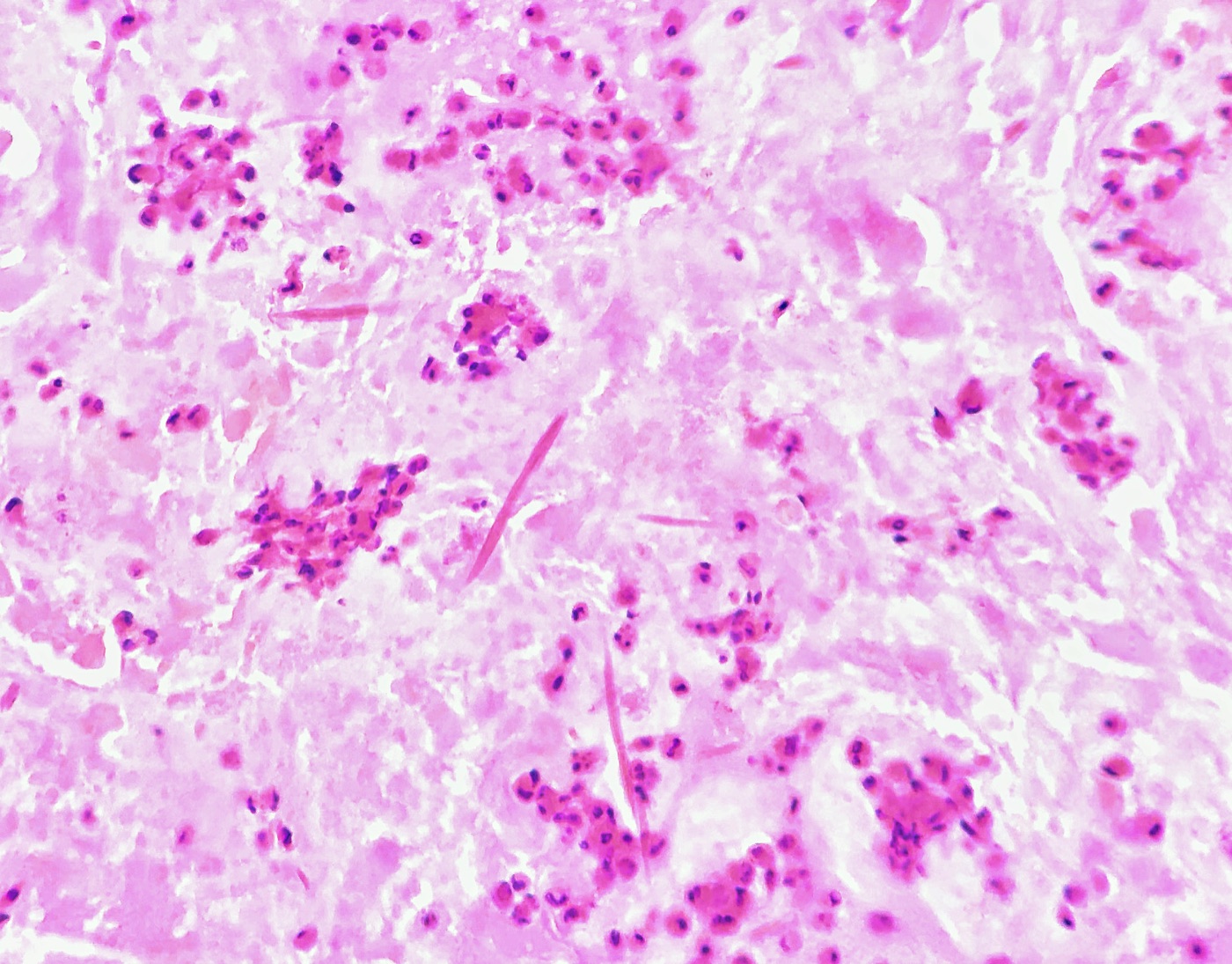
- Charcot-Leyden crystals are bipyramidal, needle-shaped eosinophilic crystals composed of galectin-10 derived from eosinophils (Persson 2019).
- Eosinophils accompany these elements.
Type II pneumocyte hyperplasia and diffuse alveolar damage in respiratory cytology
- Timing in BAL: in 62 BAL samples from 38 patients with acute respiratory distress syndrome, type II pneumocytes were present in 25% of samples from days 1–3, 44% from days 4–10 and 18% from days 21–32. None appeared after day 32 (Stanley 1992).
- Morphology: type II pneumocytes appear singly or in groups. Some resemble radiation-injured cells. Others are small, with a high N/C ratio, membrane irregularity and prominent nucleoli, and resemble adenocarcinoma (Stanley 1992).
- Diffuse alveolar damage (DAD) in BAL shows abundant macrophages and desquamated alveolar cells with variable atypia. Three-dimensional clusters have hobnailed peripheral cells. Flat "squamotized" sheets have dense cytoplasm. Dense basophilic or amphophilic amorphous material, the counterpart of hyaline membranes, accompanies both cluster types (Beskow 2000).
- Reporting: DAD is diagnosed or suggested in BAL only in the appropriate clinical setting (Beskow 2000).
Radiation and drug effect in respiratory cytology
- Radiation effect: cytomegaly with proportional nuclear enlargement, multinucleation, cytoplasmic vacuoles and polychromasia. The N/C ratio remains low. The nuclear chromatin is smudged or degenerated (IAC-IARC-WHO 2022).
- Drug-associated injury: type II cell hyperplasia accompanied fibrosis in 7 of 16 patients with amiodarone pulmonary toxicity and was not found without fibrosis (Bedrossian 1997).
- Separation from recurrent carcinoma: the history of therapy and the low N/C ratio separate treatment effect from recurrent carcinoma (IAC-IARC-WHO 2022).
Macrophage disorders in respiratory cytology
Lipoid pneumonia and lipid-laden macrophages
- Exogenous lipoid pneumonia follows aspiration of mineral, vegetable or animal oils. BAL shows lipid-laden macrophages after infection and hemorrhage are excluded (Meyer 2012).
- Oil Red O (ORO) specificity: among 50 consecutive BAL specimens from patients without vaping-associated lung injury, 45 contained ORO-positive macrophages. With a lipid-laden macrophage index of ≥85 as the threshold, 7 of 50 (14%) were positive. ORO positivity is therefore not specific for e-cigarette or vaping product use-associated lung injury (Jebastin Thangaiah 2022).
Amiodarone effect in BAL and FNA
- Foamy macrophages with lamellated cytoplasmic inclusions were present in every specimen from 16 symptomatic patients taking amiodarone. Their presence did not depend on other signs of toxicity, and they represent a routine drug effect (Bedrossian 1997).
- Controls: foamy macrophages were absent in BAL from 53 normal controls and rare in 27 patients with other interstitial lung diseases (Bedrossian 1997).
- Toxicity versus exposure: BAL cell counts and differentials did not separate amiodarone pneumonitis from other lung disease in treated patients. A reduced macrophage percentage had 82% sensitivity and 69% specificity. Amiodarone pneumonitis remains a diagnosis of exclusion (Ohar 1992).
Pulmonary alveolar proteinosis in BAL
- Definition: pulmonary alveolar proteinosis (PAP) is the accumulation of surfactant from macrophage dysfunction. Autoimmune PAP with GM-CSF autoantibodies accounts for >90% of cases. Hereditary PAP involves CSF2RA or CSF2RB mutations. Secondary and congenital forms also occur (Trapnell 2019).
- BAL findings: cloudy, milky to light brown fluid whose debris settles without centrifugation. The sediment contains PAS-positive amorphous material after hemorrhage, infection and malignancy are excluded (Meyer 2012).
- Treatment relevance: whole-lung lavage removes the excess surfactant (Trapnell 2019).
Asbestos bodies in respiratory cytology
- Morphology: asbestos bodies are ferruginous bodies with a thin, transparent core and a beaded, golden-brown iron-protein coat. The transparent core distinguishes them from other ferruginous bodies (Roggli 2010).
- BAL quantification: asbestos bodies were found in BAL from 15 of 20 occupationally exposed individuals, 9 of 31 patients with sarcoidosis and 2 of 5 with idiopathic pulmonary fibrosis. More than 1 asbestos body per 10⁶ cells indicated considerable occupational exposure. Occasional bodies were nonspecific (Roggli 1986).
- Asbestosis requires interstitial fibrosis of the appropriate pattern together with evidence of excess asbestos in tissue (Roggli 2010). BAL demonstrates exposure and does not establish asbestosis.
| Macrophage finding | Cytoplasm | Confirmatory test | Main association |
|---|---|---|---|
| Lipid-laden macrophages | Large, clear vacuoles | ORO on unfixed material | Lipoid pneumonia; low specificity for vaping injury |
| Amiodarone foamy macrophages | Fine foamy vacuoles; lamellated inclusions on electron microscopy | Drug history | Amiodarone exposure; toxicity is a diagnosis of exclusion |
| Hemosiderin-laden macrophages | Golden-brown granules | Iron stain | Chronic or occult alveolar hemorrhage |
| PAP | Few macrophages in granular acellular material | PAS | Autoimmune or secondary PAP |
| Asbestos bodies | Beaded golden bodies with a transparent core | Iron stain; quantification | Asbestos exposure |
Contaminants in transthoracic FNA and EBUS-TBNA
- Mesothelial cells form flat sheets with "windows" between cells when the needle crosses the pleura.
- Hepatocytes appear when a low posterior or lower-lobe approach crosses the diaphragm. Polygonal cells with granular cytoplasm, central round nuclei and bile pigment identify them.
- Bronchial epithelium and cartilage in EBUS-TBNA come from the airway wall that the needle crosses. These cells do not indicate metastasis to the lymph node. Source: IAC-IARC-WHO 2022. Adequacy criteria for EBUS-TBNA lymph nodes: Lung FNA Adequacy and Rapid On-Site Evaluation.
Differential diagnosis of reactive respiratory epithelium and adenocarcinoma
| Feature | Reactive bronchial cells and Creola bodies | Type II pneumocyte hyperplasia | Adenocarcinoma |
|---|---|---|---|
| Cilia or terminal bars | Present on some cells | Absent | Absent |
| Architecture | Flat sheets; Creola balls with a ciliated edge | Single cells and small groups | Three-dimensional groups, acini, papillae; many single cells |
| Nuclear features | Enlarged, uniform; smooth membranes | Enlarged; nucleoli; mild membrane irregularity | Pleomorphic; irregular membranes; macronucleoli |
| Population | Spectrum from normal to reactive | Spectrum within an injury setting | Separate, second population |
| Setting | Bronchoscopy, asthma, infection | ARDS, DAD, drug toxicity, transplant | Mass lesion on imaging |
Differential diagnosis of squamous metaplasia, oral contamination and squamous cell carcinoma in respiratory cytology
| Feature | Squamous metaplasia and repair | Oral contamination | Squamous cell carcinoma |
|---|---|---|---|
| Cells | Cohesive polygonal cells | Superficial and intermediate squames | Single keratinized cells and syncytia |
| Nuclei | Regular, smooth membranes | Small, pyknotic or vesicular | Enlarged, hyperchromatic, irregular; pyknotic in keratinized cells |
| Cytoplasm | Dense, cyanophilic | Thin, polygonal | Dense, orangeophilic keratin; bizarre shapes |
| Background | Clean or inflammatory | Bacteria, food particles | Necrosis and keratin debris |
Related page: Lung Squamous Cell Carcinoma (Cytology). Related page: Lung Adenocarcinoma (Cytology).
Respiratory Infections in Lung Cytology
Definition. Respiratory cytology identifies fungi, filamentous bacteria, viral cytopathic effect and parasites in sputum, bronchial washing, bronchoalveolar lavage (BAL), endobronchial ultrasound-guided transbronchial needle aspiration (EBUS-TBNA) and transthoracic fine-needle aspiration (FNA). An infection without a neoplasm is reported in the Benign category of the WHO Reporting System for Lung Cytopathology (Schmitt 2023). Cytology gives a rapid morphologic diagnosis, and culture, antigen tests or PCR identify the species and its susceptibility.
Related page: WHO Reporting System for Lung Cytopathology. Related page: Granulomas and Sarcoidosis on Lung and EBUS FNA.
Clinical use of cytology in respiratory infections
- BAL in immunocompromised hosts: in 604 BAL specimens from 344 patients, yeasts, pseudohyphae or hyphae occurred in 25.7%. Candida was the most frequent opportunistic fungus. Pneumocystis was found in 14 patients (4%), and herpes simplex virus (HSV) or cytomegalovirus (CMV) effect in 4 patients (1.2%) (Linder 1987).
- FNA of nodules and masses: in 15 FNAs with fungal infection, the lung was the most common site. Imaging most often suggested malignancy or infection. A genus-level diagnosis from yeast morphology was possible in 12 cases (5 Histoplasma, 4 Cryptococcus, 3 Coccidioides). The diagnosis was made within 24 hours in 9 cases, 4 of them at on-site evaluation (George 2020).
- Rapid on-site evaluation: when the on-site impression is infection, a dedicated pass is sent for culture (George 2020).
- Granulomas and mycobacteria: mycobacterial infection produces necrotizing granulomas or, in immunocompromised hosts, sheets of foamy histiocytes with numerous bacilli. Ziehl-Neelsen or auramine stains and culture identify the organisms (IAC-IARC-WHO 2022). Stain and culture workup of granulomatous inflammation: Granulomas and Sarcoidosis on Lung and EBUS FNA.
Pneumocystis jirovecii pneumonia in respiratory cytology
- Specimen of choice: Pneumocystis cannot be cultured. Bronchoscopy with BAL is the reference procedure for diagnosis (Huang 2011).
- Papanicolaou and Diff-Quik appearance: foamy alveolar casts (FACs) are frothy eosinophilic or cyanophilic aggregates that contain the organisms. HIV-infected patients had significantly more FACs and organism clusters than patients with other immunodeficiencies (Jacobs 2001).
- Background findings: half of the Pneumocystis pneumonia samples showed foamy alveolar macrophages, activated lymphocytes, plasma cells and reactive type II pneumocytes. These findings can suggest a noninfectious lung condition (Jacobs 2001).
- Stains: Grocott methenamine silver (GMS) stains the cyst wall. In 135 BAL samples from HIV-positive patients, May-Grünwald-Giemsa and toluidine blue O each detected 33.3%, Papanicolaou 31.1% and GMS 29.6%. The differences were not significant (Djamin 1998).
- BAL and brushing: BAL detected 10 infections that bronchial brushing missed. One infection was found by brushing alone. BAL with a single stain is appropriate (Djamin 1998).
- Immunofluorescence: among 47 BAL samples, Pneumocystis was detected in 1 by Papanicolaou smear, 4 by GMS stain and 8 by immunofluorescence. Five of 9 positive cases were detected by immunofluorescence only (Delvenne 1993).
- Immunohistochemistry on cell block: in 895 BAL cell blocks stained simultaneously, Pneumocystis immunohistochemistry detected 14 positive samples. GMS was read as positive in 6 of 14 (42.8%), rising to 12 of 14 after autostainer maintenance. Immunohistochemistry detected more organisms per case (Gonzalez 2017).
- PCR on BAL: a meta-analysis of 16 studies gave a pooled sensitivity of 98.3% and specificity of 91.0%. At 20% prevalence, a negative BAL PCR corresponds to a post-test probability below 0.5% (Fan 2013).
- Serum (1→3)-β-D-glucan: pooled sensitivity was 83.5% and specificity 75.5% at 80 pg/mL. At 400 pg/mL, specificity rose to 93.6% (Prosty 2025).
Filamentous fungi in respiratory cytology
Aspergillus in respiratory cytology
- Morphology: narrow, septate hyphae of uniform width with acute-angle, dichotomous branching. Conidial heads (fruiting bodies) form in cavities exposed to air (IAC-IARC-WHO 2022).
- Detection: Aspergillus was readily identified by cytologic examination and by culture in BAL (Linder 1987). Cytology and GMS stain detected the same 2 of 47 positive BAL specimens (Delvenne 1993).
- Calcium oxalate crystals: birefringent needle-like crystals in rosette or wheat-sheaf arrangements occurred in pulmonary cytology from 11 of 65 patients with Aspergillus infection and in none of 60 controls. A. niger accounted for 45.4% of crystal-associated cases. Crystals preceded positive culture by more than a year in 2 patients (Farley 1985).
- Atypia: 64% of patients with crystals also showed moderate to severe cytologic atypia (Farley 1985). Reactive atypia adjacent to a fungus ball mimics squamous cell carcinoma.
Mucorales in respiratory cytology
In a series of 83 FNAs with mucormycosis, 57 were from the lung (Shastri 2024).
- Morphology: broad, nonseptate, folded, ribbon-like hyphae with right-angle branching (Shastri 2024).
- Tissue reaction: necroinflammatory in 36 cases, bland necrosis in 22, mixed inflammation in 16, suppurative in 5 and granulomatous in 4. Immunocompromised patients more often showed mixed inflammation (Shastri 2024).
- Culture: fungal culture grew Rhizopus in only 2 of 13 cultured cases. Morphology often provides the only rapid diagnosis (Shastri 2024).
Yeasts in respiratory cytology
- Candida: budding yeasts and pseudohyphae. Small numbers in sputum and washings reflect oral contamination. Patients with clinically significant infection had many budding yeasts and pseudohyphae in BAL (Linder 1987).
- Cryptococcus: round yeasts of variable size with narrow-based budding and a thick mucoid capsule. Mucicarmine stains the capsule of typical C. neoformans. Capsule-deficient organisms are mucicarmine-negative. The Fontana-Masson stain is positive in both capsule-deficient and typical Cryptococcus and was negative in the other yeasts examined in that study (Ro 1987).
- Histoplasma: small, uniform intracellular yeasts within macrophages. Histoplasma was the most frequent genus-level yeast diagnosis in an FNA series (George 2020).
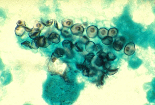
- Coccidioides: in 73 lung FNAs, smears showed abundant granular eosinophilic debris with little inflammation. Granulomas were present in 3 cases only. In that series, spherules measured about 20–200 µm, were often crushed or fractured, and contained endospores. Culture grew the organism in 9 of 44 cultured cases (Raab 1993).
- Coccidioidomycosis in other specimens: BAL showed the widest range of forms, including immature, endosporulating, empty and ruptured spherules, with acute inflammation. Lung FNA showed necrosis with little inflammation. Immunocompetent patients outnumbered immunocompromised patients (Aly 2016).
- Blastomyces: large yeasts with a thick, double-contoured wall and broad-based budding (IAC-IARC-WHO 2022).
Organisms in respiratory cytology: morphology and stains
| Organism | Form in cytology | Distinguishing feature | Helpful stain or test |
|---|---|---|---|
| Pneumocystis jirovecii | Foamy alveolar casts with small cysts | Cup-shaped cysts within frothy casts | GMS; immunofluorescence or immunohistochemistry; BAL PCR |
| Aspergillus | Narrow septate hyphae | Acute-angle branching; conidial heads; oxalate crystals | GMS, PAS; culture |
| Mucorales | Broad ribbon-like hyphae | Few or no septa; right-angle branching; folding | GMS, PAS; culture, molecular testing |
| Candida | Budding yeasts and pseudohyphae | Pseudohyphae with constrictions | GMS, PAS |
| Cryptococcus | Round yeasts of variable size | Narrow-based budding; mucoid capsule | Mucicarmine; Fontana-Masson for capsule-deficient forms |
| Histoplasma | Small intracellular yeasts | Uniform yeasts in macrophages | GMS; urine or serum antigen |
| Blastomyces | Large yeasts | Thick double-contoured wall; broad-based budding | GMS, PAS |
| Coccidioides | Spherules with endospores | Spherules about 20–200 µm; crushed forms | GMS, PAS; culture, serology |
| Actinomyces | Thin branching filaments in colonies | Sulfur granules; not acid-fast | Gram, GMS |
| Nocardia | Thin beaded branching filaments | Weakly acid-fast | Gram, GMS, modified acid-fast (Fite) |
| Strongyloides stercoralis | Larvae | Larvae among inflammatory cells in hyperinfection | Papanicolaou; stool and serology |
Sources: Raab 1993; Ro 1987; McHugh 2017; Shastri 2024; Farley 1985; IAC-IARC-WHO 2022.
Filamentous bacteria in respiratory cytology
- Shared features: Actinomyces and Nocardia both form thin, beaded, branching, Gram-positive, GMS-positive filaments that fragment into bacillary and coccoid forms at their edges (McHugh 2017).
- Separating features: the width of the filaments, the angle of branching and retention of a modified acid-fast stain separate them. Nocardia is modified acid-fast, and Actinomyces is not (McHugh 2017).
- Clinical relevance: the two genera differ in antibiotic susceptibility and in the need for early surgery, so the distinction changes management (McHugh 2017).
- Actinomyces in sputum: colonies from the oral cavity and tonsillar crypts contaminate sputum. They indicate infection only with a matching lesion and an inflammatory reaction (IAC-IARC-WHO 2022).
- Nocardia on FNA: 4 FNAs of pulmonary nocardiosis showed neutrophils, histiocytes and necrosis, with a granuloma in 1. Modified Ziehl-Neelsen stain showed long, slender, branching filaments. Three patients were immunocompromised (Sharma 2016).
Viral cytopathic effect in respiratory cytology
| Virus | Cytopathic effect | Setting | Supporting test |
|---|---|---|---|
| Herpes simplex virus | Multinucleation, nuclear molding, ground-glass chromatin with margination, Cowdry A intranuclear inclusions | Prolonged mechanical ventilation; oral-labial lesions | PCR; immunocytochemistry |
| Cytomegalovirus | Cytomegaly; single large intranuclear inclusion with a halo; granular cytoplasmic inclusions | Transplant recipients; HIV | Immunocytochemistry; in situ hybridization; culture |
| Adenovirus | Smudged, dark intranuclear inclusions | Transplant recipients, children | Immunohistochemistry; in situ hybridization |
| Measles virus | Multinucleated giant cells with intranuclear and cytoplasmic inclusions | Children with leukemia; giant cell pneumonia without rash | Culture; immunofluorescence; serology |
| Respiratory syncytial virus | Pink cytoplasmic inclusions; syncytial cells | Bone marrow and stem cell transplant recipients | Immunofluorescence; PCR |
Sources: Luyt 2007; Paradis 1988; Ohori 1995; Siegel 1990; Parham 1993; Zaman 1996.
- HSV in ventilated patients: HSV bronchopneumonitis, defined by HSV in BAL plus HSV nuclear inclusions in lavage cells or bronchial biopsies, occurred in 42 of 201 (21%) nonimmunocompromised patients who deteriorated after ≥5 days of mechanical ventilation. Oral-labial lesions, HSV in the throat and macroscopic bronchial lesions were risk factors (Luyt 2007).
- CMV sensitivity of cytology: in 71 BAL specimens from 56 immunocompromised patients, CMV inclusions were found in 3 of 14 patients with CMV pneumonia (sensitivity 21%, specificity 98%). Culture was 100% sensitive and 70% specific. Immunocytochemistry detected CMV antigen in 9 of 11 culture-positive, cytology-negative cases (Paradis 1988).
- CMV in situ hybridization: cytology detected CMV in 2 of 47 BAL specimens and in situ hybridization in 7. Every cell with an inclusion labeled with the probe (Delvenne 1993).
- Adenovirus: 4 of 308 lung transplant recipients developed adenovirus pneumonia, 3 of them children. All 4 died within 45 days. Smudged intranuclear inclusions with immunohistochemical and in situ hybridization positivity were present at autopsy (Ohori 1995).
- Measles: giant cell pneumonia is diagnosed by cytology of nasal or bronchial secretions that show multinucleated giant cells with intranuclear and intracytoplasmic inclusions. Measles was isolated from bronchial washing and sputum of a child in leukemia remission without a rash (Siegel 1990).
- RSV: BAL from transplant recipients showed discrete pink cytoplasmic inclusions, confirmed by immunofluorescence and electron microscopy (Parham 1993; Zaman 1996).
Parasites in respiratory cytology
- Strongyloides stercoralis hyperinfection affects immunocompromised patients, particularly those receiving long-term corticosteroids, and can be fatal. A bronchial washing from a 61-year-old male on corticosteroids contained abundant larvae, acute inflammatory cells and bronchial cells with basal cell hyperplasia or regenerative atypia. The larvae were recognizable in both conventional and ThinPrep preparations (Grapsa 2009).
- Regenerative bronchial atypia accompanying larvae is a reactive finding and does not indicate carcinoma (Grapsa 2009).
Sources of error in the diagnosis of respiratory infections in lung cytology
| Finding | Misinterpretation | Separating feature |
|---|---|---|
| Foamy alveolar casts | Proteinaceous fluid or mucus | GMS or immunostain shows cysts |
| Reactive atypia next to a fungus ball | Squamous cell carcinoma | Oxalate crystals and hyphae; clinical cavity |
| Oral Candida or Actinomyces in sputum | Pulmonary infection | Few organisms; no inflammatory reaction or lesion |
| Necrotic FNA with few organisms | Necrotic carcinoma | GMS on smears and cell block; culture pass |
| Degenerated ciliated cells | Viral cytopathic effect | No inclusions; ciliocytophthoria |
| Type II pneumocytes with viral inclusions | Adenocarcinoma | Inclusions; infection setting |
| Capsule-deficient Cryptococcus | Histoplasma or Candida | Fontana-Masson positive; variable size |
Related page: Normal and Reactive Findings in Respiratory Cytology.
Reporting of respiratory infections in the WHO Reporting System for Lung Cytopathology
- The Benign category covers unequivocally benign findings with or without a specific benign diagnosis, such as granulomas. Its management depends on clinical confirmation of an inflammatory lesion (Schmitt 2023).
- An infection without malignant cells is reported in this category with a specific descriptive diagnosis when the morphology allows one (IAC-IARC-WHO 2022).
- A genus-level morphologic diagnosis is stated with a recommendation for culture or molecular confirmation. Culture was negative in many morphologically diagnosed fungal infections (George 2020; Shastri 2024).
Lymphoproliferative and Spindle Cell Lesions on Lung FNA
Definition. Lymphoid and spindle cell lesions are uncommon in lung and mediastinal cytology, and both groups are frequent sources of diagnostic error. Lymphoid lesions range from reactive lymphoid hyperplasia to extranodal marginal zone lymphoma, large B-cell lymphoma and classic Hodgkin lymphoma. Spindle cell and mesenchymal lesions range from granulomas and benign tumors such as pulmonary hamartoma and solitary fibrous tumor to sarcomatoid carcinoma and sarcoma. They are classified according to the WHO Classification of Thoracic Tumours, 5th edition (WHO 2021), and reported in the five categories of the WHO Reporting System for Lung Cytopathology (Schmitt 2023).
Related page: WHO Reporting System for Lung Cytopathology.
Lymphoproliferative lesions on lung and EBUS FNA
Sampling for suspected lymphoma on EBUS-TBNA
- Meta-analysis: in 14 studies with 425 participants, EBUS-TBNA had a pooled sensitivity of 66.2% and specificity of 99.3% for lymphoma. Sensitivity was 67.1% for a new diagnosis and 77.8% for recurrence (Labarca 2019).
- Factors that raised sensitivity in recurrent lymphoma were rapid on-site evaluation, sample size and flow cytometry (Labarca 2019).
- Hodgkin lymphoma: in a series of 189 patients with isolated mediastinal lymphadenopathy, EBUS-TBNA was diagnostic in 3 of 12 procedures for suspected new lymphoma, all non-Hodgkin. No case of Hodgkin lymphoma was diagnosed by EBUS-TBNA. A negative result does not exclude lymphoma, and mediastinoscopy or another biopsy follows when suspicion is high (Erer 2017).
- Triage: when on-site evaluation shows a monotonous or atypical lymphoid population, additional passes are collected for flow cytometry and a cell block (IAC-IARC-WHO 2022).
Reactive lymphoid hyperplasia on lung and EBUS FNA
- Cytomorphology: a polymorphous mixture of small lymphocytes, centrocytes, centroblasts, immunoblasts and tingible body macrophages. Anthracotic macrophages are common in mediastinal nodes (IAC-IARC-WHO 2022).
- Flow cytometry shows polytypic B cells and a mixture of T cells (IAC-IARC-WHO 2022).
- Overlap with marginal zone lymphoma: marginal zone lymphoma is also heterogeneous, with small lymphocytes, transformed cells, plasma cells and lymphohistiocytic aggregates. Monotypic light chain expression is required to separate them (Crapanzano 2003).
Extranodal marginal zone lymphoma of the lung
- Clinical features: pulmonary marginal zone lymphoma of mucosa-associated lymphoid tissue is the most common primary pulmonary lymphoma. In 71 patients, the median age was 63 years and 60.6% were asymptomatic. A predisposing systemic disorder was present in 32.4%, most often primary Sjögren syndrome (19.7%). Consolidation and nodular or mass-like lesions were each present in 87%, often with an air bronchogram. The diagnostic rate was 33% for FNA and 72.4% for core needle biopsy (Tzilas 2025).
- Cytomorphology: in 13 FNAs of marginal zone lymphoma from several sites, intermediate-sized cells with moderate cytoplasm, slight nuclear membrane irregularity and inconspicuous nucleoli predominated. Plasmacytoid morphology was present in 10 specimens. Monocytoid cells, plasma cells, lymphohistiocytic aggregates and tingible body macrophages were variable. Lymphoepithelial lesions were not seen. Flow cytometry showed a compatible phenotype in 3 of 6 specimens and was nondiagnostic in the other 3, so a nondiagnostic flow result does not exclude marginal zone lymphoma (Crapanzano 2003).
- Pulmonary series: in 15 lung FNAs of marginal zone lymphoma, smears contained mainly small centrocyte-like cells. Marked plasma cell differentiation was present in 11. Tissue fragments with lymphoid tangles were present in every case with slides available. Multinucleated giant cells occurred in 9 cases and granulomas in 2 (Ko 2014).
- Ancillary tests: immunophenotyping confirmed B-cell clonality in all 15 cases. FISH showed a MALT1 translocation in 4 of 10 tested cases and trisomy 3 in 3 of 4 tested cases (Ko 2014).
- Granulomas do not exclude lymphoma. Giant cells and poorly formed granulomas occur in marginal zone lymphoma, and a lymphoid smear with many plasmacytoid cells and lymphoid tangles is sent for immunophenotyping (Ko 2014).
- BAL: in 3 patients with pulmonary marginal zone lymphoma, BAL showed lymphocytosis, B cells of 28–52% and monotypic light chain expression in 2 (Poletti 1995).
Large B-cell and Hodgkin lymphomas in the mediastinum and lung
- Diffuse large B-cell lymphoma shows a dispersed population of large lymphoid cells with lymphoglandular bodies in the background.
- Classic Hodgkin lymphoma contains few Hodgkin and Reed-Sternberg cells in a mixed background of small lymphocytes, eosinophils, histiocytes and plasma cells.
- Primary mediastinal large B-cell lymphoma is an anterior mediastinal large B-cell lymphoma of young adults with sclerosis (WHO 2021). Sclerotic tumors yield scant aspirates, and core biopsy supports the diagnosis.
Related page: Diffuse Large B-Cell Lymphoma (Cytology). Related page: Classic Hodgkin Lymphoma (Cytology). Related page: Small Lymphocytic Lymphoma and Chronic Lymphocytic Leukemia (Cytology). Related page: Follicular Lymphoma (Cytology). Related page: Mantle Cell Lymphoma (Cytology).
Post-transplant and EBV-driven lymphoproliferative disorders of the lung
- Lung transplant recipients: in 14 recipients who underwent EBUS-TBNA, a mean of 15 months after transplant, the aspirate was diagnostic of malignancy in 10 (71%). Ancillary studies to establish clonality in post-transplant lymphoproliferative disorder were feasible on this material (Sturgis 2015).
- Lymphomatoid granulomatosis is a rare EBV-associated B-cell lymphoproliferative disease. It always involves the lungs and shows EBV-positive atypical B cells in an angiocentric, angiodestructive T-cell infiltrate with necrosis. Grade depends on the number and density of EBV-positive B cells. Nodal or marrow involvement is extremely rare and suggests another diagnosis (Melani 2020).
| Feature | Reactive lymphoid hyperplasia | Marginal zone lymphoma | Diffuse large B-cell lymphoma | Classic Hodgkin lymphoma | Small cell carcinoma |
|---|---|---|---|---|---|
| Population | Polymorphous | Small to intermediate cells, plasmacytoid cells | Monotonous large cells | Few large atypical cells in a mixed background | Cohesive groups and single cells |
| Distinctive finding | Tingible body macrophages | Plasma cells; lymphoid tangles; giant cells | Lymphoglandular bodies | Reed-Sternberg cells; eosinophils | Molding, crush artifact, necrosis |
| Flow cytometry | Polytypic B cells | Monotypic CD5−, CD10− B cells | Monotypic or failed (fragile cells) | Usually nondiagnostic | Not applicable |
| Key immunostain or test | Mixed B and T cells | MALT1 FISH, trisomy 3 | CD20, PAX5; Ki-67 high | CD30, CD15, PAX5 (weak) | Keratin, neuroendocrine markers, TTF-1 |
Sources: Crapanzano 2003; Ko 2014; Erer 2017; WHO 2021.
Related page: Small Cell Lung Carcinoma (Cytology).
Spindle cell and mesenchymal lesions on lung FNA
Frequency of spindle cell lesions on transthoracic FNA
- In 779 percutaneous lung FNAs, 61 (7.8%) had a dominant spindle cell component (Hummel 2001).
- Reactive processes made up 33 (54%): 31 granulomas, 1 organizing pneumonia and 1 inflammatory pseudotumor (Hummel 2001).
- Benign neoplasms made up 5: 2 hamartomas, 2 solitary fibrous tumors and 1 schwannoma (Hummel 2001).
- Malignant neoplasms made up 23 (38%). The 8 primary tumors included 5 carcinomas with spindle cell or sarcomatoid features, 1 spindle cell carcinoid, 1 leiomyosarcoma and 1 synovial sarcoma. The 15 metastases included 9 melanomas (Hummel 2001).
- A specific diagnosis was made in 52 of 61 (85%), with no false-positive diagnoses. Organizing pneumonia was misread as a solitary fibrous tumor (Hummel 2001).
- Granulomas are the most common spindle cell finding. Epithelioid histiocytes in spindled aggregates are the main source of a spindle cell pattern. Related page: Granulomas and Sarcoidosis on Lung and EBUS FNA.
Pulmonary hamartoma on FNA
- Diagnostic components: in 14 fine-needle biopsies, fibromyxoid stroma was present in 93%, chondroid material in 79% and both in 71%. Chondroid material was scant in more than half. Epithelial cells were never the only prominent component, and significant epithelial atypia was absent (Wood 2008).
- S100 stained the stromal material in every case with stroma in the cell block (Wood 2008).
- Fibromyxoid fragments are wispy, discrete fragments of fibroblasts in fibrillary ground substance. They are more useful than mature cartilage, which appears in a minority of aspirates. Bronchial cells and adipocytes have no diagnostic value without fibromyxoid tissue or cartilage (Dunbar 1989).
- Performance in proficiency testing: across 766 participant responses for 19 hamartoma FNAs in the CAP nongynecologic program, the false-positive rate was 22%. The most common false-positive diagnoses were carcinoid tumor, adenocarcinoma and small cell carcinoma. Only 26% of responses gave the specific diagnosis. The specificity for a benign interpretation was 78% (Hughes 2005).
- Imaging correlation: knowledge of the radiologic appearance of the target lesion supports a confident cytologic diagnosis of hamartoma (Dunbar 1989).
Solitary fibrous tumor on FNA
- Cytomorphology: low to moderate cellularity of mixed oval, elongated, round and stellate cells with pink collagenous stroma and hypercellular clusters. Benign and malignant tumors overlapped cytologically. Nuclear pleomorphism was the most notable difference (Tani 2018).
- STAT6: nuclear STAT6 expression was present in 59 of 60 solitary fibrous tumors (98%). Other tumor types, including sarcomatoid mesotheliomas, were negative, apart from weak staining in 3 dedifferentiated liposarcomas and 1 deep fibrous histiocytoma (Doyle 2014).
- STAT6 on cytology: nuclear STAT6 staining identified solitary fibrous tumor on FNA material. Cytologic mimics showed cytoplasmic staining only (Tani 2018).
- Genetics: a NAB2::STAT6 fusion defines the tumor (Doyle 2014).
Inflammatory myofibroblastic tumor on FNA
- Site distribution: of 20 inflammatory myofibroblastic tumors with cytology material, 8 (40%) arose in the lung (Stoll 2011).
- Cytomorphology: bland spindle cells with oval nuclei and small prominent nucleoli in a background of lymphocytes and plasma cells. Focal atypia and ganglion-like cells occurred in 7 cases (Stoll 2011).
- Immunophenotype: ALK was positive in 8 of 17 (47.1%) and actin in 10 of 10. Keratin was variable and Ki-67 low. FISH confirmed ALK rearrangement in 3 of 3 tested cases (Stoll 2011).
- ALK-negative tumors: about 50% of inflammatory myofibroblastic tumors carry ALK rearrangements. ALK-negative tumors can carry ROS1 or PDGFRB fusions, and 85% of tumors sequenced in one series carried a kinase fusion (Lovly 2014).
- A spindle cell lesion with a prominent inflammatory background raises the possibility of inflammatory myofibroblastic tumor, and ALK immunostaining is performed on the cell block (Stoll 2011).
Synovial sarcoma on lung and mediastinal FNA
- Cytomorphology: in 38 FNAs, smears were highly cellular, with clusters and single cells of mixed round, oval and spindle cells. A pericapillary arrangement was frequent. Pleomorphism and mitoses were rare (Zhang 2019).
- Metastatic and recurrent tumors: aspirates of primary, recurrent, pulmonary and mediastinal synovial sarcoma showed cohesive clusters of spindle cells with ovoid hyperchromatic nuclei, inconspicuous nucleoli and scant tapering cytoplasm. Multinucleated tumor giant cells were absent. In biphasic tumors the spindle cells outnumbered the epithelial cells, which makes biphasic and monophasic tumors difficult to separate on smears (Kilpatrick 1996).
- Immunophenotype: vimentin, EMA, BCL2 and TLE1 were frequently positive. Keratins were occasionally positive in biphasic tumors (Zhang 2019).
- TLE1 specificity: TLE1 was positive in 82% of 73 synovial sarcomas. It was also positive, mostly weakly, in 15% of malignant peripheral nerve sheath tumors and 8% of solitary fibrous tumors (Foo 2011).
- Molecular confirmation: the SS18::SSX fusion was detected in every FNA by PCR or FISH (Zhang 2019).
Schwannoma, sarcomatoid carcinoma and other spindle cell lesions on lung FNA
- Schwannoma of the posterior mediastinum or chest wall yields cohesive fragments of spindle cells with wavy nuclei and fibrillary stroma. S100 and SOX10 are positive (WHO 2021).
- Pleomorphic and sarcomatoid carcinoma accounted for 5 of 8 primary malignant spindle cell lesions in one series (Hummel 2001). Keratin and TTF-1 or p40 positivity, even when focal, supports carcinoma (WHO 2021).
- Spindle cell carcinoid tumor forms loosely cohesive spindle cells with salt-and-pepper chromatin. Related page: Typical and Atypical Carcinoid Tumor of the Lung (Cytology).
- Sclerosing pneumocytoma is a benign tumor of surface and stromal cells that occurs mainly in women. In 6 cytology specimens, architecture was papillary, solid, single-cell or acinar. Atypia occurred in 4. Three were first diagnosed as adenocarcinoma. Surface and stromal cells were positive for TTF-1 and EMA. Only surface cells expressed keratin and napsin A (Policarpio-Nicolas 2025).
- Metastatic sarcoma and melanoma: Metastatic Tumors to the Lung (Cytology).
- Sarcomatoid mesothelioma: Diffuse Mesothelioma in Effusions.
Immunohistochemistry of spindle cell lesions on lung FNA
| Lesion | Keratin | S100 / SOX10 | STAT6 | ALK | Other markers | Molecular finding |
|---|---|---|---|---|---|---|
| Pulmonary hamartoma | Entrapped epithelium only | S100 in stroma | Negative | Negative | — | — |
| Solitary fibrous tumor | Negative | Negative | Nuclear positive | Negative | CD34 | NAB2::STAT6 |
| Inflammatory myofibroblastic tumor | Variable | Negative | Negative | Positive in about half | Actin | ALK rearrangement; ROS1 or PDGFRB fusion in some ALK-negative tumors |
| Synovial sarcoma | Focal; biphasic tumors | Negative | Negative | Negative | EMA, BCL2, TLE1 | SS18::SSX |
| Schwannoma | Negative | Positive | Negative | Negative | — | — |
| Sarcomatoid carcinoma | Positive, often focal | Negative | Negative | Negative | TTF-1 or p40 in some | — |
| Spindle cell carcinoid | Positive | Sustentacular cells only | Negative | Negative | Chromogranin, synaptophysin, INSM1 | — |
TLE1 also stains some malignant peripheral nerve sheath tumors and solitary fibrous tumors, mostly weakly (Foo 2011).
Sources: Wood 2008; Doyle 2014; Tani 2018; Stoll 2011; Lovly 2014; Zhang 2019; Foo 2011; WHO 2021.
Reporting of lymphoid and spindle cell lesions in the WHO Reporting System for Lung Cytopathology
- A benign neoplasm, such as pulmonary hamartoma, is reported in the Benign category with a specific diagnosis. Limited resection follows according to tumor type and symptoms (Schmitt 2023).
- Scant cells that suggest a lymphoproliferative disorder and bland spindle cell lesions without a specific diagnosis are examples of the Atypical category. Category examples and management: WHO Reporting System for Lung Cytopathology.
- A lymphoma or sarcoma confirmed by flow cytometry, immunohistochemistry or molecular testing is reported as Malignant with its WHO name (WHO 2021).
Respiratory Specimen Types, Sputum Adequacy, Inhaled Particles, Storage Disease and Alveolar Hemorrhage
Definition. Exfoliative respiratory cytology comprises sputum, bronchial washing, bronchial brushing and bronchoalveolar lavage (BAL). Together with fine-needle aspiration biopsy, these specimens are reported with the WHO Reporting System for Lung Cytopathology (Dolezal et al. 2024). Beyond tumor diagnosis, exfoliative specimens document inhaled mineral particles, lysosomal storage cells in alveolar macrophages and hemosiderin-laden macrophages of alveolar hemorrhage (Moreira et al. 2005; Borie et al. 2021; De Lassence et al. 1995). Neoplastic entities follow the WHO Classification of Thoracic Tumours, 5th edition (2021) (Nicholson et al. 2022).
Related page: Normal and Reactive Findings in Respiratory Cytology (normal cells by specimen type, BAL differential patterns, lipid-laden and amiodarone macrophages, alveolar proteinosis, asbestos bodies) Related page: Lung FNA Adequacy and Rapid On-Site Evaluation (WHO adequacy criteria for each specimen type and rapid on-site evaluation) Related page: Respiratory Infections in Lung Cytology (organisms in respiratory specimens) Related page: Granulomas and Sarcoidosis on Lung and EBUS FNA (granulomas and sarcoidosis) Related page: WHO Reporting System for Lung Cytopathology (diagnostic categories and risk of malignancy)
Respiratory specimen types and their preparation
| Specimen | Collection | Preparation | Main use |
|---|---|---|---|
| Spontaneous sputum | Deep cough, preferably early morning, after rinsing the mouth | Direct smears from selected material; or alcohol–polyethylene glycol fixation, blending and concentration (Saccomanno method) | Lung cancer diagnosis; sensitivity 71% for central and 49% for peripheral lesions |
| Induced sputum | Nebulized hypertonic saline before collection | Fresh, unfixed submission | Opportunistic infection, including Pneumocystis |
| Post-bronchoscopy sputum | Sputum produced after bronchoscopy | As for spontaneous sputum | Additional yield when bronchoscopic samples are negative |
| Bronchial washing | Saline instilled into the airway and aspirated | Cytocentrifuge, liquid-based slides or cell block | Combined with brushing and biopsy at bronchoscopy |
| Bronchial brushing | Brush scraped over a visible or fluoroscopically targeted lesion | Direct smears or brush rinse | Central and peripheral lesions |
| BAL | Wedged bronchoscope, saline instilled and recovered | As for bronchial washing; differential count | Diffuse non-neoplastic disease, opportunistic infection, peripheral tumors |
Sources: Sheaff and Singh 2013; Saccomanno et al. 1963; Gottschall et al. 2005; Rizzo et al. 1990; Schreiber and McCrory 2003; Rivera et al. 2013; Chopra et al. 1977; Kiska et al. 1998.
- Sputum storage. Early-morning sputum remains usable after overnight refrigeration (Sheaff and Singh 2013).
- Concentration method. In the Saccomanno method, sputum is collected into an alcohol fixative containing polyethylene glycol, homogenized in a blender, concentrated and smeared (Saccomanno et al. 1963; Gottschall et al. 2005). In 249 consecutive specimens, blended Saccomanno preparations contained more diagnostic cells than fresh pick-and-smear preparations, with fewer false negatives (Rizzo et al. 1990). The pellet can also be used for a cell block (Sheaff and Singh 2013).
- Pre- and post-bronchoscopy sputum. In 70 patients with proven lung cancer, bronchoscopy was diagnostic in 41 of 52 primary carcinomas (79%). Brushing and forceps biopsy each gave the highest yield (67%), and brushing reached more peripheral lesions. Pre- and post-bronchoscopy sputum was positive in 2 cases in which brushing and biopsy were negative. Bronchial washing did not add significantly to the yield (Chopra et al. 1977).
- Number of sputum samples. In 400 consecutive patients with histologically proven lung carcinoma (mean 2.8 samples each), overall sputum sensitivity was 0.58. It rose from 0.37 with one sample to 0.57 with three, and by only 0.01 more with four to six samples. Cell-block (paraffin) examination added a further 0.075 (Pilotti et al. 1982).
- Pooled performance. In a systematic review, sputum cytology had a pooled sensitivity of 66% and specificity of 99%. Sensitivity was 71% for central and 49% for peripheral lesions (Schreiber and McCrory 2003; Rivera et al. 2013).
- Induced sputum volume. Of 177 induced sputum specimens examined for Pneumocystis by direct fluorescent antibody stain, 47.4% were below the recommended 2 mL. Positivity was 18% for specimens of 2 mL or more and 19% for 0.5–1.9 mL, so low-volume specimens were acceptable (Kiska et al. 1998). Organism morphology is covered in Respiratory Infections in Lung Cytology.
- BAL quality criteria and cellular patterns are described in Normal and Reactive Findings in Respiratory Cytology.
Sputum adequacy for cytology and for culture
Sputum is screened for different purposes in the cytology and microbiology laboratories, and the two adequacy criteria are not interchangeable.
| Purpose | Criterion | Basis |
|---|---|---|
| Cytology for malignancy | At least a few alveolar macrophages, ciliated columnar bronchial cells and enough volume for at least two smears | WHO Reporting System for Lung Cytopathology (Dolezal et al. 2024) |
| Bacterial culture: rejection | More than 10 squamous epithelial cells per low-power (100×) field | Such specimens grew more than 4 species, mostly oropharyngeal flora (Murray and Washington 1975) |
| Bacterial culture: acceptance | Fewer than 10 squamous cells per low-power field | Flora resembled that of transtracheal aspirates (Murray and Washington 1975) |
| Bacterial culture: highest quality (group 5) | More than 25 leukocytes and fewer than 10 buccal squamous cells per 100× field | A potential pathogen in a group 5 sputum predicted growth in the paired transtracheal aspirate in 94% (Geckler et al. 1977) |
- Agreement with transtracheal aspirates. Sputa with fewer than 25 buccal squamous cells per 100× field agreed with paired transtracheal aspirates in 79%. Sputa with more than 25 agreed in 27% (Geckler et al. 1977).
- Macrophages and culture. Bronchial epithelial cells and alveolar macrophages in group 5 sputa did not significantly increase their value for culture (Geckler et al. 1977).
- Macrophages and cytology. Saliva can still contain tumor cells, which are reported when present (Sheaff and Singh 2013).
Inhaled mineral particles and pneumoconioses in respiratory cytology
Silica and silicate particles in BAL
- Birefringence. Crystalline silica particles are weakly birefringent under polarized light, and silicates are more strongly birefringent (Hua et al. 2025). Strong birefringence in a silicotic nodule is attributed to accompanying silicates such as mica and talc, and the absence of birefringent particles does not exclude silicosis (Ehrlich et al. 2024).
- BAL in silicosis. Silicotic patients had a marked increase in BAL macrophages containing particles by polarizing microscopy compared with healthy controls. Simple and complicated silicosis did not differ. In acute silicosis with massive exposure, more than 70% of BAL macrophages contained dust particles (Moreira et al. 2005).
- Time since exposure. The proportion of particle-laden macrophages tended to fall with longer retirement from work (Moreira et al. 2005).
Ferruginous bodies with non-asbestos cores
- Foundry workers. BAL from 5 foundry workers contained ferruginous bodies that resembled asbestos bodies at 200×. At 400×, a separate population with a thin black ribbon core was identified (Dodson et al. 1993).
- Electron microscopy. Transmission electron microscopy confirmed non-asbestos cores in most bodies with black cores. A population of non-asbestos ferruginous bodies with transparent cores also existed (Dodson et al. 1993).
- Asbestos bodies and their quantification in BAL are covered in Normal and Reactive Findings in Respiratory Cytology.
Hard metal lung disease
- Cause. Hard metal lung disease is an occupational interstitial lung disease of workers exposed to tungsten carbide dust. Its characteristic pathology is giant cell interstitial pneumonia (Takada et al. 2014). Cobalt exposure in the hard metal and bonded diamond tool industries causes the disease (Adams et al. 2017).
- BAL. Cannibalistic multinucleated giant cells in BAL are diagnostic with a history of exposure and appropriate imaging. When these cells are absent, lung biopsy is required (Adams et al. 2017).
- Lymphocytes. In 19 patients with tungsten in lung tissue, the giant cell interstitial pneumonia group had a higher BAL lymphocyte percentage (31.5%) than the fibrosis group (3.22%). The patients were younger and had shorter exposure (Tanaka et al. 2014).
- Other patterns. Cobalt-related disease also presents with usual interstitial pneumonia, desquamative interstitial pneumonia or hypersensitivity pneumonitis patterns (Adams et al. 2017).
- Elemental analysis. Mineralogical analysis is not required for most occupational lung diseases. Small quantities of mineral dust are present in healthy lungs, so quantitative or semiquantitative methods such as electron probe microanalysis are used when analysis is needed (Takada et al. 2014).
Chronic beryllium disease
- Beryllium lymphocyte proliferation test. The test is the basis of surveillance and of the diagnosis of beryllium sensitization and chronic beryllium disease (Balmes et al. 2014).
- Definitions. A confirmed abnormal test without lung disease is beryllium sensitization. Sensitization with a granulomatous inflammatory response in the lung is chronic beryllium disease (Balmes et al. 2014).
- Risk. Higher exposure and a genetic variant of the HLA-DP β chain increase the risk of progression (Balmes et al. 2014). Granulomas on cytology and their differential diagnosis are covered in Granulomas and Sarcoidosis on Lung and EBUS FNA.
| Exposure | Cytologic or BAL finding | Confirmatory method |
|---|---|---|
| Crystalline silica | Macrophages with weakly birefringent particles | Polarized light; occupational history |
| Silicates (mica, talc) | Strongly birefringent particles | Polarized light; elemental analysis |
| Foundry dust | Ferruginous bodies with black ribbon cores | Light microscopy at 400×; electron microscopy |
| Hard metal (cobalt, tungsten carbide) | Cannibalistic multinucleated giant cells; lymphocytosis | Exposure history; elemental analysis of tissue |
| Beryllium | Granulomatous inflammation | Beryllium lymphocyte proliferation test |
Sources: Ehrlich et al. 2024; Moreira et al. 2005; Dodson et al. 1993; Adams et al. 2017; Tanaka et al. 2014; Takada et al. 2014; Balmes et al. 2014.
Lysosomal storage diseases in respiratory cytology
Three lysosomal storage diseases involve the lung: Gaucher disease, Niemann-Pick disease (acid sphingomyelinase deficiency, ASMD) and Fabry disease (Borie et al. 2021).
| Disease | Lung involvement | BAL finding | Defining defect or feature |
|---|---|---|---|
| Gaucher disease | Interstitial lung disease is rare | Lipid-laden (foamy) macrophages | Glucocerebrosidase activity <15% of normal |
| ASMD (Niemann-Pick types A and B) | Interstitial lung disease in almost 90% of type B patients on CT | Large multivacuolated histiocytes with deep blue granules on May-Grünwald-Giemsa ("sea-blue" Niemann-Pick cells), more than 90% of macrophages | Deficient acid sphingomyelinase activity |
| Fabry disease | Obstructive lung disease developed in 32% of 95 adults, more often in men and smokers | Not described | Lysosomal enzyme deficiency (Borie et al. 2021) |
Sources: Borie et al. 2021; von Ranke et al. 2016.
- Gaucher disease. In a series of 411 patients, 8 had respiratory symptoms and interstitial lung disease. In a series of 95 patients, 10 of the 81 with chest radiographs (12.3%) had interstitial lung disease. Lung biopsy shows foamy histiocytes in alveoli, interstitium and subendothelial locations (Borie et al. 2021).
- Pseudo-Gaucher cells occur in some blood disorders and infections, for example myeloma with histiocytic immunoglobulin crystals. They resemble Gaucher cells in bone marrow (Borie et al. 2021).
- Niemann-Pick disease. Types A and B result from acid sphingomyelinase deficiency, with accumulation of lipid-laden Niemann-Pick cells. Type C results from defective cholesterol trafficking. Pulmonary involvement occurs in all three types and is most frequent in type B (von Ranke et al. 2016).
- BAL in ASMD. BAL shows increased cellularity (median 1.5 × 10⁶/mL), and microbiology is usually negative (Borie et al. 2021). BAL shows the characteristic Niemann-Pick cells (von Ranke et al. 2016).
- Biopsy in ASMD. Numerous finely vacuolated foamy macrophages fill the alveoli, consistent with an endogenous lipid pneumonia. Ciliated bronchial epithelium is vacuolated, and type II pneumocytes are not (Borie et al. 2021).
- Fabry disease. Obstructive lung disease developed in 32% of 95 adults, more often in men and smokers (Borie et al. 2021).
- Treatment relevance. Enzyme replacement therapy has been available for Gaucher disease since 1991 and has changed its natural history (Borie et al. 2021).
Alveolar hemorrhage and hemosiderin-laden macrophages
Clinical features of diffuse alveolar hemorrhage
- Presentation. Diffuse alveolar hemorrhage often causes respiratory failure. Hemoptysis is absent in up to one third of patients, and imaging resembles other alveolar filling processes, so BAL is often required for recognition (Lara and Schwarz 2010).
- Occult hemorrhage in anticoagulated patients. Three anticoagulated patients with dyspnea, unexplained acute anemia and alveolar infiltrates had markedly increased hemosiderin and hemoglobin in lavaged macrophages, without hemoptysis. Reversal of anticoagulation led to regression of infiltrates in two (Finley et al. 1975).
- Thrombocytopenic immunocompromised patients. Lavage showed occult pulmonary hemorrhage in 9 of 20 patients with pulmonary infiltrates. No hemorrhage was precipitated by the procedure, including 7 patients with platelet counts of 20,000/mm³ or less (Drew et al. 1977).
Time course of hemosiderin in alveolar macrophages
- Appearance. Macrophages containing hemosiderin first appeared in tracheal aspirates of 4 infants 50 hours after an acute pulmonary hemorrhage and after 72 hours in cultured macrophages (Sherman et al. 1984).
- Clearance. Hemosiderin was cleared rapidly from the lungs after an acute hemorrhage (Sherman et al. 1984).
- Baseline. A small percentage of macrophages in BAL from healthy adults and children contain hemosiderin (Sherman et al. 1984).
Quantification of hemosiderin-laden macrophages
- Golde score. The Golde score grades the hemosiderin content of individual alveolar macrophages on Prussian blue stain (De Lassence et al. 1995).
- Percentage of siderophages. In 240 BALs from 194 immunocompromised patients, the percentage of siderophages among alveolar macrophages correlated with the Golde score. Alveolar hemorrhage was defined by at least 20% siderophages and was present in 36% of samples (De Lassence et al. 1995).
- Associations. Alveolar hemorrhage was associated with thrombocytopenia below 50,000/mm³, other coagulation abnormalities, creatinine of 2.5 mg/dL or more and heavy smoking. It was present in 75% of cardiac transplant recipients and usually accompanied another cause of pneumonia (De Lassence et al. 1995).
- Interstitial disease. In a comparison of controls, 32 patients with diffuse interstitial disease and 3 with diffuse pulmonary hemorrhage, a 20% cut-off for Perls-positive macrophages had 100% sensitivity and 91.6% specificity. A hemosiderin score cut-off of 50 had 100% sensitivity and specificity (Pérez-Arellano et al. 1992).
- Diffuse alveolar damage. In 21 patients with biopsy-proven diffuse alveolar damage, the median proportion of hemosiderin-laden macrophages was 5%, but 33% had 20% or more. A value of 20% or more was associated with higher mortality and did not by itself establish diffuse alveolar hemorrhage (Maldonado et al. 2009).
| Measure | Threshold | Source |
|---|---|---|
| Siderophages among alveolar macrophages | ≥20% defines alveolar hemorrhage | De Lassence et al. 1995 |
| Perls-positive macrophages | 20%: sensitivity 100%, specificity 91.6% | Pérez-Arellano et al. 1992 |
| Hemosiderin score | 50: sensitivity and specificity 100% | Pérez-Arellano et al. 1992 |
| First appearance of siderophages after bleeding | About 50 hours | Sherman et al. 1984 |
Differential diagnosis of pigmented and vacuolated alveolar macrophages
| Finding | Leading diagnosis | Separating test or feature |
|---|---|---|
| Golden-brown granules, Prussian blue positive, ≥20% of macrophages | Alveolar hemorrhage | Clinical setting; diffuse alveolar damage can also show ≥20% |
| Weakly birefringent particles in macrophages | Silica exposure | Polarized light; occupational history |
| Strongly birefringent particles | Silicates (mica, talc) | Polarized light |
| Cannibalistic multinucleated giant cells | Hard metal lung disease | Cobalt or tungsten carbide exposure |
| Multivacuolated histiocytes with deep blue granules, >90% of macrophages | Niemann-Pick disease (ASMD) | Deficient acid sphingomyelinase activity; low HDL-cholesterol |
| Lipid-laden foamy macrophages with organomegaly and cytopenia | Gaucher disease | Glucocerebrosidase activity |
Sources: De Lassence et al. 1995; Maldonado et al. 2009; Ehrlich et al. 2024; Adams et al. 2017; Borie et al. 2021.
Reporting of non-neoplastic particle, storage and hemorrhage findings
- Category. Findings without a neoplasm are reported in the Benign category of the WHO Reporting System with a specific description when the morphology allows one (Schmitt et al. 2023).
- Hemosiderin-laden macrophages. The report gives the percentage of siderophages among alveolar macrophages on Prussian blue stain (De Lassence et al. 1995).
- Mineral particles. The report describes birefringent particles or ferruginous bodies and their cores. Exposure is established by history and, when required, quantitative elemental analysis (Takada et al. 2014; Dodson et al. 1993).
- Storage cells. Vacuolated macrophages that suggest a storage disease are reported with the suspected disease named. Early diagnosis and early enzyme replacement therapy help prevent irreversible organ damage (Borie et al. 2021).
References
- Abdalhai A, Hsiao WY, Wang Q. Application of the World Health Organization Reporting System for Lung Cytopathology: risk of malignancy and overall survival in fine-needle aspiration biopsies. Acta Cytol. 2026:1–11 (online ahead of print). doi:10.1159/000552703. PMID 42207720
- Adams TN, Butt YM, Batra K, Glazer CS. Cobalt related interstitial lung disease. Respir Med. 2017;129:91–97. doi:10.1016/j.rmed.2017.06.008. PMID 28732841
- Aly FZ, Millius R, Sobonya R, et al. Cytologic diagnosis of coccidioidomycosis: spectrum of findings in Southern Arizona patients over a 10 year period. Diagn Cytopathol. 2016;44(3):195–200. PMID 26800146. doi:10.1002/dc.23419
- Annema JT, van Meerbeeck JP, Rintoul RC, et al. Mediastinoscopy vs endosonography for mediastinal nodal staging of lung cancer: a randomized trial. JAMA. 2010;304(20):2245–2252. PMID 21098770. doi:10.1001/jama.2010.1705
- Balmes JR, Abraham JL, Dweik RA, et al. An official American Thoracic Society statement: diagnosis and management of beryllium sensitivity and chronic beryllium disease. Am J Respir Crit Care Med. 2014;190(10):e34–e59. doi:10.1164/rccm.201409-1722ST. PMID 25398119
- Bedrossian CW, Warren CJ, Ohar J, et al. Amiodarone pulmonary toxicity: cytopathology, ultrastructure, and immunocytochemistry. Ann Diagn Pathol. 1997;1(1):47–56. PMID 9869825. doi:10.1016/s1092-9134(97)80008-1
- Beskow CO, Drachenberg CB, Bourquin PM, et al. Diffuse alveolar damage. Morphologic features in bronchoalveolar lavage fluid. Acta Cytol. 2000;44(4):640–646. PMID 10934959. doi:10.1159/000328540
- Borie R, Crestani B, Guyard A, Lidove O. Interstitial lung disease in lysosomal storage disorders. Eur Respir Rev. 2021;30(160):200363. doi:10.1183/16000617.0363-2020. PMID 33927007
- Bousema JE, Dijkgraaf MGW, van der Heijden EHFM, et al. Endosonography with or without confirmatory mediastinoscopy for resectable lung cancer: a randomized clinical trial. J Clin Oncol. 2023;41(22):3805–3815. PMID 37018653. doi:10.1200/JCO.22.01728
- Canberk S, Field A, Bubendorf L, et al. A brief review of the WHO reporting system for lung cytopathology. J Am Soc Cytopathol. 2023;12(4):251–257. doi:10.1016/j.jasc.2023.04.002. PMID 37156705
- Chopra SK, Genovesi MG, Simmons DH, Gothe B. Fiberoptic bronchoscopy in the diagnosis of lung cancer: comparison of pre- and post-bronchoscopy sputa, washings, brushings and biopsies. Acta Cytol. 1977;21(4):524–527. PMID 269602
- Crapanzano JP, Lin O. Cytologic findings of marginal zone lymphoma. Cancer. 2003;99(5):301–309. PMID 14579297. doi:10.1002/cncr.11509
- De Lassence A, Fleury-Feith J, Escudier E, et al. Alveolar hemorrhage. Diagnostic criteria and results in 194 immunocompromised hosts. Am J Respir Crit Care Med. 1995;151(1):157–163. doi:10.1164/ajrccm.151.1.7812547. PMID 7812547
- Delvenne P, Arrese JE, Thiry A, et al. Detection of cytomegalovirus, Pneumocystis carinii, and aspergillus species in bronchoalveolar lavage fluid. A comparison of techniques. Am J Clin Pathol. 1993;100(4):414–418. PMID 7692721. doi:10.1093/ajcp/100.4.414
- Djamin RS, Drent M, Schreurs AJ, et al. Diagnosis of Pneumocystis carinii pneumonia in HIV-positive patients. Bronchoalveolar lavage vs. bronchial brushing. Acta Cytol. 1998;42(4):933–938. PMID 9684580. doi:10.1159/000331971
- Dodson RF, O'Sullivan M, Corn CJ, et al. Analysis of ferruginous bodies in bronchoalveolar lavage from foundry workers. Br J Ind Med. 1993;50(11):1032–1038. doi:10.1136/oem.50.11.1032. PMID 8280628
- Dolezal D, Kholová I, Cai G. The World Health Organization Reporting System for Lung Cytopathology: a review of the first edition. J Clin Transl Pathol. 2024;4(1):18–35. doi:10.14218/jctp.2023.00068. PMID 38736711
- Doyle LA, Vivero M, Fletcher CD, et al. Nuclear expression of STAT6 distinguishes solitary fibrous tumor from histologic mimics. Mod Pathol. 2014;27(3):390–395. PMID 24030747. doi:10.1038/modpathol.2013.164
- Drew WL, Finley TN, Golde DW. Diagnostic lavage and occult pulmonary hemorrhage in thrombocytopenic immunocompromised patients. Am Rev Respir Dis. 1977;116(2):215–221. doi:10.1164/arrd.1977.116.2.215. PMID 889174
- Dunbar F, Leiman G. The aspiration cytology of pulmonary hamartomas. Diagn Cytopathol. 1989;5(2):174–180. PMID 2776600. doi:10.1002/dc.2840050212
- Ehrlich R, Murray J, Said-Hartley Q, Rees D. Silicotuberculosis: a critical narrative review. Eur Respir Rev. 2024;33(174):240168. doi:10.1183/16000617.0168-2024. PMID 39694587
- Erer OF, Erol S, Anar C, et al. Diagnostic yield of EBUS-TBNA for lymphoma and review of the literature. Endosc Ultrasound. 2017;6(5):317–322. PMID 27121291. doi:10.4103/2303-9027.180762
- Evison M, Edwards T, Balata H, et al. Prevalence of nodal metastases in lymph node stations 8 & 9 in a large UK lung cancer surgical centre without routine pre-operative EUS nodal staging. Lung Cancer. 2018;115:127–130. PMID 29290254. doi:10.1016/j.lungcan.2017.11.023
- Fan LC, Lu HW, Cheng KB, et al. Evaluation of PCR in bronchoalveolar lavage fluid for diagnosis of Pneumocystis jirovecii pneumonia: a bivariate meta-analysis and systematic review. PLoS One. 2013;8(9):e73099. PMID 24023814. doi:10.1371/journal.pone.0073099
- Farley ML, Mabry L, Muñoz LA, et al. Crystals occurring in pulmonary cytology specimens. Association with Aspergillus infection. Acta Cytol. 1985;29(5):737–744. PMID 3863422
- Finley TN, Aronow A, Cosentino AM, Golde DW. Occult pulmonary hemorrhage in anticoagulated patients. Am Rev Respir Dis. 1975;112(1):23–29. doi:10.1164/arrd.1975.112.1.23. PMID 1147381
- Foo WC, Cruise MW, Wick MR, et al. Immunohistochemical staining for TLE1 distinguishes synovial sarcoma from histologic mimics. Am J Clin Pathol. 2011;135(6):839–844. PMID 21571956. doi:10.1309/AJCP45SSNAOPXYXU
- Geckler RW, Gremillion DH, McAllister CK, Ellenbogen C. Microscopic and bacteriological comparison of paired sputa and transtracheal aspirates. J Clin Microbiol. 1977;6(4):396–399. doi:10.1128/jcm.6.4.396-399.1977. PMID 334796
- George B, Rivera Rolon MDM, Clement CG. Role of fine-needle aspiration cytology in early diagnosis of fungal infections. Diagn Cytopathol. 2020;48(7):645–651. PMID 32352648. doi:10.1002/dc.24447
- Gonzalez AA, Hamele-Bena D, Wood T, et al. Pneumocystis jirovecii immunostain versus Gomori/Grocott methenamine silver stain of bronchoalveolar lavage in cell blocks: an institutional experience. J Am Soc Cytopathol. 2017;6(6):242–247. PMID 31043294. doi:10.1016/j.jasc.2017.06.207
- Gottschall EB, McGinley JN, Spoelstra N, et al. Effect of cytological fixative and environmental conditions on nuclear morphometric characteristics of squamous epithelial cells in sputum. Cytometry B Clin Cytom. 2005;67(1):19–26. doi:10.1002/cyto.b.20060. PMID 15952215
- Grapsa D, Petrakakou E, Botsoli-Stergiou E, et al. Strongyloides stercoralis in a bronchial washing specimen processed as conventional and Thin-Prep smears: report of a case and a review of the literature. Diagn Cytopathol. 2009;37(12):903–905. PMID 19530103. doi:10.1002/dc.21125
- Hiroshima K, Yoshizawa A, Takenaka A, et al. Cytology reporting system for lung cancer from the Japan Lung Cancer Society and Japanese Society of Clinical Cytology: an interobserver reproducibility study and risk of malignancy evaluation on cytology specimens. Acta Cytol. 2020;64(5):452–462. PMID 32222718. doi:10.1159/000506431
- Hua JT, Zell-Baran LM, Cool CD, et al. Quantitative lung particulate analysis links increased anthracotic pigment burden to burn pit exposure in post-9/11 veterans with distal lung disease. Sci Rep. 2025;15(1):43992. doi:10.1038/s41598-025-27793-1. PMID 41407757
- Huang L, Cattamanchi A, Davis JL, et al. HIV-associated Pneumocystis pneumonia. Proc Am Thorac Soc. 2011;8(3):294–300. PMID 21653531. doi:10.1513/pats.201009-062WR
- Hughes JH, Young NA, Wilbur DC, et al. Fine-needle aspiration of pulmonary hamartoma: a common source of false-positive diagnoses in the College of American Pathologists Interlaboratory Comparison Program in Nongynecologic Cytology. Arch Pathol Lab Med. 2005;129(1):19–22. PMID 15628903. doi:10.5858/2005-129-19-FAOPHA
- Hummel P, Cangiarella JF, Cohen JM, et al. Transthoracic fine-needle aspiration biopsy of pulmonary spindle cell and mesenchymal lesions: a study of 61 cases. Cancer. 2001;93(3):187–198. PMID 11391606. doi:10.1002/cncr.9028
- IAC-IARC-WHO Joint Editorial Board. WHO Reporting System for Lung Cytopathology. IAC-IARC-WHO Cytopathology Reporting Systems, 1st ed., vol. 1. IARC; 2022. ISBN 978-92-832-4516-2.
- Jacobs JA, Dieleman MM, Cornelissen EI, et al. Bronchoalveolar lavage fluid cytology in patients with Pneumocystis carinii pneumonia. Acta Cytol. 2001;45(3):317–326. PMID 11393061. doi:10.1159/000327625
- Jebastin Thangaiah J, Booth CN, Brainard JA, et al. Oil Red O staining of pulmonary macrophages in bronchoalveolar lavage specimens is not specific for vaping-associated lung injury. Am J Clin Pathol. 2022;158(6):723–729. PMID 36222561. doi:10.1093/ajcp/aqac118
- Kilpatrick SE, Teot LA, Stanley MW, et al. Fine-needle aspiration biopsy of synovial sarcoma. A cytomorphologic analysis of primary, recurrent, and metastatic tumors. Am J Clin Pathol. 1996;106(6):769–775. PMID 8980353. doi:10.1093/ajcp/106.6.769
- Kiska DL, Bartholoma NY, Forbes BA. Acceptability of low-volume, induced sputum specimens for diagnosis of Pneumocystis carinii pneumonia. Am J Clin Pathol. 1998;109(3):335–337. doi:10.1093/ajcp/109.3.335. PMID 9495208
- Ko HM, Geddie WR, Boerner SL, et al. Cytomorphological and clinicopathological spectrum of pulmonary marginal zone lymphoma: the utility of immunophenotyping, PCR and FISH studies. Cytopathology. 2014;25(4):250–258. PMID 24261323. doi:10.1111/cyt.12119
- Korevaar DA, Kovacevic B, Papadopoulou E, et al. ERS/ESGE/ESTS clinical practice guidelines on endobronchial and oesophageal endosonography for the diagnosis and staging of lung cancer. Eur Respir J. 2026;68(2):2600097. PMID 42167778. doi:10.1183/13993003.00097-2026
- Labarca G, Sierra-Ruiz M, Kheir F, et al. Diagnostic accuracy of endobronchial ultrasound transbronchial needle aspiration in lymphoma. A systematic review and meta-analysis. Ann Am Thorac Soc. 2019;16(11):1432–1439. PMID 31291126. doi:10.1513/AnnalsATS.201902-175OC
- Lara AR, Schwarz MI. Diffuse alveolar hemorrhage. Chest. 2010;137(5):1164–1171. doi:10.1378/chest.08-2084. PMID 20442117
- Layfield LJ, Baloch Z, Elsheikh T, et al. Standardized terminology and nomenclature for respiratory cytology: the Papanicolaou Society of Cytopathology guidelines. Diagn Cytopathol. 2016;44(5):399–409. PMID 26990836. doi:10.1002/dc.23457
- Lindeman NI, Cagle PT, Aisner DL, et al. Updated molecular testing guideline for the selection of lung cancer patients for treatment with targeted tyrosine kinase inhibitors: guideline from the College of American Pathologists, the International Association for the Study of Lung Cancer, and the Association for Molecular Pathology. Arch Pathol Lab Med. 2018;142(3):321–346. doi:10.5858/arpa.2017-0388-CP. PMID 29355391
- Lindeman NI, Cagle PT, Beasley MB, et al. Molecular testing guideline for selection of lung cancer patients for EGFR and ALK tyrosine kinase inhibitors: guideline from the College of American Pathologists, International Association for the Study of Lung Cancer, and Association for Molecular Pathology. Arch Pathol Lab Med. 2013;137(6):828–860. doi:10.5858/arpa.2012-0720-OA. PMID 23551194
- Linder J, Vaughan WP, Armitage JO, et al. Cytopathology of opportunistic infection in bronchoalveolar lavage. Am J Clin Pathol. 1987;88(4):421–428. PMID 2821792. doi:10.1093/ajcp/88.4.421
- Lovly CM, Gupta A, Lipson D, et al. Inflammatory myofibroblastic tumors harbor multiple potentially actionable kinase fusions. Cancer Discov. 2014;4(8):889–895. PMID 24875859. doi:10.1158/2159-8290.CD-14-0377
- Luyt CE, Combes A, Deback C, et al. Herpes simplex virus lung infection in patients undergoing prolonged mechanical ventilation. Am J Respir Crit Care Med. 2007;175(9):935–942. PMID 17234903. doi:10.1164/rccm.200609-1322OC
- Maldonado F, Parambil JG, Yi ES, et al. Haemosiderin-laden macrophages in the bronchoalveolar lavage fluid of patients with diffuse alveolar damage. Eur Respir J. 2009;33(6):1361–1366. doi:10.1183/09031936.00119108. PMID 19129275
- McHugh KE, Sturgis CD, Procop GW, et al. The cytopathology of Actinomyces, Nocardia, and their mimickers. Diagn Cytopathol. 2017;45(12):1105–1115. PMID 28888064. doi:10.1002/dc.23816
- Melani C, Jaffe ES, Wilson WH. Pathobiology and treatment of lymphomatoid granulomatosis, a rare EBV-driven disorder. Blood. 2020;135(16):1344–1352. PMID 32107539. doi:10.1182/blood.2019000933
- Meyer KC, Raghu G, Baughman RP, et al. An official American Thoracic Society clinical practice guideline: the clinical utility of bronchoalveolar lavage cellular analysis in interstitial lung disease. Am J Respir Crit Care Med. 2012;185(9):1004–1014. PMID 22550210. doi:10.1164/rccm.201202-0320ST
- Minami Y, Takenaka A, Hiroshima K, et al. New WHO Reporting System for Lung Cytopathology: reproducibility test of the diagnosis and usefulness of an online tutorial system for the new cytological categorization. Acta Cytol. 2025;69(4):386–398. PMID 40435969. doi:10.1159/000546179
- Moreira VB, Ferreira AS, Soares PJ, et al. The role of bronchoalveolar lavage in quantifying inhaled particles in silicosis. Rev Port Pneumol. 2005;11(5):457–475. doi:10.1016/s0873-2159(15)30522-5. PMID 16288345
- Murray PR, Washington JA. Microscopic and bacteriologic analysis of expectorated sputum. Mayo Clin Proc. 1975;50(6):339–344. PMID 1127999
- Nicholson AG, Tsao MS, Beasley MB, et al. The 2021 WHO Classification of Lung Tumors: impact of advances since 2015. J Thorac Oncol. 2022;17(3):362–387. doi:10.1016/j.jtho.2021.11.003. PMID 34808341
- Ohar JA, Jackson F, Dettenmeier PA, et al. Bronchoalveolar lavage cell count and differential are not reliable indicators of amiodarone-induced pneumonitis. Chest. 1992;102(4):999–1004. PMID 1395816. doi:10.1378/chest.102.4.999
- Ohori NP, Michaels MG, Jaffe R, et al. Adenovirus pneumonia in lung transplant recipients. Hum Pathol. 1995;26(10):1073–1079. PMID 7557939. doi:10.1016/0046-8177(95)90268-6
- Ohori NP. Epithelial cell atypia in bronchoalveolar lavage specimens from lung transplant recipients. Am J Clin Pathol. 1999;112(2):204–210. PMID 10439800. doi:10.1093/ajcp/112.2.204
- Paradis IL, Grgurich WF, Dummer JS, et al. Rapid detection of cytomegalovirus pneumonia from lung lavage cells. Am Rev Respir Dis. 1988;138(3):697–702. PMID 2849341. doi:10.1164/ajrccm/138.3.697
- Parham DM, Bozeman P, Killian C, et al. Cytologic diagnosis of respiratory syncytial virus infection in a bronchoalveolar lavage specimen from a bone marrow transplant recipient. Am J Clin Pathol. 1993;99(5):588–592. PMID 8493953. doi:10.1093/ajcp/99.5.588
- Patolia S, Tamae Kakazu M, Chami HA, et al. Bronchoalveolar lavage lymphocytes in the diagnosis of hypersensitivity pneumonitis among patients with interstitial lung disease. Ann Am Thorac Soc. 2020;17(11):1455–1467. PMID 32757946. doi:10.1513/AnnalsATS.202005-420OC
- Pérez-Arellano JL, Losa García JE, García Macías MC, et al. Hemosiderin-laden macrophages in bronchoalveolar lavage fluid. Acta Cytol. 1992;36(1):26–30. PMID 1546509
- Persson EK, Verstraete K, Heyndrickx I, et al. Protein crystallization promotes type 2 immunity and is reversible by antibody treatment. Science. 2019;364(6442):eaaw4295. PMID 31123109. doi:10.1126/science.aaw4295
- Pilotti S, Rilke F, Gribaudi G, Ravasi GL. Sputum cytology for the diagnosis of carcinoma of the lung. Acta Cytol. 1982;26(5):649–654. PMID 6293230
- Poletti V, Romagna M, Gasponi A, et al. Bronchoalveolar lavage in the diagnosis of low-grade, MALT type, B-cell lymphoma in the lung. Monaldi Arch Chest Dis. 1995;50(3):191–194. PMID 7663488
- Policarpio-Nicolas MLC, Webb S, Azzato EM, et al. Cytologic findings and ancillary tests results of sclerosing pneumocytoma: our institutional experience. Diagn Cytopathol. 2025;53(1):3–9. PMID 39162254. doi:10.1002/dc.25396
- Prosty C, Luo OD, Khalaf R, et al. Diagnostic test accuracy of the Fungitell serum (1→3)-β-D-glucan assay for the diagnosis of Pneumocystis jirovecii pneumonia: a systematic review and meta-analysis. Clin Microbiol Infect. 2025;31(4):542–550. PMID 39536824. doi:10.1016/j.cmi.2024.11.004
- Raab SS, Silverman JF, Zimmerman KG. Fine-needle aspiration biopsy of pulmonary coccidiodomycosis. Spectrum of cytologic findings in 73 patients. Am J Clin Pathol. 1993;99(5):582–587. PMID 8493952. doi:10.1093/ajcp/99.5.582
- Raghu G, Remy-Jardin M, Ryerson CJ, et al. Diagnosis of hypersensitivity pneumonitis in adults. An official ATS/JRS/ALAT clinical practice guideline. Am J Respir Crit Care Med. 2020;202(3):e36–e69. PMID 32706311. doi:10.1164/rccm.202005-2032ST
- Rivera MP, Mehta AC, Wahidi MM. Establishing the diagnosis of lung cancer: diagnosis and management of lung cancer, 3rd ed: American College of Chest Physicians evidence-based clinical practice guidelines. Chest. 2013;143(5 Suppl):e142S–e165S. PMID 23649436. doi:10.1378/chest.12-2353
- Rizzo T, Schumann GB, Riding JM. Comparison of the pick-and-smear and Saccomanno methods for sputum cytologic analysis. Acta Cytol. 1990;34(6):875–880. PMID 2175137
- Ro JY, Lee SS, Ayala AG. Advantage of Fontana-Masson stain in capsule-deficient cryptococcal infection. Arch Pathol Lab Med. 1987;111(1):53–57. PMID 3541845
- Roggli VL, Gibbs AR, Attanoos R, et al. Pathology of asbestosis—an update of the diagnostic criteria: report of the asbestosis committee of the College of American Pathologists and Pulmonary Pathology Society. Arch Pathol Lab Med. 2010;134(3):462–480. PMID 20196674. doi:10.5858/134.3.462
- Roggli VL, Piantadosi CA, Bell DY. Asbestos bodies in bronchoalveolar lavage fluid. A study of 20 asbestos-exposed individuals and comparison to patients with other chronic interstitial lung diseases. Acta Cytol. 1986;30(5):470–476. PMID 3465136
- Roy-Chowdhuri S, Dacic S, Ghofrani M, et al. Collection and handling of thoracic small biopsy and cytology specimens for ancillary studies: guideline from the College of American Pathologists in collaboration with the American College of Chest Physicians, Association for Molecular Pathology, American Society of Cytopathology, American Thoracic Society, Pulmonary Pathology Society, Papanicolaou Society of Cytopathology, Society of Interventional Radiology, and Society of Thoracic Radiology. Arch Pathol Lab Med. 2020;144(8):933–958. doi:10.5858/arpa.2020-0119-CP. PMID 32401054
- Rusch VW, Asamura H, Watanabe H, et al. The IASLC lung cancer staging project: a proposal for a new international lymph node map in the forthcoming seventh edition of the TNM classification for lung cancer. J Thorac Oncol. 2009;4(5):568–577. PMID 19357537. doi:10.1097/JTO.0b013e3181a0d82e
- Saccomanno G, Saunders RP, Ellis H, et al. Concentration of carcinoma or atypical cells in sputum. Acta Cytol. 1963;7:305–310. PMID 14063649
- Sardana R, Torous VF. Cytological characteristics and clinical significance of ciliocytophthoria in bronchoalveolar lavage specimens. Acta Cytol. 2026:1–15 (online ahead of print). PMID 41824623. doi:10.1159/000551336
- Schmitt FC, Bubendorf L, Canberk S, et al. The World Health Organization Reporting System for Lung Cytopathology. Acta Cytol. 2023;67(1):80–91. doi:10.1159/000527580. PMID 36509066
- Schreiber G, McCrory DC. Performance characteristics of different modalities for diagnosis of suspected lung cancer: summary of published evidence. Chest. 2003;123(1 Suppl):115S–128S. doi:10.1378/chest.123.1_suppl.115s. PMID 12527571
- Sharma S, Singh P, Garg R, et al. Pulmonary nocardiosis in fine-needle aspiration cytology smears: report of four cases. Diagn Cytopathol. 2016;44(4):347–350. PMID 26764039. doi:10.1002/dc.23429
- Shastri M, Srinivasan R, Kundu R, et al. Mucormycosis: cytomorphological spectrum in fine-needle aspiration cytology. J Cytol. 2024;41(1):47–52. PMID 38282815. doi:10.4103/joc.joc_107_23
- Sheaff MT, Singh N. The respiratory tract. In: Cytopathology. Springer London; 2013:49–100. doi:10.1007/978-1-4471-2419-1_3
- Sherman JM, Winnie G, Thomassen MJ, et al. Time course of hemosiderin production and clearance by human pulmonary macrophages. Chest. 1984;86(3):409–411. doi:10.1378/chest.86.3.409. PMID 6468000
- Siegel C, Johnston S, Adair S. Isolation of measles virus in primary rhesus monkey cells from a child with acute interstitial pneumonia who cytologically had giant-cell pneumonia without a rash. Am J Clin Pathol. 1990;94(4):464–469. PMID 2220674. doi:10.1093/ajcp/94.4.464
- Silvestri GA, Gonzalez AV, Jantz MA, et al. Methods for staging non-small cell lung cancer: diagnosis and management of lung cancer, 3rd ed: American College of Chest Physicians evidence-based clinical practice guidelines. Chest. 2013;143(5 Suppl):e211S–e250S. PMID 23649440. doi:10.1378/chest.12-2355
- Stanley MW, Henry-Stanley MJ, Gajl-Peczalska KJ, et al. Hyperplasia of type II pneumocytes in acute lung injury. Cytologic findings of sequential bronchoalveolar lavage. Am J Clin Pathol. 1992;97(5):669–677. PMID 1575213. doi:10.1093/ajcp/97.5.669
- Stoll LM, Li QK. Cytology of fine-needle aspiration of inflammatory myofibroblastic tumor. Diagn Cytopathol. 2011;39(9):663–672. PMID 20730898. doi:10.1002/dc.21444
- Sturgis CD, Brainard JA, Sethi S, et al. Endobronchial ultrasonography-guided transbronchial needle aspiration, an effective modality for sampling targeted thoracic lesions in adult lung transplant recipients. J Am Soc Cytopathol. 2015;4(6):321–326. PMID 31051746. doi:10.1016/j.jasc.2015.04.003
- Takada T, Moriyama H, Suzuki E. Elemental analysis of occupational and environmental lung diseases by electron probe microanalyzer with wavelength dispersive spectrometer. Respir Investig. 2014;52(1):5–13. doi:10.1016/j.resinv.2013.05.002. PMID 24388365
- Tanaka J, Moriyama H, Terada M, et al. An observational study of giant cell interstitial pneumonia and lung fibrosis in hard metal lung disease. BMJ Open. 2014;4(3):e004407. doi:10.1136/bmjopen-2013-004407. PMID 24674995
- Tani E, Wejde J, Åström K, et al. FNA cytology of solitary fibrous tumors and the diagnostic value of STAT6 immunocytochemistry. Cancer Cytopathol. 2018;126(1):36–43. PMID 28914981. doi:10.1002/cncy.21923
- Trapnell BC, Nakata K, Bonella F, et al. Pulmonary alveolar proteinosis. Nat Rev Dis Primers. 2019;5(1):16. PMID 30846703. doi:10.1038/s41572-019-0066-3
- Travis WD, Brambilla E, Noguchi M, et al. Diagnosis of lung cancer in small biopsies and cytology: implications of the 2011 International Association for the Study of Lung Cancer/American Thoracic Society/European Respiratory Society classification. Arch Pathol Lab Med. 2013;137(5):668–684. doi:10.5858/arpa.2012-0263-RA. PMID 22970842
- Tzilas V, Sykes AG, Lindell RM, et al. Pulmonary mucosa-associated lymphoid tissue lymphoma (MALToma): clinico-radiologic features, diagnosis, and outcomes. Respir Med. 2025;248:108339. PMID 40907831. doi:10.1016/j.rmed.2025.108339
- von Ranke FM, Pereira Freitas HM, Mançano AD, et al. Pulmonary involvement in Niemann-Pick disease: a state-of-the-art review. Lung. 2016;194(4):511–518. doi:10.1007/s00408-016-9893-0. PMID 27164983
- Wahidi MM, Herth F, Yasufuku K, et al. Technical aspects of endobronchial ultrasound-guided transbronchial needle aspiration: CHEST guideline and expert panel report. Chest. 2016;149(3):816–835. PMID 26402427. doi:10.1378/chest.15-1216
- WHO Classification of Tumours Editorial Board. Thoracic Tumours. WHO Classification of Tumours, 5th ed., vol. 5. IARC; 2021. ISBN 978-92-832-4506-3.
- Wood B, Swarbrick N, Frost F. Diagnosis of pulmonary hamartoma by fine needle biopsy. Acta Cytol. 2008;52(4):412–417. PMID 18702357. doi:10.1159/000325545
- Yamada Y, Yoshihara S. Creola bodies in infancy with respiratory syncytial virus bronchiolitis predict the development of asthma. Allergol Int. 2010;59(4):375–380. PMID 20864800. doi:10.2332/allergolint.09-OA-0165
- Yasufuku K, Pierre A, Darling G, et al. A prospective controlled trial of endobronchial ultrasound-guided transbronchial needle aspiration compared with mediastinoscopy for mediastinal lymph node staging of lung cancer. J Thorac Cardiovasc Surg. 2011;142(6):1393–1400.e1. PMID 21963329. doi:10.1016/j.jtcvs.2011.08.037
- Yatabe Y, Dacic S, Borczuk AC, et al. Best practices recommendations for diagnostic immunohistochemistry in lung cancer. J Thorac Oncol. 2019;14(3):377–407. doi:10.1016/j.jtho.2018.12.005. PMID 30572031
- Yoshizawa A, Hiroshima K, Takenaka A, et al. Cytology reporting system for lung cancer from the Japan Lung Cancer Society and the Japanese Society of Clinical Cytology: an extensive study containing more benign lesions. Acta Cytol. 2022;66(2):124–133. PMID 34872081. doi:10.1159/000520426
- Zaman SS, Seykora JT, Hodinka RL, et al. Cytologic manifestations of respiratory syncytial virus pneumonia in bronchoalveolar lavage fluid: a case report. Acta Cytol. 1996;40(3):546–551. PMID 8669193. doi:10.1159/000333913
- Zhang Y, Wessman S, Wejde J, et al. Diagnosing synovial sarcoma by fine-needle aspiration cytology and molecular techniques. Cytopathology. 2019;30(5):504–509. PMID 31161693. doi:10.1111/cyt.12736
Image credits
- Squamous cell carcinoma, WHO lung cytopathology category malignant. A, robotic bronchoscopic FNA of a pulmonary nodule with clusters and single malignant squamous cells, several with dense orangeophilic keratinized cytoplasm (Papanicolaou stain, 20×). B, concurrent cryobiopsy with infiltrating squamous cell carcinoma (H&E, 10×). Image: Selvaggi Suzanne M., PMC open access (Diagn Cytopathol. 2026 Apr 29;54(8):562–7. doi: 10.1002/dc.70136) FIGURE 1, CC BY.
- Charcot-Leyden crystals. Image: Ed Uthman, Wikimedia Commons, CC BY 2.0.
- Pneumocystis jirovecii cysts in a bronchoalveolar lavage smear. Image: CDC Public Health Image Library, ID 554 (content provider Dr. Russell K.), Wikimedia Commons, Public domain.

