Library › Esophageal and gastric cytology and GI wall FNA
Esophageal and gastric cytology and GI wall FNA
Last reviewed 2026-11-04 by a board-certified pathologist. 79 references.
Esophageal Brushing Cytology: Infections, Barrett Esophagus and Carcinoma
Definition. Esophageal brushing cytology is the examination of cells abraded from the esophageal mucosa with a brush passed through the endoscope. The main diagnostic targets are infectious esophagitis (Candida, herpes simplex virus and cytomegalovirus), Barrett esophagus with or without dysplasia, and squamous cell carcinoma and adenocarcinoma. Esophageal tumors are classified by the WHO Classification of Tumours of the Digestive System, 6th edition (2026), which replaced the 5th edition (2019) (Arends et al. 2026; Nagtegaal et al. 2020).
Brushing cytology and forceps biopsy of the esophagus as paired specimens
Agreement between paired specimens. In 222 consecutive paired esophageal biopsies and brushings, the same or a similar diagnosis was made in 166 pairs (74.8%). The commonest concordant diagnoses were Barrett metaplasia, Candida esophagitis and nonspecific inflammation (Geisinger 1995).
Findings only in the brushing.Candida was the commonest discordant finding: it was present in 27 brushings and absent from the concurrent biopsies. Two squamous cell carcinomas and one adenocarcinoma were diagnosed by cytology alone. One squamous cell carcinoma was found only in the biopsy (Geisinger 1995).
Surface sampling. Brushing samples a larger mucosal area than a forceps biopsy, which samples one focus. Dyscohesive cells are readily sampled by the brush (Tyagi et al. 2016).
Sources of false-negative brushings. Scant cellularity, necrosis, inflammation that obscures the epithelium and fibrosis caused by the tumor lead to false-negative reports.
Sources of false-positive brushings. Overinterpretation of the nuclear hyperchromasia of regenerating epithelium at an ulcer causes false-positive reports. Reactive atypia cannot always be separated from malignant atypia on cytology (Tyagi et al. 2016).
Invasion. Cytology cannot distinguish dysplasia from invasive carcinoma (Tyagi et al. 2016).
Pooled accuracy of brush cytology. Pooled sensitivity and specificity of brush cytology for gastrointestinal malignancy were 83.4% and 80.9% (cited by Desai et al. 2021).
Infectious esophagitis on brushing cytology
Frequency and sampling yield of esophageal infections in immunocompromised patients
Organisms in HIV infection. In 110 consecutive HIV-infected patients with esophageal symptoms, 65% had an esophageal infection. Candida alone was found in 30%, Candida with cytomegalovirus in 20%, cytomegalovirus alone in 6% and herpes simplex virus alone in 5% (Bonacini et al. 1991).
Brushing versus histology for Candida. Endoscopic brushings were more sensitive than histology for Candida esophagitis (95% versus 70%). Blind brushing through an orogastric tube had a sensitivity of 84% and a specificity of 75% for infectious esophagitis (Bonacini et al. 1991).
Ulcers in AIDS. In 48 ulcer episodes in patients with AIDS, the commonest causes were idiopathic ulcer (22), cytomegalovirus (15) and reflux (6). Brushings were positive only in patients with herpes simplex virus or incidental Candida. Viral cytopathic effect was visible on routine histology in every patient with viral esophagitis (Wilcox et al. 2004).
Paired series. In 327 paired brushings and biopsies, 48 cases of Candida were found. Sixteen of these were detected only by cytology. One of three herpes simplex cases was detected by cytology (Saad et al. 2003).
Cytologic and histologic features of esophageal infections
Organism
Cell infected and site
Diagnostic features
Sources
Candida
Superficial squamous epithelium and surface debris
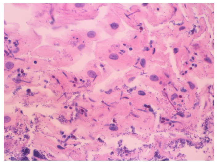
Cellular and nuclear enlargement, a large basophilic intranuclear inclusion with a clear halo, granular eosinophilic cytoplasmic inclusions
Bhaijee et al. 2011; Teot et al. 1993
Site of sampling determines the yield. Herpes simplex virus infects squamous epithelium, and biopsies for it are taken from the ulcer margin, where the cytopathic effect is most likely to be seen (Wang et al. 2016; Rosołowski and Kierzkiewicz 2013). In patients with HIV infection, cytomegalovirus esophagitis was diagnosed on biopsies taken mainly from the ulcer base, and up to 10 biopsies may be needed to exclude viral esophagitis (Wilcox, Straub and Schwartz 1996).
Coinfection. In one brushing series, numerous herpes-infected squamous cells obscured a smaller population of cytomegalovirus-infected cells. In another patient, the biopsy contained only necrotic debris while the cytology showed cytomegalovirus inclusions (Teot et al. 1993).
Clinical setting of herpes esophagitis. In 23 patients with herpes esophagitis, 22 were immunocompromised and 70% had simultaneous infections with other organisms. Culture was slightly more sensitive than microscopic examination (89% versus 76%) (McBane and Gross 1991).
Significance of Candida. In 271 esophageal biopsies with Candida, the absence of pseudohyphae, of invasion of intact epithelium or of neutrophils did not exclude clinically significant infection. Candida in an esophageal sample is treated as potentially significant and is integrated with clinical and endoscopic findings (Hissong et al. 2020).
Barrett esophagus: definition and cytologic recognition
ACG definition. The American College of Gastroenterology (ACG) 2022 guideline requires intestinal metaplasia in the tubular esophagus and columnar mucosa at least 1 cm long for a diagnosis of Barrett esophagus. Patients with a normal-appearing Z line do not undergo routine biopsy (Shaheen et al. 2022).
Other definitions. Guidelines from the British Society of Gastroenterology and the Asia-Pacific region do not require intestinal metaplasia (Shaheen et al. 2022).
Columnar cell types. The columnar-lined esophagus contains gastric fundic-type epithelium, junctional cardiac epithelium and specialized columnar epithelium with goblet cells (Shaheen et al. 2022).
Goblet cells in brushings. In a paired series of 327 cases, Barrett esophagus was diagnosed cytologically by the presence of goblet cells. Of 141 cases of nondysplastic Barrett esophagus, 52% were diagnosed by both methods, 8% by cytology alone and 40% by histology alone (Saad et al. 2003).
Second-opinion rule. The ACG recommends that dysplasia of any grade in Barrett esophagus be confirmed by a second pathologist with expertise in gastrointestinal pathology (Shaheen et al. 2022).
Detection of dysplasia and adenocarcinoma in Barrett esophagus by cytology
Study
Method
Low-grade dysplasia
High-grade dysplasia or adenocarcinoma
Specificity
Kumaravel et al. 2010 (530 patients)
Endoscopic brushing
31% (low-grade or indefinite)
82%
95% (any dysplasia)
Saad et al. 2003 (327 pairs)
Endoscopic brushing
6 of 30 detected by cytology
9 of 10 high-grade dysplasia; 22 of 23 carcinomas
Not stated
Falk et al. 1997
Endoscopic brushing
22%
11 of 11
95%
Falk et al. 1997
Nonendoscopic balloon
25%
80%
No abnormal cells in patients without dysplasia
Fritcher et al. 2008 (92 patients)
Brushing, conventional cytology
5%
32% (high-grade dysplasia); 45% (adenocarcinoma)
93%
Fritcher et al. 2008
Brushing, FISH
50%
82% (high-grade dysplasia); 100% (adenocarcinoma)
100%
Pattern across studies. Brush cytology detects high-grade dysplasia and adenocarcinoma well and detects low-grade dysplasia poorly (Kumaravel et al. 2010; Falk et al. 1997).
Cytology as a stand-alone surveillance test. Histology had a higher dysplasia detection rate than cytology (24.0% versus 15.7%). The authors judged the value of routine cytology in surveillance questionable (Kumaravel et al. 2010).
Complementary use. In 65 paired Barrett specimens, brushings showed a higher-grade lesion than histology in 13 cases, and biopsies showed the higher grade in 5. Agreement was 72%. The two methods together detected more serious lesions than either alone (Geisinger, Teot and Richter 1992).
FISH. FISH probes to 8q24 (MYC), 9p21 (CDKN2A), 17q12 (ERBB2) and 20q13 were more sensitive than conventional cytology for low-grade dysplasia, high-grade dysplasia and adenocarcinoma. All patients with a polysomic FISH result had high-grade dysplasia or adenocarcinoma within 6 months (Fritcher et al. 2008).
Nonendoscopic cell collection for Barrett esophagus screening
Devices. Swallowable devices consist of a capsule containing a compressible sponge on a string (Cytosponge, EsophaCap) or an inflatable silicone balloon (EsoCheck). The device is withdrawn through the mouth and collects an esophageal cytology sample (Shaheen et al. 2022).
Biomarkers. The sample is tested for trefoil factor 3 (TFF3), a protein expressed in intestinal metaplasia, by immunohistochemistry interpreted by a pathologist. Methylated DNA markers are an alternative and are measured by a polymerase chain reaction-based test (Shaheen et al. 2022).
ACG recommendation. A swallowable nonendoscopic capsule sponge device combined with a biomarker is an acceptable alternative to endoscopy for screening patients with chronic reflux symptoms and other risk factors (conditional recommendation, very-low-quality evidence) (Shaheen et al. 2022).
BEST3 trial design. The trial randomized 13,514 patients aged 50 years or older who had taken acid suppressants for reflux for more than 6 months in 109 English general practices. Patients with a positive Cytosponge-TFF3 test were referred for endoscopy (Fitzgerald et al. 2020).
BEST3 results. Barrett esophagus was diagnosed in 140 of 6,834 participants in the intervention group and 13 of 6,388 in the usual-care group (rate ratio 10.6). Nine intervention participants had dysplastic Barrett esophagus or stage I cancer, against none in the usual-care group (Fitzgerald et al. 2020).
Positive predictive value. Of the participants who underwent endoscopy after a positive test, 59% had Barrett esophagus or cancer (Fitzgerald et al. 2020; Shaheen et al. 2022).
Uptake and safety. Of those invited, 39% expressed interest. Among 1,654 participants who swallowed the device, one detachment required endoscopic removal (Fitzgerald et al. 2020).
Wide-area transepithelial sampling in Barrett esophagus
Method. Wide-area transepithelial sampling (WATS) uses an abrasive brush passed through the endoscope to sample deeper layers of the Barrett epithelium over a wide area. The smear is up to 150 μm thick, against 3–5 μm for a forceps biopsy section, and is analyzed with computer-assisted three-dimensional imaging (WATS-3D) (Shaheen et al. 2022).
Yield. In a meta-analysis of 11 studies, adding WATS to forceps biopsy increased the detection of Barrett esophagus by 16% in absolute terms and of dysplasia by 2%; the dysplasia detection rate rose 2.05-fold in relative terms (Suresh Kumar et al. 2020).
ACG position. The ACG made no recommendation on WATS-3D in surveillance (Shaheen et al. 2022).
Management of Barrett esophagus by dysplasia grade (ACG 2022)
Finding on confirmed histology
ACG recommendation
Nondysplastic Barrett esophagus, segment ≥3 cm
Surveillance endoscopy every 3 years
Nondysplastic Barrett esophagus, segment <3 cm
Surveillance endoscopy every 5 years
Low-grade dysplasia
Endoscopic eradication therapy; surveillance every 6 months for 1 year and then annually is an acceptable alternative
High-grade dysplasia or intramucosal carcinoma
Endoscopic eradication therapy
Visible lesion before ablation
Endoscopic resection of the lesion first
Source: Shaheen et al. 2022. Surveillance biopsies follow the Seattle protocol: biopsy of visible lesions, then four-quadrant biopsies at intervals of ≤2 cm (Shaheen et al. 2022).
Esophageal squamous cell carcinoma and adenocarcinoma on brushing cytology
Cytologic features of malignancy. On crush preparations of endoscopic biopsies, smears from invasive carcinoma are cellular. Cell groups show nuclear enlargement, pleomorphism, hyperchromasia, marked anisonucleosis, crowding, overlapping, loss of polarity and abnormal mitoses. Crush smears of in situ or intramucosal lesions showed only focal high-grade dysplasia because of low cellularity (Desai et al. 2021).
Repair versus carcinoma. In 121 consecutive esophageal brushings with companion biopsies, the most reliable criteria for malignancy were sharply angulated nuclear rims, a high nuclear-to-cytoplasmic ratio and coarsely granular chromatin. Nuclei with these features were few. Nuclear pleomorphism overlapped with repair. Multiple nucleoli, macronucleoli and loss of polarity occurred in both repair and carcinoma (Hoover and Berman 1988).
Typing. Well-differentiated and moderately differentiated squamous cell carcinomas were typed with confidence on crush cytology. Poorly differentiated tumors were difficult to type on cytology alone (Desai et al. 2021).
Grading. In 36 esophageal squamous cell carcinomas, cytologic grade matched histologic grade in 25 (69.4%) (Muniraj, Siddaraju and Sistla 2016).
Brushing before and after biopsy. In 40 esophageal carcinomas, the positive yield was 87.5% for brushing before biopsy, 90% for suction cytology, 100% for brushing after biopsy and 90% for endoscopic biopsy. Brushing after biopsy yielded significantly more material than brushing before biopsy (Singh et al. 1994).
Population screening for squamous neoplasia. In asymptomatic adults in Linxian, China, the balloon sampler had a sensitivity of 44% and a specificity of 99% for biopsy-proven squamous cell carcinoma. The sponge sampler had a sensitivity of 18%. For squamous dysplasia or carcinoma, the sensitivities were 47% and 24% (Roth et al. 1997).
Differential diagnosis of an atypical esophageal brushing
Finding
Favors
Against carcinoma
Sources
Hyperchromatic regenerating cells at an ulcer
Reactive atypia
Ulcer setting; limited atypia
Tyagi et al. 2016
Multinucleated, molded squamous nuclei with margination
Herpes simplex virus esophagitis
Viral cytopathic effect
Wang et al. 2016
Angulated nuclear rims, high nuclear-to-cytoplasmic ratio, coarsely granular chromatin
Carcinoma
Not applicable
Hoover and Berman 1988
Prominent nucleoli and loss of polarity
Either repair or carcinoma
Shared by both; not discriminating
Hoover and Berman 1988
Enlarged stromal or endothelial cells with a haloed intranuclear inclusion
Cytomegalovirus esophagitis
Inclusions; cells from the ulcer base
Teot et al. 1993
Goblet cells in columnar groups without nuclear atypia
Nondysplastic Barrett esophagus
Bland nuclei
Saad et al. 2003
Crowded, overlapping columnar groups with pleomorphism and loss of polarity
Barrett-associated high-grade dysplasia or adenocarcinoma
Not applicable
Desai et al. 2021; Kumaravel et al. 2010
Pleomorphic squamous cells in a cellular smear
Squamous cell carcinoma
Not applicable
Desai et al. 2021
Esophageal tumors in the WHO 6th edition and changes from the 5th edition
New precursor entity. Esophageal epidermoid metaplasia is a new entity in the 6th edition. It is a sharply demarcated area of epithelial hyperplasia with a prominent granular cell layer and hyperorthokeratosis and is a precursor of squamous cell carcinoma. It was not an entity in the 5th edition (Arends et al. 2026).
Carcinomas with combined squamoid and glandular components. The 6th edition groups adenosquamous carcinoma, mucoepidermoid carcinoma and adenoid cystic carcinoma in one section. Each forms <1% of primary esophageal carcinomas (Arends et al. 2026). This is an organizational change: the three entities are grouped for comparison, and none is new. For adenosquamous carcinoma, an arbitrary requirement of at least 20% of each component has been suggested; otherwise the tumor is classified by its majority component (Arends et al. 2026).
Grading of esophageal dysplasia. Esophageal dysplasia is graded as low grade or high grade in both the 5th and 6th editions. The 6th edition extends two-tier grading to all digestive precursor lesions by merging the former low-grade and intermediate-grade categories of the gallbladder and bile ducts (Nagtegaal et al. 2020; Arends et al. 2026).
5th-edition framework. The 5th edition described the inflammation–metaplasia–dysplasia–adenocarcinoma sequence of Barrett esophagus, with TP53 mutation linked to high-grade dysplasia and SMAD4 mutation preceding invasion. It recommended ERBB2 (HER2) testing of esophageal and gastric adenocarcinomas (Nagtegaal et al. 2020).
HER2 testing. The CAP/ASCP/ASCO guideline recommends HER2 assessment of tumor from every patient with advanced gastroesophageal adenocarcinoma who is a candidate for HER2-targeted therapy, before that therapy begins (Bartley et al. 2017).
Gastric Cytology: Normal Findings, Adenocarcinoma and Lymphoma
Definition. Gastric cytology comprises endoscopic brushings and crush preparations of the gastric mucosa and endoscopic ultrasound-guided fine-needle aspiration (EUS-FNA) or fine-needle biopsy (EUS-FNB) of the gastric wall. Its main targets are Helicobacter pylori gastritis, gastric adenocarcinoma, including the diffusely infiltrative linitis plastica pattern, and gastric lymphoma. Gastric epithelium also appears as a contaminant in transgastric EUS-FNA of the pancreas and adjacent organs (Wang, Lozano and Cai 2024). Gastric epithelial tumors are classified by the WHO Classification of Tumours of the Digestive System, 6th edition (2026), and gastric lymphomas by the 5th edition of the WHO Classification of Haematolymphoid Tumours (Arends et al. 2026; Alaggio et al. 2022).
Sampling methods for gastric cytology
Brushing and biopsy. In a series of 106 upper gastrointestinal brushings, eight endoscopic biopsies were nonrepresentative, and seven of these lesions were malignant (Muniraj, Siddaraju and Sistla 2016).
Crush cytology. Crush preparations are made by compressing endoscopic biopsy fragments between slides (Desai et al. 2021).
EUS-FNA of the gastrointestinal wall. In 62 EUS-FNA specimens of intramural and extramural gastrointestinal lesions, EUS-FNA had a sensitivity of 89%, a specificity of 88% and an accuracy of 89% for neoplasia. Forceps biopsy had previously failed in 43% of these patients. One gastric lymphoma and one GIST were false negatives (Vander Noot et al. 2004).
Early experience. A 7-year review of 488 gastric brushings found 80 positive for malignancy. Histologic correlation was possible in 429 gastric cases and supported the positive cytologic diagnoses (Lin and Harmata 1983).
Accuracy and reporting categories of gastrointestinal brush and crush cytology
Study
Specimens
Sensitivity
Specificity
Notes
Muniraj, Siddaraju and Sistla 2016
106 upper GI brushings
100%
96.4%
One false positive; no false negatives
Tyagi et al. 2016
101 prebiopsy GI brushings
62.7%
94%
False-negative rate 37.3%; inadequate material and regenerative atypia caused most discordance
Desai et al. 2021
Crush cytology, 451 GI lesions
97.3%
90%
100% sensitivity and specificity for esophageal, junctional and gastric lesions
Ambati et al. 2026
Crush cytology, 303 GI samples
96.1%
96.2%
Four categories: negative, atypical favors reactive, atypical suspicious for malignancy, positive
Category use in gastric samples. Of 58 gastric crush preparations, 2 were negative for malignancy, 31 atypical favors reactive, 1 atypical suspicious for malignancy and 24 positive for malignancy (Ambati et al. 2026).
Lesions needing tissue confirmation. Poorly differentiated carcinoma, non-Hodgkin lymphoma, neuroendocrine tumor and spindle cell neoplasms required biopsy and immunohistochemistry for confirmation in the Desai series. The 64 gastric lesions positive on crush cytology in that series comprised 50 adenocarcinomas, 9 poorly differentiated carcinomas, 2 lymphomas, 1 neuroendocrine tumor and 2 spindle cell neoplasms or GISTs (Desai et al. 2021).
Lesions sampled by EUS-FNA of the gastrointestinal wall. In the Vander Noot series, the neoplasms were carcinoma (24), GIST (18), neuroendocrine neoplasm (2) and lymphoma (1). False-positive diagnoses came from endometriosis, a duodenal diverticulum with smooth muscle hyperplasia and normal pancreas presenting as a periduodenal mass (Vander Noot et al. 2004).
Normal gastric epithelium and gastric contaminants
Gastric surface epithelium. Gastrointestinal contaminants typically form large sheets of regular mosaic epithelium (Wang, Lozano and Cai 2024).
Contaminants in EUS-FNA. Epithelium from the stomach and duodenum frequently contaminates EUS-FNA specimens. Gastric foveolar cells can be mistaken for the epithelium of a mucinous neoplasm (Wang, Lozano and Cai 2024).
Features of a gastrointestinal contaminant. Lesional mucinous cells appear as small clusters or single cells. Duodenal epithelium contains scattered goblet cells (Wang, Lozano and Cai 2024).
Changes shared with neoplasia. Gastric epithelium may show nuclear grooves, nuclear inclusions and subtle crowding. Gastric mucin can be mistaken for neoplastic mucin (Pitman et al. 2014; Wang, Lozano and Cai 2024).
Degenerative atypia. In one case, degenerating gastric epithelial cells in a transgastric aspirate were initially interpreted as low-grade adenocarcinoma. Review showed transitional features with normal gastric surface epithelium (Park et al. 2014).
Gastric-foveolar epithelium in cysts. The lining of a gastric-foveolar-type mucinous cyst may be indistinguishable from gastric contamination in a transgastric sample (Pitman et al. 2014).
Helicobacter pylori and benign gastric findings on brushing cytology
Brush cytology for H. pylori. In 109 dyspeptic patients, antral brushings detected H. pylori in 78%, histology in 66% and the rapid urease test in 59%. With either cytologic or histologic identification as the reference, sensitivity was 95% for brush cytology, 80.5% for histology and 72% for the rapid urease test (Mostaghni et al. 2008).
Comparison with biopsy. Brush cytology was superior to endoscopic biopsy for detecting Candida in a series of 106 upper gastrointestinal brushings (Muniraj, Siddaraju and Sistla 2016).
Gastric xanthoma. Five biopsy-proven xanthomas were initially reported on brushing as suspicious for signet-ring cell adenocarcinoma. Xanthoma cells were PAS negative, Oil red O positive and weakly positive with Masson trichrome. Signet-ring carcinoma cells were strongly PAS positive (Kumar et al. 2006).
Gastric adenocarcinoma on cytology
Lauren subtyping on cytology. In one series, 21 of 32 gastric adenocarcinomas (65.6%) were correctly subtyped on brushings as intestinal (16) or diffuse (5) (Muniraj, Siddaraju and Sistla 2016).
Dyscohesive tumors on brushing. A signet-ring cell carcinoma was not detected on a brushing whose smears contained only inflammatory cells and columnar cells (Tyagi et al. 2016).
Linitis plastica. Gastric linitis plastica is a diffusely infiltrating adenocarcinoma, and superficial mucosal biopsies have a low yield. In 34 patients with suspected linitis plastica, EUS-FNB diagnosed 9 of 10 confirmed cases. Eight of these patients had at least one earlier endoscopy with negative mucosal biopsies (Assaf et al. 2022).
Gastric adenocarcinoma in the WHO 5th and 6th editions
Topic
5th edition (2019)
6th edition (2026)
Poorly cohesive carcinoma
Heterogeneity discussed, including signet-ring cell carcinoma and poorly cohesive carcinoma NOS
Subclassified into signet-ring cell and non-signet-ring cell subtypes; concept of pure signet-ring cell carcinoma introduced
Tubular adenocarcinoma
Not addressed in Nagtegaal et al. 2020
Crawling-type adenocarcinoma recognized as a distinctive variant
Uncommon subtypes
Gastric adenocarcinoma of fundic-gland type described
Expanded spectrum, including carcinoma with lymphoid stroma, AFP-producing carcinoma, micropapillary adenocarcinoma, fundic-gland type and sarcomatoid carcinoma
Precursor lesions
Separate sections for gastric dysplasia, intestinal-type adenoma and gastric-type adenomas; two-tier (low-grade/high-grade) dysplasia grading
Gastric dysplasia, intestinal-type adenoma and gastric-type adenoma combined in one section under gastric dysplasia, mirroring the MAPS III guideline; lesions arising beneath the foveolar surface (oxyntic gland neoplasms and pyloric gland adenoma) are separated; grading remains two-tier
Biomarkers
ERBB2 (HER2) testing recommended
TCGA groups and HER2, claudin 18.2, MMR/MSI and PD-L1 treated as tools for precision oncology
Sources: Nagtegaal et al. 2020 (5th edition); Kushima 2026 and Arends et al. 2026 (6th edition).
Oxyntic gland adenoma. The 6th edition defines oxyntic gland adenoma more clearly. A pushing interface and prolapse-type misplacement are characteristic and are not evidence of adenocarcinoma of fundic-gland type (Arends et al. 2026).
Dysplasia in H. pylori-naive stomachs. The 6th edition describes a dysplastic lesion with a foveolar phenotype that develops in H. pylori-naive stomachs (Arends et al. 2026).
Etiology. Most sporadic gastric cancers arise from chronic inflammation, usually caused by H. pylori infection. Up to 10% are familial (Nagtegaal et al. 2020).
Gastric lymphoma on cytology
Classification and genetics of gastric lymphoma
Extranodal marginal zone lymphoma of mucosa-associated lymphoid tissue (EMZL). EMZL is retained as a distinct entity in WHO-HAEM5. The neoplastic cells are mature small B cells, typically negative for CD5 and CD10. Plasmacytic differentiation and reactive follicles are common (Alaggio et al. 2022).
Genetics. Trisomies of chromosomes 3 and 18 are common in marginal zone lymphomas. The t(11;18)(q21;q21) translocation, which forms the BIRC3::MALT1 fusion, recurs in gastric and pulmonary EMZL and is rare at other sites (Alaggio et al. 2022).
Response to H. pylori eradication. Eradication of H. pylori caused regression of low-grade gastric MALT lymphoma in 5 of 6 patients in the first report (Wotherspoon et al. 1993).
Predictor of nonresponse. About 20–30% of H. pylori-associated gastric MALT lymphomas do not regress after antibiotic therapy. The API2-MLT (BIRC3::MALT1) transcript was detected in 9 of 12 nonresponsive tumors and in none of 10 responsive tumors (Liu et al. 2001). WHO-HAEM5 states that BIRC3::MALT1 identifies gastric EMZL that does not respond to eradication (Alaggio et al. 2022).
Testing small biopsies. Real-time RT-PCR detected t(11;18) in formalin-fixed endoscopic biopsies from 8 of 38 patients (21%) with gastric MALT lymphoma. It was absent from 8 gastric large B-cell lymphomas and 11 cases of chronic active gastritis (Zhang, Garces and Dong 2006).
Indolent NK-cell lymphoproliferative disorder. This WHO-HAEM5 entity involves the stomach and the small and large intestines. It is superficial, usually regresses spontaneously and consists of atypical medium-sized cells with pale cytoplasm (Alaggio et al. 2022).
Diagnostic yield of cytology and ancillary tests in gastric lymphoma
Study
Specimen
Finding
Sherman et al. 1994
Gastric brushing (27 patients)
Definitive diagnosis of lymphoma in 48% overall; all cytologically diagnosed lymphomas were large cell type; no false-positive diagnoses
Almasri et al. 1997
Flow cytometry of fresh endoscopic biopsies (29 patients)
Adequate cells in 26; B-cell monoclonality in 16, of which only 8 were diagnosable as lymphoma on morphology alone
Liu, Baskota and Gonzalez 2025
EUS-FNA/FNB of gastrointestinal lymphoma (40 patients)
Concordant with the final WHO diagnosis in 72.5%; incomplete phenotyping in 17.5%; suspicious in 10.0%; cell block and flow cytometry quality affected accuracy
Light-chain restriction. Flow cytometry of fresh cell suspensions from endoscopic biopsies distinguishes monoclonal from polyclonal B-cell infiltrates by surface immunoglobulin light-chain analysis. Monoclonality was detected in five cases in which clonal B cells formed <5% of cells (Almasri et al. 1997).
Combined flow cytometry and FISH. In one gastric MALT lymphoma that could not be diagnosed on histology, flow cytometry showed a clonal population and FISH showed trisomy 18 (Matsueda et al. 2018).
Number of passes. In the EUS-FNA/FNB lymphoma series, 1 to 5 FNA passes gave better accuracy than 6 or more (Liu, Baskota and Gonzalez 2025).
Rapid on-site evaluation. Onsite adequacy assessment by cytopathologists and by cytotechnologists performed similarly in the same series (Liu, Baskota and Gonzalez 2025).
Differential diagnosis of dyscohesive cells in a gastric sample
Entity
Cytologic and ancillary findings
Sources
Signet-ring cell carcinoma
Dyscohesive cells with strong PAS-positive mucin
Kumar et al. 2006
Gastric xanthoma
Foamy cells that are PAS negative and Oil red O positive
Kumar et al. 2006
Diffuse large B-cell lymphoma
Large lymphoid cells; all lymphomas diagnosed on gastric brushings in one series were large cell type
Sherman et al. 1994
Extranodal marginal zone lymphoma
Small CD5-negative, CD10-negative B cells with plasmacytic differentiation; light-chain restriction on flow cytometry
Alaggio et al. 2022; Almasri et al. 1997
Degenerated gastric epithelium
Sheets with transitional features to normal foveolar epithelium
Park et al. 2014
GIST and Smooth Muscle Tumors of the GI Wall on EUS-FNA
Definition. Gastrointestinal stromal tumor (GIST) is a mesenchymal neoplasm that shares immunomorphologic features with the interstitial cells of Cajal (Sbaraglia et al. 2021). Most GISTs carry an activating variant of KIT or PDGFRA (Judson et al. 2025). GIST is the commonest mesenchymal tumor specific to the gastrointestinal tract, and leiomyoma is the commonest mesenchymal tumor of the esophagus (Miettinen and Lasota 2006; Stelow et al. 2003). Endoscopic ultrasound-guided fine-needle aspiration (EUS-FNA) and fine-needle biopsy sample these intramural masses and allow separation of GIST from leiomyoma, schwannoma and other mimics on cell block immunohistochemistry (Stelow et al. 2008). GIST is classified in the WHO Classification of Tumours of the Digestive System, 6th edition (2026), whose mesenchymal tumors are harmonized in definition and content with the WHO Classification of Soft Tissue and Bone Tumours, 6th edition (2026) (Arends et al. 2026).
GIST in the WHO 6th editions and changes from the 5th edition
Digestive System, 6th edition. GIST is retained in the mesenchymal tumors chapter. Succinate dehydrogenase (SDH)-deficient GIST has a separate section and is recognized as a distinct entity because of its clinical, histologic, immunohistochemical and molecular features and its syndromic association (Arends et al. 2026).
5th-edition rule. In the 5th edition, SDH-deficient GIST was a subtype of GIST (Arends et al. 2026).
Harmonization. Mesenchymal tumors of the digestive system are harmonized with the Soft Tissue and Bone 6th edition in definition and content as far as possible. Tumors with similar features at all sites, such as solitary fibrous tumor, are described in the introduction to the mesenchymal chapter (Arends et al. 2026).
Soft Tissue and Bone, 6th edition. The volume added 19 new entities and replaced the term "variant" with "morphological pattern" for features without different clinical behavior (Hornick et al. 2026).
Other changes in the digestive mesenchymal chapter.NUT-rearranged sarcoma is recognized as a distinct tumor type in the gastrointestinal tract. Gastroblastoma moved to the mesenchymal chapter under the umbrella term GLI1-altered neoplasms (Arends et al. 2026).
Clinical features and sites of GIST
Sites. GISTs arise in the stomach (60%), jejunum and ileum (30%), duodenum (4–5%), rectum (4%), colon and appendix (1–2%) and esophagus (<1%). Most patients are older than 50 years (Miettinen and Lasota 2006).
Incidence. The estimated incidence is 10–20 per million, including incidental minimal tumors (Miettinen and Lasota 2006).
Children. GISTs are rare in children (<1%) and occur almost exclusively in the stomach (Miettinen and Lasota 2006).
Neurofibromatosis type 1. Patients with NF1 are predisposed to multiple small intestinal GISTs (Miettinen and Lasota 2006).
Syndromes. Carney triad comprises gastric GIST, paraganglioma and pulmonary chondroma, and most of these tumors show SDHC promoter hypermethylation. Carney-Stratakis syndrome comprises GIST and paraganglioma with germline variants of SDHA, SDHB, SDHC or SDHD (Judson et al. 2025).
Endoscopic ultrasound sampling of subepithelial masses
Lesions <2 cm. Small asymptomatic submucosal lesions <2 cm in the upper gastrointestinal tract and small intestine may be kept under surveillance and biopsied or excised if they grow. Large retrospective studies suggest that routine surveillance of these lesions is unnecessary (Judson et al. 2025).
Lesions >2 cm. Suspected GISTs larger than 2 cm are investigated and diagnosed whenever possible. In the stomach the diagnosis is usually made by EUS-guided fine-needle aspiration or core needle biopsy (Judson et al. 2025).
Reasons for preoperative diagnosis. A preoperative diagnosis excludes leiomyosarcoma, germ cell tumor, lymphoma, benign and malignant neurogenic tumors and desmoid tumors, which need different treatment (Judson et al. 2025).
Mutational testing. When the diagnosis is GIST and systemic treatment is planned, mutational analysis is mandatory to exclude imatinib-resistant disease (Judson et al. 2025).
Immunocytochemistry on cell blocks. In 95 EUS-FNA aspirates of subepithelial mesenchymal tumors, CD117 immunoreactivity always predicted GIST at follow-up, and 15 of 16 CD34-positive cases were GISTs. Strong smooth muscle actin or desmin usually predicted leiomyoma. The exceptions were a glomus tumor and a nonneoplastic smooth muscle contaminant (Stelow et al. 2008).
Cytomorphology of GIST on FNA
Feature
Spindle cell GIST
Epithelioid GIST
Share of cases (histology)
70%
20% (mixed 10%)
Arrangement
Cohesive to loose three-dimensional clusters and dispersed cells; fascicles; palisading in some
Groups of round cells; occasional acinar-like arrangement
Cytoplasm
Delicate, fibrillary, with wispy bipolar processes; perinuclear vacuoles
Eosinophilic or clear; plasmacytoid cells; bubbly cytoplasm from multiple vacuoles
Nuclei
Elongated to wavy, with blunt to tapered ends
Round, eccentric, smooth membranes, fine chromatin
Background
Loosely fibrillary stroma, pink to magenta on May-Grünwald-Giemsa
Variable
Sources: Vij et al. 2013; Sbaraglia et al. 2021; Kapatia et al. 2020.
Perinuclear vacuoles. Perinuclear (juxtanuclear) cytoplasmic vacuoles occur most often in gastric GISTs (Sbaraglia et al. 2021). Perinuclear vacuoles, nuclear pseudoinclusions and multinucleation were seen in four cases in a cytology series of 42 patients (Kapatia et al. 2020).
Skeinoid fibers. Hyaline or fibrillary eosinophilic skeinoid fibers occur in 10–20% of GISTs, almost exclusively in the small bowel (Sbaraglia et al. 2021).
Palisading. Nuclear palisading is seen in a minority of GISTs. It also occurs in smooth muscle and neural tumors and is therefore not specific for GIST (Sbaraglia et al. 2021).
Genotype and site of epithelioid GIST. Epithelioid GISTs frequently carry PDGFRA variants, and most arise in the stomach (Sbaraglia et al. 2021).
Cytologic features of aggressive tumors. Cellular dyscohesion, nuclear pleomorphism, intranuclear pseudoinclusions, prominent nucleoli, mitoses and necrosis were more prominent in malignant, metastatic and recurrent GISTs. Epithelioid morphology was more common in malignant and metastatic tumors (Vij et al. 2013).
Epithelioid GIST. In 42 patients with GIST on FNA, 8.8% of aspirates showed predominantly epithelioid cells and 15.5% mixed morphology. All 18 cell blocks tested were strongly KIT positive. The authors advise KIT and/or DOG1 immunocytochemistry in all suspected GISTs (Kapatia et al. 2020).
Immunohistochemistry of GIST on cell block
Marker
Expression in GIST
Source
KIT (CD117)
Most cases; cytoplasmic and diffuse, dot-like accentuation in up to half; negative in about 5%
Sbaraglia et al. 2021; Judson et al. 2025
DOG1 (anoctamin 1)
Negative in about 5%; positive in about half of KIT-negative GISTs
Judson et al. 2025; Sbaraglia et al. 2021
KIT and DOG1 both negative
About 1%
Judson et al. 2025
CD34
60–70%
Sbaraglia et al. 2021
Smooth muscle actin
About 30%
Sbaraglia et al. 2021
Desmin, cytokeratin
<2%
Sbaraglia et al. 2021
SDHB
Lost in SDH-deficient GIST
Miettinen et al. 2011
DOG1 on FNA cell blocks. In 30 GISTs and 68 non-GIST tumors, DOG1 had 100% sensitivity and 100% specificity. KIT had 70% sensitivity and 76% specificity, rising to 89% specificity when adenoid cystic carcinomas were excluded. All KIT-negative GISTs were DOG1 positive (Fatima, Cohen and Siddiqui 2011).
GIST negative for KIT or DOG1. In a suspected GIST that is negative for KIT and/or DOG1, molecular analysis for typical KIT or PDGFRA variants can confirm the diagnosis (Judson et al. 2025).
Molecular classification of GIST
Genotype
Frequency
Features
Sources
KIT exon 11
About 70%
Juxtamembrane domain; deletions, insertions and missense variants
Sbaraglia et al. 2021
KIT exon 9
<10%
Extracellular domain; mutational testing allows imatinib dosing of 800 mg daily in advanced disease
Sbaraglia et al. 2021; Judson et al. 2025
KIT exons 13 and 17
Small subset
ATP-binding pocket and activation loop
Sbaraglia et al. 2021
PDGFRA
About 10%
Exons 12, 14 and 18; 70% are exon 18 p.D842V; often KIT-negative and epithelioid
Sbaraglia et al. 2021
KIT/PDGFRA wild type
10–15%
About half SDH deficient; others NF1-related, BRAF p.V600E or rare NTRK fusions
Sbaraglia et al. 2021; Judson et al. 2025
Mutual exclusivity.KIT and PDGFRA variants are mutually exclusive with very rare exceptions (Sbaraglia et al. 2021).
Testing algorithm for wild-type GIST. For KIT/PDGFRA wild-type GISTs that are not NF1-related, SDHB immunohistochemistry is performed. Tumors without SDHB loss are tested for BRAF and RAS variants. Tumors negative for all of these are tested for NTRK fusions (Judson et al. 2025).
Imatinib resistance. Tyrosine kinase inhibitors have no or limited efficacy in GISTs with PDGFRA p.D842V and in GISTs without KIT or PDGFRA variants (von Mehren and Joensuu 2018).
Natural history by genotype.KIT/PDGFRA wild-type GISTs tend to behave more indolently than KIT exon 11-variant GISTs (Judson et al. 2025).
SDH-deficient GIST
Frequency. SDH-deficient GISTs were found in 66 of 756 gastric GISTs studied (8.7%), with a separate estimated frequency of 7.5% among unselected gastric GISTs. All 378 nongastric GISTs were SDHB positive (Miettinen et al. 2011).
Age and sex. Nearly all gastric GISTs in patients younger than 20 years were SDH deficient, and only rare GISTs in older adults were. Females outnumbered males by more than 2:1 (Miettinen et al. 2011).
Morphology and immunophenotype. The tumors were often multiple, involved the muscularis propria in a plexiform pattern and had epithelioid, hypercellular morphology. They were consistently KIT and DOG1 positive and almost always smooth muscle actin negative (Miettinen et al. 2011).
Behavior. Lymph node metastases occurred in 5 patients, and lymphovascular invasion was present in more than half of the cases studied. Tumors with low mitotic rates metastasized, and many patients survived long term with metastases (Miettinen et al. 2011).
Features that prompt SDHB testing. Pediatric or young female patient, gastric location, multinodular or multifocal growth, epithelioid or mixed morphology and nodal metastases (Judson et al. 2025).
Risk models. The AFIP and contour-map risk models are not recommended for SDH-deficient GIST (Judson et al. 2025).
Risk assessment of GIST and the limits of FNA
Prognostic factors. Mitotic rate, tumor size and tumor site are prognostic factors of proven value. Tumor rupture is an adverse factor (Judson et al. 2025).
Mitotic count. The mitotic count is recommended as the number of mitoses in a total area of 5 mm² (Judson et al. 2025).
Site. Gastric GISTs ≤10 cm with ≤5 mitoses per 50 HPF have a low risk of metastasis. Intestinal GISTs >5 cm have at least moderate risk regardless of mitotic rate (Miettinen and Lasota 2006).
AFIP risk categories. The table gives the risk of recurrence by size, mitotic index and site in the AFIP (Miettinen and Lasota) scheme, as reproduced in Rutkowski, Przybył and Zdzienicki 2013. Cells that are merged across rows or columns in that reproduction are omitted.
Size
Mitotic index
Stomach
Duodenum
Small intestine
Rectum
≤2 cm
≤5/50 HPFs
0%
0%
0%
0%
>2 cm, ≤5 cm
≤5/50 HPFs
Very low (1.9%)
Low (8.3%)
Low (4.3%)
Low (8.5%)
>5 cm, ≤10 cm
≤5/50 HPFs
Low (3.6%)
High (34%)
Intermediate (24%)
High (57%)
>10 cm
≤5/50 HPFs
Intermediate (12%)
Omitted (merged cell)
Omitted (merged cell)
Omitted (merged cell)
≤2 cm
>5/50 HPFs
Insufficient data
Insufficient data
High (50%)
Omitted (merged cell)
>2 cm, ≤5 cm
>5/50 HPFs
Intermediate (16%)
Omitted (merged cell)
Omitted (merged cell)
Omitted (merged cell)
Source: Miettinen and Lasota 2006, as reproduced in Rutkowski, Przybył and Zdzienicki 2013. AFIP thresholds are expressed per 50 HPF; the British Sarcoma Group recommends expressing the mitotic count per 5 mm² (Judson et al. 2025).
TNM. The British Sarcoma Group does not recommend TNM classification for GIST (Judson et al. 2025).
Mitotic count on small samples. Mitotic count was evaluable in 9 of 12 EUS-FNA samples (75%), against 100% of tunneling bloc and forceps samples (Kobara et al. 2015).
Ki-67 on FNA. In 55 gastric GISTs, the mean Ki-67 index was 2% in EUS-FNA samples and 10% in the resection specimens. The authors judged the FNA Ki-67 index unreliable for proliferation (Seven et al. 2021).
Leiomyoma and other smooth muscle tumors of the GI wall
Leiomyoma. True leiomyomas of the gastrointestinal tract are rare. Leiomyomas and GISTs differ in clinical and FNA features, and cell block immunohistochemistry separates them (Stelow et al. 2003).
Smooth muscle immunophenotype. All 24 upper gastrointestinal leiomyomas in one series showed strong, diffuse muscle marker positivity (Taşkın et al. 2020).
Interstitial cells of Cajal in leiomyoma. KIT-positive and DOG1-positive interstitial cells of Cajal were present in all deep esophageal leiomyomas, averaging 20% of lesional cells and focally >50%. They were found in 6 of 8 gastric and 1 of 5 small-bowel leiomyomas and in no superficial esophageal or colorectal leiomyomas. No KIT or PDGFRA mutations were found (Deshpande et al. 2014).
Consequence for small samples. These cells can lead to misdiagnosis of leiomyoma as GIST, particularly on biopsy samples (Deshpande et al. 2014). Interpretation of KIT and DOG1 is especially difficult in small biopsy and cytology specimens (Taşkın et al. 2020).
Mast cells. CD117 also highlighted mast cells in all 24 upper gastrointestinal leiomyomas (Taşkın et al. 2020).
Smooth muscle contaminant. Nonneoplastic muscularis propria can be aspirated and produce a smooth muscle immunoprofile (Stelow et al. 2008).
Other mesenchymal tumors of the gastric wall on EUS-FNA
Non-GIST spectrum. Among 25 non-GIST gastric mesenchymal tumors on EUS-FNA from two institutions, there were 10 leiomyomas, 8 schwannomas, 5 glomus tumors, 1 PEComa and 1 desmoid tumor. These were selected after exclusion of 113 GISTs diagnosed on FNA (Saoud et al. 2023).
Schwannoma. In 51 gastric schwannomas, all tumors were S100 positive and most were GFAP positive. All were negative for KIT, DOG1, smooth muscle actin and desmin, and CD34 was rare. A peritumoral lymphoid cuff was present in 96%, with germinal centers in 74%, and diffuse intratumoral lymphoid infiltration in all cases. No KIT or PDGFRA mutations were found (Voltaggio et al. 2012). Schwannoma was the second commonest non-GIST gastric mesenchymal tumor on EUS-FNA (8 of 25) (Saoud et al. 2023). On EUS-FNA the abundant lymphocytes are mixed B and T cells and can be mistaken for lymphoma (Chen and Cai 2019).
Inflammatory fibroid polyp. This benign neoplasm arises most often in the stomach and is composed of short spindle and stellate cells in a myxoid or collagenous stroma with eosinophils and perivascular onion-skin fibrosis. The cells express CD34 and rarely smooth muscle actin and are negative for KIT, DOG1, desmin, S100, SOX10 and keratins. Most cases carry PDGFRA mutations; gastric tumors have exon 18 mutations, usually p.D842V (Sbaraglia et al. 2021).
Plexiform fibromyxoma. This rare tumor arises mostly in the gastric antrum. It shows plexiform growth of bland spindle to ovoid cells in a vascular myxoid stroma. The cells express smooth muscle actin, with focal desmin or caldesmon, and are negative for DOG1, KIT, S100 and CD34 (Arslan et al. 2021).
Solitary fibrous tumor. STAT6 immunohistochemistry, reflecting the NAB2::STAT6 fusion, is the most specific and sensitive marker (Sbaraglia et al. 2021).
Differential diagnosis of spindle cell aspirates from the GI wall
Tumor
KIT
DOG1
CD34
SMA/desmin
S100
Other
Sources
GIST
+ (about 95%)
+ (about 95%)
60–70%
SMA about 30%; desmin <2%
−
KIT or PDGFRA variant
Sbaraglia et al. 2021; Judson et al. 2025
Leiomyoma
Interstitial cells of Cajal and mast cells only
Interstitial cells of Cajal only
Not stated
Strong, diffuse
Not stated
No KIT/PDGFRA variant
Deshpande et al. 2014; Taşkın et al. 2020
Schwannoma
−
−
Rare
SMA −; desmin −
+
Peritumoral lymphoid cuff; polytypic lymphocytes
Voltaggio et al. 2012; Chen and Cai 2019
Inflammatory fibroid polyp
−
−
+
SMA rare; desmin −
−
Eosinophils; PDGFRA variant
Sbaraglia et al. 2021
Plexiform fibromyxoma
−
−
−
SMA +; focal desmin
−
Myxoid vascular stroma; antrum
Arslan et al. 2021
CAP cancer protocols for the esophagus, stomach, GIST and GI neuroendocrine tumors
Protocol (as listed by the College of American Pathologists)
Version
Date listed
Esophagus
4.2.0.1
June 2022
Stomach
4.4.0.0
March 2023
GIST, Resection
4.4.0.0
September 2026
GIST, Biopsy
4.4.0.0
September 2026
GIST Biomarker Reporting
1.1.0.0 on the listing; 1.2.0.0 printed in the template
September 2026
Stomach NET
5.0.0.0
December 2023
Duodenum and Ampulla NET
2.0.0.1
June 2024
Jejunum and Ileum NET
2.0.0.0
December 2023
Appendix NET
5.0.0.0
December 2023
Colon NET
5.0.0.0
December 2023
Source: College of American Pathologists, cancer protocol templates page (accessed 2026-11-04). Of the protocols in this table, only GIST has a separate biopsy template.
Scope of the GIST biopsy protocol. The College of American Pathologists GIST biopsy protocol (version 4.4.0.0, September 2026) is recommended for clinical care and is optional for accreditation. Its procedure list includes core needle, endoscopic and fine-needle aspiration biopsy. The protocol excludes resections, cytologic specimens and pediatric, familial or syndromic GIST (College of American Pathologists 2026).
Reporting elements of the GIST biopsy protocol. The case summary reports procedure, tumor site, histologic type, tumor size from the clinicoradiologic estimate, mitotic rate, histologic grade, treatment effect and risk assessment. Associated syndrome, necrosis, immunohistochemistry (KIT, DOG1, SDHA, SDHB, BRAF) and molecular studies are optional elements (College of American Pathologists 2026).
Mitotic rate in the GIST biopsy protocol. The count starts in the most mitotically active area and is made in 5 mm² of tumor. Modern wide-field microscopes require approximately 20 to 25 high-power fields to cover 5 mm², and older models require 50. Histologic grade is G1 at 5 or fewer mitoses per 5 mm² and G2 at more than 5 per 5 mm² (College of American Pathologists 2026).
Changes in GIST biopsy protocol version 4.4.0.0. The protocol now covers GIST at all sites and includes WHO 6th-edition updates (College of American Pathologists 2026).
GI wall spindle cell tumor immunostain lookup
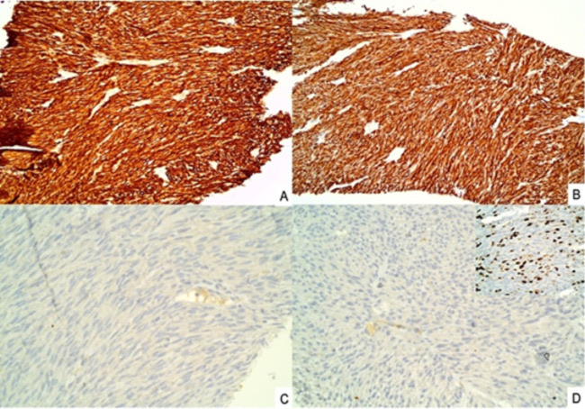
Gastrointestinal stromal tumor (rectal primary, metastatic to liver), immunohistochemistry: the spindle cells are diffusely positive for DOG1 (A) and CD117 (B) and negative for S100 (C) and SMA (D); the inset in D shows a Ki-67 index of about 10%. Magnification not stated.Image: Niranjan Gauri et al., PMC open access (Discoveries (Craiova). 2025 Mar 31;13(1):e202. doi: 10.15190/d.2025.1) Figure 3, CC BY.
Gastrointestinal Neuroendocrine Neoplasms on Cytology
Definition. Neuroendocrine neoplasms (NENs) of the digestive system are epithelial neoplasms with morphologic and immunohistochemical evidence of neuroendocrine differentiation. They are divided into well-differentiated neuroendocrine tumors (NETs) and poorly differentiated neuroendocrine carcinomas (NECs), with mixed neuroendocrine–non-neuroendocrine neoplasms (MiNENs) as a separate category (Nagtegaal et al. 2020). The WHO Classification of Tumours of the Digestive System, 6th edition (2026), places digestive NENs in one chapter and adds amphicrine-like carcinoma (Arends et al. 2026). Gastrointestinal NENs are sampled by endoscopic ultrasound-guided fine-needle aspiration (EUS-FNA) or fine-needle biopsy (EUS-FNB) of the gastric, duodenal or rectal wall and by FNA of liver and lymph node metastases.
Grading of digestive neuroendocrine tumors, the separation of NET G3 from NEC and Ki-67 grading on cell blocks are described in Solid cellular neoplasms of the pancreas on cytology. The thresholds and rules apply to every digestive site (Arends et al. 2026). In the 5th edition, the grade of digestive NETs used only the mitotic count and the Ki-67 index (Nagtegaal et al. 2020). The 6th edition also records tumor necrosis alongside the grade to gather evidence. Necrosis does not change the grade, and it supports NEC over NET without being definitive (Arends et al. 2026).
Digestive neuroendocrine neoplasms in the WHO 6th edition and changes from the 5th edition
Chapter structure. The 6th edition places NENs of all digestive sites in one chapter. The 5th edition described them in each organ-specific chapter (Arends et al. 2026).
Proof of neuroendocrine differentiation. The 6th edition emphasizes that expression of at least two neuroendocrine markers, preferably including one of chromogranin A, synaptophysin and INSM1, is required, except in highly specific and unusual circumstances such as the tuft cell variant of small cell carcinoma. CD56 is considered insufficiently specific (Arends et al. 2026). The 5th-edition summary sets no minimum number of markers (Nagtegaal et al. 2020).
Amphicrine-like carcinoma. In MiNEN, the neuroendocrine and non-neuroendocrine components form distinct zones or nodules. In amphicrine-like carcinoma (ALC), the two lines of differentiation are intimately intermixed or present in the same cells. ALCs are staged as carcinomas (Arends et al. 2026).
Recognition of amphicrine-like carcinoma. To aid recognition of ALC, the 6th edition gives detailed guidelines, for example morphologic evidence of neuroendocrine differentiation in ≥30% of neoplastic cells, confirmed by expression of at least two neuroendocrine markers. ALC is a family of tumors and is named by its non-neuroendocrine counterpart, for example amphicrine-like adenocarcinoma. Goblet cell adenocarcinoma of the appendix is the commonest ALC (Arends et al. 2026).
Gastric NET types 4 and 5. The 6th edition adds two subtypes of gastric enterochromaffin-like (ECL) cell NET to types 1 to 3 (Arends et al. 2026).
Cytomorphology of neuroendocrine neoplasms on FNA
The criteria below were described for pancreatic NENs sampled by EUS (La Rosa, Maragliano and Marchiori 2025).
Feature
NET
Large cell NEC
Small cell NEC
Cellularity
High or moderate
High or moderate
High
Arrangement
Loosely cohesive groups and rosette-like structures
Loose crowded fragments, sometimes three-dimensional, and dispersed single cells
Loose crowded fragments, sometimes three-dimensional, and dispersed single cells
Cells
Uniform, small to medium, with pale cytoplasm and indistinct borders; plasmacytoid
Large cells with abundant eosinophilic cytoplasm
Small cells with minimal to imperceptible cytoplasm
Nuclei
Eccentric, round to oval; small inconspicuous nucleoli
Angulated, pleomorphic, molded; scant nucleoli; mitoses and apoptotic bodies
Chromatin
Finely stippled "salt and pepper"
Coarsely stippled; vesicular on Papanicolaou stain
Hyperchromatic, coarsely stippled
Background
Can be hemorrhagic
Necrotic
Necrotic, with crushed cells and chromatin smearing
Source: La Rosa, Maragliano and Marchiori 2025.
Architecture of rectal NETs. Rectal NETs typically show a trabecular pattern, with trabeculae that fold back on themselves, more columnar cells and somewhat fusiform nuclei (Bellizzi 2020, Hum Pathol).
Immunohistochemistry of gastrointestinal neuroendocrine neoplasms on cell block
Keratin. NETs and NECs express keratin, which separates them from pheochromocytoma and paraganglioma. The keratins most often expressed are K8 and K18, which are detected by most broad-spectrum keratin antibodies (Bellizzi 2020, Hum Pathol).
CK7 and CK20. Neuroendocrine epithelial neoplasms are usually CK7 and CK20 negative. Up to one quarter of gastroenteropancreatic NETs are CK20 positive (Bellizzi 2020, Hum Pathol).
Somatostatin receptor 2A. SSTR2A expression is the basis of somatostatin receptor imaging and of somatostatin analogue and peptide receptor radionuclide therapy (Bellizzi 2020, Hum Pathol).
INSM1 and fixation. In 20 NETs and NECs with both formalin-fixed and CytoLyt-fixed cell blocks, all formalin-fixed blocks showed widespread positivity, while 65% of CytoLyt-fixed blocks were negative and 30% equivocal (Garlin-Politis et al. 2024).
INSM1 and grade. In pancreatic NETs, average INSM1 H-scores were 229–266 in G1 and G2 tumors and 30–33 in G3 tumors (Kim et al. 2020).
Transcription factors for the site of origin of a metastatic NET
Marker
Expression
Sources
CDX2
Tier-one marker of midgut NETs
Bellizzi 2020, Hum Pathol
Islet 1
Most sensitive pancreatic NET marker; also typical of rectal and duodenal NETs and positive in up to 10% of lung NETs
Bellizzi 2020, Hum Pathol
SATB2
96% of rectosigmoid and 79% of appendiceal NETs; 7% of other well-differentiated neoplasms; never positive in the gastric, duodenal and pancreatic NETs examined; any expression 86% sensitive and 93% specific for lower GI origin
Bellizzi 2020, Histopathology
SATB2
100% of rectal or rectosigmoid and 100% of appendiceal well-differentiated NETs in a second series
Hoskoppal et al. 2020
Occult primary. Ten to twenty percent of NETs present as metastasis of occult origin, usually to liver or bone (Bellizzi 2020, Hum Pathol).
SATB2 in NEC. SATB2 is expressed by 79% of Merkel cell carcinomas, with strong expression, and by 33% of lung and 60% of extrapulmonary visceral NECs, with weaker expression (Bellizzi 2020, Histopathology).
Gastric neuroendocrine tumors by type
Type
Setting
Clinical and pathologic features
Sources
1
Chronic atrophic gastritis of the gastric body with hypochlorhydria, hypergastrinemia and vitamin B12 deficiency
70–80% of gastric NETs; small and multiple; low risk of nodal or distant metastasis
Dell'Unto et al. 2026; Panzuto et al. 2025; Basuroy et al. 2014
2
Zollinger-Ellison syndrome in MEN1
Hypergastrinemia from a gastrinoma
Panzuto et al. 2025; Arends et al. 2026
3
Sporadic, with normal blood gastrin
Gastrin independent; managed as for gastric adenocarcinoma
Panzuto et al. 2025; Basuroy et al. 2014
4 (new in 6th edition)
Inactivating ATP4A mutation impairing acid production
Exceptionally rare; severe hypergastrinemia, prominent parietal cell hyperplasia, many NETs
Arends et al. 2026; Strosberg et al. 2026
5 (new in 6th edition)
Long-term proton pump inhibitor therapy
Moderate hypergastrinemia; may be multifocal; background parietal cell and ECL-cell hyperplasia; increasingly common; lower malignant potential than type 3
Arends et al. 2026; Strosberg et al. 2026
5th-edition rule. The 5th edition recognized gastric ECL-cell NET types 1, 2 and 3 (Arends et al. 2026).
Grade of type 1 NETs. In a multicenter study of 137 patients with type 1 NETs, 75% were G1 and 25% G2. G3 type 1 NETs formed 1.6% of type 1 NETs in one series (Panzuto et al. 2025).
Ki-67 in type 1 NETs. Unlike other gastrointestinal NETs, the Ki-67 index has no clear prognostic role in type 1 gastric NETs. A high Ki-67 index in a presumed type 1 NET prompts re-evaluation of the type, since type 3 NETs behave differently (Panzuto et al. 2025).
Type 1 NETs >1 cm. In 106 patients with type 1 NETs larger than 1 cm, 61.4% were G1 and 37.7% G2. Recurrence occurred in 22.6%, and lesions ≥24 mm predicted shorter progression-free survival (Dell'Unto et al. 2026).
Endoscopic assessment. Type 1 NETs >1 cm and type 2 NETs are assessed by EUS for invasion beyond the submucosa before endoscopic resection (Basuroy et al. 2014).
Type 3 NETs. Tumor size is the only scientifically supported criterion for endoscopic treatment of type 3 NETs, with a 1-cm threshold (Panzuto et al. 2025).
Frequency. Gastric NETs account for 0.6–2% of gastric polyps identified at endoscopy (Basuroy et al. 2014).
Clinical relevance of types 4 and 5. Recognition of these types limits unnecessary radical resection, given their lower malignant potential than sporadic type 3 NETs (Strosberg et al. 2026).
Duodenal neuroendocrine tumors
Site and presentation. Most duodenal NETs arise in the first or second part of the duodenum, and those in the second part are mostly ampullary. Most are found incidentally. Larger lesions can cause obstruction, jaundice when periampullary, or bleeding (Panzuto et al. 2025).
Functional and syndromic types. Duodenal gastrinomas are not infrequent, especially in MEN1, and present with Zollinger-Ellison syndrome. Somatostatin-expressing NETs and composite gangliocytoma/neuroma and neuroendocrine tumor (CoGNET) occur almost exclusively in the ampullary region. Periampullary NETs, including somatostatinomas, occur in neurofibromatosis type 1 (Panzuto et al. 2025).
CoGNET. The tumor formerly called gangliocytic paraganglioma is named CoGNET in the WHO 5th-edition classification of neuroendocrine tumors cited by ENETS. It is triphasic, with epithelioid neuroendocrine, Schwannian-like spindle and ganglion-like cells. Its differential diagnosis includes other NETs, paraganglioma, ganglioneuroma and GIST (Panzuto et al. 2025).
Hormone immunohistochemistry. Tissue from duodenal NETs may need hormone immunohistochemistry in addition to Ki-67, because gastrin-producing, somatostatin-producing and nonfunctioning types have different risks of lymph node metastasis (Panzuto et al. 2025).
Sampling. Representative tissue from duodenal NETs can be difficult to obtain, and periampullary lesions may need several attempts (Panzuto et al. 2025). EUS with FNA or FNB is used for local staging and for lesions with nondiagnostic endoscopic biopsies (Rossi et al. 2018).
EUS indications. EUS is used for lesions ≥1 cm to stage and exclude locoregional nodes, for periampullary lesions, and for every suspected gastrinoma regardless of size (Panzuto et al. 2025).
Rectal neuroendocrine tumors
Endoscopic appearance. Rectal NETs are mostly sessile submucosal tumors with a smooth surface and a yellowish, lipoma-like color (Panzuto et al. 2025).
Biopsy. Biopsy or polypectomy is avoided for lesions >3 mm. Cold forceps biopsy achieved R0 removal in 100% of 2–3 mm tumors, 34.3% of 4–5 mm tumors and none of 6–10 mm tumors (Panzuto et al. 2025).
Endoscopic resection. Endoscopic resection is preferred for rectal NETs <1 cm and is used in selected 1–2 cm tumors (Panzuto et al. 2025).
Differential diagnosis of neuroendocrine-appearing aspirates from the GI tract
Entity
Distinguishing findings
Sources
Well-differentiated NET
Plasmacytoid uniform cells, "salt and pepper" chromatin; keratin positive
La Rosa, Maragliano and Marchiori 2025; Bellizzi 2020, Hum Pathol
Paraganglioma
Keratin negative; GATA3 positive; chromogranin A, synaptophysin, INSM1 and SSTR2A positive
La Rosa, Maragliano and Marchiori 2025
CoGNET (ampulla)
Triphasic: epithelioid neuroendocrine, Schwannian-like spindle and ganglion-like cells
Panzuto et al. 2025
Large cell NEC
Necrosis, abundant cytoplasm, prominent nucleoli
La Rosa, Maragliano and Marchiori 2025
Small cell NEC
Crushed cells with chromatin smearing, necrosis
La Rosa, Maragliano and Marchiori 2025
Lymphoma
Negative for cytokeratin, chromogranin A, synaptophysin and INSM1
La Rosa, Maragliano and Marchiori 2025
Amphicrine-like carcinoma
Neuroendocrine and non-neuroendocrine differentiation intermixed in the same tumor
Arends et al. 2026
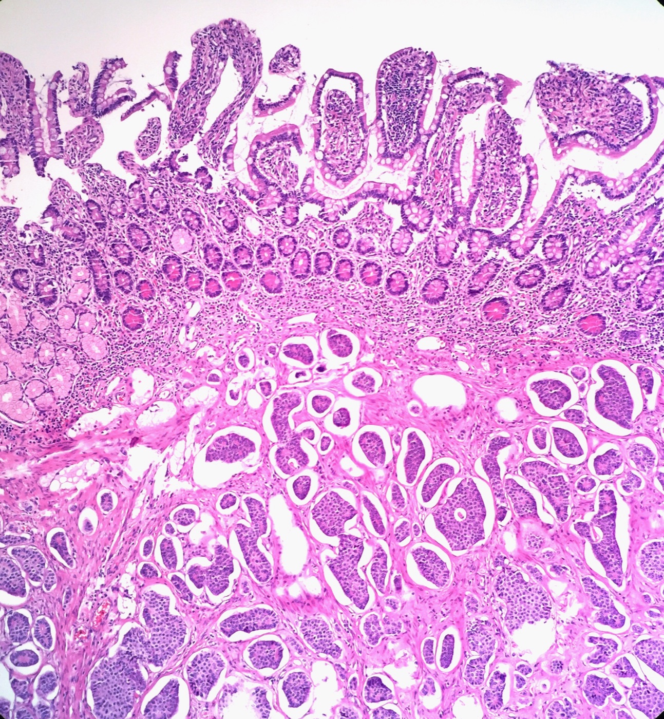
Low-grade neuroendocrine tumor of the small intestine, histology for correlation with EUS-FNA: nests and islands of uniform tumor cells, some with peripheral palisading and retraction spaces, fill the submucosa beneath intact villous mucosa. H&E, low power (magnification not stated).Image: Ed Uthman, Wikimedia Commons, CC BY 2.0.
References
Alaggio R, Amador C, Anagnostopoulos I, et al. The 5th edition of the World Health Organization Classification of Haematolymphoid Tumours: lymphoid neoplasms. Leukemia. 2022;36(7):1720–1748. doi:10.1038/s41375-022-01620-2. PMID 35732829
Almasri NM, Zaer FS, Iturraspe JA, et al. Contribution of flow cytometry to the diagnosis of gastric lymphomas in endoscopic biopsy specimens. Mod Pathol. 1997;10(7):650–656. PMID 9237173
Ambati DK, Suresh PK, Nayak R, et al. Diagnostic utility of endoscopic crush cytology in gastrointestinal malignancies. Acta Cytol. 2026 (online ahead of print). doi:10.1159/000552792. PMID 42258585
Arends MJ, Esposito I, Gill AJ, et al. Changes in the 6th edition of the World Health Organization classification of tumours of the digestive system. Histopathology. 2026;88(7):1295–1314. doi:10.1111/his.70116. PMID 41724188
Arslan ME, Li H, Fu Z, et al. Plexiform fibromyxoma: review of rare mesenchymal gastric neoplasm and its differential diagnosis. World J Gastrointest Oncol. 2021;13(5):409–423. doi:10.4251/wjgo.v13.i5.409. PMID 34040702
Assaf A, Terris B, Palmieri LJ, et al. Endoscopic ultrasound guided fine needle biopsy in patients with suspected gastric linitis plastica. Clin Res Hepatol Gastroenterol. 2022;46(5):101903. doi:10.1016/j.clinre.2022.101903. PMID 35301155
Bartley AN, Washington MK, Colasacco C, et al. HER2 testing and clinical decision making in gastroesophageal adenocarcinoma: guideline from the College of American Pathologists, American Society for Clinical Pathology, and the American Society of Clinical Oncology. J Clin Oncol. 2017;35(4):446–464. doi:10.1200/JCO.2016.69.4836. PMID 28129524
Basuroy R, Srirajaskanthan R, Prachalias A, et al. Review article: the investigation and management of gastric neuroendocrine tumours. Aliment Pharmacol Ther. 2014;39(10):1071–1084. doi:10.1111/apt.12698. PMID 24628514
Bellizzi AM. Immunohistochemistry in the diagnosis and classification of neuroendocrine neoplasms: what can brown do for you? Hum Pathol. 2020;96:8–33. doi:10.1016/j.humpath.2019.12.002. PMID 31857137
Bellizzi AM. SATB2 in neuroendocrine neoplasms: strong expression is restricted to well-differentiated tumours of lower gastrointestinal tract origin and is most frequent in Merkel cell carcinoma among poorly differentiated carcinomas. Histopathology. 2020;76(2):251–264. doi:10.1111/his.13943. PMID 31233624
Bhaijee F, Subramony C, Tang SJ, et al. Human immunodeficiency virus-associated gastrointestinal disease: common endoscopic biopsy diagnoses. Patholog Res Int. 2011;2011:247923. doi:10.4061/2011/247923. PMID 21559197
Bonacini M, Young T, Laine L. The causes of esophageal symptoms in human immunodeficiency virus infection: a prospective study of 110 patients. Arch Intern Med. 1991;151(8):1567–1572. PMID 1651690
Chen W, Cai G. Endoscopic ultrasound-guided fine-needle aspiration biopsy of gastric schwannoma: cytomorphologic features and diagnostic pitfalls. Diagn Cytopathol. 2019;47(11):1218–1222. doi:10.1002/dc.24289. PMID 31343112
Dell'Unto E, Srirajaskanthan R, Toumpanakis C, et al. Management strategies and outcomes for type 1 gastric neuroendocrine tumors measuring more than 1 cm. Clin Transl Gastroenterol. 2026;17(10):e01083. doi:10.14309/ctg.0000000000001083. PMID 42607265
Desai P, Kabrawala M, Patel C, et al. Crush cytology: an expeditious diagnostic tool for gastrointestinal tract malignancy. Endosc Int Open. 2021;9(5):E735–E740. doi:10.1055/a-1388-6479. PMID 33937515
Deshpande A, Nelson D, Corless CL, et al. Leiomyoma of the gastrointestinal tract with interstitial cells of Cajal: a mimic of gastrointestinal stromal tumor. Am J Surg Pathol. 2014;38(1):72–77. doi:10.1097/PAS.0b013e3182a0d134. PMID 24145645
Falk GW, Chittajallu R, Goldblum JR, et al. Surveillance of patients with Barrett's esophagus for dysplasia and cancer with balloon cytology. Gastroenterology. 1997;112(6):1787–1797. doi:10.1053/gast.1997.v112.pm9178668. PMID 9178668
Fatima N, Cohen C, Siddiqui MT. DOG1 utility in diagnosing gastrointestinal stromal tumors on fine-needle aspiration. Cancer Cytopathol. 2011;119(3):202–208. doi:10.1002/cncy.20149. PMID 21400668
Fitzgerald RC, di Pietro M, O'Donovan M, et al. Cytosponge-trefoil factor 3 versus usual care to identify Barrett's oesophagus in a primary care setting: a multicentre, pragmatic, randomised controlled trial. Lancet. 2020;396(10247):333–344. doi:10.1016/S0140-6736(20)31099-0. PMID 32738955
Fritcher EG, Brankley SM, Kipp BR, et al. A comparison of conventional cytology, DNA ploidy analysis, and fluorescence in situ hybridization for the detection of dysplasia and adenocarcinoma in patients with Barrett's esophagus. Hum Pathol. 2008;39(8):1128–1135. doi:10.1016/j.humpath.2008.02.003. PMID 18602665
Garlin-Politis M, Upadhyay Baskota S, Picon S, et al. CytoLyt fixation impedes insulinoma-associated protein 1 (INSM1) immunoreactivity compared to formalin fixation. J Am Soc Cytopathol. 2024;13(3):213–218. doi:10.1016/j.jasc.2024.02.002. PMID 38575468
Geisinger KR. Endoscopic biopsies and cytologic brushings of the esophagus are diagnostically complementary. Am J Clin Pathol. 1995;103(3):295–299. doi:10.1093/ajcp/103.3.295. PMID 7872251
Hissong E, Schechter S, Mowers J, et al. The diagnosis of clinically significant oesophageal Candida infections: a reappraisal of clinicopathological findings. Histopathology. 2020;76(5):748–754. doi:10.1111/his.14063. PMID 31944368
Hoover L, Berman JJ. Epithelial repair versus carcinoma in esophageal brush cytology. Diagn Cytopathol. 1988;4(3):217–223. doi:10.1002/dc.2840040308. PMID 3076105
Hornick JL, Bovée JVMG, Antonescu CR, et al. The 2026 World Health Organization Classification of Tumors of Soft Tissue and Bone: 6th edition. Am J Surg Pathol. 2026 (online ahead of print). doi:10.1097/PAS.0000000000002624. PMID 42839882
Hoskoppal D, Epstein JI, Gown AM, et al. SATB2 protein expression by immunohistochemistry is a sensitive and specific marker of appendiceal and rectosigmoid well differentiated neuroendocrine tumours. Histopathology. 2020;76(4):550–559. doi:10.1111/his.14012. PMID 31595536
Judson I, Jones RL, Wong NACS, et al. Gastrointestinal stromal tumour (GIST): British Sarcoma Group clinical practice guidelines. Br J Cancer. 2025;132(1):1–10. doi:10.1038/s41416-024-02672-0. PMID 38840030
Kapatia G, Gupta N, Saikia UN, et al. Fine needle aspiration cytology of primary and metastatic gastrointestinal stromal tumour. Cytopathology. 2020;31(2):136–143. doi:10.1111/cyt.12785. PMID 31698512
Kim D, Viswanathan K, Goyal A, et al. Insulinoma-associated protein 1 (INSM1) is a robust marker for identifying and grading pancreatic neuroendocrine tumors. Cancer Cytopathol. 2020;128(4):269–277. doi:10.1002/cncy.22242. PMID 31977134
Kobara H, Mori H, Rafiq K, et al. Analysis of the amount of tissue sample necessary for mitotic count and Ki-67 index in gastrointestinal stromal tumor sampling. Oncol Rep. 2015;33(1):215–222. doi:10.3892/or.2014.3608. PMID 25405369
Kumar PV, Monabati A, Naini MA, et al. Gastric xanthoma: a diagnostic problem on brushing cytology smears. Acta Cytol. 2006;50(1):74–79. doi:10.1159/000325898. PMID 16514844
Kumaravel A, Lopez R, Brainard J, et al. Brush cytology vs. endoscopic biopsy for the surveillance of Barrett's esophagus. Endoscopy. 2010;42(10):800–805. doi:10.1055/s-0030-1255710. PMID 20821361
Kushima R. Gastric carcinoma classification in the WHO 6th edition (2026): updated framework and emerging entities. Virchows Arch. 2026 (online ahead of print). doi:10.1007/s00428-026-04697-7. PMID 42709193
La Rosa S, Maragliano R, Marchiori D. Roadmap to challenges in limited cellularity specimens from pancreatic neuroendocrine neoplasms: diagnostic tools for the most appropriate management of limited material. Endocr Pathol. 2025;36(1):25. doi:10.1007/s12022-025-09870-3. PMID 40668487
Lin BP, Harmata PA. Gastric and esophageal brush cytology. Pathology. 1983;15(4):393–397. doi:10.3109/00313028309085165. PMID 6674869
Liu H, Ruskon-Fourmestraux A, Lavergne-Slove A, et al. Resistance of t(11;18) positive gastric mucosa-associated lymphoid tissue lymphoma to Helicobacter pylori eradication therapy. Lancet. 2001;357(9249):39–40. doi:10.1016/S0140-6736(00)03571-6. PMID 11197361
Liu T, Baskota SU, Gonzalez A. Endoscopic ultrasound-guided fine-needle aspiration and fine-needle biopsy in the diagnosis of gastrointestinal lymphomas. J Am Soc Cytopathol. 2025;14(2):102–109. doi:10.1016/j.jasc.2024.12.002. PMID 39732576
Matsueda K, Omote S, Sakata M, et al. The diagnosis of gastric mucosa-associated lymphoid tissue lymphoma by flow cytometry and fluorescence in situ hybridization of biopsy specimens. Intern Med. 2018;57(8):1081–1086. doi:10.2169/internalmedicine.9617-17. PMID 29269666
McBane RD, Gross JB Jr. Herpes esophagitis: clinical syndrome, endoscopic appearance, and diagnosis in 23 patients. Gastrointest Endosc. 1991;37(6):600–603. doi:10.1016/s0016-5107(91)70862-6. PMID 1756917
Miettinen M, Lasota J. Gastrointestinal stromal tumors: pathology and prognosis at different sites. Semin Diagn Pathol. 2006;23(2):70–83. doi:10.1053/j.semdp.2006.09.001. PMID 17193820
Miettinen M, Wang ZF, Sarlomo-Rikala M, et al. Succinate dehydrogenase-deficient GISTs: a clinicopathologic, immunohistochemical, and molecular genetic study of 66 gastric GISTs with predilection to young age. Am J Surg Pathol. 2011;35(11):1712–1721. doi:10.1097/PAS.0b013e3182260752. PMID 21997692
Mohamed AA, Lu XL, Mounmin FA. Diagnosis and treatment of esophageal candidiasis: current updates. Can J Gastroenterol Hepatol. 2019;2019:3585136. doi:10.1155/2019/3585136. PMID 31772927
Mostaghni AA, Afarid M, Eghbali S, et al. Evaluation of brushing cytology in the diagnosis of Helicobacter pylori gastritis. Acta Cytol. 2008;52(5):597–601. doi:10.1159/000325604. PMID 18833824
Muniraj F, Siddaraju N, Sistla SC. Role of brush cytology in the diagnosis of neoplastic and non-neoplastic upper gastrointestinal lesions. Cytopathology. 2016;27(6):407–417. doi:10.1111/cyt.12333. PMID 27061229
Nagtegaal ID, Odze RD, Klimstra D, et al. The 2019 WHO classification of tumours of the digestive system. Histopathology. 2020;76(2):182–188. doi:10.1111/his.13975. PMID 31433515
Panzuto F, O'Toole D, Klöppel G, et al. Controversies in NEN: an ENETS position statement on the endoscopic management of localised gastric, duodenal and rectal neuroendocrine neoplasms. J Neuroendocrinol. 2025;37(12):e70060. doi:10.1111/jne.70060. PMID 40524475
Park HS, Yoon SO, Lim BJ, et al. The limitations of endoscopic ultrasound-guided fine needle aspiration cytology in the diagnosis of pancreatic serous cystadenoma: a brief case report. Korean J Pathol. 2014;48(5):405–408. doi:10.4132/KoreanJPathol.2014.48.5.405. PMID 25366082
Pitman MB, Centeno BA, Ali SZ, et al. Standardized terminology and nomenclature for pancreatobiliary cytology: the Papanicolaou Society of Cytopathology guidelines. Cytojournal. 2014;11(Suppl 1):3. doi:10.4103/1742-6413.133343. PMID 25191517
Rosołowski M, Kierzkiewicz M. Etiology, diagnosis and treatment of infectious esophagitis. Prz Gastroenterol. 2013;8(6):333–337. doi:10.5114/pg.2013.39914. PMID 24868280
Rossi RE, Rausa E, Cavalcoli F, et al. Duodenal neuroendocrine neoplasms: a still poorly recognized clinical entity. Scand J Gastroenterol. 2018;53(7):835–842. doi:10.1080/00365521.2018.1468479. PMID 29726295
Roth MJ, Liu SF, Dawsey SM, et al. Cytologic detection of esophageal squamous cell carcinoma and precursor lesions using balloon and sponge samplers in asymptomatic adults in Linxian, China. Cancer. 1997;80(11):2047–2059. doi:10.1002/(sici)1097-0142(19971201)80:11<2047::aid-cncr3>3.0.co;2-u. PMID 9392326
Rutkowski P, Przybył J, Zdzienicki M. Extended adjuvant therapy with imatinib in patients with gastrointestinal stromal tumors: recommendations for patient selection, risk assessment, and molecular response monitoring. Mol Diagn Ther. 2013;17(1):9–19. doi:10.1007/s40291-013-0018-7. PMID 23355099
Saad RS, Mahood LK, Clary KM, et al. Role of cytology in the diagnosis of Barrett's esophagus and associated neoplasia. Diagn Cytopathol. 2003;29(3):130–135. doi:10.1002/dc.10334. PMID 12951679
Saoud C, Illei PB, Siddiqui MT, et al. Cytopathology of rare gastric mesenchymal neoplasms: a series of 25 cases and review of literature. Cytopathology. 2023;34(1):15–27. doi:10.1111/cyt.13187. PMID 36227114
Sbaraglia M, Businello G, Bellan E, et al. Mesenchymal tumours of the gastrointestinal tract. Pathologica. 2021;113(3):230–251. doi:10.32074/1591-951X-309. PMID 34294940
Seven G, Kochan K, Caglar E, et al. Evaluation of Ki67 index in endoscopic ultrasound-guided fine needle aspiration samples for the assessment of malignancy risk in gastric gastrointestinal stromal tumors. Dig Dis. 2021;39(4):407–414. doi:10.1159/000511994. PMID 33017820
Shaheen NJ, Falk GW, Iyer PG, et al. Diagnosis and management of Barrett's esophagus: an updated ACG guideline. Am J Gastroenterol. 2022;117(4):559–587. doi:10.14309/ajg.0000000000001680. PMID 35354777
Sherman ME, Anderson C, Herman LM, et al. Utility of gastric brushing in the diagnosis of malignant lymphoma. Acta Cytol. 1994;38(2):169–174. PMID 8147207
Singh T, Gupta NM, Bhasin DK, et al. Comparison of brush before biopsy, suction cytology, brush after biopsy and endoscopic biopsy in the diagnosis of carcinoma oesophagus. J Gastroenterol Hepatol. 1994;9(6):564–566. doi:10.1111/j.1440-1746.1994.tb01561.x. PMID 7865714
Stelow EB, Murad FM, Debol SM, et al. A limited immunocytochemical panel for the distinction of subepithelial gastrointestinal mesenchymal neoplasms sampled by endoscopic ultrasound-guided fine-needle aspiration. Am J Clin Pathol. 2008;129(2):219–225. doi:10.1309/NL2WYAD8EUH3XFRF. PMID 18208801
Stelow EB, Stanley MW, Mallery S, et al. Endoscopic ultrasound-guided fine-needle aspiration findings of gastrointestinal leiomyomas and gastrointestinal stromal tumors. Am J Clin Pathol. 2003;119(5):703–708. doi:10.1309/UWUV-Q001-0D9W-0HPN. PMID 12760289
Strosberg J, Chatterjee D, Lauwers GY, et al. Expansion of the World Health Organization (WHO) gastric neuroendocrine tumor classification: introducing types 4 and 5. J Natl Cancer Inst. 2026 (online ahead of print). doi:10.1093/jnci/djag320. PMID 42730652
Suresh Kumar VC, Harne P, Patthipati VS, et al. Wide-area transepithelial sampling in adjunct to forceps biopsy increases the absolute detection rates of Barrett's oesophagus and oesophageal dysplasia: a meta-analysis and systematic review. BMJ Open Gastroenterol. 2020;7(1):e000494. doi:10.1136/bmjgast-2020-000494. PMID 32928869
Taşkın OÇ, Armutlu A, Adsay V, et al. Clinicopathologic and immunohistochemical characteristics of upper gastrointestinal leiomyomas harboring interstitial cells of Cajal: a potential mimicker of gastrointestinal stromal tumor. Ann Diagn Pathol. 2020;45:151476. doi:10.1016/j.anndiagpath.2020.151476. PMID 32062475
Teot LA, Ducatman BS, Geisinger KR. Cytologic diagnosis of cytomegaloviral esophagitis: a report of three acquired immunodeficiency syndrome-related cases. Acta Cytol. 1993;37(1):93–96. PMID 8382003
Tyagi R, Kaur J, Kaur G, et al. Cytohistological discordance on gastrointestinal brushings: facts unfolded. Niger Med J. 2016;57(5):299–302. doi:10.4103/0300-1652.190597. PMID 27833250
Vander Noot MR 3rd, Eloubeidi MA, Chen VK, et al. Diagnosis of gastrointestinal tract lesions by endoscopic ultrasound-guided fine-needle aspiration biopsy. Cancer. 2004;102(3):157–163. doi:10.1002/cncr.20360. PMID 15211474
Vij M, Agrawal V, Kumar A, et al. Cytomorphology of gastrointestinal stromal tumors and extra-gastrointestinal stromal tumors: a comprehensive morphologic study. J Cytol. 2013;30(1):8–12. doi:10.4103/0970-9371.107505. PMID 23661933
Voltaggio L, Murray R, Lasota J, Miettinen M. Gastric schwannoma: a clinicopathologic study of 51 cases and critical review of the literature. Hum Pathol. 2012;43(5):650–659. doi:10.1016/j.humpath.2011.07.006. PMID 22137423
von Mehren M, Joensuu H. Gastrointestinal stromal tumors. J Clin Oncol. 2018;36(2):136–143. doi:10.1200/JCO.2017.74.9705. PMID 29220298
Wang HW, Kuo CJ, Lin WR, et al. Clinical characteristics and manifestation of herpes esophagitis: one single-center experience in Taiwan. Medicine (Baltimore). 2016;95(14):e3187. doi:10.1097/MD.0000000000003187. PMID 27057845
Wang M, Lozano MD, Cai G. The World Health Organization System for Reporting Pancreaticobiliary Cytopathology: standardized categories and practical approaches to pancreatic lesions. J Clin Transl Pathol. 2024;4(3):122–135. doi:10.14218/jctp.2024.00034. PMID 40191142
Wilcox CM, Rodgers W, Lazenby A. Prospective comparison of brush cytology, viral culture, and histology for the diagnosis of ulcerative esophagitis in AIDS. Clin Gastroenterol Hepatol. 2004;2(7):564–567. doi:10.1016/s1542-3565(04)00239-3. PMID 15224280
Wilcox CM, Straub RF, Schwartz DA. Prospective evaluation of biopsy number for the diagnosis of viral esophagitis in patients with HIV infection and esophageal ulcer. Gastrointest Endosc. 1996;44(5):587–593. doi:10.1016/s0016-5107(96)70014-7. PMID 8934167
Wotherspoon AC, Doglioni C, Diss TC, et al. Regression of primary low-grade B-cell gastric lymphoma of mucosa-associated lymphoid tissue type after eradication of Helicobacter pylori. Lancet. 1993;342(8871):575–577. doi:10.1016/0140-6736(93)91409-f. PMID 8102719
Zhang W, Garces J, Dong HY. Detection of the t(11;18) API2/MALT1 translocation associated with gastric MALT lymphoma in routine formalin-fixed, paraffin-embedded small endoscopic biopsy specimens by robust real-time RT-PCR. Am J Clin Pathol. 2006;126(6):931–940. doi:10.1309/unxjvamv77jam4fm. PMID 17153776