Library › Esophagus, stomach and small intestine pathology: esophagitis, gastritis, celiac disease, carcinoma, neuroendocrine, mesenchymal and lymphoid tumors
Esophagus, stomach and small intestine pathology: esophagitis, gastritis, celiac disease, carcinoma, neuroendocrine, mesenchymal and lymphoid tumors
Last reviewed 2026-11-03 by a board-certified pathologist. 10 references.
Barrett Esophagus and Dysplasia
Definition (US, ACG 2022). Columnar mucosa extending ≥1 cm above the gastroesophageal junction (GEJ), with intestinal metaplasia (goblet cells) on biopsy. The British (BSG) definition does not require goblet cells. Barrett esophagus is the precursor of esophageal adenocarcinoma.
Clinical features of Barrett esophagus and dysplasia
- Cause: chronic gastroesophageal reflux.
- Risk factors: male sex, age over 50 years, white race, central obesity, smoking, family history.
- Screening (ACG 2022): patients with chronic reflux and ≥3 risk factors. A swallowable nonendoscopic capsule device combined with a biomarker is an acceptable alternative to endoscopy.
- Endoscopy: salmon-colored tongues extending above the GEJ. The length is described with the Prague C & M criteria (circumferential and maximum extent). Short-segment Barrett esophagus is <3 cm, and long-segment is ≥3 cm.
- Biopsy (Seattle protocol): 4-quadrant biopsies every 2 cm, or every 1 cm with known or suspected dysplasia, plus targeted biopsies of visible lesions. ACG 2022 recommends at least 8 biopsies at screening.
- Annual risk of progression:
- non-dysplastic: about 0.2–0.5% per year to adenocarcinoma (ACG 2022)
- low-grade dysplasia (LGD): about 0.7% per year to adenocarcinoma overall (ACG 2022). Expert-confirmed LGD progressed to HGD or adenocarcinoma at 9.1% per patient-year (Curvers et al. 2010), and the SURF trial surveillance arm at 11.8% per patient-year.
- high-grade dysplasia (HGD): about 6% per year to adenocarcinoma (Rastogi et al. 2008)
Microscopic findings in Barrett esophagus and dysplasia
- Intestinal metaplasia:
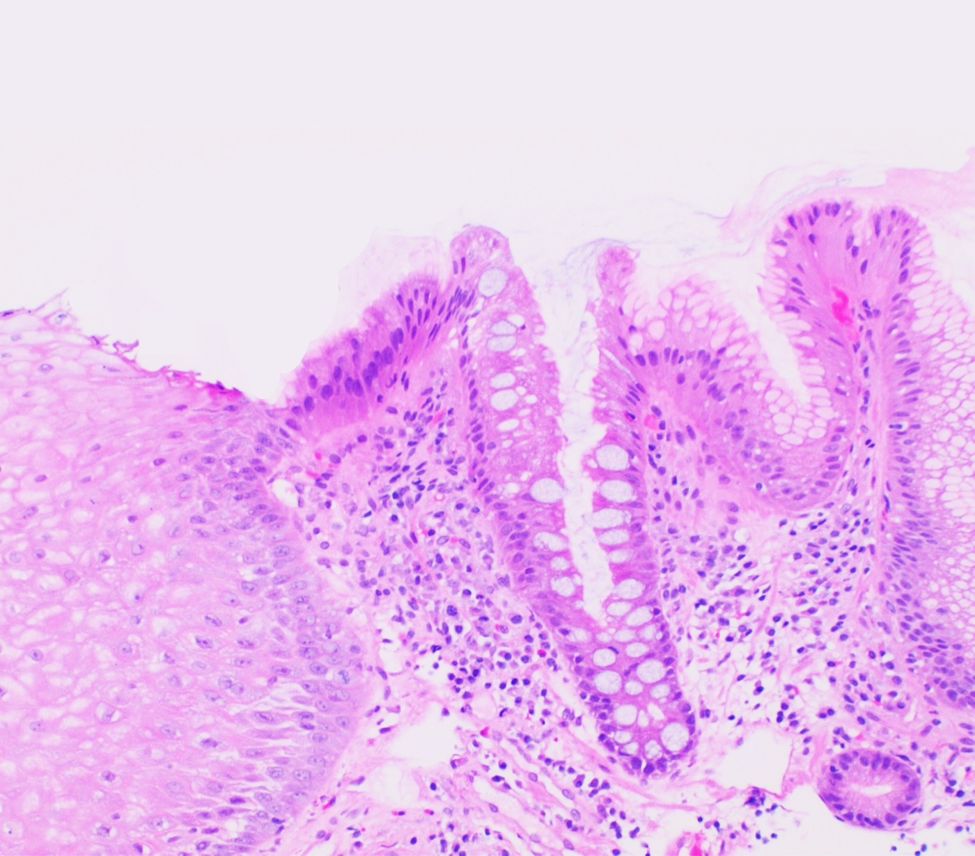
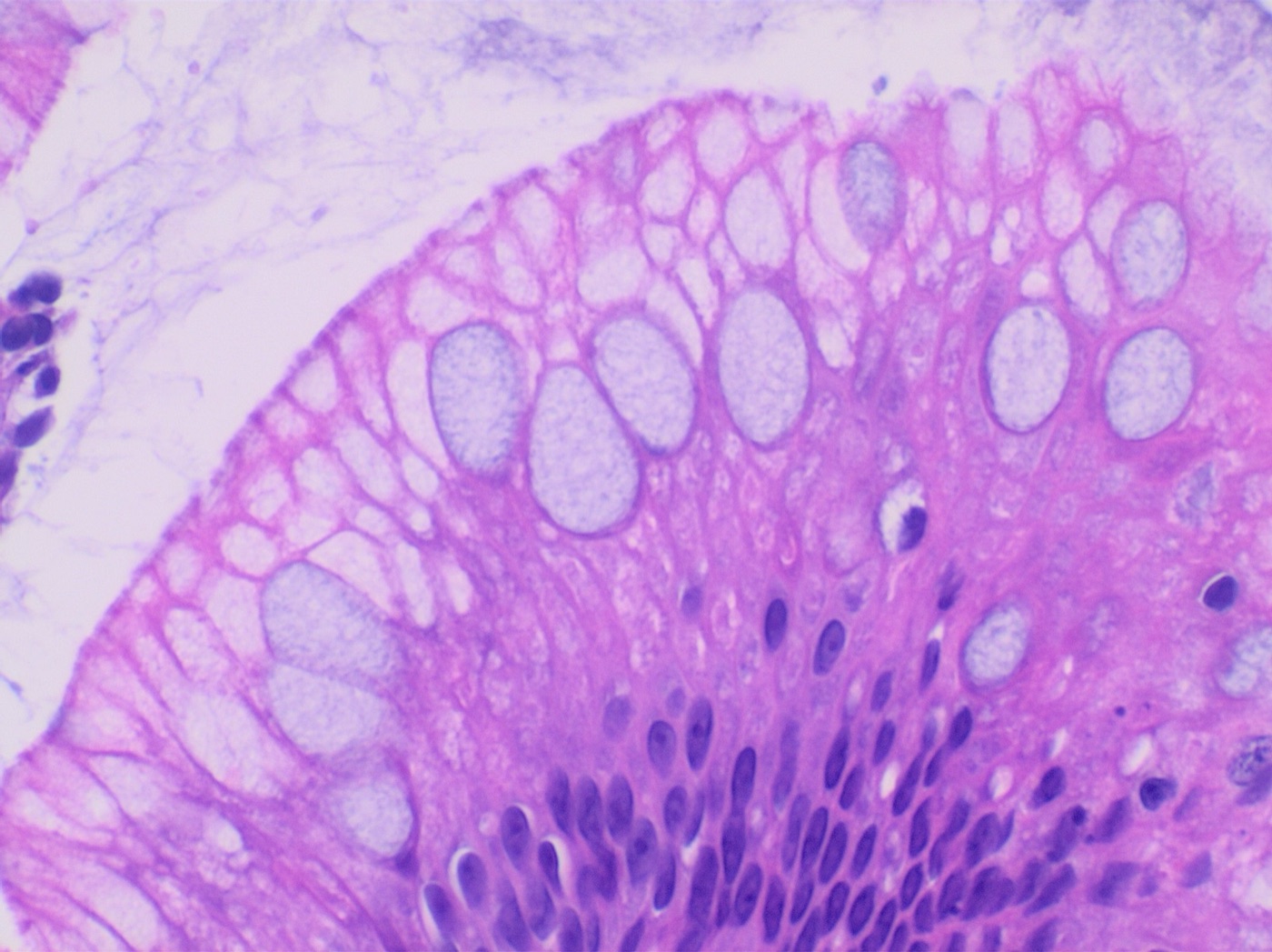
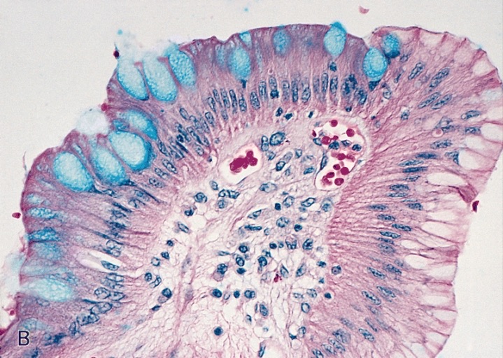
- Goblet cells: barrel-shaped mucin vacuoles that indent the nucleus; blue on Alcian blue at pH 2.5. The diagnosis is made on H&E morphology, and routine Alcian blue staining is not required.
- Pseudogoblet cells are distended foveolar cells, in a row along the surface without true goblet shape. They are not counted as intestinal metaplasia.
- Duplicated (split) muscularis mucosae is common in Barrett esophagus. Invasion into the space between the layers is still pT1a. It is not staged as submucosal invasion (pT1b).
Dysplasia grades
| Grade | Cytology | Architecture | Surface |
|---|---|---|---|
| Negative | Bland basal nuclei | Normal | Matures at the surface |
| Indefinite | Atypia that cannot be classified because of inflammation or erosion | Normal | Partial maturation |
| Low-grade (LGD) | Elongated, pencil-shaped, hyperchromatic nuclei, stratified but confined to the base of the cells; polarity preserved | Mostly preserved | Does not mature |
| High-grade (HGD) | Rounded nuclei, vesicular chromatin, prominent nucleoli, loss of polarity, nuclei reaching the luminal surface, atypical mitoses | Complex: crowding, budding, cribriform glands | No maturation |
| Intramucosal adenocarcinoma | As for HGD | Single cells, sheets, angulated or fused glands invading the lamina propria or muscularis mucosae | n/a |
- Non-conventional (foveolar or gastric-type) dysplasia also exists. Its nuclei are less pencil-like and the cytoplasm has more mucin. It is frequently underdiagnosed.
- Crypt dysplasia (basal crypt dysplasia-like atypia) shows dysplastic-appearing crypt bases with a maturing surface. It is a recognized pitfall.
Ancillary testing in Barrett esophagus and dysplasia
- p53 IHC: an aberrant pattern supports dysplasia and predicts progression (Redston et al. 2022). The aberrant patterns are strong diffuse overexpression and the "null" pattern (absent epithelial staining with a positive internal control). A wild-type pattern does not exclude dysplasia. ACG 2022 makes no recommendation on routine p53 IHC.
- Molecular: TP53 mutation occurs early. Loss of CDKN2A (p16) and aneuploidy accompany progression.
Differential diagnosis of Barrett esophagus and dysplasia
| Look-alike | Distinguishing features |
|---|---|
| Gastric cardia or GEJ intestinal metaplasia | Endoscopic location below the GEJ; classified as intestinal metaplasia of the cardia |
| Reactive or regenerative atypia | Inflammation, erosion, surface maturation, gradual transition |
| Pseudogoblet cells | No barrel shape, no indentation of the nucleus; H&E morphology is decisive, because columnar cells and pseudogoblet cells can also stain with Alcian blue |
| LGD vs HGD | HGD has lost polarity, has round nuclei with nucleoli, and complex architecture |
Staging and reporting (AJCC 8th edition)
- pTis: high-grade dysplasia
- pT1a: invasion of the lamina propria or muscularis mucosae, including the space within a duplicated muscularis mucosae
- pT1b: invasion of the submucosa
- pT2: invasion of the muscularis propria
- pT3: invasion of the adventitia
- Endoscopic resections: depth is often substaged as m1–m3 (mucosa) and sm1–sm3 (submucosa). High-risk features are poor differentiation, lymphovascular invasion, a positive deep margin and submucosal invasion >500 µm.
- Also report: dysplasia grade, depth of invasion, lymphovascular invasion, and deep and lateral margins of endoscopic resections.
Management summary (ACG 2022)
- Confirmation: dysplasia of any grade, and indefinite for dysplasia, is confirmed by a second pathologist with GI expertise.
- Non-dysplastic:
- surveillance endoscopy every 5 years if <3 cm
- every 3 years if ≥3 cm
- Indefinite: optimization of twice-daily PPI therapy and repeat endoscopy within 6 months. If indefinite for dysplasia persists, endoscopy is repeated annually.
- LGD:
- endoscopic eradication therapy (radiofrequency ablation) is suggested
- if LGD is managed by surveillance, endoscopy is performed at 6 months, then 12 months, then annually
- HGD or T1a carcinoma: endoscopic resection plus ablation.
AGA 2025 surveillance guideline for Barrett esophagus
The American Gastroenterological Association (AGA) published a surveillance guideline in 2025 (Wani et al.). ACG 2022 is the parallel US guideline. The AGA 2025 abstract states the following:
- Nondysplastic Barrett esophagus: surveillance endoscopy (conditional recommendation).
- Columnar-lined esophagus <1 cm: no endoscopic surveillance (conditional recommendation). This agrees with the ACG 2022 definition, which requires ≥1 cm of columnar mucosa.
- Endoscopic imaging: high-definition white light endoscopy combined with chromoendoscopy over white light endoscopy alone (strong recommendation).
- Biomarkers and sampling: no recommendation on p53 or TissueCypher to predict progression, or on wide-area transepithelial sampling. ACG 2022 also makes no recommendation on routine p53 IHC.
- Chemoprevention: daily proton pump inhibitor therapy over no therapy and over antireflux surgery (conditional recommendation).
- Pathology: the diagnosis of Barrett-related neoplasia is confirmed by an expert pathologist (implementation statement). ACG 2022 specifies confirmation of dysplasia of any grade, and of indefinite for dysplasia, by a second pathologist with GI expertise.
- Intervals: the AGA 2025 surveillance intervals and its management of LGD and indefinite for dysplasia are given in the full text, which was not openly available. The intervals in the management summary above are from ACG 2022.
- Endoscopic eradication therapy (AGA 2024, Rubenstein et al.): strong recommendation for eradication therapy in HGD; conditional recommendation for eradication therapy in LGD, with surveillance endoscopy a reasonable choice for patients who place more value on the potential harms of eradication therapy and less on its uncertain benefits; conditional recommendation against eradication therapy in nondysplastic Barrett esophagus. For visible lesions, focal endoscopic resection plus ablation is suggested over stepwise endoscopic mucosal resection, with endoscopic mucosal resection or endoscopic submucosal dissection chosen by lesion characteristics. ACG 2022 also suggests eradication therapy for LGD, with surveillance as the alternative.
References
- Amin MB, et al., eds. AJCC Cancer Staging Manual, 8th ed. Springer; 2017. doi:10.1007/978-3-319-40618-3
- Curvers WL, et al. Low-grade dysplasia in Barrett's esophagus: overdiagnosed and underestimated. Am J Gastroenterol. 2010;105:1523–1530. doi:10.1038/ajg.2010.171
- Fitzgerald RC, et al. British Society of Gastroenterology guidelines on the diagnosis and management of Barrett's oesophagus. Gut. 2014;63:7–42. doi:10.1136/gutjnl-2013-305372
- Odze RD. Diagnosis and grading of dysplasia in Barrett's oesophagus. J Clin Pathol. 2006;59:1029–1038. doi:10.1136/jcp.2005.035337
- Rastogi A, et al. Incidence of esophageal adenocarcinoma in patients with Barrett's esophagus and high-grade dysplasia: a meta-analysis. Gastrointest Endosc. 2008;67:394–398. doi:10.1016/j.gie.2007.07.019
- Redston M, et al. Abnormal TP53 predicts risk of progression in patients with Barrett's esophagus regardless of a diagnosis of dysplasia. Gastroenterology. 2022;162:468–481. doi:10.1053/j.gastro.2021.10.038
- Rubenstein JH, et al. AGA Clinical Practice Guideline on Endoscopic Eradication Therapy of Barrett's Esophagus and Related Neoplasia. Gastroenterology. 2024;166:1020–1055. doi:10.1053/j.gastro.2024.03.019
- Shaheen NJ, et al. Diagnosis and Management of Barrett's Esophagus: An Updated ACG Guideline. Am J Gastroenterol. 2022;117:559–587. doi:10.14309/ajg.0000000000001680
- Wani S, et al. AGA Clinical Practice Guideline on Surveillance of Barrett's Esophagus. Gastroenterology. 2025;169:1184–1231. doi:10.1053/j.gastro.2025.09.012
- WHO Classification of Tumours Editorial Board. Digestive System Tumours, 5th ed. IARC; 2019.
Image credits
- Barrett esophagus. Image: Mikael Häggström, M.D., Wikimedia Commons, CC0.
- Incomplete intestinal metaplasia in Barrett esophagus. Image: Mikael Häggström, M.D., Wikimedia Commons, CC0.
- Barrett mucosa. Image: The Armed Forces Institute of Pathology (AFIP), Wikimedia Commons, Public domain.


