Library › Pelvic washing cytology and ovarian cyst FNA
Pelvic washing cytology and ovarian cyst FNA
Last reviewed 2026-11-10 by a board-certified pathologist. 40 references.
Pelvic Washings: Benign Findings, Collagen Balls, Endosalpingiosis, Müllerian Neoplasms, Staging and Reporting
Definition. Pelvic washing cytology is the cytologic examination of washings from the female pelvis; collection, clinical use and outcome data are given in Peritoneal Washing Cytology. Washings contain cells mechanically detached from the serosa together with naturally exfoliated cells (Basset et al. 2023). In ovarian or fallopian tube carcinoma limited to one or both ovaries or tubes, malignant cells in ascites or peritoneal washings define FIGO 2014 stage IC3 (Prat 2014; Berek et al. 2021). The International System for Reporting Serous Fluid Cytopathology (ISRSFC) contains a chapter dedicated to washings (Basset et al. 2023).
Related page: Ovarian cyst fluid and FNA cytology (ovarian cyst fluid and aspiration cytology) Related page: Peritoneal Washing Cytology (collection, benign mimics, outcome data by primary site, FIGO stage IC3, gastric and pancreatic staging, ancillary panel) Related page: The International System for Reporting Serous Fluid Cytopathology (ISRSFC categories and risk of malignancy) Related page: Metastatic Müllerian Carcinoma in Serous Effusions (Müllerian carcinoma in spontaneous effusions)
Collection of pelvic washings
- Timing. The timing of collection is given in Peritoneal Washing Cytology. Tumor cells spilled during exploration and removal of the primary tumor do not reflect the natural biology of the tumor, and their significance in a washing taken afterward is unknown (Zuna 2022).
- Fluid. Spontaneous pelvic fluid is labeled as peritoneal fluid, or as ascites when abundant. For the washing, about 100 mL of saline, preferably a balanced salt solution such as peritoneal dialysis fluid, is instilled, agitated and aspirated (Zuna 2022).
- Holding specimens. Holding washings from operations for presumed benign disease, and sending them only if malignancy is found, delays processing of unfixed fluid and compromises cellular detail. A balanced salt solution is preferred when this approach is used (Zuna 2022).
- Clinical use by gynecologic oncologists. In a survey of 168 gynecologic oncologists, 63.9% routinely obtained washings during endometrial cancer staging, and 53% of these reported that a positive result could change management. Sixty-six percent treated an indeterminate result as negative (VandenBussche et al. 2025).
Benign elements in pelvic washings
- Comparison with ascites. In spontaneous ascites, mesothelial cells form three-dimensional groups of rounded cells, and a second cell population supports malignancy. In a pelvic washing, a second cell population does not by itself indicate malignancy (Zuna 2022).
- Mesothelial sheets. The sheets (Peritoneal Washing Cytology) have a honeycomb pattern and are often folded or rolled. Compressed sheets artificially distort the nuclear-to-cytoplasmic ratio. Nuclei are round to oval, sometimes bilobed or grooved, with finely granular chromatin and a single micronucleolus (Zuna 2022).
- Mesothelial polymorphism. Several mesothelial patterns coexist in one washing in pelvic inflammatory disease, ruptured ovarian cysts, cystic neoplasms and inflammatory gastrointestinal lesions such as diverticulitis or appendicitis (Zuna 2022).
- Large benign tumors. Leiomyomas and ovarian fibrothecomas, including Meigs syndrome, produce highly reactive mesothelial cells. The changes form one spectrum distributed evenly through the sample. Large fibromas produce rolled-up, pseudopapillary mesothelial sheets (Zuna 2022).
- Capillary tangles. Longitudinal capillaries wrapped in tangled fibrillar material are the residue of lysed adhesions after healed inflammation or endometriosis (Zuna 2022).
- Detached ciliary tufts. Anucleate fragments of cilia with a little cytoplasm were present in all 14 washings from women aged 21 to 47 years, 13 of whom were in the secretory phase. Washings without tufts came from 3 postmenopausal women, 7 women in the proliferative phase and 1 woman after hysterectomy and bilateral salpingo-oophorectomy. The tufts represent cyclic shedding from tubal epithelium in the luteal phase (Sidawy et al. 1987).
- Cast-like groups. Three-dimensional groups with a molded outline, a smooth common border and bland nuclei arranged at the periphery occur with benign epithelial-stromal tumors, particularly adenofibromas. They probably represent ovarian surface epithelium extracted from surface invaginations (Zuna 2022).
- Hemosiderin-laden macrophages. These follow intraperitoneal bleeding from endometriosis, ruptured tubal pregnancy, prior surgery or malignancy, and are nonspecific (Zuna 2022).
- Incision-derived tissue. Skeletal muscle fibers and, rarely, adipose tissue come from the abdominal incision (Zuna 2022).
- Bland epithelium from ruptured benign cysts. Epithelial cells from ruptured cystic endometriosis, benign cystic tumors and bowel mucosa are foreign to the peritoneal cavity. Review of the operative note clarifies their source (Zuna 2022).
Collagen balls in pelvic washings
- Morphology. The collagen core (Peritoneal Washing Cytology) stains green on Papanicolaou stain (Sasaki et al. 1995) and is homogeneous or filamentous (Zuna 2022).
- Immunophenotype. The covering cells express keratin and vimentin, and the core contains collagen types I and III. The structures are fragments of serous membrane (Sasaki et al. 1995).
- Frequency. Collagen balls were found in 32 of 174 washings (18%) (Sasaki et al. 1995). In one laboratory their recorded frequency rose from 3.2% in 1995 to 28.5% in 2002 without a change in specimen handling (Szporn et al. 2005).
- Origin. They appear to represent small protrusions from the ovarian surface (Zuna 2022).
- Synonyms. Collagen globules and serosal balls (Zuna 2022; Sasaki et al. 1995).
Endosalpingiosis and endometriosis in pelvic washings
- Endosalpingiosis. Ectopic tubal-type epithelium on the ovaries, paratubal tissues, omentum and pelvic and para-aortic lymph nodes. Psammoma bodies and microcalcifications are frequent (Zuna 2022).
- Cytology of endosalpingiosis. Small aggregates and papillary fragments of columnar cells with oval nuclei, scant cytoplasm, fine chromatin and single micronucleoli. Cilia and psammoma bodies are present in some cases, and cells surrounding psammoma bodies have a scalloped outline (Zuna 2022).
- Series of 38 benign cases. In washings from 38 women without gynecologic malignancy, 92% showed tubular or small branching papillary structures, some with psammoma bodies. Each case had 4 to 50 groups, and some groups had up to moderate atypia. All cases were PAX8 positive (Sneige et al. 2013).
- Coexistence with neoplasia. Endosalpingiosis and benign or malignant serous neoplasms coexist in some patients (Zuna 2022).
- Endometriosis. Endometriotic epithelium and stroma are rare in washings and usually follow rupture of an endometriotic cyst (Zuna 2022).
Psammoma bodies in pelvic washings
- Reporting practice. Of pathologists surveyed for the ISRSFC, 64.1% reported psammomatous calcification without epithelial cells as benign, 20.5% as atypical and 6.8% as suspicious (VandenBussche et al. 2025).
- Association with neoplasia. In 78 cytology specimens with psammoma bodies, 20.5% were pelvic washings. Of peritoneal cytology specimens with psammoma bodies, 79% were neoplastic, mostly gynecologic tumors (Braun et al. 2024).
Müllerian neoplasms in pelvic washings
- Ovarian carcinoma. In the literature summarized by Zuna (2022), peritoneal cytology at laparotomy is positive in 48–60% of women with ovarian cancer. Single-institution rates by primary site and stage are given in Peritoneal Washing Cytology. Malignant cells resemble those in effusions, often accompanied by polymorphic mesothelial sheets (Zuna 2022).
- Dislodged tumor fragments. Large fragments of tumor can be dislodged from intraperitoneal tumor while the washing is taken. This pattern is rare in spontaneous effusions (Zuna 2022).
- Comparison with the primary tumor. The abnormal cells differ from reactive mesothelium and surface reactions and match the primary tumor (Zuna 2022).
- High-grade serous carcinoma. The cytology is given in Peritoneal Washing Cytology; group borders are frayed (Zuna 2022). Serous carcinoma of the endometrium has the same appearance, and washings can be positive when the tumor is histologically confined to the uterus (Zuna 2022).
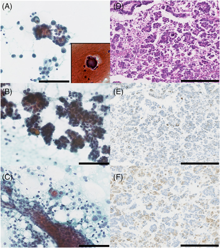
- Low-grade serous lesions. Washings with cells of a serous borderline tumor or low-grade serous carcinoma (Peritoneal Washing Cytology) are reported as low-grade serous neoplasm, and histology determines the category (Zuna 2022).
- Endometrioid carcinoma of the endometrium. Low-grade endometrioid carcinoma involves the peritoneum late and is infrequently positive in washings when confined to the uterus. Its small nuclei resemble those of reactive mesothelial groups. Haphazardly arranged columnar cells with dyspolaric nuclei distinguish it from tubal epithelium (Zuna 2022).
- Cervical squamous cell carcinoma. The resemblance of nonkeratinizing malignant squamous cells to reactive mesothelial cells (Peritoneal Washing Cytology) is greatest after pelvic radiation (Zuna 2022).
Serous borderline tumors: washings and peritoneal implants
In 101 patients with serous borderline tumors, 26 of 96 with staging biopsies had peritoneal implants (17 noninvasive and 9 invasive) and 19 had endosalpingiosis (Sneige et al. 2012).
| Measure | Value |
|---|---|
| Washings positive for serous neoplasm among patients with implants | 18 of 26 (69%) |
| Sensitivity for implants | 69% |
| Specificity | 84% |
| Positive predictive value | 62% |
| Negative predictive value | 88% |
| Recurrence with a positive washing and negative biopsies or endosalpingiosis only | None |
Source: Sneige et al. 2012.
- Invasive and noninvasive implants were not distinguished by cytology (Zuna 2022).
Washings at risk-reducing salpingo-oophorectomy
In 117 BRCA1 or BRCA2 carriers (119 washings), washings from 113 patients were benign. Two atypical washings corresponded to endosalpingiosis with cystadenofibroma and to normal histology. The suspicious and the malignant washing both corresponded to endometriosis and endosalpingiosis without carcinoma. All 9 patients with abnormal tubal or ovarian histology had benign washings (Landon et al. 2012). Further series are summarized in Peritoneal Washing Cytology.
Differential diagnosis of epithelial groups in pelvic washings
| Feature | Endosalpingiosis | Cast-like groups of benign tumors | Low-grade serous neoplasm | High-grade carcinoma |
|---|---|---|---|---|
| Number of groups | Few | Rare | Many, on several slides | Many |
| Architecture | Small cohesive papillary or tubular groups | Molded groups with a smooth border | Large three-dimensional groups | Three-dimensional groups and single cells |
| Group border | Cohesive | Smooth, molded | Cohesive | Frayed |
| Single cells | Absent | Absent | Rare | Characteristic |
| Nuclei | Normal size, fine chromatin | Bland, peripheral | Normal to variable size | Enlarged, pleomorphic, prominent nucleoli |
| Psammoma bodies | Frequent | Infrequent | Variable | Variable |
Sources: Zuna 2022; Sneige et al. 2013.
Immunocytochemistry in pelvic washings
- Epithelial markers. B72.3 is among the most useful markers of epithelial cells in peritoneal washings (Lin 2009). The MOC-31 and Ber-EP4 panel is given in Peritoneal Washing Cytology.
- Limits of mesothelial markers. WT1 and D2-40 stain serous carcinoma as well as mesothelium. Calretinin and CK5/6 are sensitive mesothelial markers but are expressed by some carcinomas (Dolezal et al. 2024).
- Müllerian markers in benign inclusions. Endosalpingiosis and endometriosis express epithelial and Müllerian markers, so a positive stain does not establish metastasis. Comparison with the histology of the primary tumor usually classifies the cells (Zuna 2022).
Pelvic washings in FIGO staging
Pelvic washings in FIGO 2014 staging of ovarian, fallopian tube and peritoneal carcinoma
- Stage II: pelvic extension below the pelvic brim, IIA (uterus, tubes or ovaries) and IIB (other pelvic intraperitoneal tissues). Cytology does not create a separate stage II substage (Berek et al. 2021).
- Stage III: cytologically or histologically confirmed spread to the peritoneum outside the pelvis or to retroperitoneal nodes (Berek et al. 2021).
- Stage IVA: pleural effusion with positive cytology (Berek et al. 2021).
- Previous rule. Before 2014, malignant cells in peritoneal washings placed ovarian carcinoma in stage IC or higher (Lin 2009). The 2014 revision divided the former single stage IC into IC1, IC2 and IC3 (Peritoneal Washing Cytology) and eliminated stage IIC (Berek et al. 2021).
- Prognosis of rupture. In a meta-analysis of 2,382 patients with early-stage epithelial ovarian cancer, preoperative capsule involvement shortened progression-free survival compared with intraoperative rupture. Intraoperative rupture did not shorten progression-free survival in completely staged patients who received platinum-based chemotherapy when indicated (Kim et al. 2013).
Peritoneal cytology in FIGO 2009 and 2023 endometrial staging
- Current rule. The removal of peritoneal cytology from endometrial staging in the FIGO 2009 revision (Pecorelli 2009) is given in Peritoneal Washing Cytology. The 2023 staging assigns pelvic peritoneal metastasis to IIIB and extrapelvic peritoneal metastasis to IVB (Berek et al. 2023).
- Previous rule. Before 2009, malignant cells in peritoneal washings placed endometrial carcinoma in at least stage IIIA (Lin 2009).
- Prognostic value. In 908 patients, 22.6% had positive cytology, which predicted recurrence in FIGO stage I and II disease (Ueno et al. 2023). The survival association in early-stage disease is given in Peritoneal Washing Cytology.
- Molecular subgroups. Positive cytology occurred in 41% of p53-abnormal and 16% of mismatch repair-deficient carcinomas. It remained an independent predictor of recurrence and death in p53-abnormal carcinoma (Villiger et al. 2024).
Reporting of pelvic washings
- ISRSFC categories. A malignant diagnosis is definitive, and the indeterminate categories are a last resort (Pinto et al. 2020). Category definitions and risk of malignancy are given in The International System for Reporting Serous Fluid Cytopathology.
- Variability. Most pathologists surveyed classified bland epithelial cells of unknown origin as benign. Most gynecologic oncologists found no use in reports of psammomatous calcifications, mucin, atypical cells favored benign, endometrial cells or iatrogenic material (VandenBussche et al. 2025).
| Washing finding | Report | Reason |
|---|---|---|
| Flat mesothelial sheets, collagen balls, detached ciliary tufts | Negative for malignancy | Benign elements of washings |
| Few small papillary groups with cilia and psammoma bodies | Negative for malignancy; endosalpingiosis | Few cohesive groups with bland nuclei |
| Psammoma bodies without epithelial cells | Negative for malignancy | Reported as benign by 64.1% of surveyed pathologists |
| Many three-dimensional groups of uniform cells on several slides with a serous borderline tumor | Low-grade serous neoplasm (Zuna 2022) | Zuna 2022 uses one term for borderline tumor and low-grade serous carcinoma cells and assigns the category from histology |
| Pleomorphic groups and single cells matching high-grade serous carcinoma | Malignant; metastatic high-grade serous carcinoma | Differs from reactive mesothelium; matches the primary |
Sources: Zuna 2022; VandenBussche et al. 2025; Sneige et al. 2013.
Peritoneal and pelvic washings in the CAP protocol for ovarian, fallopian tube and peritoneal tumors
- Protocol version and scope. The College of American Pathologists (CAP) "Protocol for the Examination of Specimens from Patients with Primary Tumors of the Ovary, Fallopian Tube, or Peritoneum" is version 1.5.0.0, posted in June 2024, with a CAP Laboratory Accreditation Program required use date of March 2025. The protocol applies to resections. It is not required for accreditation for biopsies or cytologic specimens (College of American Pathologists 2024).
- Washings in the synoptic report. Peritoneal washing, pelvic washing and ascitic fluid are listed among the procedures. The item "Peritoneal / Ascitic Fluid Involvement" has the answer options not submitted or unknown, negative for malignant cells, atypical, suspicious, malignant cells present, cannot be determined, and results pending (College of American Pathologists 2024).
- Borderline tumor cells in fluids under the CAP protocol. When ISRSFC categories are used, neoplastic cells in fluids from a patient whose ovary shows a borderline tumor are classified as atypia of undetermined significance. This applies after the surgical specimen confirms a borderline tumor without associated carcinoma. The preferred wording is "neoplastic cells present (serous neoplasm, low-grade, see corresponding surgical specimen)". The category suspicious for malignancy is reserved for malignant ovarian tumors. A T1 borderline tumor with neoplastic cells in fluids is staged as pT1c3 (College of American Pathologists 2024).
- Comparison of the two sources. The CAP protocol assigns borderline tumor cells in fluids to atypia of undetermined significance (College of American Pathologists 2024). Zuna reports borderline tumor and low-grade serous carcinoma cells in washings with the single term low-grade serous neoplasm, because cytology does not distinguish them reliably, and assigns the precise category from histology (Zuna 2022).
pelvic washing finding and report lookup
Ovarian Cyst Fluid and FNA Cytology
Definition. Ovarian cyst cytology examines fluid aspirated from a cystic adnexal lesion by laparoscopy, by ultrasound-guided fine-needle aspiration (FNA) or from the surgically removed specimen (Ganjei et al. 1996; Wojcik and Selvaggi 1994). Its main use is the recognition of functional follicular cysts in young women, which avoids unnecessary surgery (Ganjei et al. 1996). The method is highly specific for malignancy but insensitive, because many aspirates are acellular or paucicellular and borderline tumors are focal (Mulvany 1996; Cole et al. 2011). Tumor names follow the WHO Classification of Female Genital Tumours, 5th edition, published in 2020 (Höhn et al. 2021).
Related page: Pelvic washing cytology and ovarian cyst FNA (peritoneal washings, surface reactions and FIGO staging)
Clinical setting of ovarian cyst aspiration
- Imaging risk stratification. The Ovarian-Adnexal Reporting and Data System (O-RADS) US assigns adnexal lesions a numeric risk score from morphologic features. O-RADS US 4 is an appropriate cut-off for malignancy. In the 2022 update, lower-risk O-RADS US 3 lesions are followed with ultrasound if not excised, and MRI is emphasized for solid lesions and cystic lesions with solid components (Strachowski et al. 2023).
- Size. Of proven non-neoplastic cysts, 77% measured less than 8.0 cm, and 77% of benign and malignant neoplastic cysts measured more than 8.0 cm. Taking size into account raised overall diagnostic accuracy from 63% to 69% (Ganjei et al. 1996).
- Serum and imaging markers. In 67 cystic masses with histology, serum CA-125, radiographic size and architecture, and cytology were independent of one another. Only cytology had 100% specificity and 100% positive predictive value for malignancy (Cole et al. 2011).
- Ultrasound-guided FNA of ovarian masses. In 83 ovarian masses, with histology in 77, sensitivity was 83%, specificity 97% and accuracy 93%. Three of the 4 false-negative cases were borderline tumors (Ray et al. 2014).
Collection and preparation of ovarian cyst fluid
- Routes. Fluid is aspirated during laparoscopy or from the resected specimen (Ganjei et al. 1996). In a series of 103 cystic lesions, fluid came from the resected ovary in 48 cases (mean age 50 years) and by FNA in 55 (mean age 32 years) (Wojcik and Selvaggi 1994).
- Preparations. Papanicolaou-stained cytospin preparations (Ganjei et al. 1996) and liquid-based ThinPrep slides (Lu et al. 2004) have been used.
- Inadequate samples. Ten of 55 FNA samples (18%) were unsatisfactory (Wojcik and Selvaggi 1994). In 235 aspirates, 56% contained no diagnostic cells (Mulvany 1996). In 67 aspirates with histology, 6% were nondiagnostic and 33% paucicellular (Cole et al. 2011).
- Acellular fluid. Acellular cyst fluids represent benign cysts in most cases and are not regarded as nondiagnostic (Ganjei et al. 1996).
Cyst fluid estradiol in the diagnosis of follicular cysts
- Threshold. A cyst fluid estradiol concentration above 20 nmol/L identified 43 follicular cysts among aspirates without diagnostic cells, in addition to 49 follicular cysts recognized by cytology (Mulvany 1996).
- Low-estradiol acellular fluid. Acellular fluid with low estradiol came from 2 corpus luteum hematomas, 1 atretic follicular cyst, 7 endometriotic cysts, 27 serous or mucinous epithelial tumors and 51 other lesions (Mulvany 1996). Acellular fluid with low estradiol therefore does not exclude an epithelial neoplasm.
Cytology of non-neoplastic ovarian cysts
- Follicular cysts. Loose clusters of granulosa cells or luteinized granulosa cells (Wojcik and Selvaggi 1994). Follicular cysts were the most frequent diagnostic aspirate, 49 of 104 (Mulvany 1996).
- Corpus luteum cysts. Loose clusters of luteinized granulosa cells (Wojcik and Selvaggi 1994). Corpus luteum hematomas produced acellular fluid with low estradiol (Mulvany 1996).
- Endometriotic cysts. Hemosiderin-laden macrophages and endometrial cells (Wojcik and Selvaggi 1994). Sensitivity of aspiration for endometriotic cysts was only 36% (Mulvany 1996).
- Paraovarian and paratubal cysts. Six of 104 diagnostic aspirates came from paraovarian or paratubal cysts (Mulvany 1996).
- Ciliated bodies. Detached ciliated cytoplasmic fragments were found in 30 of 326 ovarian cyst fluids (9.2%), from simple serous cysts, paraovarian cysts, serous cystadenofibromas, serous cystadenomas and mature cystic teratomas. Ciliated bodies indicate ciliated columnar epithelium in the cyst wall and exclude a follicular cyst (Rivasi et al. 1993).
Cytology of neoplastic ovarian cysts
- Serous and mucinous cystadenomas. Cohesive sheets and papillary clusters of epithelial cells (Wojcik and Selvaggi 1994). Of 26 benign neoplasms (19 cystadenomas and 7 mature cystic teratomas), only 4 teratomas (15%) could be subclassified on cytology (Ganjei et al. 1996).
- Brenner tumor. A cystic Brenner tumor showed sheets of cells with grooved nuclei (Wojcik and Selvaggi 1994).
- Mature cystic teratoma. Mature squamous cells admixed with vacuolated cells of presumed sebaceous origin (Wojcik and Selvaggi 1994).
- Serous borderline tumor and carcinoma. Borderline tumors could not be distinguished from well-differentiated cystadenocarcinomas (Wojcik and Selvaggi 1994; Ganjei et al. 1996). Nine of 12 malignant neoplasms, 2 of them serous borderline tumors, were diagnosed as malignant, without false-positive results; the series grouped serous borderline tumors with cystadenocarcinomas (Ganjei et al. 1996). Sensitivity was 83% for proliferating or malignant serous tumors (Mulvany 1996).
- Mucinous neoplasms. Of 14 false-negative ThinPrep results (5 carcinomas and 9 borderline tumors), 10 were mucinous tumors. Eleven of the 14 false-negative samples lacked an epithelial component (Lu et al. 2004).
Adult granulosa cell tumor in cyst fluid
- Unilocular cystic type. Six unilocular cystic granulosa cell tumors had a mean diameter of 10 cm, a thin wall and a smooth lining. All were of adult type. Diagnostic serum tumor markers and evident endocrine activity were infrequent (Mulvany and Riley 1997).
- Morphology. A monotonous cell population, trabecular growth, nuclear grooves and low mitotic activity support the diagnosis (Mulvany and Riley 1997). In cyst fluid and FNA, regular tumor cells with or without nuclear grooves in a follicular pattern resembling Call-Exner bodies are characteristic (Ozkara and Turan 2008).
- Immunophenotype. The tumors expressed vimentin (6 of 6), α-inhibin (5 of 6), progesterone receptor (5 of 6) and smooth muscle actin (4 of 6), and were negative for epithelial membrane antigen (Mulvany and Riley 1997).
- Recurrent and metastatic tumors. In 8 aspirates, naked nuclei were present in all, prominent nucleoli in 6, Call-Exner bodies in 2 and nuclear grooves in 2. All 3 cell blocks were inhibin positive (Harbhajanka et al. 2016). In 6 metastatic aspirates, microfollicles of monomorphic cells with longitudinal grooves were the most consistent features, without anaplasia or necrosis (Saini et al. 2023).
- Sites and timing. In 8 recurrent or metastatic tumors diagnosed by FNA, metastases involved the abdominal wall (4), pelvis (1), liver (2) and lung (1), 1 to 14 years after the primary tumor (Harbhajanka et al. 2016).
- Cellularity. All aspirates were hypercellular, with large and small overlapping clusters and single cells. Cytoplasmic vacuoles were present in 6 of 8, and nuclear-to-cytoplasmic ratios were high, though lower than in small round cell tumors such as lymphoma (Harbhajanka et al. 2016).
- FOXL2. The somatic missense mutation 402C>G (p.C134W) in FOXL2 was present in 97% (86 of 89) of adult-type granulosa cell tumors, 21% of thecomas and 10% of juvenile-type granulosa cell tumors. It was absent in 49 sex cord-stromal tumors of other types and in 329 unrelated ovarian and breast tumors (Shah et al. 2009).
Molecular alterations applicable to cell blocks from ovarian cyst fluid
| Tumor | Alteration | Frequency |
|---|---|---|
| Adult granulosa cell tumor | FOXL2 p.C134W | 97% |
| High-grade serous carcinoma | TP53 mutation | 96% |
| Serous borderline tumor | BRAF or KRAS mutation | 61% |
| Low-grade serous carcinoma | BRAF or KRAS mutation | 68% |
| Clear cell carcinoma | ARID1A mutation | 46% |
| Endometrioid carcinoma | ARID1A mutation | 30% |
Sources: Shah et al. 2009; Cancer Genome Atlas Research Network 2011; Singer et al. 2003; Wiegand et al. 2010.
- Mutual exclusivity. No serous tumor carried both a BRAF and a KRAS mutation, and none of 72 high-grade serous carcinomas carried either (Singer et al. 2003).
- ARID1A in high-grade serous carcinoma. None of 76 high-grade serous carcinomas had an ARID1A mutation (Wiegand et al. 2010).
Diagnostic accuracy of ovarian cyst cytology
| Study | Material | Sensitivity | Specificity | Main limitation |
|---|---|---|---|---|
| Ganjei et al. 1996 | 81 aspirates, laparoscopic or postoperative | 75% | 100% | Benign neoplasms rarely subclassified |
| Mulvany 1996 | 235 aspirates | 83% for proliferating or malignant serous tumors (36% for endometriotic cysts) | Near 100% for most nonfollicular lesions | 56% without diagnostic cells |
| Lu et al. 2004 | 61 ThinPrep samples from resected cysts (65 aspirated; 4 acellular excluded) | 52% for nonbenign lesions | 6 benign lesions called nonbenign | Sparse cellularity; mucinous tumors |
| Cole et al. 2011 | 67 cystic masses with histology | 50% | 100% | Paucicellular fluid; focal borderline tumors; surface metastasis |
| Ray et al. 2014 | 77 ultrasound-guided FNAs with histology | 83% | 97% | Borderline tumors |
- False negatives. Low cellularity and secondary degeneration cause false-negative results (Ganjei et al. 1996). In the Cole series, the 5 missed malignancies were paucicellular (3) or metastatic tumors involving the surface (2), and borderline tumors were focal (Cole et al. 2011).
- False positives. A nonbenign ThinPrep diagnosis was given in 2 serous cystadenomas, 1 mucinous cystadenoma, 1 serous cystadenofibroma, 1 endometriosis and 1 corpus luteum cyst (Lu et al. 2004).
- Clinical follow-up. A negative aspirate is followed clinically. Aspiration cytology is not relied on alone (Ganjei et al. 1996; Mulvany 1996).
Composition of published ovarian cyst cytology series
| Study | Patients and lesions | Distribution of cytologic or final diagnoses |
|---|---|---|
| Wojcik and Selvaggi 1994 | 103 cystic lesions; 48 with histology | Of 48 with histology, 30 (62.5%) neoplastic and 18 (37.5%) non-neoplastic; 45 FNAs benign, mostly follicle, corpus luteum and endometriotic cysts |
| Ganjei et al. 1996 | 81 aspirates from 80 women aged 14–67 years; cysts 1.0–43.0 cm | 68 non-neoplastic, 4 benign neoplasms and 9 malignant neoplasms on cytology |
| Mulvany 1996 | 235 aspirates | 104 diagnostic: 49 follicular, 4 endometriotic, 45 neoplastic, 6 paraovarian or paratubal |
| Lu et al. 2004 | 61 ThinPrep samples with consensus review | 40 negative, 13 atypical, 8 suspicious or positive; 65% of negative cases benign on histology |
| Cole et al. 2011 | 67 cystic masses | 10 malignant or borderline: 3 primary carcinomas, 2 metastases, 5 borderline tumors |
| Ray et al. 2014 | 83 ultrasound-guided FNAs | 56 benign, 6 possibly benign, 3 suspicious, 18 malignant |
- Neoplasms in the Wojcik series. Serous, mucinous and Brenner tumors, germ cell neoplasms, a sex cord-stromal tumor and an undifferentiated carcinoma were represented (Wojcik and Selvaggi 1994).
- Non-neoplastic cysts in the Ray series. The 12 non-neoplastic cysts with histology were endometriotic and follicular cysts. All but 2 of 55 non-neoplastic cysts and benign tumors were benign or possibly benign on cytology (Ray et al. 2014).
Differential diagnosis of ovarian cyst aspirates
| Cytologic finding | Leading diagnosis | Alternative diagnoses | Separating features |
|---|---|---|---|
| Loose clusters of granulosa cells; high fluid estradiol | Follicular cyst | Corpus luteum cyst; adult granulosa cell tumor | Estradiol >20 nmol/L; small size; absence of grooves and Call-Exner bodies |
| Monotonous cells with grooves and Call-Exner-like spaces | Adult granulosa cell tumor | Follicular cyst | Inhibin; FOXL2 p.C134W |
| Hemosiderin-laden macrophages with endometrial cells | Endometriotic cyst | Corpus luteum hematoma | Endometrial epithelium and stroma |
| Ciliated bodies or ciliated columnar cells | Serous or paraovarian cyst; cystadenoma | Mature cystic teratoma; serous cystadenofibroma | Ciliated epithelium in the cyst wall |
| Mature squamous cells with sebaceous-type cells | Mature cystic teratoma | Endometriotic or serous cyst | Sebaceous-type vacuolated cells; absence of hemosiderin-laden macrophages and ciliated cells |
| Cells with grooved nuclei in sheets | Brenner tumor | Adult granulosa cell tumor | Sheets in Brenner tumor; Call-Exner bodies and inhibin in granulosa cell tumor |
| Atypical papillary groups | Serous borderline tumor or carcinoma | Benign serous cystadenoma | Atypia; separation of borderline from carcinoma not possible |
| Acellular fluid, low estradiol | No specific lesion | Epithelial neoplasm, endometriotic cyst, corpus luteum hematoma | Imaging; clinical follow-up |
Sources: Wojcik and Selvaggi 1994; Mulvany 1996; Rivasi et al. 1993; Mulvany and Riley 1997; Ozkara and Turan 2008; Shah et al. 2009; Ganjei et al. 1996.
Cyst rupture, spill and staging
- FIGO 2014. Intraoperative rupture (surgical spill) of a malignant tumor confined to the ovary is stage IC1. Capsule rupture before surgery or tumor on the ovarian surface is IC2 (Prat 2014).
- Prognosis of rupture. The effect of intraoperative and preoperative rupture on progression-free survival is given in Pelvic washing cytology and ovarian cyst FNA.
- Washings after rupture. Rupture of benign cystic tumors and endometriotic cysts places bland epithelial groups in peritoneal washings (Zuna 2022; Pelvic washing cytology and ovarian cyst FNA).
Reporting of ovarian cyst cytology
- Specific benign diagnosis. A specific diagnosis (follicular cyst, endometriotic cyst, mature cystic teratoma) is given when the cytology allows it (Wojcik and Selvaggi 1994).
- Acellular fluid. The report states that the fluid is acellular and includes the estradiol result when measured (Mulvany 1996).
- Categories in published series. Studies used negative, atypical, and suspicious or positive categories (Lu et al. 2004), or benign, possibly benign, suspicious of malignancy and malignant categories (Ray et al. 2014).
- Borderline tumors. Atypical papillary cells are reported with a statement that borderline tumor and carcinoma cannot be separated on cytology (Ganjei et al. 1996).
References
- Basset C, Collin L, Vielh P, Courtade-Saidi M. [Serous fluid cytopathology: international system: ancillary studies for serous fluids and special considerations for peritoneal washings]. Ann Pathol. 2023;43(6):452–461. doi:10.1016/j.annpat.2023.03.005. PMID 37640575
- Berek JS, Matias-Guiu X, Creutzberg C, et al. FIGO staging of endometrial cancer: 2023. Int J Gynaecol Obstet. 2023;162(2):383–394. doi:10.1002/ijgo.14923. PMID 37337978
- Berek JS, Renz M, Kehoe S, et al. Cancer of the ovary, fallopian tube, and peritoneum: 2021 update. Int J Gynaecol Obstet. 2021;155(Suppl 1):61–85. doi:10.1002/ijgo.13878. PMID 34669199
- Braun A, Hassan D, Findley J, Cheng L, Yan L. The clinicopathologic significance of psammoma bodies in cytology specimens: a series of 78 cases. Diagn Cytopathol. 2024;52(11):687–694. doi:10.1002/dc.25383. PMID 39030814
- Cancer Genome Atlas Research Network. Integrated genomic analyses of ovarian carcinoma. Nature. 2011;474(7353):609–615. doi:10.1038/nature10166. PMID 21720365
- Cole L, Mount S, Nuzzo E, Wong C. Aspiration cytology of ovarian cystic masses: histologic correlation and review of the literature. Acta Cytol. 2011;55(1):19–25. doi:10.1159/000320873. PMID 21135517
- College of American Pathologists. Protocol for the examination of specimens from patients with primary tumors of the ovary, fallopian tube, or peritoneum. Version 1.5.0.0. Posted June 2024. https://www.cap.org/protocols-and-guidelines/cancer-reporting-tools/cancer-protocol-templates
- Dolezal D, Kholová I, Cai G. The World Health Organization Reporting System for Lung Cytopathology: a review of the first edition. J Clin Transl Pathol. 2024;4(1):18–35. doi:10.14218/jctp.2023.00068. PMID 38736711
- Ganjei P, Dickinson B, Harrison T, Nassiri M, Lu Y. Aspiration cytology of neoplastic and non-neoplastic ovarian cysts: is it accurate? Int J Gynecol Pathol. 1996;15(2):94–101. doi:10.1097/00004347-199604000-00002. PMID 8786211
- Harbhajanka A, Bitterman P, Reddy VB, Park JW, Gattuso P. Cytomorphology and clinicopathologic correlation of the recurrent and metastatic adult granulosa cell tumor of the ovary: a retrospective review. Diagn Cytopathol. 2016;44(12):1058–1063. doi:10.1002/dc.23535. PMID 27493080
- Heravi-Moussavi A, Anglesio MS, Cheng SW, et al. Recurrent somatic DICER1 mutations in nonepithelial ovarian cancers. N Engl J Med. 2012;366(3):234–242. doi:10.1056/NEJMoa1102903. PMID 22187960
- Höhn AK, Brambs CE, Hiller GGR, et al. 2020 WHO classification of female genital tumors. Geburtshilfe Frauenheilkd. 2021;81(10):1145–1153. doi:10.1055/a-1545-4279. PMID 34629493
- Kim HS, Ahn JH, Chung HH, et al. Impact of intraoperative rupture of the ovarian capsule on prognosis in patients with early-stage epithelial ovarian cancer: a meta-analysis. Eur J Surg Oncol. 2013;39(3):279–289. doi:10.1016/j.ejso.2012.12.003. PMID 23273873
- Landon G, Stewart J, Deavers M, Lu K, Sneige N. Peritoneal washing cytology in patients with BRCA1 or BRCA2 mutations undergoing risk-reducing salpingo-oophorectomies: a 10-year experience and reappraisal of its clinical utility. Gynecol Oncol. 2012;125(3):683–686. doi:10.1016/j.ygyno.2012.03.009. PMID 22425664
- Lin O. Challenges in the interpretation of peritoneal cytologic specimens. Arch Pathol Lab Med. 2009;133(5):739–742. doi:10.5858/133.5.739. PMID 19415948
- Lu D, Davila RM, Pinto KR, Lu DW. ThinPrep evaluation of fluid samples aspirated from cystic ovarian masses. Diagn Cytopathol. 2004;30(5):320–324. doi:10.1002/dc.20046. PMID 15108229
- Mulvany NJ, Riley CB. Granulosa cell tumors of unilocular cystic type. Pathology. 1997;29(4):348–353. doi:10.1080/00313029700169295. PMID 9423213
- Mulvany NJ. Aspiration cytology of ovarian cysts and cystic neoplasms. A study of 235 aspirates. Acta Cytol. 1996;40(5):911–920. doi:10.1159/000334002. PMID 8842166
- Ozkara SK, Turan G. Cystic fluid and fine needle aspiration cytopathology of cystic adult granulosa cell tumor of the ovary: a case report. Acta Cytol. 2008;52(2):247–250. doi:10.1159/000325492. PMID 18500005
- Pecorelli S. Revised FIGO staging for carcinoma of the vulva, cervix, and endometrium. Int J Gynaecol Obstet. 2009;105(2):103–104. doi:10.1016/j.ijgo.2009.02.012. PMID 19367689
- Pinto D, Chandra A, Crothers BA, Kurtycz DFI, Schmitt F. The international system for reporting serous fluid cytopathology – diagnostic categories and clinical management. J Am Soc Cytopathol. 2020;9(6):469–477. doi:10.1016/j.jasc.2020.05.015. PMID 32620534
- Prat J; FIGO Committee on Gynecologic Oncology. Staging classification for cancer of the ovary, fallopian tube, and peritoneum. Int J Gynaecol Obstet. 2014;124(1):1–5. doi:10.1016/j.ijgo.2013.10.001. PMID 24219974
- Ray S, Gangopadhyay M, Bandyopadhyay A, et al. USG guided FNAC of ovarian mass lesions: a cyto-histopathological correlation, with emphasis on its role in pre-operative management guidelines. J Turk Ger Gynecol Assoc. 2014;15(1):6–12. doi:10.5152/jtgga.2014.10179. PMID 24790509
- Rivasi F, Gasser B, Morandi P, Philippe E. Ciliated bodies in ovarian cyst aspirates. Acta Cytol. 1993;37(4):489–493. PMID 8328243
- Saini T, Gupta P, Gupta N, et al. Cytomorphologic diagnosis of adult granulosa cell tumors at the metastatic sites with an emphasis on the cytologic mimics. Am J Transl Res. 2023;15(9):5633–5641. PMID 37854236
- Sasaki M, Wakasa K, Sakurai M, Iwa N. Serosal balls detected immunocytochemically in peritoneal lavage obtained during surgery. Diagn Cytopathol. 1995;13(2):124–127. doi:10.1002/dc.2840130208. PMID 8542790
- Shah SP, Köbel M, Senz J, et al. Mutation of FOXL2 in granulosa-cell tumors of the ovary. N Engl J Med. 2009;360(26):2719–2729. doi:10.1056/NEJMoa0902542. PMID 19516027
- Sidawy MK, Chandra P, Oertel YC. Detached ciliary tufts in female peritoneal washings. A common finding. Acta Cytol. 1987;31(6):841–844. PMID 3425144
- Singer G, Oldt R 3rd, Cohen Y, et al. Mutations in BRAF and KRAS characterize the development of low-grade ovarian serous carcinoma. J Natl Cancer Inst. 2003;95(6):484–486. doi:10.1093/jnci/95.6.484. PMID 12644542
- Sneige N, Dawlett MA, Kologinczak TL, Guo M. Endosalpingiosis in peritoneal washings in women with benign gynecologic conditions: thirty-eight cases confirmed with paired box-8 immunohistochemical staining and correlation with surgical biopsy findings. Cancer Cytopathol. 2013;121(10):582–590. doi:10.1002/cncy.21302. PMID 23674372
- Sneige N, Thomison JB, Malpica A, et al. Peritoneal washing cytologic analysis of ovarian serous tumors of low malignant potential to detect peritoneal implants and predict clinical outcome. Cancer Cytopathol. 2012;120(4):238–244. doi:10.1002/cncy.21219. PMID 22826172
- Strachowski LM, Jha P, Phillips CH, et al. O-RADS US v2022: an update from the American College of Radiology's Ovarian-Adnexal Reporting and Data System US Committee. Radiology. 2023;308(3):e230685. doi:10.1148/radiol.230685. PMID 37698472
- Szporn AH, Chen X, Wu M, et al. Increase in the incidence of peritoneal collagen balls over a 10-year period. Acta Cytol. 2005;49(4):387–390. doi:10.1159/000326171. PMID 16124167
- Ueno Y, Toyoshima M, Shigemi D, et al. Significance of positive peritoneal cytology for recurrence and survival in patients with endometrial cancer. J Obstet Gynaecol Res. 2023;49(1):304–313. doi:10.1111/jog.15457. PMID 36210139
- VandenBussche CJ, Jackson A, Li Z, et al. The International System for Reporting Serous Fluid Cytopathology survey data reveal substantial variability in the reporting and utilization of pelvic washing and peritoneal fluid specimens among pathologists and gynecologic oncologists. J Am Soc Cytopathol. 2025;14(5):309–316. doi:10.1016/j.jasc.2025.04.002. PMID 40374461
- Villiger AS, Zurbriggen S, Imboden S, et al. Reviving peritoneal cytology: exploring its role in endometrial cancer molecular classification. Gynecol Oncol. 2024;182:148–155. doi:10.1016/j.ygyno.2024.01.006. PMID 38266401
- Werling RW, Yaziji H, Bacchi CE, Gown AM. CDX2, a highly sensitive and specific marker of adenocarcinomas of intestinal origin: an immunohistochemical survey of 476 primary and metastatic carcinomas. Am J Surg Pathol. 2003;27(3):303–310. doi:10.1097/00000478-200303000-00003. PMID 12604886
- Wiegand KC, Shah SP, Al-Agha OM, et al. ARID1A mutations in endometriosis-associated ovarian carcinomas. N Engl J Med. 2010;363(16):1532–1543. doi:10.1056/NEJMoa1008433. PMID 20942669
- Wojcik EM, Selvaggi SM. Fine-needle aspiration cytology of cystic ovarian lesions. Diagn Cytopathol. 1994;11(1):9–14. doi:10.1002/dc.2840110104. PMID 7956669
- Zuna RE. Diagnostic cytopathology of peritoneal washings. Cytojournal. 2022;19:9. doi:10.25259/CMAS_02_07_2021. PMID 35510121
Image credits
- Low-grade serous carcinoma. A, intraoperative peritoneal washing with flat and papillary clusters of mildly atypical cells; inset, a psammoma body. B, tumor touch preparation with micropapillary clusters. C, high-grade serous carcinoma shown for comparison, with pleomorphic nuclei. D–F, the invasive tumor (H&E) with scattered weak p53 and partial p16 staining. Papanicolaou stain (A–C, 400×); H&E and immunostains (D–F, 200×); scale bars 100 µm (A–C) and 250 µm (D–F). Image: Yamamoto Eriko et al., PMC open access (Cancer Rep (Hoboken). 2022 Jul 8;5(10):e1676. doi: 10.1002/cnr2.1676) FIGURE 1, CC BY.
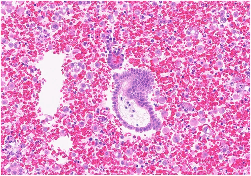
- Cell block of a pelvic washing taken at hysterectomy. Image: Htun Nyein Nyein et al., PMC open access (Diagn Cytopathol. 2026 Sep 1;54(12):E319–23. doi: 10.1002/dc.70206) FIGURE 4, CC BY.