Library › Pancreaticobiliary cytology and the WHO Reporting System
Pancreaticobiliary cytology and the WHO Reporting System
Last reviewed 2026-11-04 by a board-certified pathologist. 113 references.
The WHO Reporting System for Pancreaticobiliary Cytopathology
Definition. The WHO Reporting System for Pancreaticobiliary Cytopathology (WHO system) is the international terminology for reporting fine-needle aspiration (FNA) and biopsy cytology of the pancreas and brushing cytology of the bile ducts. It was developed by the International Academy of Cytology (IAC), the International Agency for Research on Cancer (IARC) and the WHO and was published in 2022 (Pitman et al. 2023, Acta Cytol). It places each specimen in one of seven diagnostic categories. Each category carries an associated risk of malignancy (ROM) and a management recommendation. The system revises the six-category Papanicolaou Society of Cytopathology (PSC) system of 2014–2015, and its entity names follow the WHO Classification of Digestive System Tumours, 5th edition (2019) (Pitman et al. 2023, J Am Soc Cytopathol; Nagtegaal et al. 2020). The WHO Classification of Digestive System Tumours, 6th edition (2026), adds simple mucinous cyst of the pancreas and intraductal tubulopapillary and intraductal oncocytic papillary neoplasms of the bile ducts as entities, and it removes acinar cystic transformation of the pancreas from the classification (Arends et al. 2026).
Related pages: Normal pancreas, gastrointestinal contaminants and EUS-guided sampling (specimen collection, normal elements and contaminants), Pancreatic cyst fluid chemistry and molecular testing (CEA, amylase, glucose and molecular testing), Biliary brushing cytology: technique, normal findings and reactive atypia and Biliary adenocarcinoma on brushing and ancillary testing (bile duct brushings).
Scope and principles of the WHO pancreaticobiliary reporting system
- Specimens covered: endoscopic ultrasound (EUS)-guided FNA and biopsy of solid and cystic pancreatic lesions, and endoscopic retrograde cholangiopancreatography (ERCP) brushings of bile duct strictures. EUS-FNA is the main method for pancreatic lesions. ERCP brushing is the main method for bile duct strictures (Wang et al. 2024).
- Correlation is a core rule. Each interpretation is made with the clinical and imaging findings. Ancillary results (cyst fluid CEA and amylase, molecular tests, immunocytochemistry) are incorporated into the final category (Pitman et al. 2023, Acta Cytol; Centeno et al. 2024).
- Sample reports are given for each category. A separate section of the WHO system volume states the ROM and management options for clinicians (Centeno et al. 2024).
- Low-resource settings: the system gives reporting and management options for laboratories in low- and middle-income countries, where ancillary testing is often limited (Pitman et al. 2023, Acta Cytol).
Diagnostic categories of the WHO pancreaticobiliary reporting system
| Category | Definition | Typical entities and findings |
|---|---|---|
| Insufficient/inadequate/non-diagnostic | The specimen does not permit a diagnosis of the targeted lesion, for qualitative or quantitative reasons | Preparation artifact; obscuring blood; gastrointestinal (GI) contaminant only; normal pancreatic tissue from a targeted mass; acellular FNA of a solid mass or acellular duct brushing; acellular cyst fluid without thick mucin or elevated CEA |
| Benign/negative for malignancy | Unequivocally benign findings, with or without a specific diagnosis | Acute, chronic and autoimmune pancreatitis; pseudocyst; lymphoepithelial cyst; ectopic splenic tissue; serous cystadenoma; lymphangioma; benign parenchyma when imaging shows no well-defined mass |
| Atypical | Limited nuclear or architectural atypia, insufficient in quantity or quality for any other category | Atypia exceeding reactive change; mucinous epithelium that cannot be separated from GI contaminant |
| Pancreaticobiliary neoplasm, low risk/grade (PaN-Low) | Neoplastic epithelium of an intraductal or cystic neoplasm with low-grade atypia | Intraductal papillary mucinous neoplasm (IPMN), mucinous cystic neoplasm (MCN), pancreatic intraepithelial neoplasia (PanIN), biliary intraepithelial neoplasia (BilIN) and intraductal papillary neoplasm of the bile duct, each with low-grade atypia; mucinous cyst without epithelium |
| Pancreaticobiliary neoplasm, high risk/grade (PaN-High) | Neoplastic epithelium of an intraductal or cystic neoplasm with high-grade atypia | IPMN, MCN, PanIN, BilIN and intraductal papillary neoplasm of the bile duct with high-grade atypia; intraductal oncocytic papillary neoplasm; intraductal tubulopapillary neoplasm |
| Suspicious for malignancy | Some features of malignancy, insufficient in number or quality for an unequivocal diagnosis | Scant or poorly preserved adenocarcinoma; a solid cellular neoplasm that cannot be typed for lack of cell block material |
| Malignant | Unequivocal features of malignancy; includes primary neoplasms and metastases | Ductal adenocarcinoma and variants, cholangiocarcinoma, acinar cell carcinoma, neuroendocrine tumor of any grade, neuroendocrine carcinoma, solid-pseudopapillary neoplasm, pancreatoblastoma, lymphoma, gastrointestinal stromal tumor, sarcoma, metastasis |
Sources: Pitman et al. 2023 (Acta Cytol); Centeno et al. 2024; Wang et al. 2024, Tables 1 and 2.
Differences between the WHO and Papanicolaou Society of Cytopathology systems
The PSC system had one "neoplastic" category with two subgroups. "Neoplastic: benign" held serous cystadenoma and lymphangioma. "Neoplastic: other" held premalignant intraductal and cystic neoplasms of every grade together with the low-grade malignancies pancreatic neuroendocrine tumor (PanNET) and solid-pseudopapillary neoplasm (SPN) (Pitman et al. 2014). The WHO system redistributes these entities.
| Entity | PSC category (2014–2015) | WHO category (2022) |
|---|---|---|
| Serous cystadenoma, lymphangioma | Neoplastic: benign | Benign/negative for malignancy |
| IPMN or MCN with low-grade atypia | Neoplastic: other | PaN-Low |
| IPMN or MCN with high-grade atypia | Neoplastic: other | PaN-High |
| High-grade BilIN | Suspicious for malignancy | PaN-High |
| Well-differentiated PanNET, SPN, gastrointestinal stromal tumor | Neoplastic: other | Malignant |
| Findings suggestive of PanNET or SPN, insufficient for diagnosis | Atypical | Suspicious for malignancy |
| Ductal adenocarcinoma, acinar cell carcinoma, pancreatoblastoma, metastasis | Positive/malignant | Malignant |
Sources: Pitman et al. 2023 (J Am Soc Cytopathol); Centeno et al. 2024; Wang et al. 2024.
- Reason for the change: PanNET and SPN are malignant neoplasms in the WHO Classification of Digestive System Tumours, 5th edition. The reporting categories now match the histologic classification (Centeno et al. 2024).
- Effect on stratification: the PSC "neoplastic: other" category combined lesions with widely different risks of malignancy. Splitting it into PaN-Low and PaN-High separates low-risk mucinous cysts from high-risk ones (Hoda et al. 2022; Hsiao and Wang 2024).
- Category names in the literature: the 2023 summary papers write the high category as "pancreatic neoplasm, high risk/grade". The 2024 overview and later series write "pancreaticobiliary neoplasm, high-risk/grade", abbreviated PaN-High. Some reviews use PanN-Low and PanN-High for the same categories (Pitman et al. 2023, Acta Cytol and J Am Soc Cytopathol; Centeno et al. 2024; Wang et al. 2024).
Risk of malignancy in the WHO pancreaticobiliary categories
ROM varies with the denominator (resected cases or all cases), the follow-up method and the mix of solid and cystic lesions. The values below are from pancreatic EUS-FNA cohorts.
| Category | Hoda 2022 (334 FNAs, one institution) | Hsiao and Wang 2024 (1012 FNAs) | Range in published series (Wang 2024 review) |
|---|---|---|---|
| Insufficient/non-diagnostic | 7.7% | 8.3% | 5–50% |
| Benign/negative | 1.0% | 3.2% | 0–40% |
| Atypical | 28.0% | 24.6% | 28–50% |
| PaN-Low | 4.8% | 9.1% | 5–20% |
| PaN-High | 60.0% | 46.7% | 60–90% |
| Suspicious for malignancy | 100% | 75% | 80–100% |
| Malignant | 100% | 100% | 97–100% |
- The PSC "neoplastic: other" category had a ROM of 30.3% in the Hoda cohort and 7.3% in the Hsiao cohort. When the same cases are split, the ROM of PaN-High is about 5 to 12.5 times that of PaN-Low (Hoda et al. 2022; Hsiao and Wang 2024).
- Survival: the WHO categories also stratified overall survival better than the PSC categories (Hsiao and Wang 2024).
- Solid lesions: in 521 EUS-FNAs of solid pancreatic lesions, ROM was the same in both systems: non-diagnostic 48.2%, negative 2.3%, atypical 78.1%, suspicious 100% and malignant 99.4%. The new categories change little for solid masses because the prevalence of malignancy in solid masses is high (81.76% in that series) (Vasas et al. 2024).
- Bile duct brushings: ROM is higher in the lower categories of brushing cytology. Published ranges are 28–69% for non-diagnostic, up to 30% for benign and 25–61% for atypical. No ROM data were available for PaN-High in brushings at the time of the review. ROM of suspicious brushings is 74–100% (Wang et al. 2024). Brushing categories are covered under Biliary brushing cytology: technique, normal findings and reactive atypia.
Adequacy and the insufficient category in pancreaticobiliary cytology
- No numeric threshold. There is no consensus minimum number of epithelial cells, particularly for cysts (Wang et al. 2024).
- Imaging findings determine whether a sample is representative. When imaging shows a distinct solid mass, a paucicellular or acellular sample is non-diagnostic. A sample of normal pancreatic tissue that does not explain a well-defined mass is also non-diagnostic, whatever its cellularity (Wang et al. 2024; Pitman et al. 2014).
- When imaging shows no distinct mass, benign pancreatic tissue may be reported as benign, with a note that the sample may not represent the lesion (Wang et al. 2024).
- Absence of epithelium does not by itself make a cyst sample non-diagnostic. A pseudocyst has no epithelial lining by definition. Fluid with inflammatory cells and histiocytes that fits a pseudocyst or a serous cystadenoma is placed in the benign category (Pitman et al. 2014; Wang et al. 2024).
- Mucin or elevated CEA without epithelium: thick, colloid-like mucin or cyst fluid CEA above 192 ng/mL establishes a mucinous cyst. The specimen is reported as a neoplastic mucinous cyst (PaN-Low), with a comment that the grade of atypia cannot be assessed (Wang et al. 2024).
- Thin mucin resembling tissue paper is difficult to separate from GI contaminant mucin. GI mucin forms isolated thick to thin clumps and never forms diffuse colloid-type mucin (Nagle et al. 2005; Wang et al. 2024).
- Fibrotic lesions such as chronic pancreatitis and autoimmune pancreatitis often yield scant material (Wang et al. 2024).
- Measures that reduce non-diagnostic samples: repeat sampling, FNB or core biopsy, and rapid on-site evaluation (ROSE). ROSE lowers the non-diagnostic rate (Wang et al. 2024).
Benign and atypical categories in pancreaticobiliary cytology
- A descriptive negative report (no specific benign diagnosis) means that the sample was adequate and showed no atypia. It is not a diagnosis of a benign lesion. The false-negative rate of FNA of a solid mass averages 15%. In a patient with a clinically and radiologically suspicious mass, a negative aspirate is presumed to be a false negative (Pitman et al. 2014).
- Benign neoplasms such as serous cystadenoma are reported in the benign category in the WHO system. Serous cystadenoma aspirates are often paucicellular. In one series, 63% of serous cystadenomas were initially misinterpreted and 27% were reported as non-diagnostic (Wang et al. 2024).
- Atypical is used for cellular or extracellular findings beyond recognizable normal or reactive changes. It raises the possibility of a neoplasm but does not meet the criteria for any other category (Pitman et al. 2014).
- Mucinous epithelium of uncertain origin: when lesional mucinous cells cannot be separated from GI contaminant epithelium, the cells are reported as atypical (Wang et al. 2024).
- In bile duct brushings, "atypical" means atypia beyond that of reactive conditions (Wang et al. 2024).
- Variable use: the criteria for atypical are applied inconsistently. Some low-grade IPMN and BilIN cases that belong in PaN-Low are reported as atypical (Wang et al. 2024).
Pancreaticobiliary neoplasm categories: PaN-Low and PaN-High
The two neoplasm categories are reserved mainly for intraductal neoplasms and MCN. They are graded on the cytomorphology of the epithelium (Centeno et al. 2024).
| Feature | PaN-Low | PaN-High |
|---|---|---|
| Epithelium | Mucinous epithelium with low-grade atypia, or mucinous cyst contents without epithelium | Neoplastic epithelium with high-grade atypia |
| Cells | Columnar mucinous cells | Small cells, smaller than a 12-μm duodenal enterocyte |
| Nucleus | Low nuclear-to-cytoplasmic (N:C) ratio, bland chromatin | High N:C ratio, hypochromatic or hyperchromatic abnormal chromatin, irregular contour |
| Arrangement | Sheets and papillae | Small clusters and single cells |
| Background | Thick colloid-like mucin | Necrosis |
| Pancreatic ROM | 5–20% | 60–90% |
| Usual management | Surveillance | Resection |
Sources: Wang et al. 2024; Pitman et al. 2014; Hoda et al. 2022.
- Intermediate-grade dysplasia belongs in PaN-Low. The WHO system places IPMN and MCN with low- to intermediate-grade dysplasia in PaN-Low, in line with the two-tier grading (low and high grade) of the WHO Classification of Digestive System Tumours. Two-tier grading of bile duct and pancreatic precursors applies in both the 5th edition (Nagtegaal et al. 2020) and the 6th edition (Arends et al. 2026). Former intermediate-grade dysplasia is graded as low grade (Wang et al. 2024, Table 2; Arends et al. 2026). The PSC guideline noted that intermediate-grade atypia cannot be reliably separated from high-grade atypia on cytology (Pitman et al. 2014).
- Necrosis supports high-grade atypia but does not separate high-grade dysplasia from invasive carcinoma (Wang et al. 2024).
- High-grade dysplasia versus carcinoma: cytology alone often cannot separate them. Both are usually treated by resection, so the distinction has limited effect on management (Wang et al. 2024).
- Intraductal oncocytic papillary neoplasm and intraductal tubulopapillary neoplasm are placed in PaN-High (Wang et al. 2024). The 5th edition WHO classification separates both from IPMN on genomic and morphologic grounds (Nagtegaal et al. 2020). The 6th edition also classifies intraductal tubulopapillary and intraductal oncocytic papillary neoplasms of the extrahepatic bile ducts as separate entities, which they were not in the 5th edition (Arends et al. 2026).
- Main differential for PaN-Low: gastric foveolar epithelium carried in by a transgastric needle. CEA, amylase, glucose and KRAS testing help separate lesional epithelium from contaminant (Wang et al. 2024). Cyst entities and grading are covered under Pancreatic cystic neoplasms on cytology.
Suspicious and malignant categories in pancreaticobiliary cytology
- Suspicious for malignancy applies when features suggest a malignancy but material is scant, poorly preserved, or not matched by a distinct mass on imaging (Wang et al. 2024).
- Mucinous cyst with necrosis: the suspicious category is also used for a mucinous cyst with high-grade dysplasia and necrosis, particularly with high-risk imaging findings (Wang et al. 2024).
- Three causes of a suspicious report in pancreatic adenocarcinoma: very well-differentiated tumors with subtle abnormalities, scant cellularity from tumor desmoplasia, and heavy GI contamination (Pitman et al. 2014).
- Solid cellular neoplasms without confirmation: an aspirate that suggests PanNET or SPN, but lacks material for immunocytochemistry, is suspicious for malignancy in the WHO system. The PSC system placed such cases in the atypical category (Wang et al. 2024).
- A suspicious result is not by itself an indication for resection or neoadjuvant therapy. Management depends on clinical correlation and ancillary results (Wang et al. 2024).
- Malignant includes primary pancreatic and biliary malignancies of every grade and metastases. Pancreatic ductal adenocarcinoma (PDAC) accounts for 85–90% of pancreatic malignancies (Pitman et al. 2014).
- Specificity: a malignant interpretation of pancreatic FNA or biliary brushing has a specificity of 90–95% or higher in most studies. Strict criteria raise specificity at the cost of sensitivity (Pitman et al. 2014).
Management recommendations for WHO pancreaticobiliary categories
| Category | Recommended next step |
|---|---|
| Insufficient/non-diagnostic | Repeat sampling; FNB or core biopsy; ROSE |
| Benign/negative | Clinical follow-up and disease-specific treatment; notify the clinical team when normal tissue is obtained from a mass; continued surveillance of a bile duct stricture |
| Atypical | Multidisciplinary review; molecular testing; repeat sampling with ROSE |
| PaN-Low | Conservative management and surveillance unless high-risk features are present |
| PaN-High | Surgical resection |
| Suspicious for malignancy | Clinical correlation and ancillary testing before treatment |
| Malignant | Resection when resectable; neoadjuvant therapy before resection in selected patients; chemotherapy with or without radiation when unresectable |
Source: Wang et al. 2024, summarizing the WHO system.
- Mucinous cystic neoplasm: MCN was formerly an absolute indication for resection. Recent guidelines favor surveillance unless high-risk features are present (Wang et al. 2024).
- PanNET: resection is recommended for functioning PanNETs (Wang et al. 2024). For non-functioning PanNETs, ENETS 2023 recommends active surveillance for asymptomatic tumors of 1 cm or less without main pancreatic duct dilation, individualized management for tumors larger than 1 cm and up to 2 cm, and surgery for tumors larger than 2 cm or with main duct dilation (Kos-Kudła et al. 2023).
- Second opinion: many atypical and suspicious samples are interpreted conservatively. Review by an experienced pancreatic cytopathologist can spare the patient a repeat procedure (Pitman et al. 2014).
Ancillary testing within the WHO pancreaticobiliary reporting system
- Cyst fluid: CEA, amylase and molecular tests (KRAS, GNAS) classify cysts as mucinous or non-mucinous (Pitman et al. 2023, Acta Cytol; Wang et al. 2024). Thresholds and test performance are covered under Pancreatic cyst fluid chemistry and molecular testing.
- Solid lesions: immunocytochemistry on cell blocks is used to type solid cellular neoplasms and to separate PDAC from reactive ductal epithelium (Layfield et al. 2014). Panels are tabulated under Solid cellular neoplasms of the pancreas on cytology and Pancreatic ductal adenocarcinoma and its variants on cytology.
- Bile duct brushings: fluorescence in situ hybridization (FISH) increases sensitivity without loss of specificity (Layfield et al. 2014). Details are under Biliary adenocarcinoma on brushing and ancillary testing.
- Ancillary results can change the category assigned on morphology (Wang et al. 2024).
Common errors in assigning WHO pancreaticobiliary categories
| Situation | Correct WHO category | Key point |
|---|---|---|
| Benign acinar and ductal tissue from a well-defined 2-cm mass | Insufficient/non-diagnostic | The sample does not explain the mass |
| Thick colloid-like mucin, no epithelium | PaN-Low | Comment that grade cannot be assessed |
| Cyst fluid with histiocytes and inflammatory cells, high amylase, no epithelium | Benign (pseudocyst) | Absence of epithelium is expected |
| Serous cystadenoma | Benign | PSC "neoplastic: benign" no longer exists |
| Well-differentiated PanNET, any grade | Malignant | PSC placed it in "neoplastic: other" |
| SPN | Malignant | Low-grade malignant neoplasm in WHO 5th edition |
| Neoplastic cells suggesting PanNET, no cell block for confirmation | Suspicious for malignancy | PSC placed this in atypical |
| IPMN with small cells, high N:C ratio and necrosis | PaN-High | Necrosis does not prove invasion |
| Mucinous cells indistinguishable from gastric contaminant | Atypical | Lesional origin is uncertain |
WHO Reporting System for Pancreaticobiliary Cytopathology category lookup
Normal Pancreas, Gastrointestinal Contaminants and EUS-Guided Sampling
Definition. Endoscopic ultrasound (EUS)-guided sampling is the standard method for obtaining cytology and tissue from solid and cystic pancreatic lesions. It includes fine-needle aspiration (FNA) and fine-needle biopsy (FNB) (Brugge et al. 2014; Facciorusso et al. 2025). The needle passes through the wall of the stomach or duodenum, so every pancreatic sample can contain gastrointestinal (GI) epithelium and mucin as well as normal pancreatic acinar, ductal and islet cells (Nagle et al. 2005; Mallik et al. 2021). Recognition of these normal elements and contaminants is the basis of specimen adequacy in the WHO Reporting System for Pancreaticobiliary Cytopathology (Wang et al. 2024).
Related pages: The WHO Reporting System for Pancreaticobiliary Cytopathology (adequacy and categories), Pancreatitis, pseudocyst and lymphoepithelial cyst on cytology (benign inflammatory lesions), Solid cellular neoplasms of the pancreas on cytology (neoplasms that mimic normal acinar and islet tissue), Biliary brushing cytology: technique, normal findings and reactive atypia (bile duct brushing technique).
Normal cellular components of pancreatic aspirates
| Component | Architecture | Cytoplasm | Nucleus | Main mimics |
|---|---|---|---|---|
| Acinar cells | Small cohesive acini, lobulated "raspberry-like" groups; single cells in smears made with heavy pressure | Abundant, with coarse zymogen granules | Small, round, uniform, about the size of a red blood cell; eccentric; smooth membrane; nucleolus visible | Acinar cell carcinoma, well-differentiated neuroendocrine tumor |
| Ductal cells | Flat monolayer sheets with an even "honeycomb" pattern en face and palisading at the edge; small tubules | Pale, finely vacuolated, well-demarcated borders | Round to oval, evenly spaced, no crowding | Well-differentiated ductal adenocarcinoma, duodenal and gastric epithelium |
| Islet cells | Sparse | Resemble acinar cells | Similar to acinar cells | Neuroendocrine tumor |
Sources: Conrad et al. 2013; Wang et al. 2024.
- Acinar cells predominate. They make up about 80% of the cellularity of the normal pancreas (Conrad et al. 2013).
- Bare acinar nuclei are common on smears (Conrad et al. 2013). A smear of normal acinar tissue contains few isolated cells and bare nuclei, and the groups are cohesive and raspberry-like with a fibrovascular stroma (Wang et al. 2024).
- Acinar cell carcinoma shows more single cells, larger nuclei and prominent nucleoli, and lacks the lobular organization of normal acini (Wang et al. 2024).
- Normal tissue in a mass sample: benign acinar and ductal tissue that does not explain a well-defined mass on imaging is reported as insufficient/non-diagnostic (Wang et al. 2024).
Gastrointestinal contaminants in EUS-guided pancreatic samples
- Route determines the contaminant. Head lesions are sampled through the duodenum. Body and tail lesions are sampled through the stomach (Mallik et al. 2021). Duodenal and gastric epithelium and GI mucin are therefore common on pancreatic smears.
- Frequency: in one series, contamination from the duodenum, stomach or pancreas was present in 170 of 267 EUS-FNAs: 77 positive or suspicious, 3 atypical and 90 negative (Mitsuhashi et al. 2006).
| Feature | Duodenal epithelium | Gastric foveolar epithelium | Lesional mucinous epithelium (IPMN or MCN) |
|---|---|---|---|
| Architecture | Large flat monolayer sheets | Large and small monolayer sheets; more small groups than duodenum | Small clusters, papillae and single cells |
| Key cells | Non-mucinous enterocytes with a brush border; scattered goblet cells; intraepithelial lymphocytes | Mucin-capped columnar cells | Columnar cells with apical mucin |
| Nuclei | Round, evenly spaced, no atypia | Round, evenly spaced; grooves, inclusions and slight crowding can occur | Low- or high-grade atypia |
| Mucin | Present, less than gastric | Isolated thick to thin clumps, never diffuse colloid-type mucin | Thick colloid-like extracellular mucin |
| Background | Clean; no inflammation or debris | Clean | Histiocytes, debris; necrosis with high-grade atypia |
Sources: Nagle et al. 2005; Pitman et al. 2014; Wang et al. 2024.
- Sheet size separates contaminant from lesional epithelium. GI contaminants usually form large sheets of regular mosaic epithelium. Lesional mucinous cells usually form small clusters or single cells (Wang et al. 2024).
- Goblet cells identify duodenal epithelium (Pitman et al. 2014; Wang et al. 2024). Interspersed goblet cells separate duodenal sheets from pancreatic ductal sheets (Conrad et al. 2013).
- Gastric foveolar epithelium is the main mimic of low-grade mucinous neoplasia. It can show nuclear grooves, inclusions and subtle crowding (Pitman et al. 2014). Low-grade gastric-type IPMN epithelium may be indistinguishable from gastric contaminant in a transgastric sample (Pitman et al. 2014).
- Reporting of contaminant-only samples: a specimen with only contaminant GI epithelium is non-diagnostic. When mucinous cells cannot be assigned to the lesion or to the contaminant, the specimen is reported as atypical (Wang et al. 2024).
- Ancillary tests on cyst fluid (CEA, amylase, glucose, KRAS) help separate a lesional mucinous cyst from contaminant (Wang et al. 2024). Thresholds are covered under Pancreatic cyst fluid chemistry and molecular testing.
- False-positive malignancy occurs in about 2% of EUS-FNAs of pancreatic lesions. Causes include contamination by an intervening mucosal malignancy and misinterpretation (Brugge et al. 2014).
EUS-guided fine-needle aspiration technique for pancreatic lesions
The Papanicolaou Society of Cytopathology (PSC) technique guideline describes EUS-FNA as follows (Brugge et al. 2014):
- Equipment: a linear echoendoscope is required. Prior gastric surgery, such as bypass or Whipple resection, limits access.
- Needle entry: the needle, occluded by a stylet, enters a solid lesion with one quick thrust under constant EUS guidance. The stylet is then removed and suction is applied.
- Fanning: the needle is moved back and forth within the mass in a fanning pattern to sample several areas.
- Suction: suction is turned off before the needle is withdrawn. Excessive suction increases the amount of blood in the sample. Highly vascular lesions are aspirated with minimal suction.
- Smears: each specimen is expressed onto slides. One slide is air-dried and stained with a modified Diff-Quik stain for on-site evaluation. The other is wet-fixed for a modified Papanicolaou stain. Material is also collected for a cell block.
- Core material: cores from large or biopsy needles are processed as histology. Cytology touch preparations can be made by rolling a core across a slide.
Needle gauge for EUS-guided pancreatic sampling by indication
| Needle | Typical use |
|---|---|
| 25-gauge | Highly vascular lesions; uncinate lesions; lymph nodes; suspected neuroendocrine tumors and metastatic renal cell carcinoma; serous cystadenoma and cystic neuroendocrine tumor |
| 22-gauge | Most solid targets; mucinous cysts, because the fluid is viscous; pseudocysts |
| 19-gauge core or Tru-cut | Stromal tumors, neuroendocrine tumors, lesions with poor cytology yield, suspected autoimmune pancreatitis; pseudocysts |
Source: Brugge et al. 2014.
- Smaller needles are easier to use, generally safer, and give a higher yield for pancreatic adenocarcinoma (Brugge et al. 2014).
- Number of passes: without on-site cytology, about 7 passes of a pancreatic mass are needed to maximize sensitivity. Lymph nodes need fewer passes. Stromal tumors may need 3–5 passes (Brugge et al. 2014).
Rapid on-site evaluation of EUS-guided pancreatic samples
- Purpose: rapid on-site evaluation (ROSE) confirms that lesional material is present and guides the number of passes (Brugge et al. 2014).
- Effect: ROSE reduces false-negative FNA of pancreatic masses (Brugge et al. 2014) and lowers the non-diagnostic rate (Wang et al. 2024).
- ROSE and needle type: in a meta-analysis of 15 studies (1024 patients), FNA with ROSE and FNB had similar diagnostic yield. Without ROSE, FNB gave better diagnostic adequacy for solid pancreatic lesions. FNB reached a diagnosis with fewer passes (Khan et al. 2017).
Fine-needle biopsy compared with fine-needle aspiration of pancreatic lesions
- ESGE 2025 recommendation: the European Society of Gastrointestinal Endoscopy (ESGE) recommends end-cutting FNB needles over reverse-bevel FNB needles and FNA needles for solid pancreatic lesions. FNA may still have a role when ROSE is available (Facciorusso et al. 2025).
- Antibiotics: ESGE does not recommend antibiotic prophylaxis before EUS-guided sampling of solid masses or EUS-FNA of pancreatic cysts (Facciorusso et al. 2025).
- Autoimmune pancreatitis: a meta-analysis found a pooled diagnostic yield for level 1 or 2 histologic criteria of autoimmune pancreatitis (ICDC) of 87.2% with FNB and 55.8% with FNA (P = 0.030). A 19-gauge needle performed better than a 22-gauge needle (88.9% and 60.6%) (Yoon et al. 2021).
- Specimens produced by FNB: FNB yields cores that support immunohistochemistry and architecture. Touch preparations and needle rinses from FNB still allow cytologic evaluation (Brugge et al. 2014).
| Feature | EUS-FNA | EUS-FNB |
|---|---|---|
| Product | Smears, needle rinse, cell block | Tissue cores, touch preparations, rinse |
| Dependence on ROSE | High; yield equals FNB when ROSE is used | Lower; better adequacy without ROSE |
| Passes to diagnosis | More | Fewer |
| Architecture | Limited | Preserved |
| ESGE 2025 position for solid pancreatic lesions | Role when ROSE is available | End-cutting needles recommended |
Sources: Khan et al. 2017; Facciorusso et al. 2025.
Sampling of pancreatic cysts and processing of cyst fluid
- One pass with high suction is generally used to empty a cyst. Mural nodules and adjacent masses are aspirated separately or during the cyst fluid aspiration (Brugge et al. 2014).
- Aliquot priority: when malignancy is the main concern, cytology has priority. When cyst typing is the main concern, fluid is sent first for CEA, KRAS and GNAS testing (Brugge et al. 2014).
- Centrifugation: cyst fluid is centrifuged before CEA and DNA assays (Brugge et al. 2014).
- Viscosity: thick, sticky fluid described by the endoscopist suggests a mucinous cyst. Thick fluid can be smeared directly. Thin fluid is processed by cytospin. Placing cyst fluid in preservative reduces its viscosity and can make thin mucin difficult or impossible to recognize (Pitman et al. 2014).
- Acellular fluid: an acellular cyst sample without thick mucin and without CEA above 192 ng/mL is non-diagnostic (Wang et al. 2024).
Liquid-based preparation of EUS-guided pancreatic samples
- Liquid-based cytology (LBC) of EUS-FNA material reduces bloody background, drying and crush artifact (Kwon et al. 2024).
- In acinar cell carcinoma, LBC showed a bloody background in 12.5% of cases and conventional smears in 85.7%. Prominent nucleoli and macronucleoli were seen in all LBC cases and in 28.6% and 14.3% of smears (Kwon et al. 2024).
Intrapancreatic accessory spleen on EUS-guided sampling
- Definition and site: an intrapancreatic accessory spleen (splenule) is ectopic splenic tissue in the pancreas. The pancreatic tail is the second most common site of accessory spleen (Saunders et al. 2016).
- Clinical importance: it mimics a solid neoplasm, most often a neuroendocrine tumor, on imaging. Recognition on EUS-FNA avoids resection (Tatsas et al. 2012).
- Cytology: a polymorphous population of small lymphocytes, eosinophils, plasma cells, histiocytes and red cells, with many small vessels that represent splenic sinusoids (Tatsas et al. 2012). Large platelet aggregates, lymphoglandular bodies and bland slender spindle cells are also present (Conway et al. 2013).
- Immunocytochemistry: CD8 highlights the endothelial cells of the splenic sinusoids. CD31 stains platelet aggregates (Tatsas et al. 2012; Conway et al. 2013).
- WHO category: ectopic splenic tissue is reported as benign/negative for malignancy (Wang et al. 2024).
- Epidermoid cyst in an accessory spleen yields mainly macrophages and proteinaceous debris (Tatsas et al. 2012).
| Feature | Intrapancreatic accessory spleen | Reactive lymph node | Well-differentiated neuroendocrine tumor |
|---|---|---|---|
| Cells | Polymorphous lymphocytes, eosinophils, plasma cells, histiocytes | Polymorphous lymphocytes with germinal center cells | Monotonous plasmacytoid epithelial cells |
| Vessels and platelets | Many sinusoidal vessels; large platelet aggregates | Few | Vascular cores in some |
| Tingible body macrophages | Absent | Present | Absent |
| Key stain | CD8-positive sinusoidal endothelium | — | Synaptophysin, chromogranin, INSM1 |
Sources: Tatsas et al. 2012; Conway et al. 2013; Wang et al. 2024.
Common errors in interpreting normal and contaminant elements in pancreatic samples
| Situation | Correct interpretation | Basis |
|---|---|---|
| Large flat sheets with goblet cells | Duodenal contaminant | Goblet cells and brush border |
| Mucin-capped columnar cells in a transgastric cyst sample | Gastric contaminant or low-grade mucinous epithelium; atypical when undecided | Overlap with gastric-type IPMN |
| Clumps of thick and thin mucin with clean background | GI mucin | GI mucin is never colloid-like |
| Cohesive acinar groups from a discrete mass | Non-diagnostic | Sample does not explain the mass |
| Monotonous cells with granular cytoplasm and many single cells | Suspicious for a neoplasm such as acinar cell carcinoma; immunocytochemistry indicated | Normal acini are cohesive and lobular |
| Polymorphous lymphoid cells with vessels and platelets from a tail nodule | Accessory spleen | CD8-positive sinusoids |
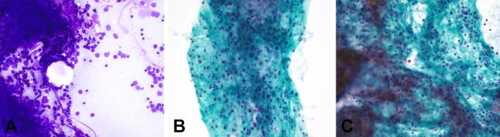
Pancreatitis, Pseudocyst and Lymphoepithelial Cyst on Cytology
Definition. Pancreatitis, pancreatic pseudocyst and lymphoepithelial cyst are non-neoplastic lesions that are sampled by endoscopic ultrasound (EUS)-guided aspiration or biopsy because they can mimic a neoplasm on imaging. All are reported in the benign/negative for malignancy category of the WHO Reporting System for Pancreaticobiliary Cytopathology (Wang et al. 2024). Chronic and autoimmune pancreatitis form masses that resemble pancreatic ductal adenocarcinoma (PDAC). A pseudocyst is a walled collection of fluid without an epithelial lining. A lymphoepithelial cyst is a true cyst lined by squamous epithelium over lymphoid tissue (Banks et al. 2013; Farooq et al. 2020).
Related pages: The WHO Reporting System for Pancreaticobiliary Cytopathology (categories), Pancreatic ductal adenocarcinoma and its variants on cytology (criteria for adenocarcinoma), Pancreatic cyst fluid chemistry and molecular testing (CEA, amylase and glucose thresholds), Pancreatic cystic neoplasms on cytology (mucinous and serous cysts).
WHO category of benign inflammatory and cystic pancreatic lesions
The WHO benign/negative category lists acute pancreatitis, chronic pancreatitis, autoimmune pancreatitis, pseudocyst, lymphoepithelial cyst, ectopic splenic tissue, lymphangioma, serous cystadenoma, and benign pancreatic parenchyma when imaging shows no well-defined mass (Wang et al. 2024, Table 2).
- A descriptive negative report is not a diagnosis of a benign lesion. A specific benign diagnosis, such as chronic pancreatitis or pseudocyst, is given when the findings support it (Pitman et al. 2014).
- Model report wording (PSC): "benign, reactive ductal epithelium and acinar tissue, acute and chronic inflammation and a background of necrotic, calcific debris consistent with chronic pancreatitis"; "cellular stromal fragments with lymphocytes and plasma cells suggestive of autoimmune pancreatitis"; "cyst fluid with inflammation and histiocytes, yellow amorphous pigment and no cyst lining epithelial cells consistent with pseudocyst fluid" (Pitman et al. 2014).
Acute pancreatitis and its local complications
The revised Atlanta classification (2012) defines the phases, severity and local complications of acute pancreatitis (Banks et al. 2013).
| Term | Definition |
|---|---|
| Mild acute pancreatitis | No organ failure and no local or systemic complications; usually resolves in the first week |
| Moderately severe acute pancreatitis | Transient organ failure, local complications or exacerbation of comorbid disease |
| Severe acute pancreatitis | Persistent organ failure, lasting more than 48 hours |
| Acute peripancreatic fluid collection | Early collection without a well-defined wall |
| Pseudocyst | Encapsulated collection of fluid with a well-defined inflammatory wall, usually outside the pancreas, with minimal or no necrosis |
| Acute necrotic collection | Early collection containing necrosis |
| Walled-off necrosis | Mature encapsulated collection of pancreatic or peripancreatic necrosis with a well-defined inflammatory wall |
Source: Banks et al. 2013.
- Timing: a pseudocyst follows interstitial edematous pancreatitis and usually needs more than 4 weeks to mature. Walled-off necrosis follows necrotizing pancreatitis (Banks et al. 2013).
- Cytology in the acute setting is seldom the primary test. Acute pancreatitis is reported in the benign category when sampled (Wang et al. 2024).
Chronic pancreatitis on EUS-guided cytology
- Cellularity: aspirates are variable and often scant because of fibrosis. Ductal cells predominate because of acinar atrophy. Islet cells may be identified because they are spared (Conrad et al. 2013).
- Background: acute and chronic inflammation with necrotic and calcific debris (Pitman et al. 2014).
- Adequacy: fibrotic lesions such as chronic pancreatitis and autoimmune pancreatitis often yield insufficient material (Wang et al. 2024).
- False-positive diagnosis: chronic pancreatitis is the most common benign lesion that causes a false-positive diagnosis of pancreatic cancer. In 367 patients with EUS-FNA interpreted as positive or suspicious, 4 had chronic pancreatitis at resection (Brugge et al. 2014).
Reactive ductal epithelium compared with well-differentiated adenocarcinoma
| Feature | Reactive ductal epithelium in chronic pancreatitis | Well-differentiated ductal adenocarcinoma |
|---|---|---|
| Sheets | Flat, honeycomb, evenly spaced | Crowded, overlapping, "drunken honeycomb" |
| Anisonucleosis within one group | Minimal | Present; more than 4:1 in a single group |
| Nuclear membrane | Smooth | Irregular, with grooves and notches |
| Chromatin | Even | Irregular distribution; parachromatin clearing |
| Single atypical cells | Rare | Present |
| p53 immunostain | Wild-type pattern (scattered, variable nuclear staining); score 0 in the Furuhata cell-block scale | Aberrant overexpression or null pattern |
| SMAD4 immunostain | Retained | Lost in about 55% |
Sources: Robins et al. 1995; Lin and Staerkel 2003; Wang et al. 2024; Layfield et al. 2014; Furuhata et al. 2017.
- Major criteria for adenocarcinoma (Robins): nuclear crowding and overlap, nuclear contour irregularity and irregular chromatin distribution. Minor criteria are nuclear enlargement, single epithelial cells, necrosis and mitoses (Robins et al. 1995).
- p53 immunocytochemistry: in a cell block study, all benign epithelium scored 0 on a semiquantitative positive-rate scale (cutoff 1+). A panel of p53, S100P and claudin 4 separated PDAC from benign epithelium (Furuhata et al. 2017).
- KRAS mutation supports malignancy in a pancreatic mass, but KRAS mutations also occur in ductal hyperplasia, ductal metaplasia and chronic pancreatitis (Layfield et al. 2014).
- Detailed criteria for adenocarcinoma are covered under Pancreatic ductal adenocarcinoma and its variants on cytology.
Autoimmune pancreatitis on EUS-guided sampling
- Classification: the International Consensus Diagnostic Criteria (ICDC) divide autoimmune pancreatitis (AIP) into type 1 and type 2. The diagnosis is based on five cardinal features: parenchymal and ductal imaging, serology, other organ involvement, pancreatic histology, and an optional response to steroids (Shimosegawa et al. 2011).
- Type 1 AIP is lymphoplasmacytic sclerosing pancreatitis. Histology shows a dense infiltrate of lymphocytes and plasma cells, with many IgG4-positive plasma cells, storiform fibrosis and obliterative phlebitis (Okazaki et al. 2022).
- Type 2 AIP is idiopathic duct-centric pancreatitis, or AIP with granulocytic epithelial lesions. It shows neutrophilic lesions and is commonly observed in Europe and the United States (Okazaki et al. 2022).
- Cytology: cellular stromal fragments with lymphocytes and plasma cells suggest AIP (Pitman et al. 2014). Smears are often scant because of fibrosis (Wang et al. 2024).
- FNB is preferred. Pooled yield for level 1 or 2 histologic criteria of AIP (ICDC) was 87.2% with FNB and 55.8% with FNA. A 19-gauge needle performed better than a 22-gauge needle (Yoon et al. 2021). Core specimens for suspected AIP are processed for histology and IgG4 immunostaining (Brugge et al. 2014).
- Findings with a Franseen FNB needle: lymphoplasmacytic infiltration in 84% and 100% of cases in two prospective studies, storiform fibrosis in 56% and 72%, and obliterative phlebitis in 24% and 43.6% (Okazaki et al. 2022).
Type 1 and type 2 autoimmune pancreatitis compared
| Feature | Type 1 AIP | Type 2 AIP |
|---|---|---|
| Histologic pattern | Lymphoplasmacytic sclerosing pancreatitis | Idiopathic duct-centric pancreatitis |
| Inflammation | Lymphocytes and plasma cells, many IgG4-positive | Neutrophils with granulocytic epithelial lesions |
| Fibrosis and vessels | Storiform fibrosis, obliterative phlebitis | — |
Sources: Shimosegawa et al. 2011; Okazaki et al. 2022.
Limits of IgG4-positive plasma cells in separating autoimmune pancreatitis from carcinoma
- Many IgG4-positive plasma cells are characteristic of type 1 AIP but are not specific. They do not distinguish AIP from pancreatic cancer (Okazaki et al. 2022).
- Biopsy findings that favor pancreatic cancer over AIP: neutrophilic infiltrates, inflammatory infiltrates with lobular edema, proliferation of plump fibroblasts, and lymphocyte-predominant infiltrates with scarce plasma cells. These are more common in pancreatic cancer than in AIP (Okazaki et al. 2022).
- KRAS mutation was found in 10 of 11 pancreatic cancers and in none of the AIP cases in one EUS-FNA study (Layfield et al. 2014).
Pancreatic pseudocyst on cytology
- Lining: a pseudocyst lacks an epithelial lining by definition (Pitman et al. 2014).
- Cytology: cyst fluid with inflammatory cells, histiocytes and yellow amorphous pigment, and an absence or paucity of epithelial cells (Pitman et al. 2014; Conrad et al. 2013).
- Adequacy: a pseudocyst sample without epithelial cells is placed in the benign category, because scant cellularity with inflammatory cells and histiocytes reflects the nature of the cyst (Wang et al. 2024).
- Cyst fluid chemistry:
- Amylase is typically in the thousands of U/L (Pitman et al. 2014). Amylase above 1,000 U/L suggests a pseudocyst. Amylase below 250 U/L makes a pseudocyst unlikely (Wang et al. 2024).
- Amylase is also elevated in cysts that communicate with the duct system, such as intraductal papillary mucinous neoplasm (IPMN) (Layfield et al. 2014).
- CEA below 5 ng/mL suggests a serous cystadenoma or pseudocyst (Wang et al. 2024).
- KRAS is wild type (Wang et al. 2024).
- Technique: pseudocysts are aspirated with a 22-gauge or 19-gauge needle so that the whole collection can be evacuated, because a residual collection may become contaminated by the aspiration (Brugge et al. 2014).
- Mucinous neoplasm resembling a pseudocyst: a mucinous neoplasm with inflammation and debris can resemble a pseudocyst. Thick colloid-like mucin or CEA above 192 ng/mL indicates a mucinous cyst (Wang et al. 2024).
Lymphoepithelial cyst of the pancreas on cytology
- Frequency and patients: lymphoepithelial cysts (LECs) make up about 0.5% of pancreatic cysts. They occur mainly in men in the 5th and 6th decades (Farooq et al. 2020).
- Histology: a true cyst lined by stratified squamous epithelium with adjacent subepithelial lymphoid tissue. Size ranges from 1.2 to 17 cm, with a mean of 4.6 cm. LEC can arise in any part of the pancreas (Farooq et al. 2020).
- Differential of squamous-lined cysts: LEC, dermoid cyst and epidermoid cyst in an intrapancreatic accessory spleen (VandenBussche and Maleki 2014).
Cytologic findings in squamous-lined pancreatic cysts
In 8 FNAs of squamous-lined cysts (7 LECs, 1 epidermoid cyst) (VandenBussche and Maleki 2014):
- All contained single nucleated and anucleate mature squamous cells, macrophages, a variable number of lymphocytes, and amorphous debris.
- Keratin was present in all but 2 cases. Cholesterol crystals were present in half.
- Benign squamous fragments were present in half.
- No nuclear atypia or necrosis was present.
- Inspissated mucin, a misleading finding, was present in 3 cases.
- Only 3 of 8 cases (38%) showed the most specific combination: squamous fragments, cholesterol crystals, keratin and squamous cells.
Causes of misdiagnosis of lymphoepithelial cyst on cytology
- Elevated CEA: LECs often have high cyst fluid CEA. Degenerated keratin debris can mimic mucin. Both findings can lead to a misdiagnosis of mucinous neoplasm (Wang et al. 2024).
- CA 125 levels have been reported as high in LEC (Layfield et al. 2014).
- Epidermoid cyst in an accessory spleen yields mainly macrophages and proteinaceous debris (Tatsas et al. 2012).
Differential diagnosis of non-neoplastic pancreatic cysts on cytology
| Feature | Pseudocyst | Lymphoepithelial cyst | Mucinous cyst (IPMN or MCN) | Serous cystadenoma |
|---|---|---|---|---|
| Lining epithelium | None | Stratified squamous | Mucinous columnar | Cuboidal, non-mucinous |
| Smear | Histiocytes, inflammation, yellow pigment | Squames, keratin, lymphocytes, cholesterol | Thick colloid-like mucin, mucinous cells | Paucicellular; rare cuboidal cells |
| Amylase | Very high | Low | Variable; high in IPMN | Low |
| CEA | Low | High | High | Low |
| KRAS | Wild type | Wild type | Mutated in many | Wild type |
| WHO category | Benign | Benign | PaN-Low or PaN-High | Benign |
Sources: Wang et al. 2024, Table 3; Pitman et al. 2014; VandenBussche and Maleki 2014. Cyst fluid thresholds are tabulated under Pancreatic cyst fluid chemistry and molecular testing.
Common errors in interpreting pancreatitis and benign cysts on cytology
| Situation | Correct interpretation | Basis |
|---|---|---|
| Pseudocyst fluid without epithelium | Benign | No lining is expected |
| Reactive ducts with mild atypia in chronic pancreatitis | Benign or atypical; malignant only when criteria are met | Chronic pancreatitis is the main cause of false-positive cancer diagnosis |
| Many IgG4-positive plasma cells in a mass biopsy | Supports type 1 AIP but does not exclude carcinoma | IgG4 plasma cells are not specific |
| Keratin debris with high CEA | Lymphoepithelial cyst until mucinous epithelium or colloid mucin is identified | Keratin mimics mucin; CEA is often high in LEC |
| Inflammatory debris with thick colloid mucin | Mucinous neoplasm | Colloid mucin is not a feature of pseudocyst |
Pancreatic Ductal Adenocarcinoma and Its Variants on Cytology
Definition. Pancreatic ductal adenocarcinoma (PDAC) is a malignant invasive gland-forming epithelial neoplasm with ductal differentiation. Most tumors form tubular glands. The variants of PDAC show other epithelial morphologies (Pitman et al. 2014). PDAC accounts for 85–90% of pancreatic malignancies (Pitman et al. 2014). Endoscopic ultrasound (EUS)-guided fine-needle aspiration (FNA) or biopsy usually establishes the diagnosis before neoadjuvant therapy, resection or palliative care (Pimenova and Rosenbaum 2026). Unequivocal adenocarcinoma is reported in the malignant category of the WHO Reporting System for Pancreaticobiliary Cytopathology (Wang et al. 2024).
Related pages: The WHO Reporting System for Pancreaticobiliary Cytopathology (categories and ROM), Pancreatitis, pseudocyst and lymphoepithelial cyst on cytology (chronic pancreatitis and reactive atypia), Pancreatic cystic neoplasms on cytology (IPMN and MCN precursors), Metastases and lymphoma in pancreatic cytology (metastatic carcinoma), Biliary adenocarcinoma on brushing and ancillary testing (cholangiocarcinoma on brushings).
Molecular pathogenesis of pancreatic ductal adenocarcinoma
- Precursors: PDAC develops from pancreatic intraepithelial neoplasia (PanIN) and intraductal papillary mucinous neoplasm (IPMN) through accumulated mutations. Invasive carcinoma can also arise in mucinous cystic neoplasm (MCN) (Molberg et al. 1998; Muraki et al. 2016). Activating KRAS mutation and telomere shortening are early events (Layfield et al. 2014). The WHO 6th edition adds simple mucinous cyst, a neoplastic cyst larger than 1.0 cm lined by gastric-type epithelium without papillae or ovarian-type stroma. Its KRAS, BRAF, TP53 and SMAD4 alterations indicate that it is likely a precursor of PDAC. It was not an entity in the 5th edition (Arends et al. 2026).
- Driver genes in invasive PDAC (Layfield et al. 2014):
| Gene | Alteration | Frequency |
|---|---|---|
| KRAS | Activating point mutation | 95% |
| CDKN2A (p16) | Inactivation | 90% |
| TP53 | Inactivation | 75% |
| SMAD4 | Inactivation | 55% |
- SMAD4 loss occurs late, in high-grade PanIN and in over half of PDAC, and is associated with a poor prognosis (Layfield et al. 2014).
- Clinical molecular testing: alterations in KRAS, TP53, BRCA1 and BRCA2 aid diagnosis, prognosis and selection of targeted therapy (Pimenova and Rosenbaum 2026).
Cytologic features of pancreatic ductal adenocarcinoma
- Cellularity and architecture: aspirates are usually hypercellular, with disorganized glandular fragments and single cells (Pimenova and Rosenbaum 2026; Wang et al. 2024).
- "Drunken honeycomb": the haphazard arrangement of tumor cells within sheets contrasts with the orderly honeycomb of benign ductal epithelium (Wang et al. 2024; Conrad et al. 2013).
- Anisonucleosis: variation in nuclear size greater than 4:1 within a single epithelial group (Wang et al. 2024).
- Nuclei: irregular contours, prominent nucleoli in many tumors, and chromatin that is hypochromatic with parachromatin clearing or hyperchromatic (Wang et al. 2024; Pimenova and Rosenbaum 2026).
- Background: necrosis in many cases (Pimenova and Rosenbaum 2026).
- Mucinous cytoplasm: some adenocarcinomas have vacuolated cytoplasm and a low nuclear-to-cytoplasmic (N:C) ratio (Wang et al. 2024).
- Cell block: small tissue fragments with embedded single atypical cells or small clusters of atypical glandular cells are diagnostic of invasive adenocarcinoma (Wang et al. 2024).
- Cholangiocarcinoma: the FNA criteria for invasive cholangiocarcinoma are the same as for PDAC. Bile duct brushings have separate criteria and a higher threshold for malignancy, covered under Biliary adenocarcinoma on brushing and ancillary testing (Pitman et al. 2014).
Cytologic criteria for adenocarcinoma in pancreatic FNA
| Study | Criteria | Performance |
|---|---|---|
| Robins et al. 1995 (90 CT-guided FNAs) | Major: nuclear crowding and overlap, nuclear contour irregularity, irregular chromatin distribution. Minor: nuclear enlargement, single epithelial cells, necrosis, mitoses | Two or more major criteria, or one major and three minor, gave 100% sensitivity and specificity; objective application of the criteria raised sensitivity from 70% to 90% |
| Lin and Staerkel 2003 (74 well-differentiated adenocarcinomas) | Anisonucleosis, nuclear membrane irregularity, nuclear crowding, overlap or three-dimensionality, nuclear enlargement greater than two red blood cells | Present in 92–99% of well-differentiated adenocarcinomas |
- Features of limited value in well-differentiated tumors: gap or confluent cell spacing, hyperchromasia, macronucleoli, mitoses, chromatin clearing and necrosis were present in only 7–38% of well-differentiated adenocarcinomas (Lin and Staerkel 2003).
- Retrospective review showed that 6 of 15 suspicious and 4 of 27 negative aspirates met the four criteria for well-differentiated adenocarcinoma at low cellularity (Lin and Staerkel 2003).
Sampling of suspected pancreatic ductal adenocarcinoma
- Route and site: head lesions are sampled through the duodenum. Body and tail lesions are sampled through the stomach. In one series of 59 aspirates reported as adenocarcinoma, 52 lesions were in the head (Mallik et al. 2021).
- Needle: smaller needles are easier to use and give a higher tissue yield for adenocarcinoma. The PSC technique guideline states that the diagnosis of adenocarcinoma is best made with aspiration cytology (Brugge et al. 2014). The 2025 European guideline recommends end-cutting biopsy needles for solid pancreatic lesions and retains a role for FNA with rapid on-site evaluation (ROSE) (Normal pancreas, gastrointestinal contaminants and EUS-guided sampling).
- Passes: without on-site evaluation, about 7 passes maximize sensitivity for a pancreatic mass. ROSE reduces false-negative results (Brugge et al. 2014).
- Cell block: cell blocks allowed an unequivocal diagnosis of malignancy in cases with hypocellular smears (Mallik et al. 2021).
- Lymph nodes: aspirates of peripancreatic lymph nodes can be examined for metastatic carcinoma during the same procedure (Mallik et al. 2021).
Diagnostic difficulties in the cytology of pancreatic ductal adenocarcinoma
The Papanicolaou Society of Cytopathology (PSC) lists three causes of suspicious reports (Pitman et al. 2014):
- Very well-differentiated adenocarcinoma with subtle abnormalities.
- Scant cellularity caused by the desmoplastic stromal response.
- Heavy gastrointestinal (GI) contamination, which can obscure tumor cells and can mimic carcinoma when injured and reactive.
- Chronic pancreatitis is the most common benign cause of a false-positive diagnosis of cancer (Brugge et al. 2014). Its features are compared with adenocarcinoma under Pancreatitis, pseudocyst and lymphoepithelial cyst on cytology.
- Test performance: in 267 EUS-FNAs with surgical correlation, sensitivity was 94.6%, specificity 100% and negative predictive value 82% (Mitsuhashi et al. 2006).
- Specificity of a malignant report is 90–95% or higher in most studies. Strict criteria keep false positives low at the cost of sensitivity (Pitman et al. 2014).
- False-negative rate: FNA of a solid mass has an average false-negative rate of 15%. A negative aspirate from a clinically and radiologically suspicious mass is presumed to be a false negative (Pitman et al. 2014).
- Liquid-based preparation reduces blood and drying artifact. In a series of acinar cell carcinomas, the two cases misinterpreted as ductal adenocarcinoma were conventional smears (Kwon et al. 2024).
Immunocytochemistry for pancreatic ductal adenocarcinoma on cell blocks
No single marker is sensitive and specific for PDAC. p53 and SMAD4 help separate PDAC from chronic pancreatitis and other mimics (Pimenova and Rosenbaum 2026).
| Marker | Result in PDAC | Use |
|---|---|---|
| CK7, CK19 | Positive | Confirms ductal differentiation |
| p53 | Aberrant: nuclear overexpression or null pattern | Supports malignancy over reactive epithelium |
| SMAD4 | Loss of nuclear expression in over half | Supports malignancy |
| Mesothelin | Positive | Supports adenocarcinoma |
| S100P, claudin 4 | Positive | Used with p53 in a panel that separates PDAC from benign epithelium |
Sources: Wang et al. 2024, Table 4; Layfield et al. 2014; Furuhata et al. 2017.
- p53, S100P and claudin 4: a classification tree using these three markers separated PDAC from benign epithelium with 100% sensitivity and 95.5% specificity on EUS-FNA cell blocks. All benign epithelium scored 0 for p53 (Furuhata et al. 2017).
- KRAS mutation supports malignancy in a pancreatic mass aspirate. KRAS mutation also occurs in ductal hyperplasia, metaplasia and chronic pancreatitis, so a positive result does not prove carcinoma (Layfield et al. 2014).
- Metastasis: an unusual cytomorphology, especially in a patient with a prior cancer, raises the possibility of metastatic carcinoma. Organ-specific panels are covered under Metastases and lymphoma in pancreatic cytology (Wang et al. 2024).
Variants of pancreatic ductal adenocarcinoma on cytology
| Variant | Definition | Frequency and behavior | Expected cytologic findings, from the histologic definition |
|---|---|---|---|
| Colloid carcinoma | Extracellular mucin pools in at least 80% of the tumor, with cuboidal cells floating in mucin | 1–3% of PDAC; most arise in association with intestinal-type IPMN; better prognosis than conventional PDAC | Abundant mucin with scant floating epithelium |
| Adenosquamous carcinoma | Malignant glandular and squamous components; at least 30% squamous component by convention | 0.38–10% of exocrine pancreatic tumors; 3-year survival after resection 14% compared with 19% for resected PDAC | Malignant squamous cells with keratinization beside malignant glands |
| Medullary carcinoma | Poor differentiation, syncytial growth, pushing borders, intense lymphoplasmacytic response | 69% KRAS wild type; 22% microsatellite instability | Syncytial groups with lymphocytes |
| Undifferentiated (anaplastic) carcinoma | Large, undifferentiated, markedly pleomorphic cells | 2–7% of PDAC | Pleomorphic giant and spindle cells; metastasis excluded by ancillary studies |
| Undifferentiated carcinoma with osteoclast-like giant cells | Mononuclear ovoid or spindle tumor cells with evenly spaced osteoclast-like giant cells | Often admixed with conventional PDAC; better prognosis than conventional PDAC in a 38-case series (5-year survival 59.1% versus 15.7%); about one-fifth arise in MCN or IPMN | Two cell populations; giant cells phagocytose mononuclear cells |
Sources: Pitman et al. 2014; Borazanci et al. 2015; Molberg et al. 1998.
- Undifferentiated carcinoma in the WHO 6th edition. For digestive system carcinomas, the 6th edition replaces the 5th-edition term "carcinoma with sarcomatoid components" with "carcinoma with mesenchymal differentiation". It also expands the undifferentiated carcinoma category to include a subset with deficiency of the SWI/SNF chromatin-remodelling complex, caused by alterations in ARID1A, SMARCA2, SMARCA4 or SMARCB1 (Arends et al. 2026).
Undifferentiated carcinoma with osteoclast-like giant cells
- Gross: large tumors, averaging 9 cm, partly or completely multicystic and hemorrhagic (Molberg et al. 1998).
- Cells: bland or pleomorphic ovoid to spindle mononuclear cells and evenly spaced osteoclast-like giant cells. Giant cells phagocytose mononuclear cells in most cases. Osteoid, bone or cartilage can occur (Molberg et al. 1998).
- Immunophenotype: the mononuclear cells expressed keratin or epithelial membrane antigen in 6 of 9 tumors and were negative for CD68. The giant cells were CD68-positive and keratin-negative in all cases. p53 and MIB-1 stained only the mononuclear cells (Molberg et al. 1998). Keratin is often negative in the mononuclear cells, so a negative keratin stain does not exclude the diagnosis (Muraki et al. 2016).
- Interpretation: the mononuclear cells are the neoplastic epithelial population. The osteoclast-like giant cells are reactive histiocytes (Molberg et al. 1998).
- Behavior: early series reported death within 1 year (Molberg et al. 1998). A larger series showed a significantly better 5-year survival than PDAC (59.1% versus 15.7%) (Muraki et al. 2016).
Adenosquamous carcinoma on cytology
- Threshold: the diagnosis requires at least 30% squamous component in the tumor. The threshold is debated (Borazanci et al. 2015).
- Cytology: an aspirate can show both malignant components but cannot measure their proportions.
- Differential: metastatic squamous cell carcinoma, and the squamoid morules of pancreatoblastoma, which are bland (Reid et al. 2019).
WHO category and reporting of pancreatic ductal adenocarcinoma on cytology
- Malignant: unequivocal adenocarcinoma, including variants (Wang et al. 2024).
- Suspicious for malignancy: features suggest adenocarcinoma, but material is scant or poorly preserved, or imaging shows no distinct mass. A suspicious report alone does not mandate resection or neoadjuvant therapy (Wang et al. 2024).
- Atypical: limited atypia, insufficient for a suspicious interpretation (Wang et al. 2024).
- PaN-High: high-grade atypia in an intraductal or cystic neoplasm. High-grade dysplasia and invasive carcinoma often cannot be separated on cytology (Wang et al. 2024).
CAP protocol for carcinoma of the exocrine pancreas
- The College of American Pathologists (CAP) Protocol for the Examination of Specimens From Patients With Carcinoma of the Pancreas, version 4.3.0.0, was posted in June 2025. It uses AJCC 8th edition staging (CAP 2025).
- Changes in version 4.3.0.0: a conditional question on pancreatic surface involvement, the item "lymphatic and/or vascular invasion", and updates to the pTNM classification (CAP 2025).
Differential diagnosis of pancreatic ductal adenocarcinoma on cytology
| Entity | Distinguishing cytologic features | Ancillary support |
|---|---|---|
| Reactive ductal epithelium (chronic pancreatitis) | Flat sheets with even spacing; smooth membranes | p53 wild-type pattern; SMAD4 retained |
| Gastric or duodenal contaminant | Large flat sheets, mucin caps, goblet cells, brush border | Morphology |
| Acinar cell carcinoma | Acinar groups, granular cytoplasm, single cells and bare nuclei, macronucleoli | Trypsin, BCL10 |
| Well-differentiated neuroendocrine tumor | Monotonous plasmacytoid single cells, salt-and-pepper chromatin | Synaptophysin, chromogranin, INSM1 |
| IPMN with high-grade dysplasia | Small cells in a mucinous background, necrosis | Imaging of an intraductal or cystic lesion |
| Metastatic adenocarcinoma | Morphology atypical for PDAC; prior cancer | Organ-specific markers |
Sources: Wang et al. 2024; Furuhata et al. 2017; Kwon et al. 2024. The solid cellular neoplasms are covered under Solid cellular neoplasms of the pancreas on cytology.
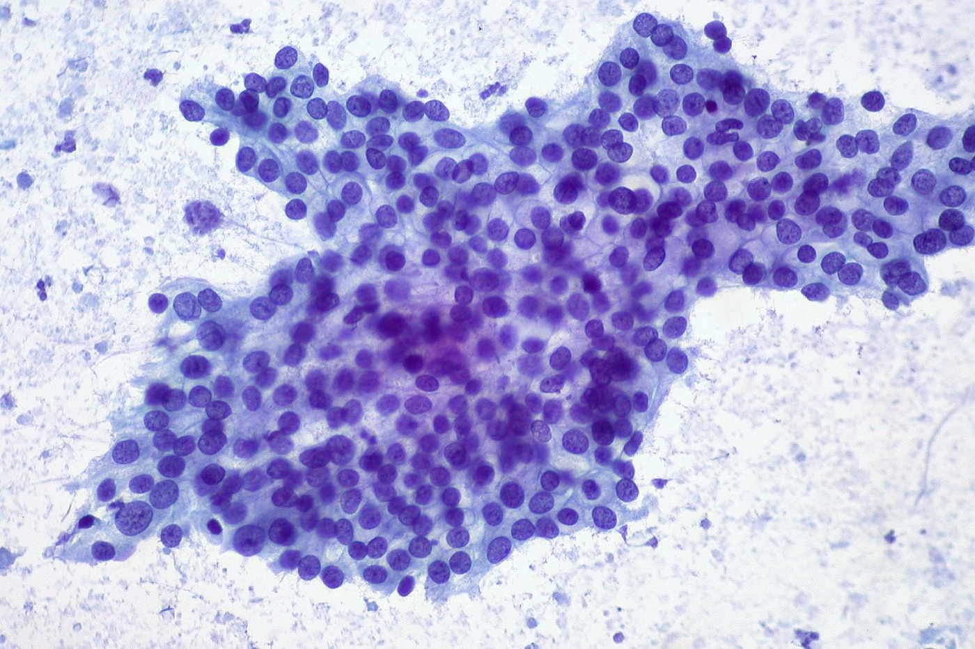
Solid Cellular Neoplasms of the Pancreas on Cytology
Definition. The solid cellular neoplasms of the pancreas are non-ductal tumors that produce highly cellular, stroma-poor aspirates of relatively uniform cells. The group comprises pancreatic neuroendocrine tumor (PanNET), solid-pseudopapillary neoplasm (SPN), acinar cell carcinoma (ACC) and pancreatoblastoma (Reid et al. 2019; Kwon et al. 2024). Their cytomorphology overlaps, so immunocytochemistry on cell block material is usually required for a specific diagnosis (Wang et al. 2024). All four are reported in the malignant category of the WHO Reporting System for Pancreaticobiliary Cytopathology. An aspirate that suggests one of them, without material for confirmation, is reported as suspicious for malignancy (Wang et al. 2024).
Related pages: The WHO Reporting System for Pancreaticobiliary Cytopathology (categories), Normal pancreas, gastrointestinal contaminants and EUS-guided sampling (normal acinar and islet cells; accessory spleen as a mimic), Pancreatic ductal adenocarcinoma and its variants on cytology (ductal adenocarcinoma), Metastases and lymphoma in pancreatic cytology (metastatic renal cell carcinoma, melanoma and lymphoma).
Clinical features of solid cellular pancreatic neoplasms
| Feature | PanNET | SPN | ACC | Pancreatoblastoma |
|---|---|---|---|---|
| Typical patient | Wide age range (16–86 years in one EUS series); both sexes | Young women | Older adults; 73% aged 60 or older in one series | Children (up to 25% of pancreatic tumors in the first decade); one-third occur in adults |
| Syndromes | MEN1, von Hippel-Lindau | — | — | Familial adenomatous polyposis, Beckwith-Wiedemann syndrome |
| Clinical findings | Functional hormone syndromes such as hypoglycemia | Can appear cystic on imaging | Lipase hypersecretion syndrome in 16%; about 50% metastatic at presentation | Elevated serum alpha-fetoprotein in up to 68% |
| Key genetic alteration | MEN1, DAXX, ATRX | CTNNB1 exon 3 (more than 95%) | BRAF fusions in about one-quarter; APC and 11p alterations | APC/β-catenin pathway; 11p loss of heterozygosity |
| WHO category | Malignant | Malignant | Malignant | Malignant |
Sources: Chatzipantelis et al. 2008; Bhatnagar et al. 2014; Kwon et al. 2024; Pitman et al. 2014; Reid et al. 2019; Nagtegaal et al. 2020; Layfield et al. 2014; Wang L et al. 2018; Wang et al. 2024.
Pancreatic neuroendocrine tumor on cytology
- Smears: highly cellular aspirates of loosely cohesive fragments with many dispersed single cells and bare nuclei. Cells are relatively uniform, epithelioid or plasmacytoid, with eccentric nuclei and salt-and-pepper chromatin. Cytoplasm is dense and granular (Wang et al. 2024).
- Most helpful findings: a richly cellular sample of a monotonous, poorly cohesive population of small or medium-sized cells with granular chromatin and plasmacytoid morphology (Chatzipantelis et al. 2008).
- Lipid-rich PanNET has fine cytoplasmic lipid droplets. Its differential includes ACC, SPN, metastatic renal cell carcinoma and ectopic adrenal cortical tissue (Wang et al. 2024).
- Cystic PanNET: 10–15% of PanNETs are cystic. PanNET belongs in the differential of every cystic pancreatic lesion (Wang et al. 2024).
- Immunocytochemistry: PanNET expresses synaptophysin, chromogranin, insulinoma-associated protein 1 (INSM1) and CD56 (Wang et al. 2024).
- Confirmation of neuroendocrine differentiation: the WHO 6th edition requires expression of at least two neuroendocrine markers, preferably including chromogranin A, synaptophysin or INSM1. It does not accept CD56 as a marker of neuroendocrine differentiation because CD56 is insufficiently specific (Arends et al. 2026). The IARC-WHO framework applied in the 5th edition named chromogranins and synaptophysin as the general neuroendocrine markers of the gastrointestinal tract and pancreas (Rindi et al. 2018; Nagtegaal et al. 2020).
- Genetics: the WHO classification treats MEN1, DAXX and ATRX mutations as entity-defining for well-differentiated neuroendocrine tumors. Neuroendocrine carcinomas usually have TP53 or RB1 mutations (Nagtegaal et al. 2020). PanNETs do not characteristically carry KRAS, TP53, CDKN2A or SMAD4 mutations (Layfield et al. 2014).
- Technique: a 25-gauge needle is often used for suspected neuroendocrine tumors because they are vascular. Core needles are also used (Brugge et al. 2014).
WHO grading of pancreatic neuroendocrine neoplasms and its use on cytology
| Category | Differentiation | Mitoses per 2 mm² | Ki-67 index |
|---|---|---|---|
| Neuroendocrine tumor, grade 1 | Well differentiated | Less than 2 | Less than 3% |
| Neuroendocrine tumor, grade 2 | Well differentiated | 2–20 | 3–20% |
| Neuroendocrine tumor, grade 3 | Well differentiated | More than 20 | More than 20% |
| Neuroendocrine carcinoma, small cell or large cell type | Poorly differentiated | More than 20 | More than 20% |
Sources: Arends et al. 2026 (WHO Classification of Digestive System Tumours, 6th edition, Table 4); Nagtegaal et al. 2020 (5th edition). The mitotic and Ki-67 thresholds are the same in both editions.
Tumor necrosis (6th edition): the presence or absence of tumor necrosis is recorded with the grade of every neuroendocrine tumor, for example "grade 2, tumor necrosis present". Only true tumor necrosis is recorded, and infarct-like necrosis is excluded (Arends et al. 2026). The 5th-edition grading table used mitotic count and Ki-67 index only (Nagtegaal et al. 2020).
Grade 2a and 2b (6th edition): the PanNET section recognizes a potential subdivision of grade 2 into grade 2a (Ki-67 index 3% to <10%) and grade 2b (Ki-67 index 10% to ≤20%). The general grading thresholds for neuroendocrine tumors are unchanged (Arends et al. 2026). The 5th edition had a single grade 2 (Nagtegaal et al. 2020).
Ki-67 counting: both editions grade on hot spots and assign the higher grade when the mitotic count and the Ki-67 index differ. The 5th edition specified a count of at least 500 cells (Nagtegaal et al. 2020). The 6th edition endorses digital image analysis of Ki-67 when it has been validated against a formal count of at least 500 tumor cells (Arends et al. 2026).
Grading on cytology uses mainly the Ki-67 index and is less reliable than grading on resection (Wang et al. 2024).
Undergrading: in one study of cell blocks, concordance with resection was 71% by visual estimate, 73% by hot spot count and 59% by complete count. All grade 1 tumors were graded correctly. Grade 2 tumors were concordant in only 36%, 41% and 9%. All grade 2 tumors were undergraded when the cell block contained fewer than 1,000 cells. With 1,000 or more cells, hot spot counting correlated best (Abi-Raad et al. 2020).
Neuroendocrine carcinomas are not graded. They are high grade by definition and are separated into small cell and large cell types (Nagtegaal et al. 2020).
Neuroendocrine tumor grade 3 compared with neuroendocrine carcinoma
| Feature | Neuroendocrine tumor, grade 3 | Neuroendocrine carcinoma |
|---|---|---|
| Morphology | Well differentiated | Poorly differentiated, small cell or large cell |
| Rb | Retained | Lost in most |
| p53 | Wild-type pattern | Aberrant in about 80–90% |
| ATRX or DAXX | Lost in about 50% | ATRX retained |
| Gene mutations | MEN1, DAXX, ATRX | TP53, RB1 |
Sources: Wang et al. 2024; Nagtegaal et al. 2020.
- Retained ATRX or retained Rb does not separate the two. Retained Rb supports grade 3 NET but does not exclude NEC. Rb loss or an aberrant p53 pattern favors carcinoma. ATRX or DAXX loss favors grade 3 neuroendocrine tumor (Wang et al. 2024).
- Separation of NET from NEC in the 6th edition: the distinction between neuroendocrine tumor and neuroendocrine carcinoma is morphological and is based on a constellation of findings, with no single defining feature. A neuroendocrine tumor can acquire NEC-like morphology (diffuse growth, high-grade cytological atypia and necrosis) over time, particularly after treatment, and rarely acquires RB1 and TP53 mutations. The 6th edition leaves open whether such a tumor is classified as "G3 NET with NEC-like morphology" or as "NEC arising from NET". This guidance is expanded from the 5th edition (Arends et al. 2026).
- Pancreatic neuroendocrine carcinoma is rare. Metastasis from the lung is excluded first. For the large cell type, extension from the ampulla is also excluded (Pitman et al. 2014).
- Management: resection is recommended for functioning PanNETs (Wang et al. 2024). For non-functioning PanNETs, ENETS 2023 recommends active surveillance for asymptomatic tumors of 1 cm or less without main pancreatic duct dilation, individualized management for tumors larger than 1 cm and up to 2 cm, and surgery for tumors larger than 2 cm or with main duct dilation (Kos-Kudła et al. 2023).
Solid-pseudopapillary neoplasm on cytology
- Patients and behavior: SPN is a low-grade malignant neoplasm, most often found in young women, with an excellent prognosis after complete resection. Resection of metastases is also preferred (Bhatnagar et al. 2014).
- Location: SPNs were more often in the body or tail than in the head in one series (Kwon et al. 2024).
- Smears: high cellularity, with thin, delicate, branching vessels in a "Chinese character" pattern, lined by one to several layers of loosely cohesive neoplastic cells (Bhatnagar et al. 2014).
- Cells: monomorphic, with round to oval or bean-shaped nuclei, nuclear grooves or indentations, finely granular evenly distributed chromatin, small inconspicuous nucleoli and indistinct cell borders. Single cells can have cytoplasmic tails. Hyaline globules support the diagnosis (Bhatnagar et al. 2014; Wang et al. 2024).
- Pseudopapillae were present in 77.8% of SPNs on liquid-based preparations and in no ACC or PanNET. All SPNs had fine chromatin (Kwon et al. 2024).
Immunophenotype and genetics of solid-pseudopapillary neoplasm
- Positive: nuclear β-catenin, CD10, CD99 in a perinuclear dot-like pattern, alpha-1-antitrypsin, progesterone receptor, SOX11, LEF1, TFE3, synaptophysin, CD56 and pancytokeratin (Bhatnagar et al. 2014; Wang et al. 2024).
- Negative: chromogranin, trypsin and BCL10 (Wang et al. 2024).
- LEF1: all SPNs, in resections and in cytology samples, showed strong diffuse nuclear LEF1 and β-catenin. Well-differentiated neuroendocrine tumors were negative. Pancreatoblastomas showed LEF1 and β-catenin mainly around and within squamoid morules (Singhi et al. 2014).
- CTNNB1: more than 95% of SPNs carry a somatic point mutation in exon 3. KRAS, CDKN2A and SMAD4 mutations have not been reported (Layfield et al. 2014).
- Misdiagnosis as PanNET: SPN expresses synaptophysin and CD56, which can lead to a misdiagnosis of PanNET when chromogranin and β-catenin are not tested (Wang et al. 2024; Layfield et al. 2014).
Acinar cell carcinoma on cytology
- Frequency and outcome: ACC accounts for about 1–2% of pancreatic neoplasms in adults (Wang L et al. 2018). About half of patients have metastases at diagnosis, and median survival was 47 months in one review (Kwon et al. 2024).
- Lipase hypersecretion syndrome is present in 16% of patients (Pitman et al. 2014).
- Smears: high cellularity, dense three-dimensional fragments and many dispersed single cells. Cells have granular cytoplasm, large nuclei and prominent nucleoli (Wang et al. 2024).
- Distinctive findings on liquid-based preparations: a necrotic background (75%), apoptotic debris (87.5%), nuclear tangles (25%), acinar structures (75%) and macronucleoli (100%). These were absent or rare in PanNET and SPN (Kwon et al. 2024).
- Single cells and bare nuclei were seen in 75% of ACCs on liquid-based preparations (Kwon et al. 2024).
- Neuroendocrine cells: scattered neuroendocrine cells are present in up to 40% of ACCs and can lead to a misdiagnosis of PanNET (Kwon et al. 2024).
- Amphicrine-like acinar cell carcinoma (6th edition): an ACC whose neuroendocrine cells are intimately admixed with the acinar cells is classified as amphicrine-like ACC. In one series, tumors reclassified as amphicrine-like ACC had been diagnosed as mixed neuroendocrine–non-neuroendocrine neoplasm (MiNEN) under the 5th edition (Kim et al. 2026). The 6th edition requires morphological evidence of neuroendocrine differentiation in ≥30% of neoplastic cells, confirmed by at least two neuroendocrine markers. Amphicrine-like carcinoma is staged as a carcinoma, and amphicrine-like ACC behaves like pure ACC (Arends et al. 2026).
- MiNEN in the 6th edition: the term is kept for tumors with morphologically distinct neuroendocrine and non-neuroendocrine zones, each forming ≥30% of the neoplasm (Chetty and Gill 2026). A neuroendocrine carcinoma whose only non-neuroendocrine component is a precursor or intramucosal lesion is not a MiNEN (Chetty and Gill 2026).
- Normal acinar tissue is more cohesive, with raspberry-like clusters, few single cells, small round nuclei and no atypia (Wang et al. 2024).
Immunophenotype and genetics of acinar cell carcinoma
- Acinar enzymes: trypsin, chymotrypsin, amylase and lipase on cell block (Layfield et al. 2014).
- BCL10: the C-terminal BCL10 antibody (clone 331.1) cross-reacts with carboxyl ester hydrolase. It was more sensitive for ACC than antibodies against other pancreatic enzymes and stained only acinar cells, ACC and pancreatic metaplasia (La Rosa et al. 2009).
- Neuroendocrine markers: synaptophysin, chromogranin and INSM1 can be focally positive (Wang et al. 2024).
- β-catenin abnormalities can be seen focally in ACC (Layfield et al. 2014).
- BRAF fusions were found in 24% of acinar-type neoplasms by fluorescence in situ hybridization. SND1 was the most common partner. The fusions are a potential therapeutic target (Wang L et al. 2018).
Pancreatoblastoma on cytology
- Definition: a rare malignant neoplasm, mainly of childhood, with acinar differentiation, neuroendocrine differentiation and squamoid morules (Pitman et al. 2014). Squamoid morules are the defining feature (Reid et al. 2019).
- Adult series: 12 FNAs from 11 adults (median age 45 years; median size 5.6 cm). Three patients had familial adenomatous polyposis or a related syndrome. Four had metastases at diagnosis (Reid et al. 2019).
- Findings present in all cases: hypercellularity, three-dimensional clusters, and single monotonous blast-like cells 1.5 to 2 times the size of a red blood cell, with a high nuclear-to-cytoplasmic ratio, fine chromatin and small distinct nucleoli (Reid et al. 2019).
- Squamoid morules were present in 70% of patients (75% of samples) and were easier to see on cell blocks. They consist of epithelioid cells with elongated, streaming nuclei and absent nucleoli, with nuclear β-catenin (Reid et al. 2019).
- Immunophenotype: synaptophysin in 10 of 10 tested cases, chromogranin in 6 of 10, trypsin in 5 of 8, and nuclear β-catenin in 6 of 8, which highlighted even subtle morules. The median Ki-67 index was 21% (Reid et al. 2019).
- Misdiagnosis: 5 of 12 samples were originally interpreted as neuroendocrine neoplasms, including poorly differentiated neuroendocrine carcinoma (Reid et al. 2019).
- Branching pseudopapillae and grooved nuclei were present in some cases and raised the possibility of SPN. The cells were more atypical than those of SPN (Reid et al. 2019).
Immunocytochemical panel for solid cellular pancreatic neoplasms
| Marker | PanNET | SPN | ACC | Pancreatoblastoma |
|---|---|---|---|---|
| Synaptophysin | Positive | Positive | Focal | Positive |
| Chromogranin | Positive | Negative | Focal | Positive in most |
| INSM1 | Positive | — | Focal | — |
| Trypsin, BCL10 | Negative | Negative | Positive | Trypsin positive in most |
| Nuclear β-catenin | Negative | Diffuse | Usually negative; focal abnormalities reported | Morules |
| LEF1 | Negative | Diffuse nuclear | Negative | Around morules |
| CD10 | — | Positive | — | — |
| CD99 | — | Perinuclear dot | — | — |
Sources: Wang et al. 2024, Table 4; Bhatnagar et al. 2014; Singhi et al. 2014; Reid et al. 2019; Layfield et al. 2014. A dash indicates that the cited sources do not state a pattern.
Differential diagnosis of solid cellular pancreatic neoplasms on cytology
| Feature | PanNET | SPN | ACC | Pancreatoblastoma |
|---|---|---|---|---|
| Architecture | Loose groups, single cells, occasional vascular cores | Branching vessels with loosely attached cells; pseudopapillae | Acinar groups, dense three-dimensional fragments, single cells | Three-dimensional clusters, single blast-like cells, morules |
| Chromatin | Salt-and-pepper | Fine, even | Irregular | Fine, immature |
| Macronucleoli | Absent | Absent | Present in all cases on liquid-based preparations | Nucleoli small and distinct |
| Special findings | Plasmacytoid cells; lipid in a variant | Grooves, cytoplasmic tails, hyaline globules | Necrosis, apoptotic debris, nuclear tangles | Squamoid morules |
| Key stain | Chromogranin | Nuclear β-catenin, LEF1 | Trypsin, BCL10 | Nuclear β-catenin in morules plus lineage markers |
Sources: Wang et al. 2024; Kwon et al. 2024; Bhatnagar et al. 2014; Reid et al. 2019; Chatzipantelis et al. 2008.
- Other mimics: intrapancreatic accessory spleen mimics PanNET on imaging (Normal pancreas, gastrointestinal contaminants and EUS-guided sampling). Metastatic renal cell carcinoma mimics lipid-rich PanNET (Metastases and lymphoma in pancreatic cytology).
- Reporting when tissue is insufficient: a solid cellular neoplasm that cannot be typed for lack of cell block material is reported as suspicious for malignancy, with the differential stated (Pitman et al. 2014; Wang et al. 2024).
Pancreatic Cystic Neoplasms on Cytology
Definition. Pancreatic cystic neoplasms are epithelial neoplasms that form cysts within the pancreas or its ducts. The three entities sampled most often by endoscopic ultrasound-guided fine-needle aspiration (EUS-FNA) are intraductal papillary mucinous neoplasm (IPMN), mucinous cystic neoplasm (MCN) and serous cystadenoma (SCA). IPMN and MCN are mucin-producing precursors of invasive adenocarcinoma. SCA is a benign neoplasm with a very low risk of malignancy. The WHO Reporting System for Pancreaticobiliary Cytopathology (IAC-IARC-WHO, 2022) places low-grade mucinous neoplasms in the category "pancreaticobiliary neoplasm, low risk/grade" (PaN-Low). It places high-grade mucinous neoplasms in "pancreaticobiliary neoplasm, high risk/grade" (PaN-High) and SCA in the benign (negative for malignancy) category (Pitman et al. 2023; Centeno et al. 2024).
Related pages: The WHO Reporting System for Pancreaticobiliary Cytopathology, Pancreatic cyst fluid chemistry and molecular testing.
Classification of pancreatic cystic neoplasms in the WHO 5th and 6th editions
The WHO Classification of Tumours, Digestive System Tumours, 5th edition (2019), changed the classification of pancreatic precursor lesions in three ways (Nagtegaal et al. 2020).
- Two-tier grading of dysplasia. Pancreatic intraepithelial neoplasia, IPMN and MCN are graded as low grade or high grade. Grading is based on the highest grade of dysplasia present. The three-tier system of the 4th edition is no longer used.
- Intraepithelial neoplasia terminology. The term "intraepithelial neoplasia" is preferred for precursor lesions in the pancreas, gallbladder and biliary tree. The term "dysplasia" is preferred in the tubular gut.
- Separate intraductal entities. Intraductal oncocytic papillary neoplasm (IOPN) and intraductal tubulopapillary neoplasm (ITPN) are classified separately from IPMN because of their distinct genomic and morphologic features (Nagtegaal et al. 2020). IOPN lacks the activating KRAS and GNAS mutations that are frequent in IPMN (Singhi et al. 2020).
The WHO Classification of Digestive System Tumours, 6th edition (2026), changed the list of pancreatic cystic lesions in two ways (Arends et al. 2026).
- Simple mucinous cyst (new entity). A simple mucinous cyst is a non-invasive cavity-forming lesion larger than 1.0 cm, lined by gastric-type epithelium, without papillary architecture or ovarian-type stroma. KRAS, BRAF, TP53 and SMAD4 alterations have been identified, so the lesion likely represents a precursor of pancreatic ductal adenocarcinoma. In the absence of dysplasia or carcinoma, the outcome is good (Arends et al. 2026). Simple mucinous cyst was not an entity in the 5th edition.
- Acinar cystic transformation removed. Acinar cystic transformation is non-neoplastic and is no longer included in the classification (Arends et al. 2026). The 5th edition listed it under that name, which replaced the former term acinar cell cystadenoma (Nagtegaal et al. 2020).
- Two-tier grading of precursors in both editions. Bile duct and pancreatic precursor lesions are graded as low grade or high grade in both the 5th edition (Nagtegaal et al. 2020) and the 6th edition (Arends et al. 2026). Former intermediate-grade dysplasia is graded as low grade (Arends et al. 2026).
The defining feature of MCN is subepithelial ovarian-type stroma. A specific cytologic diagnosis of MCN therefore requires sampling of the stroma, which is rare in fluid aspirates. MCN does not communicate with the main pancreatic duct (Crippa et al. 2008).
WHO Reporting System categories for pancreatic cystic neoplasms
| Lesion | WHO Reporting System category | PSC system category (2014) |
|---|---|---|
| Serous cystadenoma | Benign (negative for malignancy) | Neoplastic: benign |
| Lymphangioma | Benign (negative for malignancy) | Neoplastic: benign |
| IPMN with low-grade dysplasia | PaN-Low | Neoplastic: other |
| MCN with low-grade dysplasia | PaN-Low | Neoplastic: other |
| Mucinous cyst without epithelium (thick colloid-like mucin or CEA >192 ng/mL) | PaN-Low, with a comment that the grade cannot be assessed | Neoplastic: other |
| IPMN with high-grade dysplasia | PaN-High | Neoplastic: other |
| MCN with high-grade dysplasia | PaN-High | Neoplastic: other |
| IOPN and ITPN | PaN-High | Neoplastic: other |
| Cystic neuroendocrine tumor, solid-pseudopapillary neoplasm | Malignant | Neoplastic: other |
| Invasive carcinoma arising in IPMN or MCN | Malignant | Positive or malignant |
Sources: Pitman et al. 2023; Wang et al. 2024.
- Mucin without epithelium. An acellular cyst aspirate is non-diagnostic only when there is no evidence of a mucinous cyst. Thick colloid-like mucin or a cyst fluid carcinoembryonic antigen (CEA) above 192 ng/mL places the specimen in PaN-Low, with a comment that the grade of dysplasia is indeterminate (Wang et al. 2024).
- Non-mucinous cyst. A low CEA level with imaging of a simple cyst supports a report of "non-mucinous cyst" in the benign category (Wang et al. 2024).
- Risk of malignancy. In a reclassification of 334 EUS-FNAs, the absolute risk of malignancy (ROM) was 4.8% for PaN-Low and 60.0% for PaN-High (Hoda et al. 2022). The WHO system cites one study with a ROM of 4.3% for low-grade atypia and 90% for high-grade atypia, and gives PaN-High an estimated ROM of 60%–95% (Pitman et al. 2023).
Clinical and imaging features of pancreatic cystic neoplasms
| Feature | IPMN | MCN | SCA |
|---|---|---|---|
| Sex | Not stated in the cited series | Women in 95% (Crippa et al. 2008) | Women in 74% (Jais et al. 2016) |
| Age | Not stated in the cited series | Patients with invasive MCN were older than those with noninvasive MCN (55 versus 44 years) (Crippa et al. 2008) | Median 58 years (Jais et al. 2016) |
| Location | Main duct, branch ducts or both | Body and tail in 97% (Crippa et al. 2008) | Any part of the pancreas |
| Duct communication | Present | Absent | Absent |
| Lining | Mucinous epithelium of gastric, intestinal or pancreatobiliary type | Mucinous epithelium over ovarian-type stroma | Cuboidal cells with clear cytoplasm |
| Malignant potential | Precursor of invasive carcinoma | Cancer in 17.5% of resected MCNs (Crippa et al. 2008) | Three serous cystadenocarcinomas in 2622 patients; disease-related mortality 0.1% (Jais et al. 2016) |
| Cyst fluid CEA | Usually elevated | Usually elevated | Usually <5 ng/mL (Belsley et al. 2008) |
- IPMN subtypes. The pancreatobiliary subtype of IPMN is strongly associated with malignancy (European Study Group on Cystic Tumours of the Pancreas 2018).
- MCN size and nodules. In 163 resected MCNs, every MCN with cancer measured ≥40 mm or had a mural nodule. The 5-year disease-specific survival was 100% for noninvasive MCN and 57% for MCN with invasive carcinoma (Crippa et al. 2008).
- SCA management. SCA is managed conservatively in most patients. Resection is reserved for an uncertain diagnosis after full work-up, significant related symptoms, or exceptional concern for malignancy (Jais et al. 2016).
Cytologic features of intraductal papillary mucinous neoplasm
- Mucin. Thick colloid-like extracellular mucin indicates a mucinous cyst. Thin mucin that resembles tissue paper is difficult to separate from gastrointestinal contamination, although abundant thin mucin suggests a mucinous cyst (Wang et al. 2024).
- Epithelium. IPMN epithelium is gastric, intestinal or pancreatobiliary in type, or a mixture. Lesional mucinous cells often form small clusters or occur singly (Wang et al. 2024).
- Low-grade epithelium. Low-grade mucinous epithelium closely resembles gastric foveolar epithelium. Gastric contamination is therefore the main differential diagnosis of PaN-Low (Wang et al. 2024).
- Low cellularity. Many IPMN aspirates contain mucin, histiocytes and debris without epithelium. Cytology alone has a sensitivity of 27%–48% and a specificity of 83%–100% for a mucinous cyst (European Study Group on Cystic Tumours of the Pancreas 2018).
- Ancillary correlation. Cyst fluid CEA, amylase and glucose levels and KRAS and GNAS testing help separate lesional mucinous epithelium from contaminants. These tests are covered in Pancreatic cyst fluid chemistry and molecular testing.
Cytologic features of mucinous cystic neoplasm
- Epithelium and mucin. MCN aspirates contain mucin and mucinous epithelium. Neither cytology nor CEA separates MCN from IPMN (European Study Group on Cystic Tumours of the Pancreas 2018).
- Ovarian-type stroma. Fragments of ovarian-type stroma are rarely present in fluid aspirates. A forceps biopsy of the cyst wall can sample the stroma (Zhang et al. 2018).
- Stromal immunohistochemistry. Ovarian-type stroma in pancreatic MCN expresses estrogen receptor (ER), progesterone receptor (PR) and α-inhibin. Stromal density and staining are more variable in pancreatic MCN than in hepatobiliary mucinous cystic neoplasms (Lam et al. 2008).
- Clinical integration. In a woman, a unilocular or septated mucinous cyst in the pancreatic body or tail without duct communication favors MCN (Crippa et al. 2008).
- Report. Without stroma, the cytology report describes a mucinous neoplasm and grades the epithelium. The distinction between MCN and IPMN is based on imaging, demographics and molecular results.
- KRAS/GNAS-negative MCN. In a prospective cyst fluid NGS study, 7 of 10 resected MCNs, all with low-grade dysplasia, were KRAS/GNAS wild type. A negative result does not exclude MCN (Singhi et al. 2018).
Cytologic features of serous cystadenoma
- Low cellularity. About 75% of SCA aspirates are scantly cellular. The epithelial cells are small and round to cuboidal with clear cytoplasm. Plasmacytoid cells, oncocytic cells, flattened lining cells and hyalinized stromal fragments also occur (Salomao et al. 2014).
- Frequency of diagnostic epithelium. Serous epithelial cells were identified in fewer than 20% of 21 histologically confirmed SCAs. None of the EUS-guided aspirates in that series contained serous epithelium (Belsley et al. 2008).
- Hemosiderin-laden macrophages. These were present in 43% of SCA aspirates, 2% of neoplastic mucinous cyst aspirates and 9% of pseudocyst aspirates (Belsley et al. 2008).
- Gastrointestinal contamination. About 52% of EUS-guided SCA aspirates contained gastrointestinal epithelium. This epithelium was classified as atypical in two cases (Belsley et al. 2008).
- α-Inhibin. α-Inhibin was positive on cell blocks in 88% (7 of 8) of SCAs in one series and in 6 of 7 cases in another (Salomao et al. 2014; Wang et al. 2024b).
- Fine-needle biopsy. Among 15 SCAs with paired FNA and fine-needle biopsy (FNB), FNA established SCA in 1 case (7%) and FNB in 13 cases (87%) (Wang et al. 2024b).
- Cyst fluid. All 17 SCAs with CEA testing had a CEA below 192 ng/mL, and all 14 with molecular testing had wild-type KRAS (Wang et al. 2024b). VHL mutations were present in four of eight SCAs studied by whole-exome sequencing (Wu et al. 2011b).
- Category. SCA is reported in the benign category. An SCA aspirate that contains only histiocytes and inflammatory cells is also placed in the benign category, because scant cellularity is expected in SCA (Wang et al. 2024).
Mucin and gastrointestinal contaminants in pancreatic cyst aspirates
| Component | Cytologic features | Interpretation |
|---|---|---|
| Thick colloid-like mucin | Dense, homogeneous mucin; may contain histiocytes | Mucinous cyst (IPMN or MCN) |
| Thin mucin | Wispy mucin resembling tissue paper | Mucinous cyst or gastrointestinal contamination |
| Gastric foveolar epithelium | Large flat sheets with a regular mosaic pattern and apical mucin | Contaminant when sampled through the stomach |
| Duodenal epithelium | Non-mucinous enterocytes with a brush border, scattered goblet cells and intraepithelial lymphocytes | Contaminant when sampled through the duodenum |
| Lesional mucinous epithelium | Small clusters or single cells with intracellular mucin | Mucinous neoplasm |
| Keratinous debris and squames | Anucleate squames and amorphous debris | Lymphoepithelial cyst; debris can mimic mucin |
Sources: Pitman et al. 2014; Wang et al. 2024.
- When lesional cells cannot be separated from gastrointestinal contaminants, the cells are reported as atypical (Wang et al. 2024).
- The sampling route determines the type of contaminating epithelium. Transgastric sampling of a cyst in the body or tail introduces gastric epithelium. Transduodenal sampling of a cyst in the head introduces duodenal epithelium. Details are in Normal pancreas, gastrointestinal contaminants and EUS-guided sampling.
Grading of epithelial atypia in mucinous cysts
The WHO Reporting System defines high-grade atypia (HGA) as cells smaller than a 12-µm duodenal enterocyte with a high N:C ratio and abnormal chromatin, which can be hypochromatic or hyperchromatic, with or without background necrosis. HGA covers high-grade dysplasia and invasive carcinoma and places the specimen in PaN-High. Low-grade atypia represents low- to intermediate-grade dysplasia (Pitman et al. 2023). Low-grade mucinous epithelium resembles gastric foveolar epithelium (Wang et al. 2024).
| Feature of high-grade atypia | Description |
|---|---|
| Arrangement | Small clusters and single cells |
| Cell size | Smaller than a 12-µm duodenal enterocyte |
| Nuclear-to-cytoplasmic (N:C) ratio | High |
| Chromatin | Abnormal: hyperchromatic or hypochromatic |
| Nuclear contour | Irregular |
| Cytoplasmic mucin | Variable |
| Background | Necrosis present or absent |
Sources: Pitman et al. 2023; Wang et al. 2024.
- Definition of atypical epithelial cells. Atypical epithelial cells (AEC) are cells with an increased N:C ratio and nuclear hyperchromasia, with or without membrane abnormalities and cytoplasmic vacuoles, that are insufficient in quality and quantity for a malignant interpretation (Pitman et al. 2013).
- Retrospective evidence. In 112 histologically confirmed mucinous cysts, "positive" cytology had a sensitivity of 29% and a specificity of 100% for high-grade dysplasia or carcinoma. Using AEC as the threshold raised the sensitivity to 72% with a specificity of 85% and an accuracy of 80%. The AEC threshold identified 17 additional cancers (Pitman et al. 2010).
- Prospective evidence. In 66 cyst aspirates with histologic follow-up, high-grade atypia or worse had a sensitivity of 83%, a specificity of 86%, a positive predictive value of 77% and a negative predictive value of 90% for high-grade dysplasia or invasive carcinoma (Pitman et al. 2013).
- Necrosis. A necrotic background supports high-grade dysplasia. It does not reliably separate high-grade dysplasia from invasive carcinoma (Wang et al. 2024).
- High-grade dysplasia and carcinoma. Cytology often cannot separate an intraductal neoplasm with high-grade dysplasia from invasive adenocarcinoma. Both are usually treated by resection (Wang et al. 2024).
- Suspicious for malignancy. A mucinous cyst with high-grade atypia and necrosis, particularly with high-risk imaging findings, can be reported as suspicious for malignancy (Wang et al. 2024).
- Differential diagnosis of high-grade atypia. Atypical lining cells of benign cysts, cells of a neuroendocrine tumor and intermediate-grade dysplasia can mimic high-grade atypia (Wang et al. 2024).
Intraductal oncocytic papillary neoplasm and intraductal tubulopapillary neoplasm on cytology
- Category. Both entities are placed in PaN-High (Pitman et al. 2023; Wang et al. 2024).
- IOPN genetics. All 23 IOPNs of the pancreas and bile duct in one study carried ATP1B1::PRKACB, DNAJB1::PRKACA or ATP1B1::PRKACA fusions. The fusions were also detected in matched preoperative cyst fluid and bile duct brushings, and in none of 126 control pancreatobiliary lesions (Singhi et al. 2020).
- IOPN cells. IOPN epithelial cells contain numerous large mitochondria, which produce the oncocytic cytoplasm (Singhi et al. 2020).
Role of cytology in pancreatic cyst guidelines
- Kyoto guidelines (2024). The international evidence-based Kyoto guidelines for IPMN add EUS findings and EUS-FNA cytology to the assessment of high-risk stigmata and worrisome features (Ohtsuka et al. 2024). High-risk stigmata are obstructive jaundice in a patient with a cystic lesion of the pancreatic head, an enhancing mural nodule ≥5 mm or solid component, a main pancreatic duct ≥10 mm, and cytology suspicious or positive for high-grade dysplasia or adenocarcinoma (Borges et al. 2025).
- European guidelines (2018). Jaundice, an enhancing mural nodule ≥5 mm or solid mass, and cytology positive for high-grade dysplasia or cancer are absolute indications for resection of a branch-duct IPMN. MCN ≥40 mm, symptomatic MCN and MCN with a mural nodule are resected. MCN <40 mm without risk features can be managed by surveillance (European Study Group on Cystic Tumours of the Pancreas 2018).
- Targeting. When EUS-FNA is performed to detect high-grade dysplasia or cancer, any solid component or thickened cyst wall is targeted for cytology (European Study Group on Cystic Tumours of the Pancreas 2018).
- PaN-Low management. Patients with PaN-Low lesions are usually managed by surveillance (Wang et al. 2024).
Differential diagnosis of cystic pancreatic lesions on cytology
| Lesion | Key cytologic findings | Supporting tests | WHO category |
|---|---|---|---|
| IPMN, low grade | Mucin; foveolar-type mucinous epithelium | CEA >192 ng/mL; KRAS and GNAS mutations | PaN-Low |
| MCN, low grade | Mucin; mucinous epithelium; ovarian-type stroma rarely sampled | CEA >192 ng/mL; KRAS mutation; GNAS wild type (Wu et al. 2011a) | PaN-Low |
| IPMN or MCN, high grade | Small cells with increased N:C ratio and abnormal chromatin | TP53, PIK3CA or PTEN alterations (Singhi et al. 2018) | PaN-High |
| Serous cystadenoma | Scant cuboidal clear cells; hemosiderin-laden macrophages | α-Inhibin positive; CEA <5 ng/mL; VHL alteration | Benign |
| Pseudocyst | Histiocytes, debris, inflammatory cells; no epithelium | Amylase markedly elevated; CEA low | Benign |
| Lymphoepithelial cyst | Anucleate squames, keratinous debris, lymphocytes | CEA may be elevated | Benign |
| Cystic neuroendocrine tumor | Plasmacytoid cells with salt-and-pepper chromatin | Synaptophysin, chromogranin, INSM1 | Malignant |
| Solid-pseudopapillary neoplasm | Branching papillae with vascular cores; grooved nuclei | Nuclear β-catenin; CD10 | Malignant |
Sources: Pitman et al. 2023; Wang et al. 2024; Belsley et al. 2008; Salomao et al. 2014.
- About 10%–15% of pancreatic neuroendocrine tumors are cystic. Neuroendocrine tumor is included in the differential diagnosis of every cystic pancreatic lesion (Wang et al. 2024). Cystic presentations of solid tumors are covered in Solid cellular neoplasms of the pancreas on cytology.
- Pseudocyst and lymphoepithelial cyst are covered in Pancreatitis, pseudocyst and lymphoepithelial cyst on cytology.
Pancreatic Cyst Fluid Chemistry and Molecular Testing
Definition. Pancreatic cyst fluid analysis is the biochemical and molecular testing of fluid aspirated from a pancreatic cyst by endoscopic ultrasound-guided fine-needle aspiration (EUS-FNA). It addresses two questions that cytology alone often cannot resolve: whether a cyst is mucinous, and whether a mucinous cyst contains high-grade dysplasia or invasive carcinoma. The main biochemical tests are carcinoembryonic antigen (CEA), amylase and glucose. The main molecular tests detect mutations in KRAS and GNAS, which indicate a mucinous neoplasm, and alterations in TP53, PIK3CA and PTEN, which indicate advanced neoplasia. The WHO Reporting System for Pancreaticobiliary Cytopathology (IAC-IARC-WHO, 2022) recommends that biochemical and molecular results be incorporated into the final cytology report (Pitman et al. 2023).
Related pages: Pancreatic cystic neoplasms on cytology, The WHO Reporting System for Pancreaticobiliary Cytopathology.
Role of cyst fluid analysis in the WHO Reporting System for Pancreaticobiliary Cytopathology
- Neoplastic epithelium with a low CEA. When neoplastic mucinous epithelium or colloid-like mucin is present, the lesion is reported as a pancreaticobiliary neoplasm in the appropriate clinical setting, even with a CEA below 192 ng/mL (Wang et al. 2024).
- Molecular testing. Molecular testing is most useful when cytology is non-diagnostic, when fluid is insufficient for CEA, or when the CEA level is indeterminate (Wang et al. 2024).
Cyst fluid carcinoembryonic antigen
- Derivation of the 192 ng/mL cutoff. The Cooperative Pancreatic Cyst Study evaluated 341 patients, of whom 112 underwent resection. Cyst fluid CEA had the largest area under the receiver operating characteristic curve (0.79) of the tumor markers tested. The optimal cutoff was 192 ng/mL. CEA accuracy (79%) exceeded that of EUS morphology (51%) and cytology (59%). No combination of tests was more accurate than CEA alone (Brugge et al. 2004).
- Performance in later studies. A CEA ≥192 ng/mL separates mucinous from non-mucinous cysts with a sensitivity of 52%–78% and a specificity of 63%–91% (European Study Group on Cystic Tumours of the Pancreas 2018).
- Extreme values. In a pooled analysis of 450 patients, a CEA below 5 ng/mL suggested serous cystadenoma (SCA) or pseudocyst (sensitivity 50%, specificity 95%). A CEA above 800 ng/mL strongly suggested a mucinous cyst (sensitivity 48%, specificity 98%) (van der Waaij et al. 2005).
- Limits. CEA does not distinguish intraductal papillary mucinous neoplasm (IPMN) from mucinous cystic neoplasm (MCN). CEA does not separate low-grade mucinous cysts from those with high-grade dysplasia or invasive carcinoma (European Study Group on Cystic Tumours of the Pancreas 2018).
- False-positive CEA. CEA can be elevated in lymphoepithelial cysts, duplication cysts and, rarely, SCA (Wang et al. 2024). One histologically confirmed SCA had a CEA of 176.5 ng/mL (Belsley et al. 2008).
- False-negative CEA. A low CEA does not exclude a mucinous cyst (Wang et al. 2024).
- Institutional cutoffs. Optimal cutoffs differ between laboratories. A single-center study found an optimal cutoff of 48.6 ng/mL for mucinous cysts (Oh et al. 2017).
Cyst fluid amylase and lipase
- Amylase below 250 U/L. A cyst fluid amylase below 250 U/L virtually excluded a pseudocyst in pooled data (sensitivity 44%, specificity 98%) (van der Waaij et al. 2005).
- Markedly elevated amylase. A markedly elevated amylase suggests a pseudocyst. SCA usually has an amylase below 1,000 U/L. IPMN has variable amylase levels (Wang et al. 2024).
- Lipase. The European guidelines state that combined analysis of cyst fluid CEA, cyst fluid lipase and cytology provides the highest accuracy for separating mucinous from non-mucinous cysts (European Study Group on Cystic Tumours of the Pancreas 2018).
Cyst fluid glucose
- Meta-analysis. In 8 studies including 609 lesions, low cyst fluid glucose had a pooled sensitivity of 91% for a mucinous cyst. CEA alone had a sensitivity of 56%. Specificity did not differ significantly (glucose 86%, CEA 96%). Diagnostic accuracy was 94% for glucose and 85% for CEA. Adding CEA to glucose did not significantly improve accuracy (97% versus 94%) (McCarty et al. 2021).
- Cutoff. The commonly used cutoff is <50 mg/dL. Its high sensitivity makes a normal glucose useful for excluding a mucinous cyst (Wang et al. 2024).
- Status in the WHO system. Glucose has not been introduced as a standard test in the WHO Reporting System (Wang et al. 2024).
Cyst fluid CA 19-9 and the string sign
- CA 19-9. A cyst fluid CA 19-9 below 37 U/mL strongly suggested a pseudocyst or SCA (sensitivity 19%, specificity 98%) (van der Waaij et al. 2005). The European guidelines found insufficient evidence for non-CEA protein markers (European Study Group on Cystic Tumours of the Pancreas 2018).
- String sign. The string sign measures fluid viscosity. In 48 histologically or clinically confirmed cysts, the accuracy of the string sign for a mucinous cyst was 70.2%. CEA, cytology and the string sign combined reached an accuracy of 93.8% (Oh et al. 2017).
Biochemical profiles of pancreatic cysts
| Cyst type | Amylase | CEA | Glucose | Notes |
|---|---|---|---|---|
| Pseudocyst | Markedly elevated (often >1,000 U/L) | Low | Not stated in the cited sources | History of pancreatitis; covered in Pancreatitis, pseudocyst and lymphoepithelial cyst on cytology |
| Serous cystadenoma | Low (<250 U/L) | Very low (<5 ng/mL) | Usually ≥50 mg/dL (non-mucinous) | Non-mucinous |
| Lymphoepithelial cyst | Not stated in the cited sources | May be elevated | Not stated in the cited sources | Keratinous debris can mimic mucin |
| IPMN | Variable | Elevated (>192 ng/mL) in most | <50 mg/dL | Communicates with the duct system |
| MCN | Usually low; <250 U/L virtually excludes a pseudocyst (van der Waaij et al. 2005) | Elevated (>192 ng/mL) in most | <50 mg/dL | Does not communicate with the duct system |
Sources: van der Waaij et al. 2005; Wang et al. 2024; McCarty et al. 2021; Crippa et al. 2008.
Molecular alterations in pancreatic cyst fluid
- GNAS. Recurrent GNAS mutations at codon 201 were found in 66% of IPMNs. A KRAS or GNAS mutation was present in 96% of IPMNs. GNAS mutations were absent in other cystic neoplasms and in ductal adenocarcinomas unrelated to IPMN (Wu et al. 2011a).
- KRAS. A KRAS mutation supports a mucinous neoplasm. KRAS alone does not separate IPMN from MCN (Wang et al. 2024).
- RNF43, VHL and CTNNB1. Whole-exome sequencing of eight cysts of each type found RNF43 mutations in 6 of 8 IPMNs and 3 of 8 MCNs, VHL mutations in 4 of 8 SCAs, and CTNNB1 mutations in every solid-pseudopapillary neoplasm (SPN) (Wu et al. 2011b).
- Prospective next-generation sequencing (NGS). In 626 cyst fluid specimens from 595 patients, KRAS or GNAS mutations were present in 49% of cysts. Among 102 patients with surgical follow-up, these mutations were found in all 56 IPMNs and in 3 of 10 MCNs. The remaining seven MCNs, all with low-grade dysplasia, were KRAS/GNAS negative. KRAS or GNAS mutations had a sensitivity of 89% and a specificity of 100% for a mucinous cyst. Sanger sequencing of a separate cohort had a sensitivity of 65% and a specificity of 100% (Singhi et al. 2018).
- Advanced neoplasia. KRAS or GNAS mutations combined with alterations in TP53, PIK3CA or PTEN had a sensitivity of 89% and a specificity of 100% for high-grade dysplasia or invasive carcinoma. Main duct dilatation, a mural nodule and malignant cytopathology had sensitivities of 42%, 32% and 32% (Singhi et al. 2018).
- VHL in SCA. VHL alterations had a specificity of 100% for SCA in the same cohort. In one SCA, the VHL alteration was missed by Sanger sequencing of cyst fluid and detected in the resection specimen. VHL alterations can also occur in cystic pancreatic neuroendocrine tumors (Singhi et al. 2018).
- Multimarker classification. A panel of molecular markers with clinical features classified 130 resected cysts with a sensitivity of 90%–100% and a specificity of 92%–98%. The panel correctly identified 67 of 74 patients who did not require surgery (Springer et al. 2015).
- MEN1, DAXX and ATRX. Mutations in these genes are entity-defining for well-differentiated neuroendocrine tumors (Nagtegaal et al. 2020).
DNA and RNA panels for pancreatic cyst fluid
- PancreaSeq Genomic Classifier development. A 74-gene DNA and RNA panel tested mutations, fusions and gene expression. CEA messenger RNA (CEACAM5) was added by quantitative reverse transcription PCR. The classifier had a sensitivity of 95% and a specificity of 100% for a cystic precursor neoplasm, and 82% and 100% for advanced neoplasia (Nikiforova et al. 2023).
- Multi-institutional validation. In 241 prospectively tested patients, the classifier had a sensitivity of 94.6% and a specificity of 96.4% for mucinous cysts. For advanced neoplasia, the sensitivity was 86.6% and the specificity 97.9%. The classifier also identified IOPN, intraductal tubulopapillary neoplasm (ITPN) and cystic pancreatic neuroendocrine tumors (Singhi et al. 2026).
- Guideline status. The European guidelines (2018) described KRAS and GNAS analysis as promising and outside standard management (European Study Group on Cystic Tumours of the Pancreas 2018). The Kyoto guidelines (2024) review the emerging role of cyst fluid markers in separating IPMN from other cysts and in detecting high-grade dysplasia or invasive carcinoma (Ohtsuka et al. 2024).
Molecular alterations by pancreatic cyst type
| Cyst type | Characteristic alterations | Interpretation |
|---|---|---|
| IPMN | KRAS, GNAS (66%), RNF43 | GNAS supports IPMN over MCN |
| MCN | KRAS, RNF43; GNAS absent | Mucinous neoplasm |
| IPMN or MCN with advanced neoplasia | TP53, PIK3CA, PTEN with KRAS or GNAS | High-grade dysplasia or invasive carcinoma |
| SCA | VHL | Benign neoplasm |
| SPN | CTNNB1 without other alterations | Malignant (low-grade) neoplasm |
| Well-differentiated neuroendocrine tumor | MEN1, DAXX, ATRX | Malignant neoplasm |
| IOPN | PRKACA or PRKACB fusions; no KRAS | PaN-High |
| Simple mucinous cyst (new entity in the WHO 6th edition; not an entity in the 5th edition) | KRAS, BRAF, TP53, SMAD4 | Neoplastic mucinous cyst; likely precursor of ductal adenocarcinoma |
| Pseudocyst, lymphoepithelial cyst | Wild type | Non-neoplastic |
Sources: Wu et al. 2011a; Wu et al. 2011b; Singhi et al. 2018; Singhi et al. 2020; Nagtegaal et al. 2020; Wang et al. 2024; Arends et al. 2026.
Cell-free DNA in cytology supernatant and cyst fluid
- Cyst fluid as a source of DNA. Cyst fluid is tested directly by targeted NGS (Singhi et al. 2018).
- Supernatant of solid-lesion aspirates. The cytocentrifugation supernatant of pancreatic FNAs yielded amplifiable DNA suitable for mutation analysis. The supernatant showed additional mutations and higher clonality than microdissected cells. It may therefore be enriched with tumor DNA (Deftereos et al. 2014).
- Liquid-based cytology supernatant. Cell-free DNA from the supernatant of fixed liquid-based cytology samples gave a valid result in 84% (42 of 50) of pancreatic lesions. KRAS mutation detection had a sensitivity of 63.2% and a specificity of 100% and was comparable to testing of formalin-fixed tissue (Takase et al. 2023).
- Role of supernatant testing. Supernatant testing is an adjunct that reduces sampling variability. It is most useful when the clinical suspicion is high and the smears contain few atypical cells (Deftereos et al. 2014).
Integration of cyst fluid results with cytology and imaging
| Finding | Interpretation | Report |
|---|---|---|
| Mucinous epithelium with low-grade atypia; CEA elevated | Mucinous neoplasm, low grade | PaN-Low |
| Acellular; thick mucin or CEA >192 ng/mL | Mucinous neoplasm, grade indeterminate | PaN-Low with comment |
| Acellular; CEA low; simple cyst on imaging | Non-mucinous cyst | Benign |
| Acellular; no mucin; CEA not available | No evidence of the lesion | Non-diagnostic |
| Mucinous epithelium with high-grade atypia | Mucinous neoplasm, high grade | PaN-High |
| KRAS or GNAS with TP53, PIK3CA or PTEN | Advanced neoplasia likely | Integrated with cytology; high-risk result |
| VHL alteration; CEA <5 ng/mL; α-inhibin-positive cells | Serous cystadenoma | Benign |
Sources: Wang et al. 2024; Singhi et al. 2018; Pitman et al. 2023.
- Combination testing. Combinations of EUS morphology, cytology and cyst fluid CEA are more accurate for a mucinous cyst than EUS morphology or cytology alone (European Study Group on Cystic Tumours of the Pancreas 2018).
- Sensitivity of cytology. Cytologic analysis of cyst fluid had a pooled sensitivity of 42% and a specificity of 99% for a mucinous cyst (European Study Group on Cystic Tumours of the Pancreas 2018).
- Gastrointestinal contamination. CEA, amylase, glucose and KRAS testing help separate lesional mucinous epithelium from gastric contaminants, which closely resemble low-grade mucinous epithelium (Wang et al. 2024).
- Mutations and grade. KRAS and GNAS mutations indicate a mucinous neoplasm but do not indicate its grade. Advanced neoplasia is indicated by additional TP53, PIK3CA or PTEN alterations (Singhi et al. 2018).
Metastases and Lymphoma in Pancreatic Cytology
Definition. Secondary tumors of the pancreas are malignant neoplasms that arise at another site and involve the pancreas by hematogenous, lymphatic or direct spread. Primary pancreatic lymphoma is a lymphoma whose bulk of disease is located in the pancreas, with a clinical presentation centered on the pancreas, although adjacent lymph nodes and distant sites may be involved (Facchinelli et al. 2021). In the WHO Reporting System for Pancreaticobiliary Cytopathology (IAC-IARC-WHO, 2022), metastatic malignancies and pancreatic lymphomas are reported in the malignant category (Pitman et al. 2023; Wang et al. 2024). Renal cell carcinoma is the most frequent metastasis in pancreatic fine-needle aspiration (FNA) series (Olson et al. 2013; Smith et al. 2015).
Related pages: The WHO Reporting System for Pancreaticobiliary Cytopathology, Solid cellular neoplasms of the pancreas on cytology, Pancreatic ductal adenocarcinoma and its variants on cytology.
Frequency of metastases in pancreatic cytology
- Proportion of pancreatic aspirates. In two institutions, 42 of 5,495 pancreatic aspirates were metastases. Metastases formed 0.8% of all pancreatic aspirates and 1.8% of malignant aspirates (Olson et al. 2013).
- Single-institution FNA series. Of 2,327 pancreatic FNAs over 14 years, 22 showed metastatic disease (Smith et al. 2015).
- Fine-needle biopsy series. Of 1,054 pancreatic neoplasms diagnosed by EUS-guided fine-needle biopsy (FNB) over nine years, 62 (5.9%) were metastases (Cui et al. 2025).
- Literature review. Renal cell carcinoma accounted for about 35% of pancreatic metastases managed with FNA in a review of published series (Ioakim et al. 2020).
Primary sites of metastases to the pancreas in cytology series
| Series | Cases | Primary sites in order of frequency |
|---|---|---|
| DeWitt et al. 2005 (EUS-FNA, multicenter) | 24 | Kidney 10, skin 6, lung 4, colon 2, liver 1, stomach 1 |
| Olson et al. 2013 (FNA, two institutions) | 42 | Renal cell carcinoma, then melanoma, then non-small cell lung carcinoma |
| Smith et al. 2015 (FNA, single institution) | 22 | Renal cell carcinoma 14, colonic adenocarcinoma 2; one each of urothelial, non-small cell lung, ovarian serous, prostatic and papillary thyroid carcinoma and mesenchymal chondrosarcoma |
| Cui et al. 2025 (EUS-FNB) | 62 | Renal 11 (17.8%), lung 9 (14.5%), hematopoietic 9 (14.5%), cutaneous 6 (9.7%), Müllerian 6 (9.7%), mesenchymal 5 (8.1%), melanoma 3 (4.8%) |
- Olson et al. (2013) also reported two cases each of fibrolamellar hepatocellular carcinoma and solitary fibrous tumor.
- In children and young adults, in whom conventional pancreatic ductal adenocarcinoma is uncommon, a history of other tumors, including tumors with a usually indolent course, is essential to the diagnosis (Olson et al. 2013).
Clinical and imaging features of metastases to the pancreas
- Interval after the primary tumor. The median interval from the primary tumor to the pancreatic metastasis was 9 years, ranging from synchronous presentation to 21 years (Smith et al. 2015). In the FNB series, the interval ranged from synchronous presentation to 24 years (Cui et al. 2025).
- Number and size. Most metastases were unifocal (73%), with a mean greatest dimension of 3.7 cm (range 1.5–6.5 cm) (Smith et al. 2015). In the FNB series, 74% were solitary masses (Cui et al. 2025).
- Imaging. Metastases more often had well-defined margins on EUS than primary pancreatic cancer (46% versus 4%) (DeWitt et al. 2005). In the FNB series, 58% of metastases were initially interpreted as primary pancreatic tumors on imaging (Cui et al. 2025).
- Role of EUS-FNA. EUS-FNA provided the initial diagnosis of malignancy in 4 of 24 patients (17%) and the diagnosis of tumor recurrence in 16 (67%). Four metastases were first found by EUS after negative or inconclusive computed tomography (DeWitt et al. 2005).
- Rapid on-site evaluation. Of 13 metastases with on-site evaluation, 4 were recognized as metastatic, 3 as positive for malignant cells, 6 as atypical and none as negative (Smith et al. 2015).
Cytologic approach to suspected metastasis in the pancreas
- Features that raise the possibility of metastasis. A metastasis enters the differential diagnosis when the cytomorphology is unusual for a primary pancreatic tumor, especially in a patient with a history of malignancy (Wang et al. 2024).
- Mimicry of primary tumors. Metastatic renal cell carcinoma, lung adenocarcinoma, breast carcinoma, gastrointestinal adenocarcinoma, Merkel cell carcinoma and adult granulosa cell tumor closely mimicked primary pancreatic ductal adenocarcinoma, neuroendocrine neoplasms and solid-pseudopapillary neoplasm (Cui et al. 2025).
- Comparison with prior material. Clinical history and comparison with the primary tumor are required for a correct diagnosis (Olson et al. 2013; Cui et al. 2025).
- Immunocytochemistry. All diagnoses in a seven-case series were confirmed by immunostains (Ioakim et al. 2020).
- Reporting. A metastasis confirmed by morphology and immunocytochemistry is reported in the malignant category. When limited material prevents confirmation by immunocytochemistry, the specimen is reported as suspicious for malignancy (Wang et al. 2024).
Immunocytochemical panels for primary and metastatic tumors in the pancreas
| Tumor | Positive markers | Notes |
|---|---|---|
| Pancreatic ductal adenocarcinoma | CK7, CK19, mesothelin; abnormal p53; loss of SMAD4 | Primary; covered in Pancreatic ductal adenocarcinoma and its variants on cytology |
| Pancreatic neuroendocrine tumor | Synaptophysin, chromogranin, INSM1, CD56 | Primary; lipid-rich variant mimics renal cell carcinoma; CD56 does not count toward the two neuroendocrine markers required by the WHO 6th edition |
| Acinar cell carcinoma | Trypsin, chymotrypsin, BCL10 | May be focally positive for synaptophysin, chromogranin and INSM1 |
| Solid-pseudopapillary neoplasm | Nuclear β-catenin, CD10, SOX11, LEF1, TFE3, CD99, synaptophysin, CD56 | Negative for chromogranin, trypsin and BCL10 |
| Breast carcinoma | CK7, GATA3, TRPS1, mammaglobin, GCDFP-15, ER, PR | Metastatic |
| Colorectal adenocarcinoma | CK20, CDX2, SATB2; CK7 negative | Metastatic |
| Lung adenocarcinoma | CK7, TTF-1, napsin A; CK20 negative | Metastatic |
| Renal cell carcinoma | PAX8; box-like CAIX in clear cell type | Clear cell type CK7 negative; papillary type CK7 positive |
| Melanoma | SOX10, S100, Melan-A, HMB45; cytokeratin negative | Metastatic |
Source: Wang et al. 2024 (Table 4).
- Neuroendocrine markers in the WHO 6th edition. Neuroendocrine differentiation is confirmed by at least two neuroendocrine markers, preferably including chromogranin A, synaptophysin or INSM1. CD56 is insufficiently specific and is not accepted as a neuroendocrine marker (Arends et al. 2026). The IARC-WHO framework applied in the 5th edition named chromogranins and synaptophysin as the general neuroendocrine markers of the gastrointestinal tract and pancreas (Rindi et al. 2018; Nagtegaal et al. 2020).
Metastatic renal cell carcinoma and its mimics in the pancreas
- Late recurrence. A renal cell carcinoma metastasis to the pancreas can appear many years after nephrectomy (Smith et al. 2015).
- Lipid-rich neuroendocrine tumor. Neuroendocrine tumor cells can contain fine lipid droplets. The differential diagnosis of lipid-rich pancreatic neuroendocrine tumor includes metastatic renal cell carcinoma and ectopic adrenal cortical tissue, as well as acinar cell carcinoma and solid-pseudopapillary neoplasm (Wang et al. 2024).
- PAX8 antibody pitfall. Neuroendocrine tumors from many organs, including the pancreas, can be immunoreactive with the polyclonal PAX8 antibody and the MRQ50 clone, which cross-react with other PAX proteins. All neuroendocrine tumors tested were negative with two monoclonal antibodies directed to the C-terminal portion of PAX8 (PAX8R1 and BC12) (Liau et al. 2016).
- Immunopanel for a clear or vacuolated cell tumor. The panel for a clear or vacuolated cell tumor in the pancreas includes PAX8 (clone specified), CAIX, synaptophysin, chromogranin, trypsin and β-catenin (Wang et al. 2024; Liau et al. 2016).
| Feature | Metastatic clear cell renal cell carcinoma | Lipid-rich pancreatic neuroendocrine tumor |
|---|---|---|
| History | Prior renal cell carcinoma, often years earlier | No prior tumor needed |
| PAX8 | Positive | Positive with polyclonal antibody and MRQ50; negative with PAX8R1 and BC12 |
| CAIX | Box-like membranous staining | Not a defining marker |
| Synaptophysin and chromogranin | Not defining | Positive |
| CK7 | Negative | Variable |
Sources: Wang et al. 2024; Liau et al. 2016.
Non-renal metastases in pancreatic cytology
- Lung carcinoma. Lung was the second most frequent epithelial primary in the FNB series (14.5%) (Cui et al. 2025). Small cell carcinoma of other sites enters the differential diagnosis of pancreatic neuroendocrine carcinoma. A metastatic small cell carcinoma of the urinary bladder to the pancreas has been diagnosed by EUS-FNA (Ioakim et al. 2020).
- Melanoma. Melanoma was the second most common metastasis in one FNA series (Olson et al. 2013).
- Breast carcinoma. Breast carcinoma mimics pancreatic ductal adenocarcinoma (Cui et al. 2025).
- Müllerian and mesenchymal tumors. Ovarian serous carcinoma, leiomyosarcoma and mesenchymal chondrosarcoma have been diagnosed in pancreatic aspirates (Smith et al. 2015; Ioakim et al. 2020; Malacara et al. 2023).
Primary pancreatic lymphoma
- Frequency. Primary pancreatic lymphoma represents 0.1% of malignant lymphomas, 0.6% of extranodal lymphomas and 0.2% of pancreatic tumors. Secondary pancreatic involvement is more common and occurs in up to 30% of lymphomas with widespread disease (Facchinelli et al. 2021).
- Frequency in EUS-FNA practice. Primary pancreatic lymphoma accounted for 12 of 2,397 solid pancreatic masses (0.5%) sampled by EUS-FNA over 10 years (Ramesh et al. 2014).
- Patients. In a review of 266 patients, the median age was 53.1 years and 64.6% were men. Abdominal pain (75.3%) and jaundice (41.8%) were the most common symptoms. Serum lactate dehydrogenase was elevated in 50.4% and CA 19-9 in 26.9% (Facchinelli et al. 2021).
- Histologic types. Diffuse large B-cell lymphoma (DLBCL) was the most frequent type (53.6%), followed by follicular lymphoma (9.8%) and Burkitt lymphoma (7.5%). Burkitt lymphoma accounted for 52.4% of cases in patients younger than 18 years (Facchinelli et al. 2021).
- Treatment and outcome. Most patients received immunochemotherapy. Major pancreatic resection was associated with impaired survival. Central nervous system relapse or progression occurred in 20% of patients (Facchinelli et al. 2021).
Clinical and EUS features that separate pancreatic lymphoma from adenocarcinoma
| Feature | Primary pancreatic lymphoma | Pancreatic adenocarcinoma |
|---|---|---|
| Size | Larger (4.8 × 5.3 cm) | Smaller (3.2 × 3.1 cm) |
| Peripancreatic lymphadenopathy | 64% | 18% |
| Vascular invasion | 18% | 55% |
| Location in pancreatic head | 36% | 85% |
| Jaundice at presentation | 18% | 76% |
| Diagnosis by FNA cytology without flow cytometry | 28% | 91% |
Source: Johnson et al. 2014 (11 patients with lymphoma and 33 matched controls).
- Ultrasound appearance. In a 12-patient EUS series, lymphomas measured a mean of 47.5 mm. They were heterogeneous in 75% and had peripancreatic lymphadenopathy in 58%. None showed features of chronic pancreatitis or pancreatic duct dilatation (Ramesh et al. 2014).
- Location. Series differ on location. The head was involved in 83.3% of one EUS series and 63.7% of a literature review, and in 36% of a case-control study (Ramesh et al. 2014; Facchinelli et al. 2021; Johnson et al. 2014).
Cytologic diagnosis of pancreatic lymphoma
- Differential diagnosis. On cytology, pancreatic lymphoma can be mistaken for well-differentiated adenocarcinoma, acinar cell carcinoma, well-differentiated neuroendocrine tumor and autoimmune pancreatitis (Savari et al. 2020).
- Rapid on-site evaluation. On-site evaluation showed atypical lymphocytes in 92% of primary pancreatic lymphomas. This finding prompts collection of a dedicated pass for flow cytometry (Ramesh et al. 2014).
- Flow cytometry. EUS-FNA with flow cytometry had a sensitivity of 84.6% for non-Hodgkin lymphoma, compared with 30.8% without flow cytometry (Khashab et al. 2010). Flow cytometry confirmed the diagnosis in 75% of cases in another series (Ramesh et al. 2014). Adding flow cytometry raised the diagnostic rate from 28% to 100% in a case-control study (Johnson et al. 2014).
- Tissue for classification. Cytology has limits in subclassifying lymphoma (Savari et al. 2020). Tissue architecture, in addition to cytomorphology, contributes to the diagnosis of lymphoma. A non-invasive diagnostic approach is preferred to major resection (Facchinelli et al. 2021). Classification of specific lymphoma types is described in Diffuse Large B-Cell Lymphoma (Cytology).
Differential diagnosis of a dyscohesive round cell population in pancreatic FNA
| Entity | Key cytologic features | Confirmatory test |
|---|---|---|
| Lymphoma | Dyscohesive atypical lymphoid cells | Flow cytometry; CD20 and other lymphoid markers on cell block |
| Well-differentiated neuroendocrine tumor | Loosely cohesive fragments, single plasmacytoid cells, salt-and-pepper chromatin | Synaptophysin, chromogranin, INSM1 |
| Acinar cell carcinoma | Dense fragments and single cells with granular cytoplasm and prominent nucleoli | Trypsin, chymotrypsin, BCL10 |
| Solid-pseudopapillary neoplasm | Branching papillae with vascular cores; grooved nuclei; hyaline globules | Nuclear β-catenin, CD10, LEF1 |
| Metastatic melanoma | Variable; history of melanoma | SOX10, S100, Melan-A; cytokeratin negative |
| Metastatic small cell carcinoma | History of a primary tumor at another site | Neuroendocrine markers; correlation with the primary tumor |
Sources: Wang et al. 2024; Savari et al. 2020.
Biliary Brushing Cytology: Technique, Normal Findings and Reactive Atypia
Definition. Biliary brushing cytology is the cytologic examination of epithelial cells scraped from a bile duct stricture with a brush passed over a guidewire during endoscopic retrograde cholangiopancreatography (ERCP). It is the standard first sampling method for bile duct strictures (Brugge et al. 2014; Wang et al. 2024). Its main purpose is the detection of malignancy. Its main difficulty is the separation of malignant cells from the marked reactive atypia caused by primary sclerosing cholangitis (PSC), stones and stents (Pitman et al. 2014). Biliary brushings are reported with the WHO Reporting System for Pancreaticobiliary Cytopathology (IAC-IARC-WHO, 2022), which uses the same seven categories as pancreatic FNA with category-specific risks of malignancy (Pitman et al. 2023).
Related pages: The WHO Reporting System for Pancreaticobiliary Cytopathology, Biliary adenocarcinoma on brushing and ancillary testing.
Indications for biliary brushing cytology
- Biliary strictures. ERCP with bile duct brushing is the preferred method for evaluating bile duct strictures.
- Presentations. Brushings are taken for jaundice, biliary strictures, ductal dilatation, suspected pancreaticobiliary lesions on imaging, and stent placement or replacement (Selvaggi 2023).
- Surveillance in PSC. Patients with PSC undergo repeated brushings, often with fluorescence in situ hybridization (FISH), as surveillance for cholangiocarcinoma (Selvaggi 2023).
Techniques for sampling the bile duct
| Method | Description | Yield |
|---|---|---|
| Bile aspiration | A catheter is placed in the bile duct and bile is aspirated; only exfoliated cells are retrieved | Lower than brushing; rarely used alone |
| Brushing | A brush is advanced over a wire across the stricture and scrapes the superficial mucosa | Traditional method; repeated brushings improve yield |
| Retrieved stent | The removed biliary stent is sent for cytology | Lower than brushing |
| EUS-FNA | Needle aspiration of a mass or thickened duct wall | Supplemental technique for the bile duct |
Source: Brugge et al. 2014.
- Brush design. A brush 3 mm in diameter and 5 cm long with stiffer bristles was compared with a standard cytology brush in the same patients. The cancer detection rate did not differ significantly between the two brushes (Brugge et al. 2014).
- Dilation before sampling. Stricture dilation before bile acquisition has been suggested to improve sensitivity, but significant complications have been reported (Brugge et al. 2014).
- Brushing and histology. Cytologic analysis of bile duct tissue provided a more accurate diagnosis than histologic processing of the same material in one study (Brugge et al. 2014).
Handling of biliary brushing specimens
- In the procedure room. The brush is advanced out of the sheath to expose the bristles. Material is smeared directly onto several slides. The brush is then cut from the catheter, placed in a tube of fixative and agitated to release more cells (Brugge et al. 2014).
- Liquid-based processing. In one laboratory, the brush is placed in 30 mL of Hank's balanced salt solution and agitated. The specimen is centrifuged at 600 g for 10 minutes, resuspended in CytoLyt, transferred to PreservCyt and processed on a ThinPrep 5000 processor with Papanicolaou staining (Selvaggi 2023).
- Choice of preparation. In 1,118 duct brushings, the ThinPrep method had higher sensitivity and accuracy than other methods. The combination of direct smear and ThinPrep had the highest sensitivity (Volmar et al. 2006).
- Degeneration. Biliary specimens degenerate rapidly because of enzymatic activity and bile salts. Specimens are refrigerated when transport is delayed (Gupta et al. 2013).
- Residual material. Residual brush material is used for FISH and next-generation sequencing (NGS). These tests are covered in Biliary adenocarcinoma on brushing and ancillary testing.
Performance of biliary brushing cytology
- Large single-institution series. Of 1,118 bile and pancreatic duct brushings, 53.5% were negative, 16.5% malignant, 18.2% suspicious, 11% atypical or inconclusive and 0.8% inadequate. Sensitivity was 52.6%, specificity 99.4%, positive predictive value 98.9% and negative predictive value 67.1% (Volmar et al. 2006).
- Lesion type. Ductal narrowing without a mass had the lowest rate of malignancy. Patient age and sex, lesion site, stones and prior stents were not significantly associated with diagnostic agreement (Volmar et al. 2006).
- Range in the literature. Brush cytology has a specificity of 80%–100% and a sensitivity of 20%–45% (Khan et al. 2022). The Papanicolaou Society of Cytopathology guidelines quote a sensitivity of 40%–61% (Layfield et al. 2014).
- Reasons for low sensitivity. Diagnostic tissue is often subepithelial, entrapped in desmoplastic stroma or degenerated. The threshold for malignancy is high because PSC and stents cause marked reactive atypia (Pitman et al. 2014).
Adequacy of biliary brushing specimens
- No numeric threshold. There is no consensus on the minimum number of epithelial cells required for adequacy (Wang et al. 2024).
- Non-diagnostic specimens. A brushing is non-diagnostic when it is acellular, or when preparation artifact (degeneration or stain precipitate), obscuring blood or contaminating gastrointestinal epithelium prevents evaluation (Wang et al. 2024).
- Management of a non-diagnostic brushing. Repeat sampling is usually recommended. Fine-needle aspiration or biopsy is an alternative way to obtain sufficient material (Wang et al. 2024).
Normal biliary epithelium in brushings
- Architecture. Benign ductal cells form flat honeycomb sheets of cuboidal or columnar cells (Selvaggi 2023; Gupta et al. 2013).
- Nuclei. Nuclei are round to oval and evenly spaced, with evenly distributed fine chromatin and smooth nuclear membranes. The nuclear-to-cytoplasmic (N:C) ratio is low (Selvaggi 2023; Gupta et al. 2013).
- Duodenal contamination. Duodenal enterocytes are non-mucinous and have a brush border. Scattered goblet cells and intraepithelial lymphocytes identify duodenal epithelium (Pitman et al. 2014).
- Background. Bile, mucus, inflammatory cells and degenerated cells are common. Bile-stained cytoplasm was present in reactive cells in one series (Gupta et al. 2013).
Reactive atypia in biliary brushings
- Features of reactive cells. Reactive cells show slight nuclear crowding, mild nuclear enlargement and mild hyperchromasia with preserved polarity. They also show slight variability in nuclear size and inconspicuous nucleoli (Gupta et al. 2013).
- Causes. Major causes of reactive atypia and diagnostic error include PSC, pancreatitis, reactive papillary change, degeneration due to bile or duodenal contents, bile duct stones, stents and post-surgical strictures (Selvaggi 2023).
- Squamous metaplasia. Bile duct stones and stents can cause atypical squamous metaplasia (Pitman et al. 2014).
- Obscuring epithelium and sampling error. Benign epithelium can obscure malignant cells, and the brush can miss the lesion (Pitman et al. 2014).
- Mimicry in PSC. In PSC, reactive cells can form three-dimensional glandular clusters and single cells with a high N:C ratio, hyperchromatic granular chromatin, prominent nucleoli, nuclear membrane irregularity and nuclear molding. These features overlap with adenocarcinoma (Selvaggi 2023).
- False-positive rates. False-positive rates in biliary cytology have been as high as 5% overall and 8% in PSC. In the series of Volmar et al. (2006), reported as 1,000 cases and comprising 1,118 specimens, two cytologically malignant cases had no carcinoma on resection; one was from a patient with PSC (Selvaggi 2023).
Cytologic features that separate reactive, atypical and malignant biliary cells
| Feature | Reactive | Atypical | Malignant |
|---|---|---|---|
| Architecture | Flat honeycomb sheets with preserved polarity | Clusters and groups (Gupta); cohesive clusters or sheets (Selvaggi) | Three-dimensional clusters with crowding and loss of polarity; single cells |
| Nuclear size | Mild enlargement; slight variation | Moderate enlargement | Marked enlargement and pleomorphism |
| N:C ratio | Low to slightly increased | Moderately increased | High |
| Chromatin | Fine, evenly distributed | Coarse (Gupta); mildly hyperchromatic (Selvaggi) | Hyperchromatic, clumped |
| Nuclear membranes | Smooth | Irregular | Irregular; nuclear molding |
| Nucleoli | Inconspicuous or small | Occasionally prominent | Prominent |
Sources: Gupta et al. 2013; Selvaggi 2023; Cohen et al. 1995.
- The diagnosis of adenocarcinoma is covered in Biliary adenocarcinoma on brushing and ancillary testing.
Benign causes of bile duct strictures
- Inflammatory strictures. IgG4-related sclerosing cholangitis, eosinophilic cholangitis and follicular cholangitis cause benign strictures that often mimic malignancy (Nguyen Canh and Harada 2016).
- Primary sclerosing cholangitis. PSC is a chronic progressive cholestatic disease with inflammation, fibrosis and strictures of the intrahepatic and extrahepatic ducts. It carries a lifetime risk of cholangiocarcinoma of 7%–14% (Selvaggi 2023).
- Stones and stents. Stones and stents produce inflammatory and reactive changes (Pitman et al. 2014; Selvaggi 2023).
- Clinical correlation. A biliary stricture is more likely malignant in an older symptomatic man without a history of stones (Pitman et al. 2014).
Infections of the biliary tract in cytology specimens
- Liver flukes. Clonorchis sinensis infects the liver, gallbladder and bile ducts. Humans acquire it by eating raw or undercooked freshwater fish or crustaceans in endemic areas (Chen et al. 2022). Adult flukes inhabit small and medium-sized bile ducts. Complications include cholangitis, cholelithiasis, cholecystitis and cholangiocarcinoma (CDC DPDx: Clonorchiasis).
- Fluke eggs. C. sinensis eggs measure 27–35 µm by 11–20 µm. They are oval, with a convex operculum seated on shoulders at the narrow end and often a small knob at the opposite end. They are nearly indistinguishable from Opisthorchis eggs (CDC DPDx: Clonorchiasis). In bile duct brushings, the eggs are minute and easily overlooked in specimens of limited cellularity (Chen et al. 2022).
- Carcinogenicity. The International Agency for Research on Cancer classifies infection with Opisthorchis viverrini and Clonorchis sinensis as carcinogenic to humans (Group 1) (Bouvard et al. 2009).
- Fasciola hepatica. Adult flukes also inhabit the bile ducts. The eggs are much larger (130–150 µm by 60–90 µm) and operculated (CDC DPDx: Fascioliasis).
- AIDS cholangiopathy. AIDS cholangiopathy occurs with advanced disease and poor access to, or non-adherence with, antiretroviral therapy. Cytomegalovirus, Cryptosporidium parvum and microsporidia were identified in reported patients (Ahmed et al. 2022).
| Organism | Finding in bile or brushings | Size |
|---|---|---|
| Clonorchis sinensis; Opisthorchis species have nearly identical eggs | Operculated eggs with shoulders and an abopercular knob | 27–35 × 11–20 µm |
| Fasciola hepatica | Large ellipsoidal operculated eggs | 130–150 × 60–90 µm |
| Echinococcus granulosus | Protoscolices, free hooklets and calcareous corpuscles in hydatid cyst fluid; no eggs in human tissue | Not applicable |
| Giardia duodenalis | Pear-shaped trophozoites with two nuclei in duodenal contents | 10–20 µm |
Sources: CDC DPDx pages on clonorchiasis, fascioliasis, echinococcosis and giardiasis.
WHO Reporting System categories for biliary brushings without carcinoma
| Category | Biliary brushing criteria | Risk of malignancy in bile duct brushings | Management |
|---|---|---|---|
| Insufficient/inadequate/non-diagnostic | Acellular brushing; obscuring blood, preparation artifact or degeneration | 28%–69% | Repeat sampling |
| Benign (negative for malignancy) | Normal or reactive ductal cells | Up to 55% in retrospective studies (Pitman et al. 2023); up to 30% (Wang et al. 2024) | Continued surveillance of the stricture |
| Atypical | Atypia beyond reactive changes, insufficient for a neoplasm or malignancy | 25%–77% (Pitman et al. 2023); 25%–61% (Wang et al. 2024) | Multidisciplinary review, molecular testing, repeat sampling |
Sources: Pitman et al. 2023; Wang et al. 2024.
Definition of atypical in the Papanicolaou Society system. The Papanicolaou Society guidelines define atypical as cells with cytoplasmic, nuclear or architectural features that are inconsistent with normal or reactive changes of the pancreas or bile ducts and insufficient for a neoplasm or a suspicious interpretation (Pitman et al. 2014).
Risk of malignancy of atypical brushings. In one study cited by the Papanicolaou Society guidelines, the risk of malignancy of the atypical category for pancreatic and bile duct brushings was approximately 44% (Pitman et al. 2014).
Low-grade biliary neoplasia. The Papanicolaou Society system kept low-grade mucinous change of biliary epithelium in the atypical category (Pitman et al. 2014). The WHO system includes low-grade BilIN and low-grade intraductal papillary neoplasm of the bile duct in PaN-Low. In bile duct brushings, however, these lesions lack well-defined criteria and their atypia is mild, so the most appropriate category is atypical (Pitman et al. 2023).
Biliary Adenocarcinoma on Brushing and Ancillary Testing
Definition. Biliary adenocarcinoma (cholangiocarcinoma) is a malignant epithelial neoplasm with biliary differentiation arising in the bile ducts. In brushing cytology it is sampled mainly as a stricture of the perihilar or distal extrahepatic bile duct. Large duct intrahepatic cholangiocarcinoma resembles extrahepatic cholangiocarcinoma. Small duct intrahepatic cholangiocarcinoma shares features with hepatocellular carcinoma (Nagtegaal et al. 2020). The WHO 6th edition classifies small duct and large duct intrahepatic cholangiocarcinoma as separate entities (Arends et al. 2026). The 5th edition treated them as two subtypes of intrahepatic cholangiocarcinoma (Nagtegaal et al. 2020). The WHO Reporting System for Pancreaticobiliary Cytopathology (IAC-IARC-WHO, 2022) reports cholangiocarcinoma in the malignant category and its intraductal precursors in the two neoplasm categories (Pitman et al. 2023). Brushing cytology has high specificity and low sensitivity for malignancy. Fluorescence in situ hybridization (FISH) and next-generation sequencing (NGS) of brushing material increase sensitivity (Barr Fritcher et al. 2015; Singhi et al. 2020).
Related pages: Biliary brushing cytology: technique, normal findings and reactive atypia, The WHO Reporting System for Pancreaticobiliary Cytopathology.
WHO Reporting System categories for neoplastic biliary brushings
| Category | Biliary lesions | Risk of malignancy in bile duct brushings |
|---|---|---|
| Pancreaticobiliary neoplasm, low risk/grade (PaN-Low) | Biliary intraepithelial neoplasia (BilIN), low grade; intraductal papillary neoplasm of the bile duct (IPNB), low grade. In bile duct brushings these lesions lack well-defined criteria and the atypia is mild, so the most appropriate category is atypical (Pitman et al. 2023) | Not available |
| Pancreaticobiliary neoplasm, high risk/grade (PaN-High) | BilIN, high grade; IPNB, high grade; intraductal oncocytic papillary neoplasm | No data available; treated as a high-risk result |
| Suspicious for malignancy | Features suggestive of carcinoma that are insufficient in quantity or quality | 74%–100% |
| Malignant | Cholangiocarcinoma; metastatic carcinoma | 96%–100% (Pitman et al. 2023); 88%–100% (Wang et al. 2024) |
Sources: Pitman et al. 2023; Wang et al. 2024; Luis et al. 2024.
Intraductal neoplasms of the bile ducts in the WHO 6th edition. Intraductal papillary neoplasm of the bile duct (IPNB), intraductal tubulopapillary neoplasm (ITPN) and intraductal oncocytic papillary neoplasm (IOPN) are separate entities in the extrahepatic bile ducts. ITPN and IOPN of the bile duct are new entities in the 6th edition and were not separate bile duct entities in the 5th edition. Biliary ITPN is a non-invasive intraluminal neoplasm without significant mucin, composed mainly of cells with high-grade dysplasia that form back-to-back tubules. Biliary IOPN forms exophytic nodular projections of cells with abundant eosinophilic cytoplasm (Arends et al. 2026).
Change from the Papanicolaou Society system. The Papanicolaou Society of Cytopathology system placed high-grade BilIN in the suspicious category. The WHO system places it in PaN-High (Wang et al. 2024).
High-grade intraepithelial neoplasia and carcinoma. Cytology alone often cannot separate bile duct neoplasia with high-grade dysplasia from cholangiocarcinoma. The distinction is less critical because both are usually treated surgically (Wang et al. 2024).
Suspicious category in brushings. The suspicious category is frequently used for brushings with marked architectural and cytologic changes against an inflammatory background caused by stents, stones or primary sclerosing cholangitis (PSC) (Wang et al. 2024).
Specificity of a malignant interpretation. The specificity of a positive interpretation exceeds 90%–95% in most studies of biliary brushings and pancreatic FNA (Pitman et al. 2014).
Cytologic features of adenocarcinoma in biliary brushings
- Cohen criteria. In 90 biliary brushings from 80 patients, logistic regression identified three features that separated malignant from benign strictures: nuclear molding, chromatin clumping and an increased nuclear-to-cytoplasmic (N:C) ratio. The presence of two of the three had a sensitivity of 83% and a specificity of 98% (Cohen et al. 1995).
- Architecture. Features favoring malignancy include poorly cohesive single cells and three-dimensional clusters with marked nuclear crowding and loss of polarity (Selvaggi 2023). Malignant brushings showed three-dimensional clusters and papillary fragments with overlapping nuclei in 80% of cases in another series (Gupta et al. 2013).
- Nuclei. Marked nuclear hyperchromasia, chromatin clumping, prominent nucleoli and nuclear molding favor malignancy (Selvaggi 2023).
- Pancreaticobiliary adenocarcinoma pattern. Ductal adenocarcinoma and cholangiocarcinoma often show a haphazard "drunken honeycomb" arrangement, irregular nuclear contours and anisonucleosis, defined as variation in nuclear size greater than 4:1 within a single epithelial group (Wang et al. 2024).
- Bile pigment. Bile pigment was present in reactive cells and absent in atypical and malignant cells in one series (Gupta et al. 2013).
| Feature | Favors reactive change | Favors adenocarcinoma |
|---|---|---|
| Architecture | Flat honeycomb sheets with preserved polarity | Three-dimensional clusters, crowding, loss of polarity, single cells |
| N:C ratio | Low to slightly increased | Increased |
| Chromatin | Fine, evenly distributed | Clumped, hyperchromatic |
| Nuclear contour | Smooth | Irregular; nuclear molding |
| Nuclear size variation | Slight | Anisonucleosis greater than 4:1 in one group |
| Nucleoli | Inconspicuous or small | Prominent |
Sources: Cohen et al. 1995; Gupta et al. 2013; Selvaggi 2023; Wang et al. 2024.
False-positive biliary brushings in primary sclerosing cholangitis
- Primary sclerosing cholangitis. Reactive epithelium in PSC can show three-dimensional clusters, prominent nucleoli and nuclear molding (Selvaggi 2023). The full description is in Biliary brushing cytology: technique, normal findings and reactive atypia.
- False positives. Two of 12 PSC brushings reported as suspicious or positive had no carcinoma on follow-up histology, and both were FISH negative (Selvaggi 2023).
Fluorescence in situ hybridization of biliary brushings
- UroVysion probes. An early FISH assay for biliary brushings used centromeric probes for chromosomes 3, 7 and 17 and a locus-specific probe for 9p21. A case was positive when five or more cells showed polysomy. In 131 patients, the sensitivity of brushing cytology was 15% and that of FISH was 34%; specificity was 98% for cytology and 91% for FISH (Kipp et al. 2004).
- Definition of polysomy. Polysomy is a gain of more than two signals for at least two of the four probes (Layfield et al. 2014).
- Pancreatobiliary FISH probe set. A set of four probes targeting 1q21 (MCL1), 7p12 (EGFR), 8q24 (MYC) and 9p21 (CDKN2A) identified cancer in tissue with a sensitivity of 93% and a specificity of 100% (Barr Fritcher et al. 2015; Selvaggi 2023).
- Validation in brushings. In 183 validation brushings, pancreatobiliary FISH had a sensitivity of 64.7%, compared with 45.9% for UroVysion and 18.8% for routine cytology. Specificities were 92.9%, 90.8% and 100%, respectively (Barr Fritcher et al. 2015).
- Independent predictors of carcinoma. Polysomy by pancreatobiliary FISH, a mass on cross-sectional imaging, cancer cells on cytology and absence of PSC were independently associated with carcinoma (Barr Fritcher et al. 2015).
- Combined testing. In 193 brushings with FISH, sensitivity was 65% for cytology, 72% for FISH and 85% for the combination. The negative predictive value rose from 49% to 74%. Specificity of the combination fell to 42% when atypical cytology was counted as positive (Khan et al. 2022).
- Polysomy and trisomy. Among FISH-positive cases, the risk of malignancy was 82% for polysomy and 32% for trisomy 7 (Khan et al. 2022).
Biliary FISH result categories and their significance
| FISH result | Definition | Significance |
|---|---|---|
| Disomy | No abnormality | Negative |
| Polysomy | Gains of two or more probes in at least the cutoff number of cells | Consistent with carcinoma or high-grade dysplasia; ROM 82% in one series |
| Trisomy 7 | Gain of chromosome 7 alone (UroVysion) | ROM 32% in one series |
| Tetrasomy | Exactly four copies of each probe | Grouped with nonpolysomy results (Ji et al. 2022); tetrasomy in ≥10% of epithelial cells has been reported as highly suspicious for malignancy or high-grade dysplasia (Selvaggi 2023) |
| Single-locus gain, with or without 9p21 loss | Gain of one locus | Nonpolysomy category |
| Homozygous 9p21 loss | Loss of both CDKN2A signals | Nonpolysomy category |
| Multifocal polysomy | Polysomy in brushings from two or more areas of the biliary tree (UroVysion) | Strongest FISH predictor of cholangiocarcinoma in PSC |
Sources: Ji et al. 2022; Khan et al. 2022; Eaton et al. 2015; Barr Fritcher et al. 2015; Selvaggi 2023.
- Multifocal polysomy in PSC. In 371 patients with PSC without a mass, tested with UroVysion FISH, multifocal polysomy was associated with cholangiocarcinoma with a hazard ratio of 82.42. Suspicious cytology (hazard ratio 26.31) and unifocal polysomy (hazard ratio 13.27) also predicted cholangiocarcinoma. Patients with multifocal polysomy more often developed serial polysomy (91% versus 35%) (Eaton et al. 2015).
- Prognosis. In de novo cholangiocarcinoma, FISH alterations were associated with worse survival. Statistical significance was lost after adjustment for confounding variables (Ji et al. 2022).
- Cotesting. FISH is used as an adjunct to cytology. Cholangiocarcinoma and biliary high-grade dysplasia share the same chromosomal abnormalities. A positive FISH result with negative cytology is interpreted with caution (Selvaggi 2023).
Next-generation sequencing of biliary specimens
- BiliSeq. A 28-gene NGS panel applied to 346 ERCP specimens from 252 patients had a sensitivity of 73% and a specificity of 100% for malignant strictures. Pathologic evaluation had a sensitivity of 48% and a specificity of 99%. The combination reached a sensitivity of 83% with a specificity of 99% (Singhi et al. 2020).
- Brushings and PSC. BiliSeq raised the sensitivity of brushing evaluation from 35% to 77%. Among patients with PSC, BiliSeq had a sensitivity of 83% and pathologic evaluation 8% (Singhi et al. 2020).
- Targetable alterations. Therapeutically relevant alterations were found in 8% of patients in the 2020 study. Two patients with ERBB2-amplified cholangiocarcinoma responded to trastuzumab-based treatment (Singhi et al. 2020). In the later multi-institutional study, actionable alterations were found in 20% of BiliSeq version 3-positive neoplasms and modified management in 30% of these (Das et al. 2026).
- Multi-institutional DNA and RNA testing. In 2,908 brushings, biopsies and bile specimens from 2,116 patients at 28 institutions, BiliSeq versions 2 and 3 had a sensitivity of 82% and a specificity of 98% for a neoplastic stricture. Pathologic assessment had a sensitivity of 44% and a specificity of 99%. Combined testing reached a sensitivity of 88% and a specificity of 97%. In PSC and other high-risk groups, sensitivity rose from 26%–50% with pathology to 74%–86% with BiliSeq (Das et al. 2026).
- KRAS alone. KRAS mutations support a neoplasm in bile duct brushings. They are found in 30% of high-grade BilIN and 56% of IPNB and are not necessarily correlated with the grade of dysplasia (Wang et al. 2024).
- IOPN of the bile duct. PRKACA and PRKACB fusions characteristic of intraductal oncocytic papillary neoplasm were detected in bile duct brushings (Singhi et al. 2020b).
Immunocytochemistry of biliary brushings
- Proposed markers. S100P, von Hippel-Lindau protein (pVHL), IMP3 and CD10 have been studied in duct brushings (Layfield et al. 2014).
- Staining patterns. Total loss of pVHL occurred in 37 of 40 adenocarcinomas. About 70% of adenocarcinomas showed the pattern S100P positive, IMP3 positive and pVHL negative. Benign biopsies were negative for S100P and 94% were pVHL positive (Layfield et al. 2014).
- Pooled sensitivity. In a literature review, the sensitivity for pancreaticobiliary malignancy was 80.4% for IMP3, 86.4% for IMP3 with cytology, 77.8% for S100P, 54.2% for FISH and 47.0% for KRAS mutation analysis (Burnett et al. 2014).
Cholangioscopy in primary sclerosing cholangitis
In a meta-analysis of patients with PSC, single-operator cholangioscopy with targeted biopsy was the most accurate ERCP-based method (96%), with a pooled sensitivity of 65% and a specificity of 97% (Njei et al. 2016).
Differential diagnosis of malignant-appearing cells in biliary brushings
| Entity | Cytologic features | Ancillary support |
|---|---|---|
| Cholangiocarcinoma | Three-dimensional clusters, molding, chromatin clumping, high N:C ratio | Polysomy by FISH; pathogenic alterations by NGS |
| Reactive atypia in PSC or after stenting | Can show molding and prominent nucleoli in an inflammatory background | Disomy by FISH; no pathogenic alterations by NGS |
| High-grade BilIN or IPNB | Cytologically similar to carcinoma | FISH polysomy can occur; resection required for distinction |
| Pancreatic ductal adenocarcinoma involving the distal duct | Same cytologic features | Imaging of a pancreatic head mass; covered in Pancreatic ductal adenocarcinoma and its variants on cytology |
| Intraductal oncocytic papillary neoplasm | Oncocytic papillary groups | PRKACA or PRKACB fusion |
Sources: Selvaggi 2023; Barr Fritcher et al. 2015; Singhi et al. 2020; Singhi et al. 2020b; Wang et al. 2024.
CAP protocols for bile duct carcinoma resections
The College of American Pathologists (CAP) publishes separate cancer protocols for carcinomas of the perihilar bile ducts and of the distal extrahepatic bile ducts (CAP cancer protocol templates).
References
- Abi-Raad R, Lavik JP, Barbieri AL, et al. Grading pancreatic neuroendocrine tumors by Ki-67 index evaluated on fine-needle aspiration cell block material. Am J Clin Pathol. 2020;153(1):74–81. doi:10.1093/ajcp/aqz110. PMID 31415691
- Ahmed Y, Rahman MU, Khattak ZE, et al. Acquired immune deficiency syndrome cholangiopathy: case series of three patients and literature review. J Med Cases. 2022;13(9):462–470. doi:10.14740/jmc3998. PMID 36258703
- Arends MJ, Esposito I, Gill AJ, et al. Changes in the 6th edition of the World Health Organization classification of tumours of the digestive system. Histopathology. 2026;88(7):1295–1314. doi:10.1111/his.70116. PMID 41724188
- Banks PA, Bollen TL, Dervenis C, et al. Classification of acute pancreatitis—2012: revision of the Atlanta classification and definitions by international consensus. Gut. 2013;62(1):102–111. doi:10.1136/gutjnl-2012-302779. PMID 23100216
- Barr Fritcher EG, Voss JS, Brankley SM, et al. An optimized set of fluorescence in situ hybridization probes for detection of pancreatobiliary tract cancer in cytology brush samples. Gastroenterology. 2015;149(7):1813–1824.e1. doi:10.1053/j.gastro.2015.08.046. PMID 26327129
- Belsley NA, Pitman MB, Lauwers GY, Brugge WR, Deshpande V. Serous cystadenoma of the pancreas: limitations and pitfalls of endoscopic ultrasound-guided fine-needle aspiration biopsy. Cancer. 2008;114(2):102–110. doi:10.1002/cncr.23346. PMID 18260088
- Bhatnagar R, Olson MT, Fishman EK, et al. Solid-pseudopapillary neoplasm of the pancreas: cytomorphologic findings and literature review. Acta Cytol. 2014;58(4):347–355. doi:10.1159/000363546. PMID 24969629
- Borazanci E, Millis SZ, Korn R, et al. Adenosquamous carcinoma of the pancreas: molecular characterization of 23 patients along with a literature review. World J Gastrointest Oncol. 2015;7(9):132–140. doi:10.4251/wjgo.v7.i9.132. PMID 26380056
- Borges MPF, Rebelo P, Vilas-Boas F, et al. Assessing the accuracy of the International Evidence-Based Kyoto Guidelines for detecting malignancy in intraductal papillary mucinous neoplasms. GE Port J Gastroenterol. 2025. doi:10.1159/000548886. PMID 41409549
- Bouvard V, Baan R, Straif K, et al. A review of human carcinogens—Part B: biological agents. Lancet Oncol. 2009;10(4):321–322. doi:10.1016/s1470-2045(09)70096-8. PMID 19350698
- Brugge WR, De Witt J, Klapman JB, et al. Techniques for cytologic sampling of pancreatic and bile duct lesions: the Papanicolaou Society of Cytopathology guidelines. Cytojournal. 2014;11(Suppl 1):2. doi:10.4103/1742-6413.133311. PMID 25191516
- Brugge WR, Lewandrowski K, Lee-Lewandrowski E, et al. Diagnosis of pancreatic cystic neoplasms: a report of the cooperative pancreatic cyst study. Gastroenterology. 2004;126(5):1330–1336. doi:10.1053/j.gastro.2004.02.013. PMID 15131794
- Burnett AS, Bailey J, Oliver JB, Ahlawat S, Chokshi RJ. Sensitivity of alternative testing for pancreaticobiliary cancer: a 10-y review of the literature. J Surg Res. 2014;190(2):535–547. doi:10.1016/j.jss.2014.04.014. PMID 24969546
- Centeno BA, Saieg M, Siddiqui MT, et al. The World Health Organization Reporting System for Pancreaticobiliary Cytopathology: overview and summary. Cancer Cytopathol. 2024;132(7):396–418. doi:10.1002/cncy.22806. PMID 38709670
- Centers for Disease Control and Prevention. DPDx: Clonorchiasis. https://www.cdc.gov/dpdx/clonorchiasis/index.html (accessed 2026-11-04).
- Centers for Disease Control and Prevention. DPDx: Echinococcosis. https://www.cdc.gov/dpdx/echinococcosis/index.html (accessed 2026-11-04).
- Centers for Disease Control and Prevention. DPDx: Fascioliasis. https://www.cdc.gov/dpdx/fascioliasis/index.html (accessed 2026-11-04).
- Centers for Disease Control and Prevention. DPDx: Giardiasis. https://www.cdc.gov/dpdx/giardiasis/index.html (accessed 2026-11-04).
- Centers for Disease Control and Prevention. DPDx: Schistosomiasis. https://www.cdc.gov/dpdx/schistosomiasis/index.html (accessed 2026-11-04).
- Chatzipantelis P, Salla C, Konstantinou P, et al. Endoscopic ultrasound-guided fine-needle aspiration cytology of pancreatic neuroendocrine tumors: a study of 48 cases. Cancer. 2008;114(4):255–262. doi:10.1002/cncr.23637. PMID 18618505
- Chen F, Aguero-Rosenfeld M, Simsir A, Brandler TC. Liver fluke eggs in bile duct brush cytology: an unexpected diagnosis during evaluation of a biliary stricture. Diagn Cytopathol. 2022;50(12):E386–E389. doi:10.1002/dc.25044. PMID 35984297
- Chetty R, Gill AJ. Mixed neuroendocrine-non-neuroendocrine neoplasms, amphicrine-like carcinoma and related lesions: a review of changes in classification and current concepts. Histopathology. 2026. doi:10.1111/his.70291. PMID 42815537
- Cohen MB, Wittchow RJ, Johlin FC, Bottles K, Raab SS. Brush cytology of the extrahepatic biliary tract: comparison of cytologic features of adenocarcinoma and benign biliary strictures. Mod Pathol. 1995;8(5):498–502. PMID 7675767
- College of American Pathologists. Cancer protocol templates. https://www.cap.org/protocols-and-guidelines/cancer-reporting-tools/cancer-protocol-templates (accessed 2026-11-04).
- College of American Pathologists. Protocol for the Examination of Specimens From Patients With Carcinoma of the Pancreas, version 4.3.0.0. Northfield, IL: College of American Pathologists; June 2025. https://www.cap.org/protocols-and-guidelines/cancer-reporting-tools/cancer-protocol-templates
- Conrad R, Castelino-Prabhu S, Cobb C, Raza A. Cytopathology of the pancreatobiliary tract: the agony, and sometimes, the ease of it. J Gastrointest Oncol. 2013;4(2):210–219. doi:10.3978/j.issn.2078-6891.2012.022. PMID 23730518
- Conway AB, Cook SM, Samad A, Attam R, Pambuccian SE. Large platelet aggregates in endoscopic ultrasound-guided fine-needle aspiration of the pancreas and peripancreatic region: a clue for the diagnosis of intrapancreatic or accessory spleen. Diagn Cytopathol. 2013;41(8):661–672. doi:10.1002/dc.21832. PMID 22045629
- Crippa S, Salvia R, Warshaw AL, et al. Mucinous cystic neoplasm of the pancreas is not an aggressive entity: lessons from 163 resected patients. Ann Surg. 2008;247(4):571–579. doi:10.1097/SLA.0b013e31811f4449. PMID 18362619
- Cui M, Amed M, Reid MD, et al. Secondary pancreatic tumors in endoscopic ultrasound-guided fine-needle biopsy: clinicopathologic characteristics and morphological diagnostic challenges. Hum Pathol. 2025;161:105869. doi:10.1016/j.humpath.2025.105869. PMID 40617520
- Das R, Kleinberger J, Sawas T, et al. DNA/RNA-based next-generation sequencing improves the early diagnosis and management of neoplastic bile duct strictures: a 6-year, prospective, multi-institutional, real-time study. Gastroenterology. 2026;171(2):325–341. doi:10.1053/j.gastro.2026.02.040. PMID 41905432
- Deftereos G, Finkelstein SD, Jackson SA, et al. The value of mutational profiling of the cytocentrifugation supernatant fluid from fine-needle aspiration of pancreatic solid mass lesions. Mod Pathol. 2014;27(4):594–601. doi:10.1038/modpathol.2013.147. PMID 24051700
- DeWitt J, Jowell P, Leblanc J, et al. EUS-guided FNA of pancreatic metastases: a multicenter experience. Gastrointest Endosc. 2005;61(6):689–696. doi:10.1016/s0016-5107(05)00287-7. PMID 15855973
- Eaton JE, Barr Fritcher EG, Gores GJ, et al. Biliary multifocal chromosomal polysomy and cholangiocarcinoma in primary sclerosing cholangitis. Am J Gastroenterol. 2015;110(2):299–309. doi:10.1038/ajg.2014.433. PMID 25623660
- European Study Group on Cystic Tumours of the Pancreas. European evidence-based guidelines on pancreatic cystic neoplasms. Gut. 2018;67(5):789–804. doi:10.1136/gutjnl-2018-316027. PMID 29574408
- Facchinelli D, Boninsegna E, Visco C, Tecchio C. Primary pancreatic lymphoma: recommendations for diagnosis and management. J Blood Med. 2021;12:257–267. doi:10.2147/JBM.S273095. PMID 33981170
- Facciorusso A, Arvanitakis M, Crinò SF, et al. Endoscopic ultrasound-guided tissue sampling: European Society of Gastrointestinal Endoscopy (ESGE) Technical and Technology Review. Endoscopy. 2025;57(4):390–418. doi:10.1055/a-2524-2596. PMID 40015316
- Farooq A, Evans JJ, Hagen CE, Hartley CP. Lymphoepithelial cyst of the pancreas: a challenging diagnosis on fine needle aspiration. Ann Diagn Pathol. 2020;49:151603. doi:10.1016/j.anndiagpath.2020.151603. PMID 32949892
- Fratoni S, Zanelli M, Zizzo M, et al. The broad landscape of follicular lymphoma: Part I. Pathologica. 2020;112(1):1–16. doi:10.32074/1591-951X-35-19. PMID 32031179
- Furuhata A, Minamiguchi S, Shirahase H, et al. Immunohistochemical antibody panel for the differential diagnosis of pancreatic ductal carcinoma from gastrointestinal contamination and benign pancreatic duct epithelium in endoscopic ultrasound-guided fine-needle aspiration. Pancreas. 2017;46(4):531–538. doi:10.1097/MPA.0000000000000774. PMID 28099249
- Gupta M, Pai RR, Dileep D, Gopal S, Shenoy S. Role of biliary tract cytology in the evaluation of extrahepatic cholestatic jaundice. J Cytol. 2013;30(3):162–168. doi:10.4103/0970-9371.117657. PMID 24130407
- Hoda RS, Arpin RN 3rd, Rosenbaum MW, Pitman MB. Risk of malignancy associated with diagnostic categories of the proposed World Health Organization International System for Reporting Pancreaticobiliary Cytopathology. Cancer Cytopathol. 2022;130(3):195–201. doi:10.1002/cncy.22514. PMID 34623767
- Hsiao WY, Wang Q. Risk of malignancy and overall survival associated with the diagnostic categories in the World Health Organization Reporting System for Pancreaticobiliary Cytopathology. Cancer Cytopathol. 2024;132(10):635–645. doi:10.1002/cncy.22880. PMID 38944695
- Ioakim KJ, Sydney GI, Michaelides C, et al. Evaluation of metastases to the pancreas with fine needle aspiration: a case series from a single centre with review of the literature. Cytopathology. 2020;31(2):96–105. doi:10.1111/cyt.12793. PMID 31788890
- Jais B, Rebours V, Malleo G, et al. Serous cystic neoplasm of the pancreas: a multinational study of 2622 patients under the auspices of the International Association of Pancreatology and European Pancreatic Club. Gut. 2016;65(2):305–312. doi:10.1136/gutjnl-2015-309638. PMID 26045140
- Ji H, Barr Fritcher EG, Yin J, et al. Evaluating the significance of pancreatobiliary fluorescence in situ hybridization polysomy on prognosis in de novo cholangiocarcinoma. Clin Transl Gastroenterol. 2022;13(10):e00523. doi:10.14309/ctg.0000000000000523. PMID 36000989
- Johnson EA, Benson ME, Guda N, et al. Differentiating primary pancreatic lymphoma from adenocarcinoma using endoscopic ultrasound characteristics and flow cytometry: a case-control study. Endosc Ultrasound. 2014;3(4):221–225. doi:10.4103/2303-9027.144530. PMID 25485269
- Khan J, la Sancha C, Saad M, et al. The role of fluorescence in situ hybridization in pancreatobiliary brushing cytology: a large retrospective review with histologic correlation. Diagnostics (Basel). 2022;12(10):2486. doi:10.3390/diagnostics12102486. PMID 36292175
- Khan MA, Grimm IS, Ali B, et al. A meta-analysis of endoscopic ultrasound-fine-needle aspiration compared to endoscopic ultrasound-fine-needle biopsy: diagnostic yield and the value of onsite cytopathological assessment. Endosc Int Open. 2017;5(5):E363–E375. doi:10.1055/s-0043-101693. PMID 28497108
- Khashab M, Mokadem M, DeWitt J, et al. Endoscopic ultrasound-guided fine-needle aspiration with or without flow cytometry for the diagnosis of primary pancreatic lymphoma: a case series. Endoscopy. 2010;42(3):228–231. doi:10.1055/s-0029-1243859. PMID 20101569
- Kim C, Yun HN, Kim D, et al. Molecular profiling of pancreatic acinar cell carcinoma and amphicrine-like carcinoma: high frequency of homologous recombination deficiency and molecular heterogeneity. Histopathology. 2026. doi:10.1111/his.70274. PMID 42697596
- Kipp BR, Stadheim LM, Halling SA, et al. A comparison of routine cytology and fluorescence in situ hybridization for the detection of malignant bile duct strictures. Am J Gastroenterol. 2004;99(9):1675–1681. doi:10.1111/j.1572-0241.2004.30281.x. PMID 15330900
- Kos-Kudła B, Castaño JP, Denecke T, et al. European Neuroendocrine Tumour Society (ENETS) 2023 guidance paper for nonfunctioning pancreatic neuroendocrine tumours. J Neuroendocrinol. 2023;35(12):e13343. doi:10.1111/jne.13343. PMID 37877341
- Küppers R, Engert A, Hansmann ML. Hodgkin lymphoma. J Clin Invest. 2012;122(10):3439–3447. doi:10.1172/JCI61245. PMID 23023715
- Kwon M, Hong SM, Lee K, Kim H. Liquid-based cytology features of pancreatic acinar cell carcinoma: comparison with other non-ductal neoplasms of the pancreas. J Pathol Transl Med. 2024;58(4):182–190. doi:10.4132/jptm.2024.06.25. PMID 38973328
- La Rosa S, Franzi F, Marchet S, et al. The monoclonal anti-BCL10 antibody (clone 331.1) is a sensitive and specific marker of pancreatic acinar cell carcinoma and pancreatic metaplasia. Virchows Arch. 2009;454(2):133–142. doi:10.1007/s00428-008-0710-x. PMID 19066953
- Lam MM, Swanson PE, Upton MP, Yeh MM. Ovarian-type stroma in hepatobiliary cystadenomas and pancreatic mucinous cystic neoplasms: an immunohistochemical study. Am J Clin Pathol. 2008;129(2):211–218. doi:10.1309/U2BBP4EMBAHCM6E6. PMID 18208800
- Layfield LJ, Ehya H, Filie AC, et al. Utilization of ancillary studies in the cytologic diagnosis of biliary and pancreatic lesions: the Papanicolaou Society of Cytopathology guidelines for pancreatobiliary cytology. Diagn Cytopathol. 2014;42(4):351–362. doi:10.1002/dc.23093. PMID 24639398
- Layfield LJ, Ehya H, Filie AC, et al. Utilization of ancillary studies in the cytologic diagnosis of biliary and pancreatic lesions: the Papanicolaou Society of Cytopathology guidelines. Cytojournal. 2014;11(Suppl 1):4. doi:10.4103/1742-6413.133352. PMID 25191518
- Liau JY, Tsai JH, Jeng YM, et al. The diagnostic utility of PAX8 for neuroendocrine tumors: an immunohistochemical reappraisal. Appl Immunohistochem Mol Morphol. 2016;24(1):57–63. doi:10.1097/PAI.0000000000000149. PMID 25710581
- Lin F, Staerkel G. Cytologic criteria for well differentiated adenocarcinoma of the pancreas in fine-needle aspiration biopsy specimens. Cancer. 2003;99(1):44–50. doi:10.1002/cncr.11012. PMID 12589645
- Luis R, Thirunavukkarasu B, Jain D, Canberk S. Welcoming the new, revisiting the old: a brief glance at cytopathology reporting systems for lung, pancreas, and thyroid. J Pathol Transl Med. 2024;58(4):165–173. doi:10.4132/jptm.2024.06.11. PMID 39026441
- Malacara VC, Limón CGL, Quintana OB, Macías GSG. Metastases to pancreas diagnosed by endoscopic ultrasound-guided fine-needle aspiration: a case series and review of imaging and cytologic features. Cytojournal. 2023;20:18. doi:10.25259/Cytojournal_55_2022. PMID 37681083
- Mallik MK, Kapila K, Mohanty AK, et al. Endoscopic ultrasound-guided fine needle aspiration cytology of pancreatic adenocarcinomas revisited: a detailed cytological analysis. J Cytol. 2021;38(1):31–37. doi:10.4103/JOC.JOC_177_20. PMID 33935389
- McCarty TR, Garg R, Rustagi T. Pancreatic cyst fluid glucose in differentiating mucinous from nonmucinous pancreatic cysts: a systematic review and meta-analysis. Gastrointest Endosc. 2021;94(4):698–712.e6. doi:10.1016/j.gie.2021.04.025. PMID 33964311
- Mitsuhashi T, Ghafari S, Chang CY, Gu M. Endoscopic ultrasound-guided fine needle aspiration of the pancreas: cytomorphological evaluation with emphasis on adequacy assessment, diagnostic criteria and contamination from the gastrointestinal tract. Cytopathology. 2006;17(1):34–41. doi:10.1111/j.1365-2303.2006.00277.x. PMID 16417563
- Molberg KH, Heffess C, Delgado R, Albores-Saavedra J. Undifferentiated carcinoma with osteoclast-like giant cells of the pancreas and periampullary region. Cancer. 1998;82(7):1279–1287. PMID 9529019
- Muraki T, Reid MD, Basturk O, et al. Undifferentiated carcinoma with osteoclastic giant cells of the pancreas: clinicopathologic analysis of 38 cases highlights a more protracted clinical course than currently appreciated. Am J Surg Pathol. 2016;40(9):1203–1216. doi:10.1097/PAS.0000000000000689. PMID 27508975
- Nagle JA, Wilbur DC, Pitman MB. Cytomorphology of gastric and duodenal epithelium and reactivity to B72.3: a baseline for comparison to pancreatic lesions aspirated by EUS-FNAB. Diagn Cytopathol. 2005;33(6):381–386. doi:10.1002/dc.20343. PMID 16299750
- Nagtegaal ID, Odze RD, Klimstra D, et al. The 2019 WHO classification of tumours of the digestive system. Histopathology. 2020;76(2):182–188. doi:10.1111/his.13975. PMID 31433515
- Nguyen Canh H, Harada K. Adult bile duct strictures: differentiating benign biliary stenosis from cholangiocarcinoma. Med Mol Morphol. 2016;49(4):189–202. doi:10.1007/s00795-016-0143-6. PMID 27350291
- Nikiforova MN, Wald AI, Spagnolo DM, et al. A combined DNA/RNA-based next-generation sequencing platform to improve the classification of pancreatic cysts and early detection of pancreatic cancer arising from pancreatic cysts. Ann Surg. 2023;278(4):e789–e797. doi:10.1097/SLA.0000000000005904. PMID 37212422
- Njei B, McCarty TR, Varadarajulu S, Navaneethan U. Systematic review with meta-analysis: endoscopic retrograde cholangiopancreatography-based modalities for the diagnosis of cholangiocarcinoma in primary sclerosing cholangitis. Aliment Pharmacol Ther. 2016;44(11-12):1139–1151. doi:10.1111/apt.13817. PMID 27696456
- Oh SH, Lee JK, Lee KT, et al. The combination of cyst fluid carcinoembryonic antigen, cytology and viscosity increases the diagnostic accuracy of mucinous pancreatic cysts. Gut Liver. 2017;11(2):283–289. doi:10.5009/gnl15650. PMID 27609484
- Ohtsuka T, Fernandez-Del Castillo C, Furukawa T, et al. International evidence-based Kyoto guidelines for the management of intraductal papillary mucinous neoplasm of the pancreas. Pancreatology. 2024;24(2):255–270. doi:10.1016/j.pan.2023.12.009. PMID 38182527
- Okazaki K, Kawa S, Kamisawa T, et al. Amendment of the Japanese consensus guidelines for autoimmune pancreatitis, 2020. J Gastroenterol. 2022;57(4):225–245. doi:10.1007/s00535-022-01857-9. PMID 35192048
- Olson MT, Wakely PE Jr, Ali SZ. Metastases to the pancreas diagnosed by fine-needle aspiration. Acta Cytol. 2013;57(5):473–480. doi:10.1159/000352006. PMID 24021904
- Pimenova M, Rosenbaum MW. Cytology and small biopsy diagnosis of pancreatic ductal adenocarcinoma. Semin Diagn Pathol. 2026;43(2):150992. doi:10.1016/j.semdp.2026.150992. PMID 41653565
- Pitman MB, Centeno BA, Ali SZ, et al. Standardized terminology and nomenclature for pancreatobiliary cytology: the Papanicolaou Society of Cytopathology guidelines. Cytojournal. 2014;11(Suppl 1):3. doi:10.4103/1742-6413.133343. PMID 25191517
- Pitman MB, Centeno BA, Reid MD, et al. A brief review of the WHO reporting system for pancreaticobiliary cytopathology. J Am Soc Cytopathol. 2023;12(4):243–250. doi:10.1016/j.jasc.2023.03.002. PMID 37003924
- Pitman MB, Centeno BA, Reid MD, et al. The World Health Organization Reporting System for Pancreaticobiliary Cytopathology. Acta Cytol. 2023;67(3):304–320. doi:10.1159/000527912. PMID 36516741
- Pitman MB, Genevay M, Yaeger K, et al. High-grade atypical epithelial cells in pancreatic mucinous cysts are a more accurate predictor of malignancy than "positive" cytology. Cancer Cytopathol. 2010;118(6):434–440. doi:10.1002/cncy.20118. PMID 20931638
- Pitman MB, Yaeger KA, Brugge WR, Mino-Kenudson M. Prospective analysis of atypical epithelial cells as a high-risk cytologic feature for malignancy in pancreatic cysts. Cancer Cytopathol. 2013;121(1):29–36. doi:10.1002/cncy.21242. PMID 23132817
- Ramesh J, Hebert-Magee S, Kim H, Trevino J, Varadarajulu S. Frequency of occurrence and characteristics of primary pancreatic lymphoma during endoscopic ultrasound guided fine needle aspiration: a retrospective study. Dig Liver Dis. 2014;46(5):470–473. doi:10.1016/j.dld.2013.12.016. PMID 24560534
- Reid MD, Bhattarai S, Graham RP, et al. Pancreatoblastoma: cytologic and histologic analysis of 12 adult cases reveals helpful criteria in their diagnosis and distinction from common mimics. Cancer Cytopathol. 2019;127(11):708–719. doi:10.1002/cncy.22187. PMID 31581358
- Rindi G, Klimstra DS, Abedi-Ardekani B, et al. A common classification framework for neuroendocrine neoplasms: an International Agency for Research on Cancer (IARC) and World Health Organization (WHO) expert consensus proposal. Mod Pathol. 2018;31(12):1770–1786. doi:10.1038/s41379-018-0110-y. PMID 30140036
- Robins DB, Katz RL, Evans DB, Atkinson EN, Green L. Fine needle aspiration of the pancreas: in quest of accuracy. Acta Cytol. 1995;39(1):1–10. PMID 7846994
- Salomao M, Remotti H, Allendorf JD, et al. Fine-needle aspirations of pancreatic serous cystadenomas: improving diagnostic yield with cell blocks and α-inhibin immunohistochemistry. Cancer Cytopathol. 2014;122(1):33–39. doi:10.1002/cncy.21347. PMID 23939868
- Saunders TA, Miller TR, Khanafshar E. Intrapancreatic accessory spleen: utilization of fine needle aspiration for diagnosis of a potential mimic of a pancreatic neoplasm. J Gastrointest Oncol. 2016;7(Suppl 1):S62–S65. doi:10.3978/j.issn.2078-6891.2015.030. PMID 27034814
- Savari O, Al-Duwal Z, Wang Z, et al. Pancreatic lymphoma: a cytologic diagnosis challenge. Diagn Cytopathol. 2020;48(4):350–355. doi:10.1002/dc.24349. PMID 31774250
- Selvaggi SM. Diagnostic pitfalls and the value of fluorescence in situ hybridization as an adjunct to cytologic evaluation of bile duct brushings in patients with primary sclerosing cholangitis. Diagn Cytopathol. 2023;51(2):117–122. doi:10.1002/dc.25059. PMID 36181472
- Shimosegawa T, Chari ST, Frulloni L, et al. International consensus diagnostic criteria for autoimmune pancreatitis: guidelines of the International Association of Pancreatology. Pancreas. 2011;40(3):352–358. doi:10.1097/MPA.0b013e3182142fd2. PMID 21412117
- Singhi AD, Lilo M, Hruban RH, et al. Overexpression of lymphoid enhancer-binding factor 1 (LEF1) in solid-pseudopapillary neoplasms of the pancreas. Mod Pathol. 2014;27(10):1355–1363. doi:10.1038/modpathol.2014.40. PMID 24658583
- Singhi AD, McGrath K, Brand RE, et al. Preoperative next-generation sequencing of pancreatic cyst fluid is highly accurate in cyst classification and detection of advanced neoplasia. Gut. 2018;67(12):2131–2141. doi:10.1136/gutjnl-2016-313586. PMID 28970292
- Singhi AD, Nikiforova MN, Chennat J, et al. Integrating next-generation sequencing to endoscopic retrograde cholangiopancreatography (ERCP)-obtained biliary specimens improves the detection and management of patients with malignant bile duct strictures. Gut. 2020;69(1):52–61. doi:10.1136/gutjnl-2018-317817. PMID 30971436
- Singhi AD, Wald AI, Smith K, et al. PancreaSeq Genomic Classifier (PancreaSeq GC) improves pancreatic cyst classification and detection of advanced neoplasia: a multi-institutional validation study. Ann Surg Oncol. 2026;33(4):2865–2875. doi:10.1245/s10434-025-18848-8. PMID 41388172
- Singhi AD, Wood LD, Parks E, et al. Recurrent rearrangements in PRKACA and PRKACB in intraductal oncocytic papillary neoplasms of the pancreas and bile duct. Gastroenterology. 2020;158(3):573–582.e2. doi:10.1053/j.gastro.2019.10.028. PMID 31678302
- Smith AL, Odronic SI, Springer BS, Reynolds JP. Solid tumor metastases to the pancreas diagnosed by FNA: a single-institution experience and review of the literature. Cancer Cytopathol. 2015;123(6):347–355. doi:10.1002/cncy.21541. PMID 25828394
- Springer S, Wang Y, Dal Molin M, et al. A combination of molecular markers and clinical features improve the classification of pancreatic cysts. Gastroenterology. 2015;149(6):1501–1510. doi:10.1053/j.gastro.2015.07.041. PMID 26253305
- Takase Y, Naito Y, Kawahara A, et al. KRAS mutation analysis using cell-free DNA of pancreatic cancer. Anticancer Res. 2023;43(6):2683–2690. doi:10.21873/anticanres.16434. PMID 37247923
- Tatsas AD, Owens CL, Siddiqui MT, Hruban RH, Ali SZ. Fine-needle aspiration of intrapancreatic accessory spleen: cytomorphologic features and differential diagnosis. Cancer Cytopathol. 2012;120(4):261–268. doi:10.1002/cncy.21185. PMID 22298506
- van der Waaij LA, van Dullemen HM, Porte RJ. Cyst fluid analysis in the differential diagnosis of pancreatic cystic lesions: a pooled analysis. Gastrointest Endosc. 2005;62(3):383–389. doi:10.1016/s0016-5107(05)01581-6. PMID 16111956
- VandenBussche CJ, Maleki Z. Fine-needle aspiration of squamous-lined cysts of the pancreas. Diagn Cytopathol. 2014;42(7):592–599. doi:10.1002/dc.23080. PMID 24376211
- Vasas B, Fábián A, Bősze Z, et al. Comparison of risk of malignancy and predictive value of diagnostic categories defined by Papanicolaou Society of Cytopathology system and WHO reporting system for pancreaticobiliary cytopathology in solid pancreatic lesions. Therap Adv Gastroenterol. 2024;17:17562848241271958. doi:10.1177/17562848241271958. PMID 39377014
- Volmar KE, Vollmer RT, Routbort MJ, Creager AJ. Pancreatic and bile duct brushing cytology in 1000 cases: review of findings and comparison of preparation methods. Cancer. 2006;108(4):231–238. doi:10.1002/cncr.21842. PMID 16541448
- Wang L, Basturk O, Wang J, et al. A FISH assay efficiently screens for BRAF gene rearrangements in pancreatic acinar-type neoplasms. Mod Pathol. 2018;31(1):132–140. doi:10.1038/modpathol.2017.106. PMID 28884748
- Wang M, Lozano MD, Cai G. The World Health Organization System for Reporting Pancreaticobiliary Cytopathology: standardized categories and practical approaches to pancreatic lesions. J Clin Transl Pathol. 2024;4(3):122–135. doi:10.14218/jctp.2024.00034. PMID 40191142
- Wang X, Zhang X, Hui P, Cai G. Improving diagnostic yield of pancreatic serous cystadenoma with cyst fluid ancillary testing, adjunct immunohistochemistry, and additional fine-needle biopsy sampling. Cancer Cytopathol. 2024;132(7):425–434. doi:10.1002/cncy.22808. PMID 38450993 (cited as Wang et al. 2024b)
- WHO Classification of Tumours Editorial Board. Digestive System Tumours. WHO Classification of Tumours, 5th ed., vol. 1. Lyon: IARC; 2019. ISBN 978-92-832-4499-8.
- Wotherspoon A. Pathology of extranodal marginal zone lymphoma at different anatomic sites. Ann Lymphoma. 2020;4:15. doi:10.21037/aol-20-14
- Wu J, Jiao Y, Dal Molin M, et al. Whole-exome sequencing of neoplastic cysts of the pancreas reveals recurrent mutations in components of ubiquitin-dependent pathways. Proc Natl Acad Sci U S A. 2011;108(52):21188–21193. doi:10.1073/pnas.1118046108. PMID 22158988 (cited as Wu et al. 2011b)
- Wu J, Matthaei H, Maitra A, et al. Recurrent GNAS mutations define an unexpected pathway for pancreatic cyst development. Sci Transl Med. 2011;3(92):92ra66. doi:10.1126/scitranslmed.3002543. PMID 21775669 (cited as Wu et al. 2011a)
- Yoon SB, Moon SH, Song TJ, Kim JH, Kim MH. Endoscopic ultrasound-guided fine needle aspiration versus biopsy for diagnosis of autoimmune pancreatitis: systematic review and comparative meta-analysis. Dig Endosc. 2021;33(7):1024–1033. doi:10.1111/den.13866. PMID 33030283
- Zhang ML, Arpin RN, Brugge WR, et al. Moray micro forceps biopsy improves the diagnosis of specific pancreatic cysts. Cancer Cytopathol. 2018;126(6):414–420. doi:10.1002/cncy.21988. PMID 29660844
Image credits
- Intrapancreatic accessory spleen on EUS-FNA smears. A, a loosely cohesive cluster of mixed, polymorphous lymphocytes (Diff-Quik). B and C, polymorphous lymphoid cells with scattered eosinophils (Papanicolaou stain). 400×. Image: Akinsanya Adeyinka et al., PMC open access (Diagn Cytopathol. 2026 Feb 2;54(5):327–31. doi: 10.1002/dc.70093) FIGURE 1, CC BY.
- Pancreatic ductal adenocarcinoma, a better-differentiated area on FNA. Image: Ed Uthman, Wikimedia Commons, CC BY 2.0.
- Well-differentiated pancreatic neuroendocrine tumor on FNA. Image: Mikael Häggström, M.D., Wikimedia Commons, CC0.

