Library › Breast fine-needle aspiration and the IAC Yokohama System
Breast fine-needle aspiration and the IAC Yokohama System
Last reviewed 2026-11-04 by a board-certified pathologist. 144 references.
The IAC Yokohama System for Reporting Breast Fine-Needle Aspiration Biopsy Cytopathology
Definition. The International Academy of Cytology (IAC) Yokohama System for Reporting Breast Fine-Needle Aspiration Biopsy (FNAB) Cytopathology is the international standardized system for reporting breast fine-needle aspiration (FNA). It places each aspirate in one of five categories: insufficient/inadequate, benign, atypical, suspicious of malignancy and malignant. Each category has a descriptive term, a definition, a risk of malignancy (ROM) and a suggested management algorithm (Field et al. 2019). The system was first published in Acta Cytologica in 2019 (Field et al. 2019). A companion atlas followed in 2020 (Field, Raymond and Schmitt 2020). A WHO Reporting System for Breast Cytopathology, developed by the IAC with the International Agency for Research on Cancer (IARC) and the WHO, was described as nearing completion in 2026 (Field et al. 2026).
Origin. The IAC convened a group of breast cytopathologists and clinicians at the 2016 International Congress of Cytology in Yokohama (Field et al. 2019).
Proposal. An IAC proposal for standardized breast FNAB reporting was published in 2017. It linked structured reports to management algorithms and acknowledged that local resources for imaging, biopsy and treatment influence the diagnostic workup (Field, Schmitt and Vielh 2017).
Survey. A web-based survey of 186 questions, answered by 265 respondents in April to June 2018, assessed the draft category structure, definitions and management recommendations. The results informed the final text (Field et al. 2021, survey).
Earlier consensus. The preceding consensus on breast FNAB came from the 1996 National Cancer Institute conference, published in 1997 as the uniform approach to breast FNA biopsy (NIH Consensus Development Conference 1997; Field, Schmitt and Vielh 2017).
Requirements for diagnostic breast FNAB. The system states two requirements: a high standard of FNAB performance and direct-smear preparation, and interpretation by trained, experienced cytopathologists (Field et al. 2019).
Management options. The management algorithm gives alternative pathways because practice differs between countries that use the "triple test" of clinical, imaging and FNAB assessment and low- and middle-income countries with limited access to core needle biopsy (CNB) and imaging (Field et al. 2019).
Diagnostic categories of the IAC Yokohama System
Category
Definition
ROM reported in the system
Suggested management
1. Insufficient/inadequate
Paucity of cells, poor smearing or suboptimal fixation that prevents a cytomorphologic diagnosis
2.6–4.8%
Repeat FNAB when imaging appears benign; repeat FNAB or CNB when clinical or imaging findings are uncertain or suspicious
2. Benign
Unequivocally benign cytologic findings; a specific benign diagnosis is given when possible
1.4–2.3%
No further tissue biopsy when clinical and imaging findings are benign (concordant triple test); repeat FNAB or CNB when they are equivocal or suspicious
3. Atypical
Predominantly benign features with rare findings that can occur in malignancy
13–15.7%
Repeat FNAB when the atypia is attributed to technical factors; repeat FNAB or CNB when atypia persists in an adequate smear
4. Suspicious of malignancy
Malignant cytologic features that are insufficient in quantity or quality for a definitive diagnosis
84.6–97.1%
CNB after review of clinical and imaging findings
5. Malignant
Unequivocal malignant features; the tumor type is stated when possible
99.0–100%
Definitive treatment when the triple test indicates malignancy; CNB when cytology and clinical or imaging findings are discordant
Sources: Field et al. 2019 (ROM ranges); Kim, Lee and Kim 2025 (definitions and management, summarizing Field et al. 2019).
Source of the ROM ranges. The system states that its ranges come from two recent publications (Field et al. 2019). The two single-institution series published with the system in 2019 report the following values. Their abstracts do not contain the benign upper bound (2.3%) or the malignant lower bound (99.0%).
Wong et al. 2019 (579 cases with matched histology): 2.6%, 1.7%, 15.7%, 84.6% and 99.5% for categories 1 to 5.
Montezuma et al. 2019 (776 cases with histology among 3,625 aspirates): 4.8%, 1.4%, 13%, 97.1% and 100%.
Predictive values. The Wong series had a positive predictive value of 96.4% and a negative predictive value of 97.6% (Wong et al. 2019). When only malignant cases were counted as positive, the Montezuma series had a sensitivity of 97.56%, a specificity of 100% and a diagnostic accuracy of 99.11% (Montezuma et al. 2019).
Category use. In the Montezuma series the categories were distributed as insufficient 5.77%, benign 73.38%, atypical 13.74%, suspicious 1.57% and malignant 5.54% (Montezuma et al. 2019).
In situ versus invasive carcinoma. FNAB cannot reliably distinguish carcinoma in situ from invasive carcinoma. Ductal carcinoma in situ is a source of both false-negative and false-positive reports (Kim, Lee and Kim 2025).
Other sources of error. Complex sclerosing lesions, fibroadenomas, papillomas, fat necrosis and collagenous spherulosis cause false-positive reports. Pauci-cellular lobular carcinoma causes false-negative reports (Kim, Lee and Kim 2025).
Suspicious and malignant reports name the lesion when possible. A description of the suspected malignancy is encouraged in the suspicious category (Kim, Lee and Kim 2025).
Structured report components in the IAC Yokohama System
Structured reporting is intended to improve the quality and reproducibility of reports across departments, cities and countries. It also provides a basis for quality assurance and facilitates research (Field, Schmitt and Vielh 2017).
Report element
Content
Source
Category
One of the five categories, by its descriptive term
Field et al. 2019
Specific diagnosis
A specific benign diagnosis when possible; the tumor type in the malignant category when possible
Kim, Lee and Kim 2025
Description of the suspected malignancy
Encouraged in the suspicious of malignancy category
Kim, Lee and Kim 2025
Explanatory comment
Comments such as "favor benign" or "cannot exclude malignancy" in the atypical category
Kim, Lee and Kim 2025
Management
The suggested management linked to the category, interpreted with the clinical and imaging findings
Field et al. 2019; Field et al. 2020
Risk of malignancy in the Yokohama categories across published studies
Study
Design
Insufficient
Benign
Atypical
Suspicious
Malignant
Hoda and Brachtel 2019
Pooled literature, 1997–2017; 33,341 FNABs from 27 studies
30.3%
4.7%
51.5%
85.4%
98.7%
Wong et al. 2019
Single institution; 579 with histology
2.6%
1.7%
15.7%
84.6%
99.5%
Montezuma et al. 2019
Single institution; 776 with histology
4.8%
1.4%
13%
97.1%
100%
Nikas et al. 2023
Meta-analysis of 18 studies; 7,969 cases with follow-up
17%
1%
20%
86%
100%
Oosthuizen et al. 2021
Male patients; 1,532 FNABs from 1,350 men
11%
3%
28%
56%
100%
Pre-Yokohama data overstate the ROM of the indeterminate categories. The pooled 1997–2017 literature gave an atypical ROM of 51.5% (Hoda and Brachtel 2019). Series reported with the Yokohama criteria give 13–20% (Wong et al. 2019; Montezuma et al. 2019; Nikas et al. 2023).
The insufficient category shows the widest variation. Its pooled ROM was 17% (95% CI 10–28%), and heterogeneity was higher in the insufficient and benign categories than in the other three (Nikas et al. 2023).
Most data come from a few countries. Eleven of the 18 studies in the Nikas meta-analysis were from India. Sixteen were retrospective (Nikas et al. 2023).
The ROM depends on which outcomes count as malignant. The Nikas meta-analysis counted carcinoma in situ and invasive carcinoma as malignant outcomes. It counted borderline phyllodes tumors as benign and malignant phyllodes tumors as malignant (Nikas et al. 2023).
Male breast aspirates have a high insufficient rate. In the largest male series, 40% of aspirates were inadequate and 57% benign. With only malignant cases counted as positive, sensitivity was 63% and specificity 100% (Oosthuizen et al. 2021).
Diagnostic accuracy of breast FNA reported with the Yokohama System
Cut-off counted as positive
Sensitivity
Specificity or false-positive rate
Source
Atypical and above
97.8%
Specificity 83.2%
Paul et al. 2023
Suspicious and above
91.6%
Specificity 98.3%
Paul et al. 2023
Suspicious and above
91%
False-positive rate 2.33%
Nikas et al. 2023
Malignant only
76.3%
Specificity 99.9%
Paul et al. 2023
Malignant only
76.61%
False-positive rate 0.75%
Nikas et al. 2023
Area under the curve. The pooled area under the summary receiver operating characteristic curve was 0.975 in 22 studies with 10,886 subjects (Paul et al. 2023) and 0.973 in 18 studies (Nikas et al. 2023).
Choice of cut-off. Counting the atypical category as positive maximizes sensitivity. Counting only the suspicious and malignant categories as positive maximizes specificity (Paul et al. 2023).
Imaging guidance and inadequacy. In the Paul meta-analysis, FNAB without imaging guidance was associated with a lower inadequacy rate (Paul et al. 2023).
Reclassification of older reports. Reclassification of 1,080 aspirates from one center reduced the insufficient rate from 13.1% to 11.7% and the atypical rate from 23.8% to 20.1%. Diagnostic performance improved, including for fibroepithelial lesions, papillary lesions and low-grade carcinomas (Marabi et al. 2021).
Reproducibility. Pathologists with intermediate experience agreed more closely with an expert than pathologists with short experience (κ 0.838 versus 0.634) (Marabi et al. 2021).
The atypical category in the IAC Yokohama System
The atypical category covers aspirates with predominantly benign features and rare features that occur in malignancy (Kim, Lee and Kim 2025). The 2017 IAC proposal listed specific scenarios for this category (Field, Schmitt and Vielh 2017, as summarized by Pai 2024).
Scenario
Differential diagnosis
Related unit
Epithelial hyperplasia with marked dispersal of columnar cells and minimal nuclear atypia
Epithelial hyperplasia or low-grade intraductal carcinoma
Atypia from technical factors. Atypia attributed to poor smearing or fixation leads to a repeat FNAB (Kim, Lee and Kim 2025).
Explanatory comments. Comments such as "favor benign" or "cannot exclude malignancy" convey the level of concern in an atypical report (Kim, Lee and Kim 2025).
Lesions found in the atypical category on follow-up. In one series of 104 aspirates, histology of atypical aspirates included fibroadenoma with epithelial hyperplasia, benign and borderline phyllodes tumor, atypical ductal hyperplasia, metaplastic carcinoma and invasive ductal carcinoma. The ROM of the atypical category in that series was 47% (Rohilla et al. 2025).
The insufficient/inadequate category and lesions excluded from it
Definition. The category applies when paucity of cells, poor smearing or suboptimal fixation prevents a cytomorphologic diagnosis (Kim, Lee and Kim 2025).
Lesions with few epithelial cells. Aspirates from cysts, abscesses, fat necrosis and intramammary lymph nodes are reported as benign when their findings are diagnostic (Pai 2024).
Cyst fluid. Breast cyst aspirates usually yield histiocytes without epithelial cells. Reporting a collapsed cyst as inadequate complicates correlation with the clinical findings (Mendoza et al. 2011).
Rapid on-site evaluation within the IAC Yokohama System
Effect on category distribution. In a series of 2,696 breast FNABs, rapid on-site evaluation (ROSE) reduced the insufficient rate from 17.1% to 4.0%. It increased the malignant rate from 17.9% to 39.0% (Wong et al. 2019).
Recommendation. The system recommends ROSE whenever possible. ROSE reduces the insufficient rate, increases specific benign and malignant diagnoses and allows immediate triage to CNB and ancillary testing (Field et al. 2020).
Effect on accuracy. In one series reported with the Yokohama categories, ROSE improved concordance between cytology and histology from 76.9% to 90.2% (Agrawal et al., as summarized by Kim, Lee and Kim 2025).
Relation of the IAC Yokohama System to the WHO Reporting System for Breast Cytopathology
Joint program. The IAC, IARC and WHO have developed reporting systems for lung, pancreaticobiliary, lymph node, thymus and spleen, and soft tissue cytopathology, published between 2022 and 2025 (Field et al. 2026).
Status of the breast system. In 2026 four systems were described as nearing completion: breast, liver, kidney and adrenal, and head and neck (Field et al. 2026). The WHO Classification of Breast Tumours, 6th edition, also describes the breast cytopathology reporting system as in progress (Quinn et al. 2026).
Continuity with Yokohama. The proposed IAC-IARC-WHO system adopts and updates the five-category Yokohama System (Nigam et al. 2026).
Common features of the WHO systems. Each system integrates the clinical and imaging context with a diagnostic category linked to a hierarchical ROM. Each system gives management recommendations, diagnostic criteria and guidance on ancillary testing and specimen handling (Field et al. 2026).
Open questions named by the WHO editors. These include the role of FNAB versus CNB, the definition of adequacy, the use of ancillary testing and standardized cytopathologic terminology (Field et al. 2026).
Breast FNA in the WHO Classification of Breast Tumours, 6th edition
Small diagnostic samples. A new section covers FNAB, CNB and vacuum-assisted biopsy. It emphasizes standardized reporting, such as the B coding system for core biopsies, and triple assessment (Quinn et al. 2026).
Role of FNAB. FNAB is less used in high-income countries and retains an important role in resource-constrained settings (Quinn et al. 2026).
Typical entities in each Yokohama category
Category
Typical entities
Related unit
Insufficient/inadequate
Hypocellular, poorly spread or poorly fixed smears; blood only; nonrepresentative fat from a solid mass
Sources: Pai 2024; Rohilla et al. 2025; Field, Schmitt and Vielh 2017.
Common errors in assigning Yokohama categories
Situation
Correct category
Reason
Cyst fluid with histiocytes only, cyst collapsed, benign imaging
Benign
Cysts are excluded from the insufficient category when findings are diagnostic (Pai 2024)
Fat and blood only from a solid, suspicious mass
Insufficient/inadequate
The sample is nonrepresentative of the lesion
Typical fibroadenoma pattern with hypercellular bland stroma
Atypical
Low-grade phyllodes tumor is in the differential diagnosis (Field, Schmitt and Vielh 2017)
Cells with malignant features in scant number or with poor preservation
Suspicious of malignancy
Malignant features insufficient in quantity or quality (Kim, Lee and Kim 2025)
Benign cytology with a BI-RADS 5 mass
Benign, with repeat FNAB or CNB
The triple test is discordant (Kim, Lee and Kim 2025)
IAC Yokohama System for Reporting Breast FNA Biopsy Cytopathology category lookup
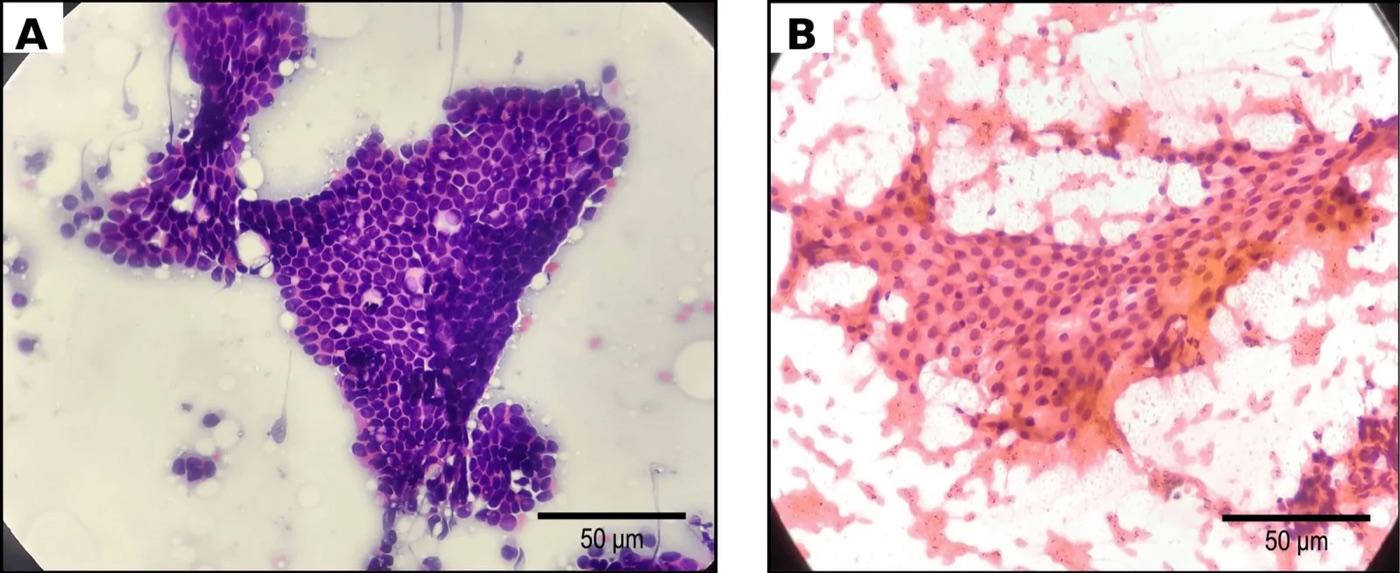
Benign breast FNA, Yokohama category 2. A, cohesive sheets of monomorphic ductal epithelial cells with overlying myoepithelial nuclei (H&E, 400×). B, numerous bare bipolar myoepithelial nuclei in the background, part of the benign biphasic pattern (Papanicolaou stain, 400×).Image: Rathore Aditi et al., PMC open access (Cureus. 2026 Jul 28;18(7):e113490. doi: 10.7759/cureus.113490) Figure 5, CC BY.
Breast Fine-Needle Aspiration: Technique, Adequacy and Normal and Physiologic Findings
Definition. Breast fine-needle aspiration (FNA) biopsy samples a palpable or image-detected breast lesion with a fine needle and produces direct smears, liquid-based preparations or cell blocks for cytologic diagnosis. It is interpreted with the clinical and imaging findings as part of the triple test (Field et al. 2019). Accurate breast FNA depends on aspiration and smear-making technique, on an adequate sample and on recognition of normal and physiologic cell populations. Physiologic changes of pregnancy and lactation, and gynecomastia in males, are the main non-neoplastic states that produce atypical-appearing epithelium (Mendoza et al. 2011; Siddiqui et al. 2002).
Role of breast FNA in relation to core needle biopsy
Current use. Core needle biopsy (CNB) has become the main diagnostic method for breast lesions in high-income countries. FNA remains the primary diagnostic tool in many low- and middle-income countries (Kim, Lee and Kim 2025).
Settings in which FNA has advantages. FNA is useful for small lesions, lesions just beneath the skin or above the chest wall, patients with breast implants and patients receiving anticoagulants. It is also used for axillary lymph nodes and distant metastases (Kim, Lee and Kim 2025).
Limits of FNA. FNA cannot reliably separate in situ from invasive carcinoma. Its accuracy is lower for papillary lesions, atypical hyperplasia and lobular carcinoma, and for nonpalpable lesions and lesions under 10 mm (Kim, Lee and Kim 2025).
Combined FNA and CNB. In one study sensitivity for carcinoma was 80% with FNA alone, 88% with CNB alone and 100% with both (Kim, Lee and Kim 2025).
Direct smears. The IAC Yokohama System bases reporting on direct smears. It emphasizes a high standard of FNAB technique and of smear preparation (Field et al. 2019; Field et al. 2020).
Operator experience. Inadequate aspirates are fewer and accuracy is higher when the same person aspirates and reports the specimen. The inadequacy rate of aspirates performed by nonpathologists was twice that of aspirates performed by pathologists (Mendoza et al. 2011).
Rapid on-site evaluation (ROSE). ROSE reduced the insufficient rate from 17.1% to 4.0% in a series of 2,696 breast FNABs (Wong et al. 2019). It also selects patients for immediate CNB and collects material for ancillary tests (Field et al. 2020). ROSE can reduce the insufficient rate to below 1% (Kim, Lee and Kim 2025).
Observations recorded during ROSE. The consistency of the lesion (soft, firm or hard), change in lesion size after aspiration, the color and consistency of any fluid, and smear cellularity are assessed (Kim, Lee and Kim 2025).
Liquid-based cytology (LBC). About 40% of laboratories in a College of American Pathologists survey used LBC for breast FNA. LBC and conventional smears detect carcinoma with comparable accuracy. LBC lowered the inadequate rate in one Yokohama reproducibility study (Kim, Lee and Kim 2025).
Imaging guidance. In a meta-analysis of 22 studies, FNAB without imaging guidance was associated with a lower inadequacy rate (Paul et al. 2023).
Aspiration and non-aspiration sampling in breast FNA
Needle and syringe. Published breast aspirates were obtained with a 22- or 23-gauge needle and a 10-mL syringe (Gupta et al. 2004; Agnihotri et al. 2020). Negative pressure is maintained during aspiration (Gupta et al. 2004).
Number of passes. In the M. D. Anderson series of palpable masses, adequate and inadequate false-negative aspirates had a mean of 3.7 and 3.0 passes, respectively. The difference was not significant (Boerner and Sneige 1998).
Non-aspiration (capillary) sampling. The needle is passed without suction, and material enters by capillary action (Dey and Ray 1993).
Comparison in 127 breast lumps. Non-aspiration sampling of 127 breast lumps caused less discomfort and less blood staining than aspiration. Its overall cellular yield was unsatisfactory, but the yield was comparable for malignant lesions (Kumarasinghe and Sheriffdeen 1995).
Comparison in 145 breast masses. Non-aspiration sampling gave more diagnostically superior samples. Aspiration gave more diagnostically adequate smears and fewer unsuitable smears (Baksh et al. 2004).
Lesions unsuited to capillary sampling. Non-suction sampling is not advocated for cystic, bony or fibrous lesions (Dey and Ray 1993).
Choice of technique. The choice depends on the site, the clinical situation and the experience and preference of the operator (Kumarasinghe and Sheriffdeen 1995).
Smear handling. Smears are air-dried for Giemsa-type stains and wet-fixed in alcohol for Papanicolaou stain (Agnihotri et al. 2020). Needle and syringe rinses can be processed as filter preparations and cell blocks (Gupta et al. 2004).
Contraindications and histologic complications of breast FNA
Contraindications. Breast FNA has no absolute contraindications (Kim, Lee and Kim 2025).
Changes in the lesion after FNA. Among 184 breast excisions after FNA, 17 (9.2%) showed changes attributable to the procedure. These were near-total destruction of the lesion by hemorrhage (1 case) or infarction (2 cases), focal hemosiderin deposition (7 cases) and peripheral hemorrhage with organization (6 cases) (Lee et al. 1994).
Epithelial displacement. One papilloma showed benign epithelial implantation in granulation tissue after FNA, simulating invasive carcinoma. Myoepithelial cells confirmed its benign nature (Lee et al. 1994).
Consequence for histology. Extensive necrosis after FNA is rare and can prevent a definitive diagnosis on the excision specimen (Lee et al. 1994).
Specimen adequacy criteria in breast FNA
Approach
Criterion
Source
Qualitative (NCI consensus)
A specimen is adequate when it resolves the problem presented by the lesion in that patient
NIH Consensus Development Conference 1997, as summarized by Mendoza et al. 2011
Quantitative (M. D. Anderson)
Six or more epithelial cell clusters, each of 5–10 well-preserved cells, on all slides
Boerner and Sneige 1998
IAC Yokohama System
Insufficient/inadequate when paucity of cells, poor smearing or suboptimal fixation prevents a cytomorphologic diagnosis
Kim, Lee and Kim 2025, summarizing Field et al. 2019
Effect of an epithelial cluster count. Among 41 false-negative aspirates of palpable masses, 48.8% had fewer than six epithelial cell clusters. Applying the count could reduce false-negative diagnoses by about half (Boerner and Sneige 1998).
A cluster count does not prevent all false negatives. The adequate false-negative aspirates contained 8 to 100 clusters (Boerner and Sneige 1998).
Invasive lobular carcinoma was more common among false-negative smears with fewer than six clusters (Boerner and Sneige 1998).
Cost of a strict count. A fixed count would reclassify 35–40% of true-negative aspirates as inadequate. Cysts, fibrotic scars and other hypocellular lesions yield few epithelial cells (Mendoza et al. 2011).
Lesions with few epithelial cells. Cysts, abscesses, fat necrosis and intramammary lymph nodes are reported in the benign category when their findings are diagnostic (Pai 2024).
Inadequacy rates. Reported inadequacy rates range from 0.7% to 25.3% (Mendoza et al. 2011). Rates of 34–58% have been reported for nonpalpable lesions (Mendoza et al. 2011). In a series of 1,532 male breast FNABs, 40% were inadequate (Oosthuizen et al. 2021).
Causes of inadequacy. The nature of the lesion accounted for 68% of inadequate aspirates and operator experience for 32% in one analysis (Mendoza et al. 2011).
Causes of false-negative breast FNA
Frequency. False-negative rates range from 1.2% to 10.6% (Mendoza et al. 2011).
Sampling causes. Poor technique, mislocalization of the lesion, small size and nonpalpable lesions produce true sampling errors (Mendoza et al. 2011).
Interpretive causes. Low-grade carcinomas with minimal atypia and lobular carcinoma, whose small cells can resemble lymphocytes, account for many interpretive errors (Mendoza et al. 2011).
Triple test. Benign triple-test results do not entirely exclude carcinoma. Such cases require periodic follow-up (Boerner and Sneige 1998).
False-positive diagnoses. False-positive rates range from 0% to 2%. Ductal or lobular hyperplasia, fibrocystic change and pregnancy-related masses are the usual sources (Mendoza et al. 2011).
Normal breast elements in FNA smears
Element
Cytologic appearance
Significance
Ductal epithelial cells
Cohesive flat sheets and duct-like configurations with evenly spaced nuclei
Main epithelial component of benign aspirates
Myoepithelial cells
Small dark nuclei within and on epithelial sheets
Part of the benign smear pattern
Bipolar bare nuclei
Oval naked nuclei scattered in the background
Characteristic of benign aspirates; numerous in fibroadenoma
Fibrocystic change; degenerated apocrine cells can appear atypical
Foam cells (macrophages)
Vacuolated cytoplasm, small eccentric nuclei
Cyst contents and papillomas
Adipose tissue
Fragments of mature adipocytes
Normal breast; from a solid mass it indicates a nonrepresentative sample
Sources: Mendoza et al. 2011; Das et al. 1995.
Benign pattern. Benign smears are usually cellular and show duct configurations, myoepithelial cells and bipolar nuclei. An inflammatory background is common (Mendoza et al. 2011).
Atypical features in an otherwise benign smear. Cellular crowding, pleomorphism and discohesion in a benign-pattern smear support a report in the atypical category (Mendoza et al. 2011).
Lactational change and lactating adenoma on breast FNA
Clinical setting. A discrete nodule that appears during pregnancy or lactation can be evaluated by FNA, which spares the patient an excision (Mendoza et al. 2011).
Cytologic features. Lactational cells are large, with enlarged nuclei, eosinophilic nucleoli and vacuolated, wispy cytoplasm in a proteinaceous background. The nuclear features can appear worrisome (Mendoza et al. 2011).
Features on liquid-based preparations. Lactating adenoma shows lacy fragments, tissue paper-like texture, globular clumps of milky background material and disrupted architecture. Singly dispersed bare nuclei contain cherry-red macronucleoli. Cellular morphology is better preserved than on direct smears (Heymann et al. 2015).
Frequency. Lactating adenoma was the diagnosis in 78.5% of 28 breast masses aspirated in pregnant and lactating patients. The patients were between 23 weeks of pregnancy and 10 months postpartum, and the masses measured 1.0–4.5 cm (Heymann et al. 2015).
Carcinoma during pregnancy and lactation. Malignancy was present in 3 of 28 cases (11%), including a false-negative angiosarcoma (Heymann et al. 2015).
Errors in both directions. Hormonally induced atypia in benign lesions can be overdiagnosed as carcinoma. Carcinoma in a lactating breast can be underdiagnosed when its atypia is attributed to hormonal change (Mehdi et al. 2019).
Secretory carcinoma. Secretory carcinoma can show cytologic features almost identical to lactational change. Clinical information is decisive in this distinction (Vesoulis and Kashkari 1998).
Requirement for history. Recognition of lactational change depends on the clinical history of pregnancy or lactation (Mendoza et al. 2011).
Galactocele on breast FNA
Definition and setting. A galactocele is a benign milk-containing cystic lesion that occurs during pregnancy and lactation (Varshney et al. 2021). It results from an obstructed duct with distension of proximal lobular segments (Joshi et al. 2013).
Imaging. A mass with a fat-fluid level on the lateral mammographic projection is a diagnostic sign of galactocele in the appropriate clinical setting (Joshi et al. 2013).
Cytologic findings. Aspiration yields milky fluid. Smears are paucicellular, with foamy macrophages and occasional apocrine cells in a proteinaceous background (Umasankar et al. 2018).
Therapeutic aspiration. FNA serves both to diagnose and to treat a galactocele (Varshney et al. 2021; Jaseem Hassan et al. 2018).
Crystallizing galactocele. Long-standing galactoceles can form crystals (Varshney et al. 2021). Aspiration yields thick, milky, chalky material. Smears show numerous semitransparent crystals of varying size with angulated borders in granular, amorphous debris. The crystals show positive birefringence (Jaseem Hassan et al. 2018).
Gynecomastia on breast FNA
Frequency. Gynecomastia is the most common lesion in male breast aspirates. It accounted for 353 of 614 male breast FNAs in a three-institution series. Male aspirates were 4.3% of all breast FNAs in that series (Siddiqui et al. 2002).
Cytologic features. In 132 cases, gynecomastia showed cohesive sheets of bland cells (100%), bipolar bare nuclei (76.5%) and columnar cells (38.6%). Mild nuclear atypia was present in seven cases (Das et al. 1995).
Features of male breast carcinoma. Carcinoma showed dyshesive groups of ductal cells with moderate to severe nuclear atypia and absence of bare nuclei and columnar cells (Das et al. 1995).
Florid gynecomastia. Florid duct atypia in gynecomastia can mimic adenocarcinoma. A higher threshold for a malignant interpretation applies in males (Siddiqui et al. 2002).
Drug-induced atypia. Gynecomastia induced by chemotherapeutic drugs produced epithelial atypia that was misinterpreted as carcinoma. The changes were reparative or regenerative, and the clinical information had been inadequate (Pinedo et al. 1991).
Nipple discharge. Nipple discharge in florid gynecomastia can be cellular, with papillary fragments, foam cells and mild to moderate atypia (Kapila and Verma 2003). Nipple discharge cytology is covered under papillary lesions (Papillary Lesions of the Breast and Nipple Discharge Cytology).
Performance of FNA in males. FNA of palpable male breast lesions was 95.8% sensitive and 100% specific in a series of 74 aspirates. Cytopathologists performed 73% of the aspirations, 93.2% were adequate and 28.4% were malignant (Hoda et al. 2019).
Inadequacy rate in males. The three-institution series had an inadequacy rate above 15% (Siddiqui et al. 2002).
Yokohama categories in males. In 1,532 male FNABs, 40% were inadequate, 57% benign, 0.6% atypical, 0.7% suspicious and 1.6% malignant. ROMs were 11%, 3%, 28%, 56% and 100% (Oosthuizen et al. 2021).
Differential diagnosis of physiologic and male breast aspirates
Finding
Physiologic or benign interpretation
Malignant mimic or alternative
Distinguishing points
Large cells with prominent nucleoli in a proteinaceous background
Lactational change, lactating adenoma
Invasive carcinoma; secretory carcinoma
History of pregnancy or lactation; vacuolated wispy cytoplasm (Mendoza et al. 2011; Vesoulis and Kashkari 1998)
Cohesive sheets with bipolar bare nuclei in a male
Gynecomastia
Male breast carcinoma
Carcinoma has dyshesive groups, moderate to severe atypia and no bare nuclei (Das et al. 1995)
Atypical ductal cells in a male receiving chemotherapy
Gynecomastia with reparative atypia
Carcinoma
Drug history; reparative features (Pinedo et al. 1991)
Granular cells with round nuclei and distinct nucleoli
Apocrine metaplasia
Apocrine carcinoma
Benign background; degenerated apocrine cells show anisonucleosis (Mendoza et al. 2011)
Fat and blood only from a solid mass
Normal breast tissue
Missed lesion
Insufficient/inadequate category (Kim, Lee and Kim 2025)
Benign Non-Neoplastic Breast Lesions on Fine-Needle Aspiration
Definition. Benign non-neoplastic breast lesions are cystic, inflammatory, reactive and foreign-body processes that form a palpable or radiologic mass without a neoplasm. On fine-needle aspiration (FNA) they include fibrocystic change, acute mastitis and breast abscess, subareolar abscess, fat necrosis, granulomatous mastitis and silicone granuloma. Several of them mimic carcinoma clinically and on imaging (Agnihotri et al. 2020; Khan et al. 2024).
Cyst aspirates. Cyst fluid usually contains macrophages mixed with other inflammatory cells. Ductal and myoepithelial cells form small balls and clusters among the macrophages (Mendoza et al. 2011).
Apocrine cells. Apocrine cells exfoliated from the cyst lining show eosinophilic cytoplasm and round nuclei with distinct nucleoli (Mendoza et al. 2011).
Nonproliferative pattern. Apocrine cells, macrophages and ductal cells in a scant aspirate are the features of nonproliferative fibrocystic change (Mendoza et al. 2011).
Proliferative pattern. A significant proliferative component yields prominent sheets and tight clusters. Atypia in these clusters is assessed by nuclear spacing, multiple nucleoli and chromatin. Intraductal papilloma and fibroadenoma are in the differential diagnosis (Mendoza et al. 2011).
Papillary apocrine change can produce papillary clusters with apocrine cytoplasmic and nuclear features (Mendoza et al. 2011).
Degenerated apocrine cells. Degenerated apocrine cells develop chromatin clumping and anisonucleosis. They can be misinterpreted as suspicious for malignancy (Mendoza et al. 2011).
Infected or ruptured cysts. The fluid becomes turbid or milky, with degenerated cells, debris and abundant inflammatory cells. The rare squamous cell carcinoma can present with similar features (Mendoza et al. 2011).
Indications for cytologic examination of breast cyst fluid
Fluid or clinical finding
Cytology
Source
Clear fluid; mass collapses completely
Not routinely required
Ciatto et al. 1987; Mendoza et al. 2011
Blood-stained fluid
Indicated
Ciatto et al. 1987
Cloudy or turbid fluid
Indicated
Mendoza et al. 2011
Mass remains palpable after aspiration
Indicated
Mendoza et al. 2011
Evidence. Among 6,782 cyst aspirates from 4,105 women, routine cytology detected five clinically and radiologically inapparent intracystic papillomas (0.1%). All five produced blood-stained fluid. Blood-stained fluid accounted for 2% of the series. Cytology identified two of the five papillomas, missed two and gave a false-positive result in one (Ciatto et al. 1987).
Acute mastitis and breast abscess on FNA
Puerperal mastitis. In 59 cases of puerperal mastitis, 44.1% developed an abscess. Symptoms began within the first 8 weeks postpartum in 37 of 59 cases (Lukassek et al. 2019).
Organisms.Staphylococcus aureus was the most common pathogen, followed by coagulase-negative staphylococci. Methicillin-resistant S. aureus was found in one case (Lukassek et al. 2019).
Needle aspiration as treatment. Ultrasound-guided needle aspiration was used in 61.5% of abscesses, and it is progressively replacing surgical incision (Lukassek et al. 2019).
Cytologic findings. Abscess aspirates contain abundant neutrophils, degenerated cells and debris (Mendoza et al. 2011).
Subareolar abscess on breast FNA
Pathology. Subareolar abscess arises from rupture of dilated lactiferous ducts with squamous metaplasia. It shows acute and chronic inflammation and a foreign-body reaction (Silverman et al. 1986).
Diagnostic findings. Anucleate squames with numerous neutrophils are diagnostic. Keratinous debris, cholesterol crystals, parakeratosis and strips of squamous epithelium are also seen (Silverman et al. 1986).
Foreign-body reaction. Sheets of histiocytes and multinucleated foreign-body giant cells are present in some cases (Silverman et al. 1986).
Sources of false-positive diagnoses. Groups of atypical ductal cells, squamous atypia and fragments of exuberant granulation tissue can suggest malignancy (Silverman et al. 1986).
Squamous cells in breast FNA. Of 15 breast aspirates with a discernible number of squamous cells, 5 were metaplastic carcinoma. The rest included subareolar abscess, fibroepithelial tumors, duct ectasia, sclerosed papilloma and a benign cyst (Ng and Kong 2003).
Feature
Benign squamous cells (abscess, cyst, metaplasia)
Malignant squamous cells (metaplastic carcinoma)
Nuclear features
Bland; good maturation
Pleomorphic; bizarre shapes
Anucleate squames
Often present
Variable
Mitoses
Absent
Present
Dyskeratosis and cell cannibalism
Rare
Obvious
Background
Abundant foamy macrophages favor a benign lesion
Tumor diathesis in some cases
Source: Ng and Kong 2003.
Fat necrosis on breast FNA
Etiology. Fat necrosis follows accidental trauma, surgery, biopsy or radiation therapy (Agnihotri et al. 2020).
Clinical and imaging presentation. The presentation ranges from an incidental benign finding to a hard mass suggestive of carcinoma. Mammographic appearances range from a lipid cyst to an ill-defined spiculated mass (Agnihotri et al. 2020).
Cytologic findings. Smears are usually hypocellular. They show histiocytes with vacuolated cytoplasm, multinucleated histiocytic giant cells with lipid vacuoles and inflammatory cells in a dirty necrotic background with fat vacuoles (Agnihotri et al. 2020).
Inflammatory cells by stage. Neutrophils predominate in the acute stage and lymphocytes and plasma cells in the chronic stage. Capillary proliferation characterizes the organizing stage (Agnihotri et al. 2020).
Epithelial cells. Ductal cells are usually absent. A few reactive epithelial cells in a necrotic background can be misinterpreted as suspicious for malignancy (Agnihotri et al. 2020).
Separation from carcinoma. Inflammatory cells outnumber ductal cells, and foamy histiocytes and giant cells form the background (Agnihotri et al. 2020).
Frequency in males. Fat necrosis was the second most common benign diagnosis among 614 male breast aspirates (21 cases) (Siddiqui et al. 2002).
Interobserver variation. In a reproducibility study, one of three observers placed a case of fat necrosis in the atypical category because of degenerated epithelial cells (Boler et al. 2022).
Radiation-induced atypia on breast FNA
Setting. Among more than 800 patients treated by breast-conserving therapy, 41 developed a palpable lesion that was aspirated. Six had recurrent carcinoma (Peterse et al. 1989).
Patterns in nonneoplastic lesions. The other aspirates showed epithelial atypia (14 cases), fat necrosis (10 cases) or poorly cellular smears without atypia or fat necrosis (13 cases) (Peterse et al. 1989).
Cytology of radiation atypia. Atypical cells can show marked anisocytosis and anisonucleosis. The nuclear-to-cytoplasmic ratio remains normal, and bipolar cells are admixed (Peterse et al. 1989).
Features absent in radiation atypia. Cell dissociation and necrotic cell debris, frequent in carcinoma smears, were not seen in aspirates of irradiated nonneoplastic breast (Peterse et al. 1989).
Seromas at irradiated lumpectomy sites. In 38 seroma aspirates, 21 showed mildly atypical degenerated cells with mild hyperchromasia and abundant cytoplasm, with macrophages, inflammatory cells and apocrine cells. Four showed highly atypical cells with hyperchromatic nuclei and nucleoli, and core biopsy found no residual tumor in all four. Two contained recurrent carcinoma (Gupta et al. 2004).
Source of false-positive diagnoses. Radiation-induced atypia can lead to a misdiagnosis of recurrent carcinoma (Peterse et al. 1989). Patients with clinical signs of recurrence or with severe atypia undergo biopsy (Gupta et al. 2004).
Granulomatous mastitis on breast FNA
Idiopathic granulomatous mastitis
Patients. Idiopathic granulomatous mastitis affects parous women of reproductive age. It mimics carcinoma clinically and on imaging (D'Alfonso et al. 2015; Johnstone et al. 2017).
Cytologic findings. Aspirates range from epithelioid cell granulomas to loose collections of histiocytes, with multinucleated giant cells, neutrophils, plasma cells and lymphocytes. Emperipolesis of neutrophils was prominent in 4 of 13 cases in one series (Gautam et al. 2026).
Diagnosis by exclusion. The cytologic diagnosis requires exclusion of tuberculosis, fungal infection and Rosai-Dorfman disease (Gautam et al. 2026).
Neutrophil-rich granulomas without necrosis. Epithelioid granulomas with predominant neutrophils, without necrosis and without acid-fast bacilli, suggest idiopathic granulomatous mastitis (Chandanwale et al. 2020).
Cystic neutrophilic granulomatous mastitis
Histologic pattern. Cystic neutrophilic granulomatous mastitis (CNGM) shows lobulocentric granulomas with mixed inflammation and clear vacuoles lined by neutrophils within the granulomas (D'Alfonso et al. 2015).
Organisms. Gram-positive bacilli are found within the clear vacuoles in some cases (D'Alfonso et al. 2015; Johnstone et al. 2017). Corynebacterium species, including Corynebacterium kroppenstedtii, were detected by culture or 16S rRNA gene sequencing of surgical and FNA specimens in 15 cases (Johnstone et al. 2017).
Limits of microbiology. No microbial culture grew bacteria in one series of 12 cases, although gram-positive bacilli were seen on histology in 5 (D'Alfonso et al. 2015). Gram stain and culture have limited sensitivity for Corynebacterium (Johnstone et al. 2017).
Patients. Patients were 25 to 49 years old (median 34 years). Imaging was highly suspicious for malignancy (BI-RADS 5) in 2 of 9 cases (D'Alfonso et al. 2015).
Clinical course. The disease worsened after core biopsy in 4 of 12 patients, with draining sinuses in 2 (D'Alfonso et al. 2015).
Tuberculous mastitis
Cytologic findings. Epithelioid granulomas with caseous necrosis, with or without acid-fast bacilli, are diagnostic of tuberculosis on smears (Chandanwale et al. 2020).
Frequency. In a series of 33 cases of granulomatous mastitis from India, 27 were tuberculous mastitis. Ziehl-Neelsen staining showed acid-fast bacilli in 13 smears, and mycobacterial culture was positive in 8 of 17 cases (Chandanwale et al. 2020). In another series, caseous necrosis was present in 7 of 22 cases and acid-fast bacilli in 7 (Khan et al. 2024).
Ancillary studies. Ziehl-Neelsen staining and mycobacterial culture or molecular testing are performed on granulomatous aspirates (Chandanwale et al. 2020; Gautam et al. 2026).
Entity
Granulomas
Necrosis
Organisms
Other clues
Idiopathic granulomatous mastitis
Epithelioid granulomas or loose histiocytes
Usually absent
None identified
Neutrophils, giant cells, plasma cells (Gautam et al. 2026; Chandanwale et al. 2020)
Cystic neutrophilic granulomatous mastitis
Lobulocentric granulomas
Absent
Gram-positive bacilli in clear vacuoles; Corynebacterium
Neutrophil-lined clear vacuoles (D'Alfonso et al. 2015)
Tuberculous mastitis
Epithelioid granulomas
Caseous necrosis
Acid-fast bacilli in some cases
Endemic setting (Chandanwale et al. 2020)
Fat necrosis
Lipid-laden giant cells; poorly formed granulomas
Fat necrosis
None
Foamy histiocytes, fat vacuoles (Agnihotri et al. 2020)
Silicone granuloma
Foreign-body giant cells
Absent
None
Vacuoles of various sizes (Dodd et al. 1993)
Silicone and foreign-body reactions on breast FNA
Clinical setting. Silicone granulomas follow silicone gel-filled implants or liquid silicone injection (Dodd et al. 1993).
Cytologic findings. Smears are modestly cellular, with loosely cohesive histiocytes containing vacuoles of various sizes. Multinucleated foreign-body giant cells, fragments of dense amorphous fibrous tissue and stripped fibroblast nuclei are also present (Dodd et al. 1993).
Specific diagnosis. The findings support a specific diagnosis when there is a history of augmentation (Dodd et al. 1993).
Distinction from fat necrosis. Histiocytes in silicone granuloma contain larger vacuoles than those of fat necrosis and often have a signet-ring appearance (Agnihotri et al. 2020).
Distinction from lipid-rich carcinoma. Lipid-rich carcinoma has vacuolated cytoplasm with marked nuclear atypia (Agnihotri et al. 2020).
Necrosis, acid-fast bacilli, gram-positive bacilli in vacuoles (Chandanwale et al. 2020; D'Alfonso et al. 2015)
Yokohama category of benign non-neoplastic lesions
Benign category. Fat necrosis, abscess and granulomatous mastitis are reported as benign. A specific benign diagnosis is given when possible (Pai 2024; Kim, Lee and Kim 2025).
Excluded from the insufficient category. Cysts, abscesses, fat necrosis and intramammary lymph nodes are excluded from the insufficient category when their findings are diagnostic (Pai 2024).
Triple-test correlation. A benign report with suspicious clinical or imaging findings leads to repeat FNA or core needle biopsy (Kim, Lee and Kim 2025). This applies to fat necrosis and granulomatous mastitis, which mimic carcinoma on imaging (Agnihotri et al. 2020; Khan et al. 2024).
Fibroadenoma and Phyllodes Tumor on Fine-Needle Aspiration
Definition. Fibroepithelial lesions of the breast are biphasic tumors composed of epithelial and stromal elements. The group comprises fibroadenoma, the most common benign breast tumor in women, and phyllodes tumor, which is rare and can recur, progress in grade and metastasize (Tan 2021). Phyllodes tumors are graded as benign, borderline or malignant on stromal features in the resection specimen (Quinn et al. 2026). On fine-needle aspiration (FNA), fibroadenoma has a characteristic pattern. The cytologic features of phyllodes tumor overlap with those of fibroadenoma, and FNA does not reliably separate the two (Wang et al. 2018).
Classification of fibroepithelial lesions in the WHO Classification of Breast Tumours
Feature (resection)
Fibroadenoma
Benign phyllodes tumor
Borderline phyllodes tumor
Malignant phyllodes tumor
Tumor border
Well defined
Well defined
Well defined, may be focally permeative
Permeative
Stromal cellularity
Variable, scant to uncommonly cellular, usually uniform
Cellular, usually mild, may be non-uniform or diffuse
Cellular, usually moderate, may be non-uniform or diffuse
Cellular, usually marked and diffuse
Stromal atypia
None
Mild or none
Mild or moderate
Marked
Mitotic activity
Usually none, rarely low
Usually <2.5 mitoses/mm² (<5 per 10 HPF)
2.5 to <5 mitoses/mm² (5–9 per 10 HPF)
≥5 mitoses/mm² (≥10 per 10 HPF)
Stromal overgrowth
Absent
Absent
Absent or very focal
Often present
Malignant heterologous elements
Absent
Absent
Absent
May be present
Proportion of all phyllodes tumors
Not applicable
60–75%
15–26%
8–20%
Source: Quinn et al. 2026 (WHO Classification of Breast Tumours, 6th edition). HPF, high-power field of 0.5 mm diameter and 0.2 mm² area.
Change in the 6th edition. The 4th and 5th editions required all five adverse features for malignant phyllodes tumor: marked stromal atypia, stromal overgrowth, ≥5 mitoses/mm², marked diffuse stromal cellularity and an infiltrative border. The 6th edition accepts four of the five (Quinn et al. 2026).
Reason for the change. Some tumors classified as borderline by the full criteria metastasized (Quinn et al. 2026).
Heterologous elements. Malignant heterologous elements, except well-differentiated liposarcoma, allow a malignant grade without other adverse features (Quinn et al. 2026).
Sampling of resections. Large or worrisome tumors are sampled with at least 2 blocks per 1 cm of the largest dimension (Quinn et al. 2026).
Fibroadenomas in young patients may have increased stromal cellularity and mitoses (Quinn et al. 2026).
Tumors with features of two grades. A tumor with mostly benign features and slight deviation toward borderline criteria, such as focal border irregularity, can be reported as favoring benign phyllodes tumor with a comment. A tumor with mostly borderline features and fewer than four adverse criteria is reported as borderline with an explanatory comment (Quinn et al. 2026).
Tubular adenoma is histologically and molecularly separate from fibroepithelial lesions and is a distinct entity on FNA (Li et al. 2023).
Fibroadenoma on breast FNA
Variants. Recognized histologic variants of fibroadenoma are the cellular, complex, juvenile and myxoid forms (Tan 2021). Fibroadenomas in young patients may have increased stromal cellularity and mitoses (Quinn et al. 2026).
Clinical setting. Fibroadenoma is the most common solid breast lesion in women under 40 years. It is a well-defined, low-density mass on imaging. In older patients the mass may show popcorn calcification. Multiple fibroadenomas occur in 15–20% of patients (Mendoza et al. 2011).
Cellularity. Aspirates are hypercellular (Mendoza et al. 2011).
Epithelial sheets. Monolayer sheets of benign epithelial cells mixed with myoepithelial cells have a branching "staghorn" or antler-horn configuration at their edges. The pattern reflects the configuration of ducts in the tumor (Mendoza et al. 2011).
Stroma. Fibrillar stromal fragments vary in cellularity and can show myxoid change (Mendoza et al. 2011).
Background. Numerous bipolar bare nuclei form the background. A large number of bipolar bare nuclei is a reliable feature in favor of fibroadenoma (Mendoza et al. 2011).
Growth pattern. Branching sheets are more common in aspirates from intracanalicular fibroadenomas. Aspirates from pericanalicular fibroadenomas can show less pronounced staghorn configurations (Mendoza et al. 2011).
Diagnostic triad. Fibromyxoid stroma, staghorn clusters and numerous single bare nuclei were present in 92.7%, 73.6% and 73.6% of fibroadenomas in one series (Mendoza et al. 2011).
Giant cells. Stromal multinucleated giant cells were present in 31.8% of fibroadenoma aspirates in one series. They are more common in phyllodes tumor and have little prognostic significance (Mendoza et al. 2011).
Accuracy. FNA sensitivity for fibroadenoma is 68–97%. Most diagnostic errors occur in older patients (Mendoza et al. 2011).
Causes of missed fibroadenoma. Absence of components of the triad and low cellularity are the common causes (Mendoza et al. 2011).
Phyllodes tumor on breast FNA
Frequency. Phyllodes tumors accounted for 0.2% of breast cancers in Australia and 6.92% in one Singaporean institutional study (Pang et al. 2026).
Germline variants. Pathogenic or likely pathogenic germline variants were present in about 14% of 234 women with phyllodes tumors (Pang et al. 2026).
Clinical clues. In one comparison, median age was 23.0 years for fibroadenoma and 39.0 years for phyllodes tumor. Median size was 2.0 cm and 5.0 cm, respectively (Tummidi et al. 2020). In a second series, mean age was 26.05 years for cellular fibroadenoma and 36.94 years for phyllodes tumor, and mean size was 3.7 cm and 6.4 cm, respectively (Rana et al. 2022).
Stromal fragments. Phyllodes tumor yields more and larger stromal fragments, with higher cellularity and more atypia than fibroadenoma (Maritz and Michelow 2017; Wang et al. 2018).
Three features with high frequency in phyllodes tumor. Fibromyxoid stromal fragments with spindle nuclei were present in 90% of cases, fibroblastic pavements in 93% and appreciable numbers of dispersed spindle fibroblastic cells in 100%. Recognition of these features raised detection of phyllodes tumor from 40% to 100% in that series (El Hag et al. 2010).
Epithelial component. El Hag found no significant difference in the epithelial component between phyllodes tumor and fibroadenoma (El Hag et al. 2010). Tummidi found large, opened-out, folded epithelial sheets to be a significant predictor of phyllodes tumor (Tummidi et al. 2020).
Stromal-to-epithelial ratio. Fibroadenomas had more epithelial fragments, fewer stromal fragments and a lower stromal-to-epithelial ratio (Maritz and Michelow 2017). In another series the inverse measure, the median epithelial-to-stromal ratio, was 3.4 for fibroadenoma and 2.6 for phyllodes tumor (Tummidi et al. 2020).
Stromal contours. Frayed and irregular stromal fragment contours, spindle stromal nuclei, background spindle nuclei and background atypia were significant predictors of phyllodes tumor. A regression model based on these features had an accuracy of 78.9% (Tummidi et al. 2020).
Atypia and grade. The frequency and degree of atypia in the dispersed cells correlated with the histologic grade of phyllodes tumor (El Hag et al. 2010).
Multivariable predictors. Age over 30 years, BI-RADS 4 or higher, marked stromal cellularity, more than 30% spindle cells in the background and traversing blood vessels in stromal fragments increased the odds of phyllodes tumor over cellular fibroadenoma. The model predicted phyllodes tumor correctly in 87.2% of cases (Rana et al. 2022).
Limits. In a four-observer study of 26 phyllodes tumors and 78 fibroadenomas, no single cytologic feature identified phyllodes tumor across all observers. The authors concluded that FNA cannot reliably distinguish the two (Wang et al. 2018).
Cytologic features that separate phyllodes tumor from fibroadenoma
Feature
Favors fibroadenoma
Favors phyllodes tumor
Source
Stromal fragments
Fewer; less cellular; smooth contours
More numerous; hypercellular; frayed and irregular contours
Maritz and Michelow 2017; Tummidi et al. 2020
Stromal nuclei
Bland
Spindle nuclei; atypia
El Hag et al. 2010; Wang et al. 2018
Fibroblastic pavements
Absent
Present in 93%
El Hag et al. 2010
Background single cells
Bipolar bare nuclei
Spindle fibroblastic cells, more than 30% of the background
El Hag et al. 2010; Rana et al. 2022
Vessels in stromal fragments
Uncommon
Traversing blood vessels
Rana et al. 2022
Epithelial sheets
Staghorn monolayer sheets
Large, opened-out, folded sheets
Mendoza et al. 2011; Tummidi et al. 2020
Stromal-to-epithelial ratio
Lower
Higher
Maritz and Michelow 2017
Age and size
Younger; smaller
Older; larger
Tummidi et al. 2020; Rana et al. 2022
Molecular findings in fibroadenoma and phyllodes tumor
MED12.MED12 mutations occur in both fibroadenoma and phyllodes tumor and are early events. Most affect exon 2, especially codon 44 (Pang et al. 2026).
MED12 by grade.MED12 variants were reported in 66% of benign, 57% of borderline and 33% of malignant phyllodes tumors (Pang et al. 2026).
TERT promoter.TERT promoter alterations are exceedingly rare in fibroadenoma. They occur in 34% of benign, 59% of borderline and 56% of malignant phyllodes tumors (Pang et al. 2026).
Pathways. Phyllodes tumors can arise from MED12-mutated fibroadenomas through acquisition of TERT promoter alterations, or de novo without MED12 mutation (Pang et al. 2026).
Distinction from cellular fibroadenoma.TERT promoter mutations help separate phyllodes tumors with fibroadenoma-like morphology from cellular fibroadenomas (Fortun et al. 2026).
Grade progression. Malignant phyllodes tumors show greater genomic complexity, with alterations of TP53, RB1, EGFR, CDKN2A/CDKN2B, NF1, PTEN and PIK3CA. These are rare in fibroadenoma (Pang et al. 2026).
Distinction from metaplastic carcinoma.MED12 mutations are not identified in metaplastic carcinoma or sarcoma, and RARA and TERT promoter mutations are not seen in metaplastic carcinoma (Pang et al. 2026).
Routine use. Routine molecular testing of fibroepithelial lesions has low diagnostic yield. It is reserved for selected diagnostic dilemmas (Pang et al. 2026).
Yokohama categories for fibroepithelial lesions
Fibroadenoma. A typical fibroadenoma is reported in the benign category with a specific diagnosis (Pai 2024).
Cellular stroma. An otherwise typical fibroadenoma with stromal hypercellularity, without necrosis or nuclear atypia, is an atypical-category scenario because phyllodes tumor is in the differential diagnosis (Field, Schmitt and Vielh 2017, as summarized by Pai 2024).
Suspected phyllodes tumor. Suspected phyllodes tumors are reported in the atypical category in published series (Pai 2024; Rohilla et al. 2025).
Follow-up of the atypical category. Histology of atypical aspirates included fibroadenoma with epithelial hyperplasia and benign phyllodes tumor (Rohilla et al. 2025).
Effect of the system. Reclassification of 1,080 aspirates with the Yokohama System improved diagnostic performance for fibroepithelial lesions (Marabi et al. 2021).
ROM calculations. The Nikas meta-analysis counted borderline phyllodes tumors as benign and malignant phyllodes tumors as malignant outcomes (Nikas et al. 2023).
Management. An atypical report leads to repeat FNA or core needle biopsy (Kim, Lee and Kim 2025).
Differential diagnosis of fibroepithelial aspirates
Tubules and tubular fragments; fibroadenoma has large epithelial fragments and naked stromal fragments
Li et al. 2023
Intraductal papilloma
Fronds resembling staghorn clusters
Foam cells with papillary fronds; cell balls; nipple discharge
Mendoza et al. 2011
Proliferative fibrocystic change
Sheets and tight clusters
Apocrine cells and macrophages; no stromal fragments
Mendoza et al. 2011
Metaplastic carcinoma
Spindle cells
Malignant epithelial or squamous component; no MED12 mutation
Ng and Kong 2003; Pang et al. 2026
Lactating adenoma or fibroadenoma with lactational change
Cellular epithelium
Vacuolated cells, prominent nucleoli, proteinaceous background, pregnancy or lactation
Heymann et al. 2015
Sources of error in fibroepithelial aspirates
False-positive diagnoses. Fibroadenomas, complex sclerosing lesions and papillomas are recognized sources of false-positive diagnoses (Kim, Lee and Kim 2025).
Myxoid stroma. A mucinous carcinoma with scant mucin was diagnosed as fibroadenoma with myxoid stroma by three observers in one reproducibility study (Boler et al. 2022).
Spindle cell lesions. Bland spindle cell lesions of the breast include mesenchymal tumors of fibroblastic and myofibroblastic derivation. Clinicoradiologic correlation and ancillary tests guide their diagnosis (Michelow and Field 2019).
Squamous cells. Fibroepithelial tumors can yield squamous cells, which place metaplastic carcinoma in the differential diagnosis (Ng and Kong 2003).
Papillary Lesions of the Breast and Nipple Discharge Cytology
Definition. Papillary lesions of the breast are intraluminal proliferations with branching fibrovascular cores. The group ranges from the benign intraductal papilloma to papillary ductal carcinoma in situ (DCIS), encapsulated papillary carcinoma, solid papillary carcinoma and invasive papillary carcinoma (Kulka et al. 2022). In fine-needle aspiration (FNA) material, benign and malignant papillary lesions share cellular smears, papillary fragments and cell balls. Low-grade papillary carcinomas can lack necrosis and myoepithelial cell loss, the features that usually mark malignancy on breast FNA (Jamidi et al. 2021). Nipple discharge cytology is the examination of fluid expressed from the nipple. Intraductal papilloma is the lesion most often associated with pathologic nipple discharge (Kalu et al. 2012).
WHO classification of papillary lesions of the breast
The WHO Classification of Breast Tumours, 6th edition (2026), is the current classification. The distribution of myoepithelial cells defines each entity in tissue.
Entity
Defining histologic features
Myoepithelial cells
Staging and management
Intraductal papilloma
Arborizing fibrovascular cores covered by basal and luminal cells
Present along the cores and at the periphery
Benign; papilloma fragments on core biopsy are coded B3 in the European system, and excision has been the usual recommendation; surveillance is considered appropriate after complete removal by vacuum-assisted biopsy
Intraductal papilloma with atypical ductal hyperplasia (ADH) or DCIS
Low-grade atypical epithelial proliferation within a papilloma: <3 mm is ADH, ≥3 mm is DCIS
Present in the papilloma; absent within the atypical proliferation
Classified by the extent of atypia
Papillary DCIS
DCIS with fibrovascular cores, low-grade nuclei and diffuse strong ER; segmental disease of small or large ducts
Absent within the papillae (p63 negative)
Staged and managed as DCIS
Encapsulated papillary carcinoma (EPC)
Papillary, cribriform or solid growth within a cyst with a fibrous capsule; pushing borders
Usually absent along the cores and at the periphery
Classic low- to intermediate-grade, ER-positive EPC without frank invasion is staged and managed as DCIS
Solid papillary carcinoma (SPC)
Solid growth with delicate fibrovascular cores; cells with eosinophilic granular cytoplasm; neuroendocrine differentiation and mucin production are very common
May be completely absent
6th edition: SPC in situ (circumscribed nodules with smooth contours, classified as in situ even without myoepithelial cells); SPC in situ with invasion (SPC in situ with a conventional invasive carcinoma, usually hypercellular mucinous carcinoma or IBC-NST, staged as invasive carcinoma); invasive SPC (nodules with ragged, jagged contours in a geographic jigsaw pattern, without myoepithelial rims, in desmoplastic stroma). 5th edition: SPC in situ, and SPC with frank invasion classified as invasive
Invasive papillary carcinoma
Invasive carcinoma with papillary architecture
Absent
Staged as invasive carcinoma
Sources: Quinn et al. 2026; Kulka et al. 2022.
Changes between the 5th and 6th WHO editions for papillary lesions
Topic
5th edition (2019)
6th edition (2026)
Solid papillary carcinoma with invasion
Tumors with features of SPC and frank invasion classified as invasive
Separated into SPC in situ with invasion (SPC plus a conventional invasive carcinoma) and invasive SPC
EPC with atypical features
EPC with high nuclear grade, increased mitotic activity, or a triple-negative or HER2-positive phenotype graded, staged and managed as invasive disease
Very rare EPC with high-grade features or ER negativity may still be categorized as EPC; excision, expert consultation and multidisciplinary review are used in difficult cases
Absent myoepithelial cells and basement membrane
Stated for EPC and SPC: absence of myoepithelial cells does not preclude in situ disease
Extended to DCIS: lack of both myoepithelial markers and basement membrane does not establish invasive carcinoma without a recognizable invasive pattern
Mimics
Not listed as an ILC pattern
Solid papillary ILC, a solid pattern of invasive lobular carcinoma with a pseudocapsule, is recognized to avoid confusion with papillary lesions
Source: Quinn et al. 2026; 5th-edition rules from Kulka et al. 2022.
Encapsulated papillary carcinoma is usually positive for estrogen receptor (ER) and progesterone receptor (PR) and lacks HER2 amplification. Solid papillary carcinoma shows diffuse strong ER expression (Kulka et al. 2022).
Rare tumors with papillary architecture include tall cell carcinoma with reversed polarity, mucinous cystadenocarcinoma, adenomyoepithelioma and secretory carcinoma (Kulka et al. 2022).
Solid papillary ILC is a well-delimited solid papillary pattern of invasive lobular carcinoma with a pseudocapsule and peripheral satellite foci. It is in the differential diagnosis of papillary lesions in small samples (Quinn et al. 2026).
Intraductal papillomas arising in the axillary tail are in the differential diagnosis of papillary hidradenoma of sweat gland origin (Kulka et al. 2022).
Clinical and imaging features of papillary breast lesions
Frequency: papillary lesions make up less than 10% of benign breast neoplasms and about 1–2% of breast carcinomas (Kaushik et al. 2026).
Central papillomas arise in large ducts and are more common than peripheral papillomas. Serous or bloody nipple discharge is often present (Kulka et al. 2022).
Peripheral papillomas are smaller, often multiple and usually asymptomatic. Usual ductal hyperplasia, ADH, atypical lobular hyperplasia and DCIS are more common within peripheral papillomas (Kulka et al. 2022).
Multiple papillomas (more than 5) carry about a threefold relative risk of subsequent breast cancer. The relative risk rises to about sevenfold when the papillomas are associated with ADH or lobular neoplasia (Kulka et al. 2022).
Encapsulated papillary carcinoma presents as a slowly growing palpable mass with bloody discharge in postmenopausal women and rarely in men. On imaging it is commonly a well-circumscribed round or oval retroareolar lesion (Kulka et al. 2022).
Solid papillary carcinoma is usually unifocal and well circumscribed. It is often central and causes nipple discharge (Kulka et al. 2022).
Imaging: in one cytology series of 20 papillary neoplasms, the most common mammographic category was BI-RADS 4 (8 of 16 patients). Two patients with BI-RADS 2 lesions had encapsulated or solid papillary carcinoma (Kaushik et al. 2026).
Cytologic features of papillary lesions on breast FNA
Smears from papillary lesions are usually cellular and contain three-dimensional papillary fragments, cell balls without fibrovascular cores, single cells and cyst contents. These elements occur in both papillomas and papillary carcinomas (Tse et al. 2008). In a series of 23 papillomas and 11 papillary carcinomas, the quantity of these elements did not separate benign from malignant lesions. Atypia in cell balls and single cells and long slender papillae with ramifying edges favored carcinoma (Tse et al. 2008).
A systematic study of 153 papillary lesions with histologic follow-up identified the parameters below (Jamidi et al. 2021).
Solid and cribriform patterns; complex and slender papillae
Nuclei
No significant atypia
Atypical nuclear features; mitoses
Background
Features of cystic change
Hemorrhage; infiltrating neutrophils
Cytoplasm
Scant
Increased cytoplasm; cytoplasmic granules; neuroendocrine features
A four-parameter rule separated benign from malignant lesions. The presence of any one of the following predicted malignancy with a sensitivity of 95.1% and a specificity of 100%: absence of myoepithelial cells within epithelial structures, cytoplasmic granules, an increased amount of cytoplasm, and a nuclear to cytoplasmic (N/C) ratio greater than 0.7 (Jamidi et al. 2021).
Myoepithelial cells were seen in 30% (6 of 20) of papillary neoplasms in a later cytology series. Benign neoplasms showed cohesive clusters and a clean background. Atypical and malignant lesions showed loss of myoepithelial cells, non-cohesive clusters and a necrotic or dirty background (Kaushik et al. 2026).
Cyst macrophages were present in 55% (11 of 20) of papillary neoplasms in the same series (Kaushik et al. 2026).
Glycogen vacuoles were found in 63.6% of papillary carcinomas, a higher proportion than in carcinoma of no special type, apocrine carcinoma and medullary-pattern carcinoma. Loose cohesive clusters and acinar formation separated papillary carcinoma from benign papillary lesions (Haji et al. 2007).
Diagnostic accuracy of FNA for papillary breast lesions
False-positive results outnumber false-negative results. In the series of Gümrükçü et al., 12 cases were false positive and 1 was false negative (Gümrükçü et al. 2022).
Papillary lesions are a recognized source of false-positive breast FNA, together with complex sclerosing lesions and fibroadenomas (Gomes Pinto and Schmitt 2024).
Sampling error produces false negatives. An invasive carcinoma with papillary DCIS was reported as benign in one Yokohama series (Kaushik et al. 2026).
Reporting papillary lesions in the IAC Yokohama System
The IAC Yokohama System has five categories: insufficient/inadequate, benign, atypical, suspicious of malignancy and malignant. The ROM ranges published with the system were 1.4–2.3% for benign, 13–15.7% for atypical, 84.6–97.1% for suspicious of malignancy and 99.0–100% for malignant (Field et al. 2019). A meta-analysis of 7,969 cases gave pooled ROMs of 1% for benign, 20% for atypical, 86% for suspicious and 100% for malignant (Nikas et al. 2023).
Cytologic finding
Usual Yokohama category in published series
Basis
Papillary lesion without atypia, myoepithelial cells present, monolayer sheets and cyst contents
Benign or atypical
Benign features of Jamidi et al. 2021; papillary lesions are placed in the indeterminate zone of reporting systems (Gümrükçü et al. 2022)
Papillary lesion with cellular crowding or mild atypia
Atypical
The atypical category includes papillary lesions (Pai 2024)
Discohesive papillary neoplasm with atypia, absent myoepithelial cells or increased N/C ratio, insufficient for a definite diagnosis
Suspicious of malignancy
Malignant parameters of Jamidi et al. 2021
Papillary neoplasm with unequivocal malignant features
Malignant
Malignant parameters of Jamidi et al. 2021
A malignant FNA does not separate in situ from invasive papillary carcinoma. High-grade DCIS cannot be distinguished from invasive carcinoma on FNA (Gomes Pinto and Schmitt 2024). Histologic examination of the excised lesion establishes invasion.
WHO Reporting System for Breast Cytopathology: a joint WHO-IARC and IAC system is in development. FNA keeps an important role in resource-constrained settings (Quinn et al. 2026).
Management follows the Yokohama algorithm for the assigned category and the triple test of clinical, imaging and cytologic findings (Field et al. 2019; Gümrükçü et al. 2022).
Immunocytochemistry and cell blocks in papillary breast lesions
Myoepithelial markers on FNA smears: p63-positive cells and p63-positive clusters were more frequent in benign lesions (11 of 22) than in malignant lesions (3 of 34) (Hoshikawa et al. 2016).
High-molecular-weight keratin (34βE12): 34βE12-positive clusters were significantly more frequent in intraductal papilloma than in DCIS (P = 0.001). All 8 papillomas had positive scores (Hoshikawa et al. 2016).
CK5 and CK14 in tissue: the atypical epithelial proliferation in a papilloma with ADH or DCIS lacks CK5 and shows strong uniform ER expression. Usual hyperplasia in a papilloma shows CK5 positivity and heterogeneous ER (Kulka et al. 2022).
Cellular smear with extracellular hyaline globules
Bland cells around the globules; overlaps with adenoid cystic carcinoma on FNA
Jain et al. 2003; Kulka et al. 2022
Mucinous carcinoma was mistaken for a papillary lesion in six cases of one series because of papilla-like structures (Gümrükçü et al. 2022).
Tubular carcinoma composed of monotonous columnar cells was misinterpreted as a papillary neoplasm in two cases of the same series (Gümrükçü et al. 2022).
Nipple discharge cytology
Indications and clinical setting of nipple discharge cytology
Physiologic discharge is common and does not require imaging. Pathologic discharge requires breast imaging in women, men and transgender patients (American College of Radiology [ACR] 2022).
Imaging by age (ACR 2022): mammography or digital breast tomosynthesis first at 40 years or older, with ultrasound usually added; ultrasound first in women younger than 30 years; mammography, tomosynthesis or ultrasound by institutional preference in women aged 30–39 years. Men 25 years and older are evaluated with mammography or tomosynthesis.
Men with pathologic discharge have a high incidence of breast cancer (ACR 2022).
Occult malignancy: without an imaging abnormality, the incidence of occult malignancy in pathologic nipple discharge is <3% (Kalu et al. 2012).
Bloody discharge is significantly associated with malignancy (P < 0.001) (Al Nemer and Kussaibi 2021).
Performance of nipple discharge cytology
Study
Patients or samples
Sensitivity
Specificity
Other findings
Gupta et al. 2004
1,948 smears, 1,530 patients
Not calculated
Not calculated
492 smears inadequate despite repeat sampling; 65 of 67 malignant diagnoses and 18 of 22 suspicious diagnoses were carcinoma on biopsy
Kooistra et al. 2009
618 patients, 163 biopsied
16.7%
66.1%
Bloody color as the test had a sensitivity of 60.6%
Kalu et al. 2012
89 patients with excision
74.5%
30%
Positive predictive value 68%, rising to 85% with abnormal imaging
Al Nemer and Kussaibi 2021
228 with tissue diagnosis
34.6% (atypical included)
100%
10.5% of patients had breast cancer
Castellano et al. 2017
139 with surgery
58%
32 of 32 malignant diagnoses confirmed
HER2 immunocytochemistry on smears matched surgical tissue in all tested cases
A malignant nipple discharge cytology result is highly specific. All 32 malignant diagnoses were confirmed histologically in one series. Positive and suspicious results influenced surgery on the nipple-areola complex (Castellano et al. 2017).
A negative result does not exclude carcinoma. Sensitivity ranged from 16.7% to 74.5% across the series above. Negative cytology with negative imaging is insufficient to avoid surgery when the clinical presentation is suspicious (Kalu et al. 2012).
Cytology alone rarely detects a malignancy missed by other tests. In the series of Kooistra et al., cytology alone identified 1 malignant lesion (unique sensitivity 2.8%) (Kooistra et al. 2009).
HER2-positive DCIS and the nipple: malignant nipple discharge cytology was strongly associated with nipple involvement by HER2-positive DCIS (P < 0.001) (Castellano et al. 2017).
Cytologic categories and findings in nipple discharge smears
Categories used in published series include inadequate, benign, inflammatory, papillary lesion not otherwise specified, suspicious and malignant (Gupta et al. 2004). Another series used negative, atypical, suspicious and papillary categories (Kalu et al. 2012).
Papillary lesion not otherwise specified: 229 of 1,948 smears in one series. Biopsy in 53 of these patients showed a papilloma in 41 (Gupta et al. 2004).
Discharge smears with collagenous spherulosis or adenoid cystic carcinoma: in three patients with hemorrhagic discharge, the smears showed foam cells and erythrocytes. One smear contained epithelial clusters with hyaline material (Jain et al. 2003).
Paget disease of the nipple on scrape cytology
Mammary Paget disease accounts for 1–4% of female breast cancers. About 90% of cases are associated with an underlying in situ or invasive carcinoma (Boșoteanu et al. 2023).
Cytologic findings of Paget disease in nipple scrapings
Diagnostic performance: scrape cytology identified all 8 cases of Paget disease among 14 patients with nipple skin change, with no false-negative or false-positive results (Lucarotti et al. 1994). An unequivocal cytodiagnosis was made in 11 cases of unilateral Paget disease, all confirmed histologically (Gupta et al. 1996).
Cells: dense tumor cell cytoplasm and a background of keratinous debris can give a squamous-like appearance. Three-dimensional aggregates, papillary-like groups, acinar-like collections and cells with vacuolated cytoplasm and eccentric nuclei indicate glandular differentiation (Samarasinghe et al. 1993).
Squamous mimicry: scrapings contain reactive squamous cells mixed with malignant glandular cells. One case was initially interpreted as squamous cell carcinoma (Vohra et al. 2012).
A negative scraping does not exclude Paget disease, and a full-thickness biopsy may be needed when clinically indicated (Herbst et al. 2020).
Immunoprofile of Paget cells
Marker
Paget disease
Source
CK7
Positive in >90%; rare cases are CK7 negative
Boșoteanu et al. 2023; Anja et al. 2021
HER2
Positive in 80–100%
Boșoteanu et al. 2023
GATA3
Positive in 95% of complex cases, including CK7-negative cases
Boșoteanu et al. 2023
ER
Positive in 10–40%
Boșoteanu et al. 2023
S100
Positive in 0–26%
Boșoteanu et al. 2023
Ki-67
Strongly expressed
Park and Suh 2009
Cytology smears: c-erbB-2 (HER2) staining was positive in the one scrape case tested. HER2, CEA or mucin helps separate Paget disease from melanoma and squamous cell carcinoma in situ (Samarasinghe et al. 1993).
Differential diagnosis of Paget disease of the nipple
Mimic
Distinguishing features
Source
Melanoma in situ
Atypical melanocytes in groups confined to the epidermis; no lumens or mucin; S100 positive in about 100%, with MelanA and HMB45
Boșoteanu et al. 2023
Toker cells
Small bland nuclei; CK7 positive; HER2 negative; Ki-67 absent or 1–3 positive cells per high-power field; CK7-positive cells in 152 of 173 (88.4%) mastectomy nipples, forming clusters in 18.5%
Park and Suh 2009; Nofech-Mozes and Hanna 2009
Squamous cell carcinoma in situ (Bowen disease)
Squamous differentiation; HER2, CEA and mucin negative
Samarasinghe et al. 1993; Boșoteanu et al. 2023
Pemphigus vulgaris
Loose clusters of cells with basophilic cytoplasm and prominent nucleoli; IgG on cell membranes by immunoperoxidase
Kobayashi et al. 1997
Toker cell receptors: two series differ on ER. Park and Suh found Toker cells consistently ER positive, while Nofech-Mozes and Hanna found clustered Toker cells ER and PR negative. Both found them HER2 negative (Park and Suh 2009; Nofech-Mozes and Hanna 2009).
Invasive Breast Carcinoma of No Special Type and Invasive Lobular Carcinoma on FNA
Definition. Invasive breast carcinoma of no special type (IBC-NST) is the WHO term for invasive carcinomas that cannot be classified morphologically as any special histological type (Cserni 2020). The WHO Classification of Breast Tumours, 6th edition (2026), is current and retains IBC-NST and ILC as breast cancer types (Quinn et al. 2026). Invasive lobular carcinoma (ILC) is defined by the WHO as a breast carcinoma composed of dyscohesive cells that are mostly individually dispersed or arranged in single files (Christgen et al. 2021). ILC is the most common special type and accounts for 10–15% of breast cancers. Inactivation of the CDH1 gene and loss of its protein, E-cadherin, explain the loss of cell cohesion (Christgen et al. 2021). On fine-needle aspiration (FNA), IBC-NST usually yields cellular smears of overtly malignant cells. ILC is a recognized source of false-negative breast FNA because aspirates are often paucicellular and the cells are small and bland (Gomes Pinto and Schmitt 2024; Crasta et al. 2005).
WHO classification of IBC-NST and invasive lobular carcinoma
Subtype, pattern and variant in the 6th WHO edition
Subtype: a tumor that differs from the main type in at least one dimension (clinical, histological or genetic), preferably with a different treatment or outcome (Quinn et al. 2026).
Pattern: a tumor that differs from the main type in morphology only (Quinn et al. 2026).
Variant: reserved for molecular or genetic alterations. Its use for morphologic forms is discouraged (Quinn et al. 2026). The 5th edition listed solid, alveolar, pleomorphic and tubulolobular ILC as variants (Christgen et al. 2021).
Category
6th edition (2026)
5th edition (2019), where different
Source
IBC-NST
Carcinoma that cannot be classified as a special type
Same
Cserni 2020; Quinn et al. 2026
Uncertain IBC-NST versus ILC after IHC
IBC-NST with "lobular features/lobular growth pattern"
Mixed IBC-NST and ILC, or ductulolobular carcinoma (ILC chapter)
Quinn et al. 2026
IBC-NST with neuroendocrine (NE) differentiation
Morphological pattern of IBC-NST when NE markers are moderate to strong, or diffuse and weak but unequivocal; carcinomas with NE morphology and negative or focal weak markers are IBC-NST; large cell NE carcinoma is not a separate entity; primary NET is diagnosed only with typical NET morphology and strong diffuse NE markers after metastasis is excluded; routine NE staining of IBC-NST is not recommended
IBC-NST with NE differentiation was a pattern of IBC-NST, but tumors with NE morphology and NE marker expression were classified as NET (grade 1 or 2) or NEC (small cell or large cell) under the unified NEN model
Quinn et al. 2026; Cserni 2020
ILC architectural patterns
Solid, alveolar and trabecular; solid papillary ILC is a form of the solid pattern
Solid, alveolar, pleomorphic and tubulolobular variants
Quinn et al. 2026; Christgen et al. 2021
ILC cytological features
Signet ring cell, apocrine, histiocytoid and pleomorphic
Histiocytoid differentiation and signet ring cell morphology described
Quinn et al. 2026; Christgen et al. 2021
ILC with extracellular mucin (ILCEM)
New ILC subtype, distinguished from classic ILC and mucinous carcinoma because of more aggressive behavior
Mentioned under mucinous carcinoma only
Quinn et al. 2026; Cserni 2020
E-cadherin-negative tumor with tubules and lobular cells
Tubulolobular carcinoma, regarded as ILC with a very good prognosis
Tubulolobular variant of ILC
Quinn et al. 2026
E-cadherin-negative ILC with E-cadherin-positive tubules
Mixed ILC and tubular carcinoma
Not addressed in the sources reviewed
Quinn et al. 2026
Mixed IBC-NST and special type
Not restated in the 6th-edition summary
Special type in 10–90%, proportion stated; <10% classified as IBC-NST; >90% pure special type
Cserni 2020
Ductulolobular carcinoma
Not restated in the 6th-edition summary
Tumors with both IBC-NST and ILC features, about 5% of breast carcinomas
Cserni 2020
Treatment: there are currently no major treatment differences between IBC-NST and ILC. Treatment is based on biomarkers, predictive assays, stage and grade (Quinn et al. 2026).
Aberrant E-cadherin: some ILCs show aberrant E-cadherin or retained membranous E-cadherin with loss of p120 or β-catenin membrane staining (Quinn et al. 2026).
Clinical features of IBC-NST and invasive lobular carcinoma relevant to FNA
ILC is less conspicuous than IBC-NST clinically and radiologically. Lobular carcinomas have a propensity to remain occult on imaging (Cserni 2020).
Metastatic pattern of ILC: metastasis to the digestive tract, ovaries, bones, leptomeninges, orbital soft tissue and skin occurs more often in ILC (Christgen et al. 2021). These sites are sampled by cytology in patients with known breast cancer.
Association with lobular carcinoma in situ (LCIS): ILC is associated with LCIS in about 50% of cases (Christgen et al. 2021).
Circumscribed high-grade carcinomas: some grade 3 IBC-NSTs, including those with a medullary pattern, can be circumscribed and mimic benign lesions on imaging (Cserni 2020).
Cytologic features of invasive carcinoma of no special type on FNA
The malignant category of the IAC Yokohama System is applied when the cytologic features are diagnostic of carcinoma. The ROM range published with the system was 99.0–100% for malignant and 84.6–97.1% for suspicious of malignancy (Field et al. 2019). A meta-analysis gave pooled ROMs of 100% for malignant and 86% for suspicious (Nikas et al. 2023).
Feature
Typical finding in IBC-NST
Source
Cellularity
Usually high
Pai 2024; Kaushik et al. 2026
Arrangement
Single cells and loose or dyscohesive clusters
Pai 2024; Kaushik et al. 2026
Nuclei
Hyperchromasia, high nuclear to cytoplasmic (N/C) ratio, irregular nuclear contours, conspicuous nucleoli
Pai 2024
Chromatin
Clumped in higher-grade tumors
Pai 2024; Robinson et al. 1994
Background
Necrotic debris in some cases
Kaushik et al. 2026
In situ versus invasive carcinoma: high-grade DCIS cannot be distinguished from invasive carcinoma on FNA. DCIS causes both false-negative and false-positive diagnoses (Gomes Pinto and Schmitt 2024).
Category assignment: cytologic atypia that is quantitatively or qualitatively insufficient for a definite diagnosis is reported as suspicious of malignancy (Field et al. 2019).
Cytologic grading of breast carcinoma on FNA
Robinson and colleagues described a three-tier cytologic grade for wet-fixed Papanicolaou-stained aspirates of ductal carcinoma. Cytologic grade corresponded well with the Elston modification of the Bloom-Richardson histologic grade in 286 carcinomas of no special type. All six features carried equal weight on regression analysis (Robinson et al. 1994).
Robinson feature
Score 1
Score 2
Score 3
Cell dissociation
Cells mostly in clusters
Single cells and clusters
Cells mostly single
Cell size
1–2 × red blood cell
3–4 × red blood cell
≥5 × red blood cell
Cell uniformity
Monomorphic
Mildly pleomorphic
Pleomorphic
Nucleoli
Indistinct
Noticeable
Prominent or pleomorphic
Nuclear margin
Smooth
Folds
Buds or clefts
Chromatin
Vesicular
Granular
Clumped and cleared
Source: Robinson et al. 1994, as tabulated by Pai 2024.
Agreement with histologic grade: among seven three-tier cytologic grading systems applied to 117 carcinomas, the Robinson method had the highest concordance with the Scarff-Bloom-Richardson grade (84.61%; κ = 0.701) (Pai and Murthy 2023).
In a Yokohama series, Robinson grading of 30 malignant aspirates showed 83.3% concordance with histologic grade (κ = 0.661) (Pai 2024).
Clinical use: cytologic grade allows grading before neoadjuvant therapy (Robinson et al. 1994).
Cytologic features of invasive lobular carcinoma on FNA
Feature
Classic ILC
Pleomorphic ILC
Source
Cellularity
Low
Higher than classic ILC
Crasta et al. 2005; Menet et al. 2008
Arrangement
Dispersed dyscohesive cells; short single files ("Indian filing")
Dyscohesive cells
Crasta et al. 2005
Cell size
Small cells
Large, lobulated, indented, hyperchromatic nuclei; abundant eosinophilic or granular cytoplasm
Crasta et al. 2005; Christgen et al. 2021
Nuclei
Mild pleomorphism; nuclear molding
Nuclear grade 3 in some definitions
Crasta et al. 2005; Christgen et al. 2021
Cytoplasm
Intracytoplasmic lumina or mucin vacuoles in some cells; signet ring cells in signet ring cell–rich ILC
Apocrine differentiation in a subset
Christgen et al. 2021
Weight of individual features: in aspirates of 11 classic ILCs, low cellularity, dispersed dyscohesive small cells, mild pleomorphism, nuclear molding and Indian filing were the most useful features. Intracytoplasmic lumina were of little diagnostic significance (Crasta et al. 2005).
Signet ring cells: small intracytoplasmic lumina without nuclear displacement were described in ILC by Gad and Azzopardi in 1975. Larger mucin vacuoles displace the nucleus and form signet ring cells (Christgen et al. 2021).
Histiocytoid ILC shows apocrine differentiation (Christgen et al. 2021). Its cells can be mistaken for histiocytes.
ILC with extracellular mucin: dyscohesive pleomorphic cells, some suspended in extracellular mucin, usually with a non-mucinous component. It mimics mucinous carcinoma on FNA (Quinn et al. 2026; Special-Type Breast Carcinomas on FNA).
Pleomorphic ILC may lose ER and overexpress HER2. TP53 mutations are present in 11–42% of pleomorphic ILCs and in <10% of classic ILCs (Christgen et al. 2021).
Diagnostic performance of FNA in invasive lobular carcinoma
Diagnostic yield: in 555 consecutive aspirates of palpable ILC, malignancy was diagnosed in 68.8% of specimens (Menet et al. 2008).
Observer experience: individual pathologists diagnosed malignancy in 44.4% to 81.1% of specimens, depending on experience (Menet et al. 2008).
Tumor factors: diagnostic performance correlated with clinical tumor size, histologic grade and subtype. Correct diagnoses were significantly more frequent in pleomorphic ILC than in classic ILC (Menet et al. 2008).
Review lowers the false-negative rate: re-examination of all 32 negative specimens reduced the false-negative rate from 5.8% to 3.8% (Menet et al. 2008).
Sentinel node imprints: metastatic lobular carcinoma resembles lymphocytes because of its low-grade cytomorphology (Howard-McNatt et al. 2012).
Immunocytochemistry in IBC-NST and invasive lobular carcinoma
Marker
IBC-NST
ILC
Comment
Source
E-cadherin
Usually membranous
Usually lost
A minority of CDH1-mutant ILCs retain E-cadherin staining: 5% with antibody NCH-38 and 23% with clone 36 in two series
Christgen et al. 2021
β-catenin
Membranous
Loss supports ILC
Used to show a non-functional adherens junction
Christgen et al. 2021
p120-catenin
Membranous
Aberrant (cytoplasmic) expression
Used to validate ILC when E-cadherin is partly retained
Christgen et al. 2021
ER and PR
Variable
Nearly always positive in classic ILC
Pleomorphic ILC may be ER negative
Christgen et al. 2021
GATA3
Positive in luminal tumors
Positive
Luminal differentiation marker
Christgen et al. 2021
E-cadherin on cell blocks is used when smears show dyscohesive single cells and the distinction between IBC-NST and ILC affects management or the interpretation of later metastases. Some ILCs retain E-cadherin, and fragmented staining or aberrant p120-catenin supports ILC in that setting (Christgen et al. 2021).
CTNNB1 mutations are consistently absent in ILC (Christgen et al. 2021).
Ki-67 on breast cytology and cell block material
International Ki67 in Breast Cancer Working Group recommendations (2021)
Preanalytic handling: samples for Ki-67 are processed in line with the ASCO/CAP guidelines for HER2 and hormone receptors. Core needle biopsies are preferred to excision specimens (Nielsen et al. 2021).
Ethanol-fixed or decalcified preparations should not be used for Ki-67 testing (Nielsen et al. 2021).
Fixation effects: Ki-67 index values decrease with fixatives other than neutral buffered formalin, with delays of 16 hours or more before fixation, and with overly short (3 hours) or long (14 days) fixation (Nielsen et al. 2021).
Scoring: all positive invasive carcinoma cells are counted within a region where all nuclei are stained, the result is the percentage of positive cells among all invasive cancer cells, and intensity is not interpreted. Global scoring had higher reproducibility than hot spot scoring in the working group's studies, although the difference was not statistically significant (Nielsen et al. 2021).
Clinical use: in T1–2, N0–1, ER-positive, HER2-negative breast cancer, Ki-67 of 5% or less or 30% or more can be used to estimate prognosis and to identify patients who do not need adjuvant chemotherapy. The working group does not recommend this decision for values between 5% and 30%, because concordance was less than acceptable (Nielsen et al. 2021).
Published Ki-67 results on breast cytology specimens
Study
Material
Finding
Dalquen et al. 1997
51 FNAs and 83 frozen tissue smears from 119 breast cancers
MIB-1 labeling index on smears correlated with mitotic activity, histologic grade and S-phase fraction (P < .0001 for each)
Robertson et al. 2018
385 tumors with FNA immunocytochemistry and resection IHC
Ki-67 varied substantially between cytology and histology; 121 of 360 tumors switched between low and high proliferation; high Ki-67 in resections was associated with poor overall survival, and high Ki-67 in cytology showed no such association
ER concordance in the same cohorts: ER was discordant between cytology and histology in only 1.5% of tumors (Robertson et al. 2018).
ER, PR and HER2 testing on breast cytology specimens
ASCO/CAP ER, PR and HER2 requirements for breast cytology specimens
Requirement
Recommendation
Source
ER and PR interpretation
1–100% of tumor nuclei positive is positive; ER with 1–10% positive nuclei is reported as ER Low Positive with a recommended comment; <1% or 0% is negative
Allison et al. 2020
Uninterpretable ER or PR result
Inadequate sample, failed external or internal controls, or preanalytic variables that interfere with accuracy
Allison et al. 2020
Preferred specimen
Large, preferably multiple, core biopsies that represent the tumor
Allison et al. 2020
Fixation for ER and PR
10% neutral buffered formalin (NBF) for 6–72 hours; cold ischemia time, fixative and time in fixative recorded
Allison et al. 2020
Unstained slides
Slides cut more than 6 weeks before analysis are not recommended
Allison et al. 2020
Cell count for cytology
At least 100 cells counted or used to estimate the percentage of positive cells when a cytology specimen is tested
Hammond et al. 2010
Alcohol-fixed cytology
Cytology samples fixed in alcohol are an example of a circumstance that may lead to an uninterpretable ER or PR result unless the laboratory has validated the method
Hammond et al. 2010
Negative or low-positive result without internal controls
Interpreted and reported with the comment "No internal controls are present, but external controls are appropriately positive"; testing of another specimen that contains internal controls may be warranted. The 2010 guideline required repeat testing and an uninterpretable report
Allison et al. 2020; Hammond et al. 2010
Internal controls present but unstained
Assay repeated and causes investigated; if internal controls remain negative, the result is reported as cannot be determined (uninterpretable)
Allison et al. 2020
HER2 specimen handling
Any specimen used for HER2 testing, including cytologic specimens, is placed in fixative within 1 hour and fixed in 10% NBF for 6–72 hours
Wolff et al. 2014
HER2 IHC 2+
Weak to moderate complete membrane staining in >10% of tumor cells
Wolff et al. 2018
HER2 IHC 0 reporting (WHO 6th edition)
The staining pattern in IHC 0 cases is specified (0 with absent membrane staining versus 0+ with membrane staining); the terms HER2-low and HER2-ultralow are not required. The 5th edition (2019) used the 2018 ASCO/CAP categories, with IHC 0 not subdivided; the HER2 reporting update is described as the most clinically relevant biomarker change since that edition
Quinn et al. 2026
Dual-probe ISH groups 2–4
Require concomitant IHC review to assign HER2 status
Wolff et al. 2018
Current HER2 guideline
The 2018 recommendations are affirmed; IHC 0 versus 1+ is now clinically relevant because trastuzumab deruxtecan is indicated for IHC 1+ or 2+ without amplification
Wolff et al. 2023
The 2020 ER/PR update retained the 2010 preanalytic recommendations (10% NBF, 6–72 hours) and contains no cytology-specific recommendations. The 2010 statements on counting at least 100 cells and on alcohol-fixed cytology samples are therefore still cited. The 2010 rule on negative results without intrinsic controls was replaced (Allison et al. 2020; Hammond et al. 2010).
The reason for an uninterpretable result is specified in the report, and an alternative sample is suggested (Hammond et al. 2010).
Published performance of ER, PR and HER2 testing on breast cytology material
Study
Material
Finding
Kinsella et al. 2013
50 formalin-fixed cell blocks (82% metastatic) compared with subsequent tissue
ER correlation excellent (cc = 0.82); PR modest (cc = 0.433); HER2 IHC detection not improved by formalin fixation (cc = 0.439). An earlier study by the same group found alcohol fixation did not affect ER results
Pareja et al. 2017
65 paired primary carcinomas and metastatic cell blocks
Concordance 78.5% for ER, 58.5% for PR and 96.9% for HER2; PR concordance 45.2% in formalin-fixed and 70.6% in CytoLyt-fixed cell blocks
Monaco et al. 2013
70 metastatic carcinomas on FNA
Concordance with the primary: ER 81%, PR 65%, HER2 71%; HER2 amplification in 20% of IHC 2+ and 64% of IHC 3+ cases
Doxtader et al. 2018
54 patients with HER2 FISH on cytology and histology
89% overall concordance; most discordant cases had years between tests or genetic heterogeneity
Receptor changes between primary and metastasis are well documented. PR is the receptor most often discordant, usually switching from positive to negative (Pareja et al. 2017).
The fixative and processing of a cytology specimen are recorded with the result, because the guidelines link interpretability to validated preanalytic conditions (Hammond et al. 2010; Wolff et al. 2014).
Differential diagnosis of dyscohesive malignant cells on breast FNA
Entity
Features that support it
Features against it
Source
IBC-NST
Cellular smear; loose clusters and single cells with high N/C ratio and nucleoli
Bland uniform small cells in single files
Pai 2024
Classic ILC
Low cellularity; small dyscohesive cells; Indian filing; nuclear molding; E-cadherin loss
Cohesive clusters with membranous E-cadherin
Crasta et al. 2005; Christgen et al. 2021
Pleomorphic ILC
Large dyscohesive cells with abundant cytoplasm; E-cadherin loss
Definition. Special-type breast carcinomas are invasive carcinomas with a distinctive morphology, biology or genetic alteration that the WHO classifies separately from invasive breast carcinoma of no special type (IBC-NST) (Cserni 2020). The WHO Classification of Breast Tumours, 6th edition (2026), is the current classification (Quinn et al. 2026). Several special types have recognizable fine-needle aspiration (FNA) patterns: mucinous, tubular, apocrine, metaplastic, secretory and adenoid cystic carcinoma (Haji et al. 2007; Stanley et al. 1993; Gupta et al. 2000). The special type is clinically relevant because some special types have an excellent prognosis (tubular carcinoma, pure mucinous carcinoma) and others are triple negative with distinct behavior (metaplastic, secretory and adenoid cystic carcinoma) (Cserni 2020; Barrientos-Toro et al. 2024; Loo et al. 2022). FNA alone often cannot establish the purity of a special type, which requires examination of the excised tumor (Cyrta et al. 2013).
WHO classification rules for special-type breast carcinomas
Topic
6th edition (2026)
5th edition (2019), where different
Source
Terminology
"Subtype" differs from the main type clinically, histologically or genetically; "pattern" differs in morphology only; "variant" is reserved for molecular or genetic alterations
"Variant" used for morphologic forms
Quinn et al. 2026
Mucinous carcinoma
Reserved for mucin-secreting carcinomas with grade 1 or 2 morphology and a favorable biomarker profile (ER positive, HER2 negative)
Defined by extracellular mucin; hypocellular (type A) and cellular (type B) forms
Quinn et al. 2026; Cserni 2020
Mucin-producing carcinoma with high nuclear grade, ER negativity or HER2 positivity
IBC-NST with mucin production
—
Quinn et al. 2026
Micropapillary morphology in mucinous carcinoma
Noted in the report without designation as a subtype
Mucinous carcinoma with micropapillary features discussed in the mucinous and micropapillary chapters
Quinn et al. 2026; Cserni 2020
Hypercellular mucinous carcinoma
Distinct special type with neuroendocrine differentiation; excluded from the neuroendocrine tumor and carcinoma categories
Same: a distinct special type, excluded from the NET and NEC categories
Quinn et al. 2026; Cserni 2020
Invasive lobular carcinoma with extracellular mucin
New ILC subtype with more aggressive behavior than classic ILC and mucinous carcinoma
Described in the mucinous carcinoma chapter
Quinn et al. 2026; Cserni 2020
Tubular carcinoma
Purity rule not restated in the 6th-edition summary
>90% tubular morphology for a pure tubular carcinoma
Cserni 2020
Invasive cribriform carcinoma
Purity rule not restated in the 6th-edition summary
>90% cribriform morphology
Cserni 2020
Carcinoma with apocrine differentiation
Not restated in the 6th-edition summary
Distinct cell borders, abundant granular eosinophilic or vacuolated cytoplasm, large nuclei with prominent nucleoli; GCDFP-15 positive, ER negative, PR negative, AR positive
Cserni 2020
Medullary morphology
Not restated in the 6th-edition summary
IBC-NST with medullary pattern
Cserni 2020
Mucinous carcinoma on FNA
Frequency: mucinous carcinoma accounts for 1–6% of breast cancers. It includes pure tumors and mixed carcinomas with a mucinous component (Cyrta et al. 2013).
Pure versus mixed tumors on FNA: in 22 aspirates (5 pure, 17 mixed), three of five pure carcinomas showed at least two of abundant mucin, small nuclei and regular nuclear outlines. Sparse mucin, large nuclei, irregular nuclear outlines or nucleoli occurred in 7 of 17 mixed carcinomas and in no pure tumor (Cyrta et al. 2013).
Signet ring cells and mucin pools: signet ring cells (62.5%) and background pools of mucin (87.5%) were significantly more frequent in mucinous carcinoma than in carcinoma of no special type and other special types (Haji et al. 2007).
Biomarkers: pure mucinous carcinoma is classically ER positive and HER2 negative (Quinn et al. 2026).
Micropapillary morphology: mucinous carcinomas with prominent micropapillary morphology have been associated with younger age, more frequent lymph node metastases and worse prognosis. These differences may reflect higher nuclear grade and HER2 positivity in the reported tumors. No clinically significant threshold of micropapillary morphology is supported (Quinn et al. 2026).
EMA staining: peripheral epithelial membrane antigen (EMA) and MUC1 staining is frequent in both classic and micropapillary mucinous carcinoma and does not separate them (Quinn et al. 2026).
Metastasis: the 6th edition emphasizes excluding a metastasis from another primary site before a mucinous carcinoma of the breast is diagnosed (Quinn et al. 2026).
Papillary mimicry: six mucinous carcinomas were misinterpreted as papillary lesions because of papilla-like structures in one FNA series (Gümrükçü et al. 2022).
Mucinous lesion in the differential
Key features
Source
Pure mucinous carcinoma
Abundant extracellular mucin; clusters of small cells with regular nuclei; ER positive, HER2 negative; grade 1 or 2
Cyrta et al. 2013; Quinn et al. 2026
Mixed mucinous carcinoma
Sparse mucin; larger irregular nuclei with nucleoli
Cyrta et al. 2013
IBC-NST with mucin production (6th edition)
Mucin with high nuclear grade, ER negativity or HER2 positivity
Quinn et al. 2026
Invasive lobular carcinoma with extracellular mucin
Dyscohesive pleomorphic cells in mucin; non-mucinous component usually present
Quinn et al. 2026
Mucocele-like lesion
Mucin with epithelium; can be associated with ductal carcinoma in situ and was diagnosed as mucinous carcinoma on FNA in a reported case
Kikuchi et al. 2012; Joneja and Palazzo 2023
Solid papillary carcinoma with mucin
Solid papillary nodules; hypercellular mucinous carcinoma is the usual invasive component of SPC in situ with invasion
Fibroadenoma is the main cytologic mimic. Fibroadenomas showed no significant atypia, minimal angular clusters, rare single epithelial cells and prominent myoepithelial cells in every case (Dawson et al. 1994).
Smooth muscle actin: epithelial fragments of tubular carcinoma were negative in 8 of 14 cases and focally positive (<10% of fragments) in 6. Fibroadenomas and fibrocystic change showed a reticulated staining pattern in almost all fragments (Fischler et al. 1994).
Diagnostic triad: angular epithelial groups, single epithelial cells and nuclear atypia suggest tubular carcinoma (Dawson et al. 1994). In both series, many aspirates were initially reported as atypia or suspicious for carcinoma (Dawson et al. 1994; Fischler et al. 1994).
Prognosis: tubular carcinoma has an excellent prognosis, and adjuvant systemic and axillary treatment can be omitted in most patients (Cserni 2020).
Papillary mimicry: monotonous columnar cells of tubular carcinoma were misinterpreted as a papillary neoplasm in two cases of one series (Gümrükçü et al. 2022).
Carcinoma with apocrine differentiation and medullary pattern on FNA
Apocrine carcinoma: abnormal apocrine cells characterized all nine apocrine carcinomas in one series and were significantly less frequent in other carcinoma types (Haji et al. 2007).
Immunoprofile: the 5th edition defined apocrine differentiation as GCDFP-15 positive, ER negative, PR negative and androgen receptor (AR) positive (Cserni 2020).
Medullary pattern: lymphomononuclear infiltration (100%), moderate to marked nuclear pleomorphism (100%) and nuclear irregularity (77.8%) characterized nine carcinomas with medullary features (Haji et al. 2007).
BRCA1 and triple-negative phenotype: tumors with medullary features are associated with BRCA1 germline mutation and are frequently triple negative (Cserni 2020).
Pattern
FNA features
Main mimic
Source
Apocrine carcinoma
Abnormal apocrine cells with abundant granular cytoplasm and prominent nucleoli
Syncytial pleomorphic cells with a dense lymphoplasmacytic background
Other carcinomas with lymphoid stroma
Haji et al. 2007; Cserni 2020
Metaplastic carcinoma on FNA
Definition and frequency: metaplastic carcinomas are a heterogeneous group of rare invasive carcinomas. They represent less than 1% of invasive breast carcinomas and are predominantly triple negative (Barrientos-Toro et al. 2024).
Components: spindle cell, squamous, chondroid, matrix-producing and osseous differentiation occur (Barrientos-Toro et al. 2024).
Immunohistochemistry: metaplastic carcinomas express high-molecular-weight keratins (CK5/6 and 34βE12) and p63. p63 stains both the epithelial and spindle cell components. Metaplastic carcinomas do not express CD34, which helps separate them from other spindle cell lesions (Barrientos-Toro et al. 2024).
FNA performance: in eight cases, mesenchymal components were detected cytologically in all cases that had them. Squamous-like changes led to an accurate diagnosis in only one of three cases, and two cases with only malignant glandular cells on FNA had squamous differentiation in the mastectomy (Lale et al. 2011).
Matrix-producing carcinoma: the diagnosis was suspected in 25% of FNAs and rendered in 40% of core biopsies among 13 pure tumors (Soler Monsó et al. 2014).
Genetics: secretory carcinoma carries the ETV6::NTRK3 fusion, which activates NTRK3 kinase (Loo et al. 2022).
Triple-negative mimics of secretory carcinoma include adenoid cystic, matrix-producing metaplastic, apocrine and acinic cell-like carcinoma (acinic cell carcinoma in the 5th edition) (Ye et al. 2024; Quinn et al. 2026).
Pan-TRK immunohistochemistry: all 14 secretory carcinomas of breast and salivary gland showed moderate to strong nuclear pan-TRK staining. None of 338 non-secretory invasive breast carcinomas showed nuclear staining (Ye et al. 2024).
Patients: secretory carcinoma was first described in children and adolescents and occurs at all ages, including in men (Alenda et al. 2005). It is among the rare malignant breast lesions in pediatric FNA series (Teles and Schmitt 2026).
Cytology: cellular smears show diffuse, prominent intracytoplasmic vacuoles and secretions, with occasional signet ring–like forms. The vacuoles are alcian blue positive and periodic acid-Schiff positive after diastase (Gupta et al. 2000).
Cohesive sheets of monotonous cells with round nuclei and small nucleoli, most containing intracytoplasmic vacuoles, were seen in a 79-year-old male (Alenda et al. 2005).
Lactational mimicry: the cytologic features can be nearly identical to lactational change. The clinical history establishes the distinction (Vesoulis and Kashkari 1998).
Test
Result in secretory carcinoma
Source
ER, PR, HER2
Usually triple negative; low ER expression and an ER-negative, PR-positive phenotype occur
Kulka et al. 2022
Pan-TRK IHC
Nuclear staining, highly specific
Ye et al. 2024
ETV6 break-apart FISH or fusion testing
ETV6::NTRK3
Loo et al. 2022
Mucin stains
Alcian blue and diastase-resistant PAS positive vacuoles
Gupta et al. 2000
Rare and salivary gland-type breast tumors and neuroendocrine morphology in the 6th WHO edition
Topic
6th edition (2026)
5th edition (2019)
Source
Acinic cell carcinoma of the breast
Renamed acinic cell-like carcinoma; breast tumors lack the t(4;9)(q13;q31) rearrangement and NR4A3 overexpression of salivary acinic cell carcinoma and resemble conventional triple-negative carcinoma
Acinic cell carcinoma
Quinn et al. 2026
Polymorphous adenocarcinoma
Removed as a breast tumor type
Listed
Quinn et al. 2026
Neuroendocrine neoplasms
Unified NEN model judged difficult to apply; solid papillary carcinoma and hypercellular mucinous carcinoma are distinct special types; carcinomas with NE morphology and negative or focal weak NE markers are IBC-NST; moderate-to-strong or diffuse weak but unequivocal NE marker expression defines IBC-NST with NE differentiation, a pattern of IBC-NST that includes former large cell NEC; primary NET is diagnosed only with typical NET morphology and strong diffuse NE markers after metastasis, SPC and IBC-NST with NE differentiation are excluded; "carcinoid tumour" is not recommended; small cell NE carcinoma is a growth pattern of invasive carcinoma once metastasis is excluded; routine NE staining of IBC-NST is not recommended
NEN terminology (NET and NEC) aligned with the 2018 IARC-WHO unified model
Quinn et al. 2026
Neuroendocrine features on FNA: cytoplasmic granules and neuroendocrine features are associated with malignant papillary lesions on breast FNA (Jamidi et al. 2021).
Metastatic neuroendocrine tumor is excluded before a primary breast tumor with neuroendocrine morphology is diagnosed (Quinn et al. 2026).
Acinic cell-like carcinoma is a triple-negative mimic of secretory carcinoma. It lacked nuclear pan-TRK staining in the 2 cases tested (Ye et al. 2024). Its cells are atypical, with granular cytoplasm and coarse eosinophilic granules in some cells (Quinn et al. 2026).
Adenoid cystic carcinoma on FNA
Cytology: aspirates are highly cellular and show extracellular spheres of metachromatic material surrounded by small, uniform cells with bland nuclei and very little cytoplasm. Similar cells occur singly in the background (Stanley et al. 1993).
Morphologic identity with the salivary tumor: breast adenoid cystic carcinoma on FNA is identical to its salivary gland counterpart (Stanley et al. 1993). Breast adenoid cystic carcinoma has a better prognosis than salivary adenoid cystic carcinoma (Cserni 2020).
Genetics: the MYB::NFIB fusion is the genetic driver of breast adenoid cystic carcinoma (Loo et al. 2022). MYB rearrangement was detected by FISH in 6 of 16 adenoid cystic carcinomas (37%) and in none of 11 cases of collagenous spherulosis (Warm et al. 2024).
Mimics with hyaline globules: collagenous spherulosis, IBC-NST, and inspissated secretions or stromal fragments in fibrocystic change and fibroadenoma can produce similar extracellular material (Stanley et al. 1993; Jain et al. 2003).
Feature
Adenoid cystic carcinoma
Collagenous spherulosis
Source
Patient age
30–90 years
40–60 years
Warm et al. 2024
MYB rearrangement
37%
0%
Warm et al. 2024
ER in luminal cells
Usually negative
Positive
Warm et al. 2024
GATA3 in luminal cells
27%
92%
Warm et al. 2024
CD10 and calponin in basal cells
Less frequent
Positive
Warm et al. 2024
Associated lobular carcinoma in situ
Not characteristic
54%
Warm et al. 2024
Reporting special-type carcinomas in the IAC Yokohama System
The IAC Yokohama System places breast aspirates in five categories: insufficient/inadequate, benign, atypical, suspicious of malignancy and malignant. The ROM ranges published with the system were 13–15.7% for atypical, 84.6–97.1% for suspicious of malignancy and 99.0–100% for malignant (Field et al. 2019). Low-grade special types are frequent sources of indeterminate reports: 12 of 24 tubular carcinomas were reported as atypia in one series, and 8 of 20 were reported as suspicious in another (Dawson et al. 1994; Fischler et al. 1994). FNA identified pure mucinous carcinoma in only a limited number of cases, and purity is established on the excised tumor (Cyrta et al. 2013). Category definitions and management are given in The IAC Yokohama System for Reporting Breast FNA Biopsy Cytopathology.
Metastases, Lymphoma, Breast Implant-Associated ALCL and Axillary Node Cytology
Definition. Non-mammary malignancies sampled by breast cytology include metastases from extramammary sites and lymphomas. Metastases to the breast from extramammary malignancies are rare, and fine-needle aspiration (FNA) can establish the diagnosis and spare unnecessary surgery (David et al. 2002). Lymphomas involving the breast account for about 2% of extranodal and <1% of all non-Hodgkin lymphomas (Talwalkar et al. 2008). Breast implant-associated anaplastic large cell lymphoma (BIA-ALCL) is a T-cell lymphoma that arises around breast implants and usually presents as a periprosthetic effusion (Jaffe et al. 2020; Julien et al. 2020). Axillary lymph node FNA and intraoperative imprint cytology of sentinel nodes stage the axilla in patients with breast carcinoma (Houssami et al. 2011; Yang et al. 2023).
Metastases to the breast from extramammary malignancies
Frequency and clinical features of metastases to the breast
Frequency: 18 non-hematologic metastases to the breast were diagnosed over 10 years in one center, 0.3% of malignant mammary tumors (Lee 2007).
Primary sites: most metastases to the breast are from melanoma and lymphoma, followed by lung carcinoma, gynecologic carcinoma and sarcoma (Huayanay Espinoza et al. 2023).
Imaging: metastases are usually single, round and discrete, without the spiculation of primary carcinoma (David et al. 2002). In a radiology series of 16 metastases, 6 of 8 lesions with breast imaging (75%) were oval with circumscribed or microlobulated margins, and 13 of 16 (81%) were asymptomatic and found on systemic staging (Choudhery et al. 2021).
First manifestation of cancer: in two of six patients in one FNA series, the breast mass was the first manifestation of the extramammary malignancy (Domanski 1996).
WHO 6th edition: the classification emphasizes exclusion of metastasis, in particular before a primary neuroendocrine neoplasm or small cell carcinoma of the breast is diagnosed (Quinn et al. 2026).
Primary site
Cases in a single-institution radiology series of 16
Cytologic and immunohistochemical clues to a metastasis in a breast aspirate
Approach: FNA is the preferred first investigation for a breast mass that is clinically or radiologically atypical for a primary breast tumor (David et al. 2002). Comparison with previous cytologic or histologic material from the known primary prevents misdiagnosis (Domanski 1996).
Morphology unusual for a breast primary: two-thirds of metastases in one series had features suggesting metastasis, such as clear cell carcinoma suggesting renal origin and small cell carcinoma suggesting pulmonary origin (Lee 2007).
Features of a breast primary: elastosis and carcinoma in situ were seen only in primary mammary cancers (Lee 2007).
Examples diagnosed by FNA: squamous carcinoma of the cervix, endometrial adenocarcinoma, gastric signet ring cell carcinoma, small cell and large cell lung carcinoma and plasma cell myeloma (Domanski 1996); mucoepidermoid carcinoma of salivary gland and small cell carcinoma of the rectum (David et al. 2002).
Clinical history is essential in some cases. Immunohistochemistry is supplementary (Lee 2007).
A male patient whose breast mass was first thought to be primary on clinical, cytologic and immunocytochemical grounds had metastatic lung carcinoma (David et al. 2002).
Suspected primary
Supporting immunostains
Source
Breast carcinoma
Estrogen receptor, GCDFP-15
Lee 2007
Lung carcinoma
TTF-1
Lee 2007
Melanoma
S100, HMB45, melan-A
Lee 2007
Ovarian serous carcinoma
WT1
Lee 2007
Lymphoma of the breast on FNA
Patients: a series of 106 breast lymphomas included 105 women and 1 man, with follow-up of 4–252 months (Talwalkar et al. 2008).
Presentation: almost all patients (97%) presented with a palpable breast mass or masses (Talwalkar et al. 2008).
Localized disease: diffuse large B-cell lymphoma (DLBCL) was the most frequent type (32 of 50, 64%). Localized lymphomas presented as solitary masses (Talwalkar et al. 2008).
Disseminated disease: follicular lymphoma was the most frequent type and was confined to this group. Disseminated lymphomas commonly presented as multifocal masses (Talwalkar et al. 2008).
Mucosa-associated lymphoid tissue (MALT) lymphoma occurred in both groups without a significant difference (Talwalkar et al. 2008).
Other types, including other B-cell and T-cell lymphomas and classic Hodgkin lymphoma, involved the breast at much lower frequency and mostly as part of disseminated disease (Talwalkar et al. 2008).
Outcome: disease-free survival differed significantly between localized and disseminated DLBCL (Talwalkar et al. 2008).
WHO 6th edition: the breast volume describes only hematolymphoid tumors that arise specifically in the breast, such as BIA-ALCL, or have tropism for breast parenchyma, such as extranodal marginal zone lymphoma. The 6th edition restricted the hematolymphoid chapter to these tumor types, and other lymphomas are classified in the WHO haematolymphoid volume (Quinn et al. 2026).
Lymphoma type
Localized breast disease
Disseminated disease
Source
DLBCL
Most frequent (64%)
Present
Talwalkar et al. 2008
Follicular lymphoma
Absent
Most frequent
Talwalkar et al. 2008
MALT lymphoma
Present
Present
Talwalkar et al. 2008
Classic Hodgkin and other lymphomas
Rare
Most cases
Talwalkar et al. 2008
Triage: an aspirate of dispersed atypical lymphoid cells is triaged for flow cytometry and a cell block.
Breast implant-associated anaplastic large cell lymphoma
Classification and epidemiology of BIA-ALCL
WHO-HAEM5: BIA-ALCL is an entity distinct from other ALK-negative ALCL. It is usually non-invasive, arises in association with textured-surface implants and has an excellent outcome. Invasion of adjacent structures worsens the prognosis (Alaggio et al. 2022).
WHO breast 6th edition: BIA-ALCL is included in the breast volume as a hematolymphoid tumor that arises specifically in the breast (Quinn et al. 2026).
FDA medical device reports (to June 30, 2024): 1,380 unique cases and 64 deaths worldwide. Textured implants were reported in 1,010 cases (73%), smooth implants in 40 (3%) and an unspecified surface in 330 (24%). The median time from the last implant to diagnosis was 8 years (range 0–40) (FDA 2025).
Timing: the median time to diagnosis is 8–10 years, and the effusion typically occurs more than 1 year after implantation (Jaffe et al. 2020).
Pathogenesis: a T-helper 2 allergic inflammatory response, 9p24.1 amplification with PD-L1 overexpression in over 50% of cases, and constitutive JAK-STAT activation through STAT3, STAT5B, JAK1 and JAK2 mutations and loss of SOCS1 and SOCS3 (Alaggio et al. 2022).
Clinical presentation of BIA-ALCL
Effusion: most patients have an effusion around the implant confined by the fibrous capsule (Jaffe et al. 2020). A late-onset implant-associated effusion is the most common initial presentation (Julien et al. 2020).
Mass: less commonly, patients present with a grossly detected mass. A mass is sampled by incisional, excisional or core biopsy (Jaffe et al. 2020).
Symptoms: swelling, a mass or pain near the implant, often years after placement (FDA 2019).
Specimen handling and cytology of BIA-ALCL effusions
Step
Recommendation
Source
Volume
A minimum of 10–50 mL of fluid for cytology, cell block and, when possible, flow cytometry and molecular studies
Jaffe et al. 2020
First priority
Cytocentrifugation and filtration of fresh, unfixed fluid with Romanowsky-type staining
Jaffe et al. 2020
Cell block
Desirable for H&E sections, immunohistochemistry and T-cell receptor (TCR) gene rearrangement testing
Jaffe et al. 2020
Leftover fresh fluid
Flow cytometry or molecular genetic analysis
Jaffe et al. 2020
Prior drainage
Serial drainages may lower the tumor burden in later samples
Jaffe et al. 2020
Capsulectomy specimen
Fixed and mapped; 2 sections on edge from each of 6 aspects (12 sections); CD30 immunohistochemistry to detect microscopic involvement
Jaffe et al. 2020
Lymph nodes
Excisional biopsy material is used to assess nodal involvement; FNA is not recommended for this purpose
Jaffe et al. 2020
Limited fluid
Cell block takes priority over flow cytometry, which is second line
Jaffe et al. 2020
FDA advice to providers
Fresh seroma fluid and representative portions of the capsule are sent for pathology, with CD30 and ALK testing
FDA 2019
Cytology: BIA-ALCL cells are discohesive, large and pleomorphic, several times larger than a small lymphocyte. A subpopulation of "hallmark cells" with horseshoe- or kidney-shaped nuclei is typically present. Reactive histiocytes are about half the size of the lymphoma cells or smaller (Jaffe et al. 2020).
Effusion cytology provides the rapid diagnosis. Atypical cells are an indication for ancillary studies (Julien et al. 2020).
Testing every seroma for CD30 by immunocytochemistry or flow cytometry, regardless of morphology, remains a matter of debate (Julien et al. 2020).
Marker or test
Result in BIA-ALCL
Source
CD30
Strong and uniform
Jaffe et al. 2020
ALK
Negative
Jaffe et al. 2020
Pan-T-cell antigens
Incompletely expressed
Jaffe et al. 2020
Cytotoxic molecules
Frequently expressed
Jaffe et al. 2020
Epithelial membrane antigen
Variable
Jaffe et al. 2020
TCR gene rearrangement
Clonal in nearly all reported cases; interpreted with clinical and pathologic features
Jaffe et al. 2020
CD30 alone is not diagnostic. CD30 is an activation antigen expressed by reactive T-cell subsets, so CD30 expression on T cells is interpreted with light scatter and cytology (Jaffe et al. 2020). A late seroma 22 years after reconstruction contained an expanded population of CD30-positive activated T cells that was nonmalignant (Kadin et al. 2017).
Feature
BIA-ALCL
Benign late seroma
Source
Cells
Large pleomorphic cells with hallmark nuclei
Reactive lymphocytes, histiocytes and activated T cells
Jaffe et al. 2020; Kadin et al. 2017
CD30
Strong and uniform on the large cells
May be present on reactive T cells
Jaffe et al. 2020; Kadin et al. 2017
TCR
Clonal
Polyclonal background; expanded T-cell families possible
Jaffe et al. 2020; Kadin et al. 2017
Treatment of BIA-ALCL
Surgery: the NCCN consensus guidelines emphasize complete surgical ablation. Adjunct treatment and chemotherapy are suggested for advanced disease with lymph node involvement (Clemens et al. 2019).
En bloc resection is the standard of care (DeCoster et al. 2021).
FDA statement: patients with confirmed BIA-ALCL undergo implant removal with removal of the surrounding scar capsule (FDA 2019).
Prognosis: when diagnosed and treated according to NCCN guidelines, BIA-ALCL has an excellent prognosis (DeCoster et al. 2021).
Axillary lymph node FNA and sentinel node imprint cytology
Meta-analysis of 31 studies: ultrasound-guided needle biopsy of axillary nodes had a sensitivity of 79.6%, a specificity of 98.3% and a positive predictive value of 97.1%. The median insufficiency rate was 4.1% (Houssami et al. 2011).
Triage effect: a median of 19.8% of women were triaged directly to axillary dissection on the basis of the biopsy. A median of 55.2% of women with metastatic nodes could be triaged, rising to 65.6% in studies with a median tumor size ≥21 mm (Houssami et al. 2011).
Sensitivity by node selection: sensitivity was higher in studies that sampled ultrasound-suspicious nodes than in those that sampled any visible node (Houssami et al. 2011).
Invasive lobular carcinoma: axillary ultrasound with FNA identified 39% of node-positive patients with ILC, similar to reports in IBC-NST (Boughey et al. 2007).
WHO lymph node cytology category
Axillary FNAs in invasive breast carcinoma (n = 290)
ROM
Non-diagnostic
6 (2.1%)
50.0%
Benign
99 (34.1%)
17.0%
Atypical
2 (0.7%)
100%
Suspicious for malignancy
7 (2.4%)
85.7%
Malignant
176 (60.7%)
99.2%
Source: Canepa et al. 2026.
Performance: excluding cases without histology, axillary FNA had a sensitivity of 86.5%, a specificity of 97.4% and a negative predictive value of 79.2% (Canepa et al. 2026).
Indeterminate results were associated with scant cellularity, acellular cell blocks, obscuring blood or fibrin, and equivocal immunohistochemistry (Canepa et al. 2026).
A benign axillary FNA does not exclude metastasis. Most false-negative benign results reflected sampling limitations, and five cases had only a single sentinel node metastasis <5 mm (Canepa et al. 2026).
Intraoperative touch imprint cytology of sentinel lymph nodes
Method: sentinel nodes are bisected. Imprints are made of each cut surface and stained with H&E and Diff-Quik. Permanent sections are examined with up to four H&E levels and cytokeratin immunohistochemistry (Howard-McNatt et al. 2012).
Lobular carcinoma: metastatic lobular carcinoma is difficult to identify because of its low-grade cytomorphology and resemblance to lymphocytes. Sensitivity for lobular carcinoma did not differ significantly from that for ductal carcinoma (IBC-NST) in that series and improved from 52% to 71% after 2004 (Howard-McNatt et al. 2012).
Likelihood ratios: the pooled positive likelihood ratio of imprint cytology was 76.15 and the negative likelihood ratio 0.23. The area under the summary ROC curve was 0.97 (Yang et al. 2023).
A negative imprint lowers the probability of nodal metastasis. Permanent sections remain the reference standard (Howard-McNatt et al. 2012).
Low-resource settings: imprint cytology is a rapid and economical substitute for frozen section (Bharath et al. 2023).
Breast implant-associated anaplastic large cell lymphoma in periprosthetic effusion fluid. A–D, atypical lymphoid cells of varying size, often large, with basophilic, finely vacuolated cytoplasm and visible nucleoli; the cell in D is multinucleated and resembles a Reed-Sternberg cell. E and F, atypical mitoses. G and H, apoptotic cells. May-Grünwald-Giemsa stain; scale bar 10 µm.Image: Davanzo Veronica et al., PMC open access (Int J Mol Sci. 2024 Mar 20;25(6):3518. doi: 10.3390/ijms25063518) Figure 2, CC BY.
References
Agnihotri MA, Kothari KS, Naik LP. Fat necrosis of the breast masquerading as malignancy diagnosed on fine-needle aspiration cytology. J Midlife Health. 2020;11(1):49–50. doi:10.4103/jmh.JMH_60_19. PMID 32684729
Al Nemer A, Kussaibi H. The accuracy of nipple discharge cytology in detecting breast cancer. Diagnosis (Berl). 2021;8(2):269–273. doi:10.1515/dx-2020-0026. PMID 32589597
Alaggio R, Amador C, Anagnostopoulos I, et al. The 5th edition of the World Health Organization Classification of Haematolymphoid Tumours: lymphoid neoplasms. Leukemia. 2022;36(7):1720–1748. doi:10.1038/s41375-022-01620-2. PMID 35732829
Alenda C, Aranda FI, Seguí FJ, Laforga J. Secretory carcinoma of the male breast: correlation of aspiration cytology and pathology. Diagn Cytopathol. 2005;32(1):47–50. doi:10.1002/dc.20157. PMID 15584046
Allison KH, Hammond MEH, Dowsett M, et al. Estrogen and progesterone receptor testing in breast cancer: ASCO/CAP guideline update. J Clin Oncol. 2020;38(12):1346–1366. doi:10.1200/JCO.19.02309. PMID 31928404
Anja K, Hélène D, Pascal VE, Liliane M. Not all cases of mammary Paget's disease are cytokeratin-7 positive: a challenging diagnosis! Int J Surg Pathol. 2021;29(6):631–634. doi:10.1177/10668969211002920. PMID 33750236
Baksh S, Masih K, Singh S, Das S. Diagnostic utility of fine needle non-aspiration cytology versus fine needle aspiration cytology in breast masses. Indian J Pathol Microbiol. 2004;47(3):319–321. PMID 16295414
Barrientos-Toro EN, Ding Q, Raso MG. Translational aspects in metaplastic breast carcinoma. Cancers (Basel). 2024;16(7):1433. doi:10.3390/cancers16071433. PMID 38611109
Berx G, Cleton-Jansen AM, Nollet F, et al. E-cadherin is a tumour/invasion suppressor gene mutated in human lobular breast cancers. EMBO J. 1995;14(24):6107–6115. doi:10.1002/j.1460-2075.1995.tb00301.x. PMID 8557030
Bharath S, Sharma D, Yadav SK, et al. A systematic review and meta-analysis of touch imprint cytology and frozen section biopsy and their comparison for evaluation of sentinel lymph node in breast cancer. World J Surg. 2023;47(2):478–488. doi:10.1007/s00268-022-06800-w. PMID 36310323
Boerner S, Sneige N. Specimen adequacy and false-negative diagnosis rate in fine-needle aspirates of palpable breast masses. Cancer. 1998;84(6):344–348. PMID 9915135
Boler AK, Roy S, Chakraborty A, Bandyopadhyay A. Reproducibility of the "International Academy of Cytology Yokohama System for Reporting Breast Cytology": a retrospective analysis of 70 cases. J Cytol. 2022;39(4):159–162. doi:10.4103/joc.joc_203_21. PMID 36605872
Boșoteanu LA, Așchie M, Orǎșanu CI, Boșoteanu M. HER2 negative mammary Paget's disease or in situ melanoma? A case report and review of the literature. Case Rep Pathol. 2023;2023:1101130. doi:10.1155/2023/1101130. PMID 37260825
Boughey JC, Middleton LP, Harker L, et al. Utility of ultrasound and fine-needle aspiration biopsy of the axilla in the assessment of invasive lobular carcinoma of the breast. Am J Surg. 2007;194(4):450–455. doi:10.1016/j.amjsurg.2007.06.017. PMID 17826054
Canepa M, Chang A, Graff SL, et al. Clinical applicability of the WHO reporting system for axillary lymph node fine-needle aspiration in invasive breast carcinoma. Cancers (Basel). 2026;18(17):2847. doi:10.3390/cancers18172847. PMID 42738368
Castellano I, Metovic J, Balmativola D, et al. The impact of malignant nipple discharge cytology (NDc) in surgical management of breast cancer patients. PLoS One. 2017;12(8):e0182073. doi:10.1371/journal.pone.0182073. PMID 28806416
Chandanwale S, Naragude P, Shetty A, et al. Cytomorphological spectrum of granulomatous mastitis: a study of 33 cases. Eur J Breast Health. 2020;16(2):146–151. doi:10.5152/ejbh.2020.5185. PMID 32285037
Choudhery S, Xiao L, Zingula S. Review of nonmammary metastases to the breast: imaging and clinical presentation. Curr Probl Diagn Radiol. 2021;50(4):495–498. doi:10.1067/j.cpradiol.2020.04.010. PMID 32534793
Christgen M, Cserni G, Floris G, et al. Lobular breast cancer: histomorphology and different concepts of a special spectrum of tumors. Cancers (Basel). 2021;13(15):3695. doi:10.3390/cancers13153695. PMID 34359596
Ciatto S, Cariaggi P, Bulgaresi P. The value of routine cytologic examination of breast cyst fluids. Acta Cytol. 1987;31(3):301–304. PMID 3109184
Clemens MW, Jacobsen ED, Horwitz SM. 2019 NCCN consensus guidelines on the diagnosis and treatment of breast implant-associated anaplastic large cell lymphoma (BIA-ALCL). Aesthet Surg J. 2019;39(Suppl 1):S3–S13. doi:10.1093/asj/sjy331. PMID 30715173
Crasta JA, Makhija P, Kumar KR, Sheriff S. Cytological features of lobular carcinoma of breast: how important are the intracytoplasmic lumina? Indian J Pathol Microbiol. 2005;48(2):170–172. PMID 16758656
Cserni G. Histological type and typing of breast carcinomas and the WHO classification changes over time. Pathologica. 2020;112(1):25–41. doi:10.32074/1591-951X-1-20. PMID 32202537
Cyrta J, Andreiuolo F, Azoulay S, et al. Pure and mixed mucinous carcinoma of the breast: fine needle aspiration cytology findings and review of the literature. Cytopathology. 2013;24(6):377–384. doi:10.1111/cyt.12016. PMID 22994403
D'Alfonso TM, Moo TA, Arleo EK, Cheng E, Antonio LB, Hoda SA. Cystic neutrophilic granulomatous mastitis: further characterization of a distinctive histopathologic entity not always demonstrably attributable to Corynebacterium infection. Am J Surg Pathol. 2015;39(10):1440–1447. doi:10.1097/PAS.0000000000000479. PMID 26200100
Dalquen P, Baschiera B, Chaffard R, et al. MIB-1 (Ki-67) immunostaining of breast cancer cells in cytologic smears. Acta Cytol. 1997;41(2):229–237. doi:10.1159/000332448. PMID 9100748
Das DK, Junaid TA, Mathews SB, et al. Fine needle aspiration cytology diagnosis of male breast lesions: a study of 185 cases. Acta Cytol. 1995;39(5):870–876. PMID 7571962
David O, Gattuso P, Razan W, et al. Unusual cases of metastases to the breast: a report of 17 cases diagnosed by fine needle aspiration. Acta Cytol. 2002;46(2):377–385. doi:10.1159/000326737. PMID 11917589
Dawson AE, Logan-Young W, Mulford DK. Aspiration cytology of tubular carcinoma: diagnostic features with mammographic correlation. Am J Clin Pathol. 1994;101(4):488–492. doi:10.1093/ajcp/101.4.488. PMID 8160643
DeCoster RC, Lynch EB, Bonaroti AR, et al. Breast implant-associated anaplastic large cell lymphoma: an evidence-based systematic review. Ann Surg. 2021;273(3):449–458. doi:10.1097/SLA.0000000000004365. PMID 33234792
Dey P, Ray R. Comparison of fine needle sampling by capillary action and fine needle aspiration. Cytopathology. 1993;4(5):299–303. doi:10.1111/j.1365-2303.1993.tb00105.x. PMID 8274668
Dodd LG, Sneige N, Reece GP, Fornage B. Fine-needle aspiration cytology of silicone granulomas in the augmented breast. Diagn Cytopathol. 1993;9(5):498–502. doi:10.1002/dc.2840090505. PMID 8287755
Domanski HA. Metastases to the breast from extramammary neoplasms: a report of six cases with diagnosis by fine needle aspiration cytology. Acta Cytol. 1996;40(6):1293–1300. doi:10.1159/000334024. PMID 8960043
Doxtader EE, Calhoun BC, Sturgis CD, et al. HER2 FISH concordance in breast cancer patients with both cytology and surgical pathology specimens. J Am Soc Cytopathol. 2018;7(1):31–36. doi:10.1016/j.jasc.2017.09.003. PMID 31043248
El Hag IA, Aodah A, Kollur SM, Attallah A, Mohamed AA, Al-Hussaini H. Cytological clues in the distinction between phyllodes tumor and fibroadenoma. Cancer Cytopathol. 2010;118(1):33–40. doi:10.1002/cncy.20057. PMID 20094997
Expert Panel on Breast Imaging; Sanford MF, Slanetz PJ, Lewin AA, et al. ACR Appropriateness Criteria® Evaluation of Nipple Discharge: 2022 update. J Am Coll Radiol. 2022;19(11S):S304–S318. doi:10.1016/j.jacr.2022.09.020. PMID 36436958
Field AS, Kurtycz DFI, Raymond WA, Schmitt F. The International Academy of Cytology Yokohama System for Reporting Breast Fine Needle Aspiration Biopsy Cytopathology: analysis and discussion of the response to a web-based survey. Cancer Cytopathol. 2021;129(6):450–459. doi:10.1002/cncy.22397. PMID 33369266
Field AS, Pitman MB, Rekhi B, Cree IA, Lokuhetty D, Schmitt F. WHO Reporting Systems in cytopathology: a review of current status and the future. Acta Cytol. 2026 (online ahead of print). doi:10.1159/acy/abjag010. PMID 42814652
Field AS, Raymond WA, Rickard M, et al. The International Academy of Cytology Yokohama System for Reporting Breast Fine-Needle Aspiration Biopsy Cytopathology. Acta Cytol. 2019;63(4):257–273. doi:10.1159/000499509. PMID 31112942
Field AS, Raymond WA, Rickard M, Schmitt F. Breast fine needle aspiration biopsy cytology: the potential impact of the International Academy of Cytology Yokohama System for Reporting Breast Fine Needle Aspiration Biopsy Cytopathology and the use of rapid on-site evaluation. J Am Soc Cytopathol. 2020;9(2):103–111. doi:10.1016/j.jasc.2019.10.004. PMID 32044283
Field AS, Raymond WA, Schmitt F, eds. The International Academy of Cytology Yokohama System for Reporting Breast Fine Needle Aspiration Biopsy Cytopathology. Springer; 2020. doi:10.1007/978-3-030-26883-1
Field AS, Schmitt F, Vielh P. IAC standardized reporting of breast fine-needle aspiration biopsy cytology. Acta Cytol. 2017;61(1):3–6. doi:10.1159/000450880. PMID 27806362
Fischler DF, Sneige N, Ordóñez NG, Fornage BD. Tubular carcinoma of the breast: cytologic features in fine-needle aspirations and application of monoclonal anti-alpha-smooth muscle actin in diagnosis. Diagn Cytopathol. 1994;10(2):120–125. doi:10.1002/dc.2840100205. PMID 8187589
Fortun A, Vernaeve H, Dekairelle AF, et al. TERT promoter mutations are useful to distinguish phyllodes tumours with a fibroadenoma-like morphology from cellular fibroadenomas. Histopathology. 2026;88(2):538–541. doi:10.1111/his.15552. PMID 40903417
Gautam P, Gupta B, Singh D, et al. Spectrum of cytological findings in idiopathic granulomatous mastitis with clinical and radiological correlation: a case series from a North Indian tertiary center. Diagn Cytopathol. 2026;54(2):118–124. doi:10.1002/dc.70043. PMID 41186119
Ghannam SM, Carter GJ, Villatoro TM, Berg WA. Granular cell tumor of the breast: radiologic-pathologic correlation. J Breast Imaging. 2021;3(4):473–481. doi:10.1093/jbi/wbab041. PMID 38424797
Gomes Pinto D, Schmitt FC. Overcoming pitfalls in breast fine-needle aspiration cytology: a practical review. Acta Cytol. 2024;68(3):206–218. doi:10.1159/000539692. PMID 38861943
Gümrükçü G, Doğan M, Gürsan N, et al. How accurately FNAC reflects the breast papillary lesions? J Cytol. 2022;39(1):30–36. doi:10.4103/joc.joc_129_21. PMID 35341114
Gupta RK, Gaskell D, Dowle C, Naran S, Lallu S, Fauck R. Fine needle aspiration cytology of seromas of the breast from irradiated lumpectomy sites. Acta Cytol. 2004;48(4):478–480. doi:10.1159/000326407. PMID 15296336
Gupta RK, Gaskell D, Dowle CS, et al. The role of nipple discharge cytology in the diagnosis of breast disease: a study of 1948 nipple discharge smears from 1530 patients. Cytopathology. 2004;15(6):326–330. doi:10.1111/j.1365-2303.2004.00169.x. PMID 15606366
Gupta RK, Kenwright D, Naran S, et al. Fine needle aspiration cytodiagnosis of secretory carcinoma of the breast. Cytopathology. 2000;11(6):496–502. doi:10.1046/j.1365-2303.2000.00283.x. PMID 11194081
Gupta RK, Simpson J, Dowle C. The role of cytology in the diagnosis of Paget's disease of the nipple. Pathology. 1996;28(3):248–250. doi:10.1080/00313029600169084. PMID 8912355
Haji BE, Das DK, Al-Ayadhy B, et al. Fine-needle aspiration cytologic features of four special types of breast cancers: mucinous, medullary, apocrine, and papillary. Diagn Cytopathol. 2007;35(7):408–416. doi:10.1002/dc.20661. PMID 17580344
Hammond MEH, Hayes DF, Dowsett M, et al. American Society of Clinical Oncology/College of American Pathologists guideline recommendations for immunohistochemical testing of estrogen and progesterone receptors in breast cancer. Arch Pathol Lab Med. 2010;134(6):907–922. doi:10.5858/134.6.907. PMID 20524868
Herbst E, Tismenetsky M, McIntosh VM. Unusual case of Paget disease of the nipple diagnosed by nipple scraping cytology. Dermatol Online J. 2020;26(3). PMID 32609452
Heymann JJ, Halligan AM, Hoda SA, Facey KE, Hoda RS. Fine needle aspiration of breast masses in pregnant and lactating women: experience with 28 cases emphasizing ThinPrep findings. Diagn Cytopathol. 2015;43(3):188–194. doi:10.1002/dc.23197. PMID 24976078
Hoda RS, Arpin RN III, Gottumukkala RV, Hughes KS, Ly A, Brachtel EF. Diagnostic value of fine-needle aspiration in male breast lesions. Acta Cytol. 2019;63(4):319–327. doi:10.1159/000494486. PMID 30904908
Hoda RS, Brachtel EF. International Academy of Cytology Yokohama System for Reporting Breast Fine-Needle Aspiration Biopsy Cytopathology: a review of predictive values and risks of malignancy. Acta Cytol. 2019;63(4):292–301. doi:10.1159/000500704. PMID 31141809
Hoshikawa S, Sano T, Hirato J, et al. Immunocytochemical analysis of p63 and 34βE12 in fine needle aspiration cytology specimens for breast lesions: a potentially useful discriminatory marker between intraductal papilloma and ductal carcinoma in situ. Cytopathology. 2016;27(2):108–114. doi:10.1111/cyt.12244. PMID 25810244
Houssami N, Ciatto S, Turner RM, et al. Preoperative ultrasound-guided needle biopsy of axillary nodes in invasive breast cancer: meta-analysis of its accuracy and utility in staging the axilla. Ann Surg. 2011;254(2):243–251. doi:10.1097/SLA.0b013e31821f1564. PMID 21597359
Howard-McNatt M, Geisinger KR, Stewart JH 4th, et al. Is intraoperative imprint cytology evaluation still feasible for the evaluation of sentinel lymph nodes for lobular carcinoma of the breast? Ann Surg Oncol. 2012;19(3):929–934. doi:10.1245/s10434-011-2038-z. PMID 21879268
Huayanay Espinoza JL, Mego Ramírez FN, Guerra Miller H, et al. A spectrum of metastases to the breast: radiologic-pathologic correlation. J Breast Imaging. 2023;5(2):209–229. doi:10.1093/jbi/wbac083. PMID 38416928
Jaffe ES, Ashar BS, Clemens MW, et al. Best practices guideline for the pathologic diagnosis of breast implant-associated anaplastic large-cell lymphoma. J Clin Oncol. 2020;38(10):1102–1111. doi:10.1200/JCO.19.02778. PMID 32045544
Jain S, Gupta S, Kumar N, Sodhani P. Extracellular hyaline material in association with other cytologic features in aspirates from collagenous spherulosis and adenoid cystic carcinoma of the breast. Acta Cytol. 2003;47(3):381–386. doi:10.1159/000326537. PMID 12789918
Jamidi SK, Li JJX, Aphivatanasiri C, et al. Papillary lesions of the breast: a systematic evaluation of cytologic parameters. Cancer Cytopathol. 2021;129(8):649–661. doi:10.1002/cncy.22412. PMID 33561323
Jaseem Hassan M, Sharma M, Khetrapal S, Khan S, Jetley S. Cytological diagnosis of crystallizing galactocele: report of an unusual case. Breast Dis. 2018;37(3):159–161. doi:10.3233/BD-170295. PMID 29286912
Johnstone KJ, Robson J, Cherian SG, Wan Sai Cheong J, Kerr K, Bligh JF. Cystic neutrophilic granulomatous mastitis associated with Corynebacterium including Corynebacterium kroppenstedtii. Pathology. 2017;49(4):405–412. doi:10.1016/j.pathol.2017.01.006. PMID 28442140
Joneja U, Palazzo J. The spectrum of mucinous lesions of the breast. Arch Pathol Lab Med. 2023;147(1):19–29. doi:10.5858/arpa.2022-0054-RA. PMID 36577093
Joshi S, Dialani V, Marotti J, Mehta TS, Slanetz PJ. Breast disease in the pregnant and lactating patient: radiological-pathological correlation. Insights Imaging. 2013;4(5):527–538. doi:10.1007/s13244-012-0211-y. PMID 23881348
Julien LA, Michel RP, Auger M. Breast implant-associated anaplastic large cell lymphoma and effusions: a review with emphasis on the role of cytopathology. Cancer Cytopathol. 2020;128(7):440–451. doi:10.1002/cncy.22233. PMID 31899606
Kadin ME, Morgan J, Xu H, et al. CD30+ T cells in late seroma may not be diagnostic of breast implant-associated anaplastic large cell lymphoma. Aesthet Surg J. 2017;37(7):771–775. doi:10.1093/asj/sjw286. PMID 28402493
Kalu ON, Chow C, Wheeler A, et al. The diagnostic value of nipple discharge cytology: breast imaging complements predictive value of nipple discharge cytology. J Surg Oncol. 2012;106(4):381–385. doi:10.1002/jso.23091. PMID 22396104
Kapila K, Verma K. Cytology of nipple discharge in florid gynecomastia. Acta Cytol. 2003;47(1):36–40. doi:10.1159/000326472. PMID 12585028
Kaushik V, Garg S, Udupa CBK, et al. A comprehensive cytomorphological analysis of papillary neoplasms of the breast. J Cytol. 2026;43(1):31–38. doi:10.4103/joc.joc_49_25. PMID 42253706
Khan AA, Ahuja S, Zaheer S, et al. Granulomatous mastitis: a diagnostic challenge—3 year single institutional experience. Diagn Cytopathol. 2024;52(1):50–57. doi:10.1002/dc.25241. PMID 37830385
Kikuchi S, Nishimura R, Osako T, et al. Mucocele-like tumor associated with ductal carcinoma in situ diagnosed as mucinous carcinoma by fine-needle aspiration cytology: report of a case. Surg Today. 2012;42(3):280–284. doi:10.1007/s00595-011-0063-y. PMID 22237901
Kim A, Lee HJ, Kim JY. Breast fine-needle aspiration cytology in the era of core-needle biopsy: what is its role? J Pathol Transl Med. 2025;59(1):26–38. doi:10.4132/jptm.2024.11.01. PMID 39815743
Kinsella MD, Birdsong GG, Siddiqui MT, et al. Immunohistochemical detection of estrogen receptor, progesterone receptor and human epidermal growth factor receptor 2 in formalin-fixed breast carcinoma cell block preparations: correlation of results to corresponding tissue block (needle core and excision) samples. Diagn Cytopathol. 2013;41(3):192–198. doi:10.1002/dc.21815. PMID 22611048
Kobayashi TK, Ueda M, Nishino T, et al. Scrape cytology of pemphigus vulgaris of the nipple, a mimicker of Paget's disease. Diagn Cytopathol. 1997;16(2):156–159. PMID 9067110
Kooistra BW, Wauters C, van de Ven S, Strobbe L. The diagnostic value of nipple discharge cytology in 618 consecutive patients. Eur J Surg Oncol. 2009;35(6):573–577. doi:10.1016/j.ejso.2008.09.009. PMID 18986790
Kulka J, Madaras L, Floris G, Lax SF. Papillary lesions of the breast. Virchows Arch. 2022;480(1):65–84. doi:10.1007/s00428-021-03182-7. PMID 34734332
Kumarasinghe MP, Sheriffdeen AH. Fine needle sampling without aspiration. Pathology. 1995;27(4):330–332. doi:10.1080/00313029500169243. PMID 8771150
Lale S, Kure K, Lingamfelter D. Challenges to diagnose metaplastic carcinoma of the breast through cytologic methods: an eight-case series. Diagn Pathol. 2011;6:7. doi:10.1186/1746-1596-6-7. PMID 21244696
Lee AH. The histological diagnosis of metastases to the breast from extramammary malignancies. J Clin Pathol. 2007;60(12):1333–1341. doi:10.1136/jcp.2006.046078. PMID 18042689
Lee KC, Chan JK, Ho LC. Histologic changes in the breast after fine-needle aspiration. Am J Surg Pathol. 1994;18(10):1039–1047. doi:10.1097/00000478-199410000-00007. PMID 8092395
Li JJX, Ng JKM, Lai BSW, et al. Tubular adenomas of the breast are cytologically distinct from fibroadenomas. Acta Cytol. 2023;67(3):219–229. doi:10.1159/000527773. PMID 36516736
Loo SK, Yates ME, Yang S, et al. Fusion-associated carcinomas of the breast: diagnostic, prognostic, and therapeutic significance. Genes Chromosomes Cancer. 2022;61(5):261–273. doi:10.1002/gcc.23029. PMID 35106856
Lucarotti ME, Dunn JM, Webb AJ. Scrape cytology in the diagnosis of Paget's disease of the breast. Cytopathology. 1994;5(5):301–305. doi:10.1111/j.1365-2303.1994.tb00434.x. PMID 7529588
Lukassek J, Ignatov A, Faerber J, Costa SD, Eggemann H. Puerperal mastitis in the past decade: results of a single institution analysis. Arch Gynecol Obstet. 2019;300(6):1637–1644. doi:10.1007/s00404-019-05333-3. PMID 31631250
Marabi M, Aphivatanasiri C, Jamidi SK, et al. The International Academy of Cytology Yokohama System for Reporting Breast Cytopathology showed improved diagnostic accuracy. Cancer Cytopathol. 2021;129(11):852–864. doi:10.1002/cncy.22451. PMID 34029453
Maritz RM, Michelow PM. Cytological criteria to distinguish phyllodes tumour of the breast from fibroadenoma. Acta Cytol. 2017;61(6):418–424. doi:10.1159/000477573. PMID 28738381
Mehdi HK, Raju K, Prasad C, Shah MB. Pitfalls in fine-needle aspiration cytology diagnosis of ductal carcinoma in a lactating breast. Int J Appl Basic Med Res. 2019;9(2):107–110. doi:10.4103/ijabmr.IJABMR_147_18. PMID 31041174
Mendoza P, Lacambra M, Tan PH, Tse GM. Fine needle aspiration cytology of the breast: the nonmalignant categories. Patholog Res Int. 2011;2011:547580. doi:10.4061/2011/547580. PMID 21660275
Menet E, Becette V, Briffod M. Cytologic diagnosis of lobular carcinoma of the breast: experience with 555 patients in the Rene Huguenin Cancer Center. Cancer. 2008;114(2):111–117. doi:10.1002/cncr.23347. PMID 18300231
Michelow P, Field AS. Spindle cell lesions of the breast on fine-needle aspiration biopsy: a miscellany of masses. Acta Cytol. 2019;63(4):328–339. doi:10.1159/000500703. PMID 31117069
Monaco SE, Wu Y, Teot LA, Cai G. Assessment of estrogen receptor (ER), progesterone receptor (PR), and human epidermal growth factor receptor 2 (HER2) status in the fine needle aspirates of metastatic breast carcinomas. Diagn Cytopathol. 2013;41(4):308–315. doi:10.1002/dc.21841. PMID 22045649
Montezuma D, Malheiros D, Schmitt FC. Breast fine needle aspiration biopsy cytology using the newly proposed IAC Yokohama System for Reporting Breast Cytopathology: the experience of a single institution. Acta Cytol. 2019;63(4):274–279. doi:10.1159/000492638. PMID 30783035
Ng WK, Kong JH. Significance of squamous cells in fine needle aspiration cytology of the breast: a review of cases in a seven-year period. Acta Cytol. 2003;47(1):27–35. doi:10.1159/000326471. PMID 12585027
Nielsen TO, Leung SCY, Rimm DL, et al. Assessment of Ki67 in breast cancer: updated recommendations from the International Ki67 in Breast Cancer Working Group. J Natl Cancer Inst. 2021;113(7):808–819. doi:10.1093/jnci/djaa201. PMID 33369635
Nigam JS, Rath A, Bharti JN, Pradeep I. Breast lesions in women ≤40 years: diagnostic accuracy and risk of malignancy of fine needle aspiration cytology based on the World Health Organization Breast Cytology Reporting System—a systematic review and meta-analysis. Diagn Cytopathol. 2026;54(8):617–625. doi:10.1002/dc.70152. PMID 42204730
Nikas IP, Vey JA, Proctor T, et al. The use of the International Academy of Cytology Yokohama System for Reporting Breast Fine-Needle Aspiration Biopsy. Am J Clin Pathol. 2023;159(2):138–145. doi:10.1093/ajcp/aqac132. PMID 36370120
Oosthuizen M, Razack R, Edge J, Schubert PT. Classification of male breast lesions according to the IAC Yokohama System for Reporting Breast Cytopathology. Acta Cytol. 2021;65(2):132–139. doi:10.1159/000512041. PMID 33333512
Pai S, Murthy VS. Correlative evaluation of seven cytological 3-tier grading systems of breast carcinoma with the standard histological grading: a 4 and ½ year study. Acta Cytol. 2023;67(5):482–492. doi:10.1159/000531463. PMID 37331335
Pai S. International Academy of Cytology standardized reporting of breast fine-needle aspiration cytology with cyto-histopathological correlation of breast carcinoma. J Pathol Transl Med. 2024;58(5):241–248. doi:10.4132/jptm.2024.07.14. PMID 39322185
Pang JB, Gorringe KL, Tan PH, Fox SB. Molecular pathology of phyllodes tumours of the breast—much more than MED12. Histopathology. 2026;88(6):1115–1125. doi:10.1111/his.70098. PMID 41611646
Pareja F, Murray MP, Jean RD, et al. Cytologic assessment of estrogen receptor, progesterone receptor, and HER2 status in metastatic breast carcinoma. J Am Soc Cytopathol. 2017;6(1):33–40. doi:10.1016/j.jasc.2016.10.001. PMID 28529880
Park S, Suh YL. Useful immunohistochemical markers for distinguishing Paget cells from Toker cells. Pathology. 2009;41(7):640–644. doi:10.3109/00313020903273092. PMID 20001343
Paul P, Azad S, Agrawal S, Rao S, Chowdhury N. Systematic review and meta-analysis of the diagnostic accuracy of the International Academy of Cytology Yokohama System for Reporting Breast Fine-Needle Aspiration Biopsy in diagnosing breast cancer. Acta Cytol. 2023;67(1):1–16. doi:10.1159/000527346. PMID 36412573
Persson M, Andrén Y, Mark J, Horlings HM, Persson F, Stenman G. Recurrent fusion of MYB and NFIB transcription factor genes in carcinomas of the breast and head and neck. Proc Natl Acad Sci U S A. 2009;106(44):18740–18744. doi:10.1073/pnas.0909114106. PMID 19841262
Peterse JL, Thunnissen FB, van Heerde P. Fine needle aspiration cytology of radiation-induced changes in nonneoplastic breast lesions: possible pitfalls in cytodiagnosis. Acta Cytol. 1989;33(2):176–180. PMID 2929217
Pinedo F, Vargas J, de Agustín P, et al. Epithelial atypia in gynecomastia induced by chemotherapeutic drugs: a possible pitfall in fine needle aspiration biopsy. Acta Cytol. 1991;35(2):229–233. PMID 1709324
Quinn C, Tan PH, Allison KH, et al. World Health Organization classification of tumours of the breast 6th edition 2026. Histopathology. 2026;89(2):199–218. doi:10.1111/his.70149. PMID 42011085
Rana C, Kamal N, Mishra P, et al. Cellular fibroadenoma versus phyllodes tumors: a pre-operative diagnostic approach based on radiological and cytological features. Diagn Cytopathol. 2022;50(8):375–385. doi:10.1002/dc.24965. PMID 35429232
Robertson S, Stålhammar G, Darai-Ramqvist E, et al. Prognostic value of Ki67 analysed by cytology or histology in primary breast cancer. J Clin Pathol. 2018;71(9):787–794. doi:10.1136/jclinpath-2017-204976. PMID 29588372
Robinson IA, McKee G, Nicholson A, et al. Prognostic value of cytological grading of fine-needle aspirates from breast carcinomas. Lancet. 1994;343(8903):947–949. doi:10.1016/s0140-6736(94)90066-3. PMID 7909010
Rohilla V, Kundu P, Gathwal M, et al. Categorization of breast fine needle aspirates using Yokohama classification and its correlation with histopathological findings. Eur J Breast Health. 2025;21(3):237–245. doi:10.4274/ejbh.galenos.2025.2024-11-4. PMID 40313066
Rutland CD, Kingsley L, Wang A, et al. RNA in situ hybridization detection of CRTC1/3::MAML2 fusions and LINC00473 in mucoepidermoid carcinomas and hidradenomas of breast, salivary glands, and skin. Mod Pathol. 2025;38(7):100756. doi:10.1016/j.modpat.2025.100756. PMID 40107452
Samarasinghe D, Frost F, Sterrett G, Whitaker D. Cytological diagnosis of Paget's disease of the nipple by scrape smears: a report of five cases. Diagn Cytopathol. 1993;9(3):291–295. doi:10.1002/dc.2840090309. PMID 8390930
Siddiqui MT, Zakowski MF, Ashfaq R, Ali SZ. Breast masses in males: multi-institutional experience on fine-needle aspiration. Diagn Cytopathol. 2002;26(2):87–91. doi:10.1002/dc.10066. PMID 11813324
Silverman JF, Lannin DR, Unverferth M, Norris HT. Fine needle aspiration cytology of subareolar abscess of the breast: spectrum of cytomorphologic findings and potential diagnostic pitfalls. Acta Cytol. 1986;30(4):413–419. PMID 3461650
Soler Monsó MT, Català I, Terricabras M, et al. Metaplastic carcinoma of the breast with chondroid differentiation (matrix-producing carcinoma): study of the diagnostic cost-effectiveness of fine-needle aspiration biopsy and needle core biopsy. Acta Cytol. 2014;58(1):9–14. doi:10.1159/000354383. PMID 24246799
Stanley MW, Tani EM, Rutquist LE, Skoog L. Adenoid cystic carcinoma of the breast: diagnosis by fine-needle aspiration. Diagn Cytopathol. 1993;9(2):184–187. doi:10.1002/dc.2840090215. PMID 8390344
Talwalkar SS, Miranda RN, Valbuena JR, et al. Lymphomas involving the breast: a study of 106 cases comparing localized and disseminated neoplasms. Am J Surg Pathol. 2008;32(9):1299–1309. doi:10.1097/PAS.0b013e318165eb50. PMID 18636016
Tan PH. Fibroepithelial lesions revisited: implications for diagnosis and management. Mod Pathol. 2021;34(Suppl 1):15–37. doi:10.1038/s41379-020-0583-3. PMID 32461622
Teles AE, Schmitt FC. Cytological assessment of paediatric breast lesions: a systematic review. Acta Cytol. 2026;70(3):299–309. doi:10.1159/000549243. PMID 41343429
The uniform approach to breast fine-needle aspiration biopsy. NIH Consensus Development Conference. Am J Surg. 1997;174(4):371–385. doi:10.1016/s0002-9610(97)00119-0. PMID 9337157
Tognon C, Knezevich SR, Huntsman D, et al. Expression of the ETV6-NTRK3 gene fusion as a primary event in human secretory breast carcinoma. Cancer Cell. 2002;2(5):367–376. doi:10.1016/s1535-6108(02)00180-0. PMID 12450792
Tse GM, Ma TK, Lui PC, et al. Fine needle aspiration cytology of papillary lesions of the breast: how accurate is the diagnosis? J Clin Pathol. 2008;61(8):945–949. doi:10.1136/jcp.2008.057489. PMID 18552172
Tummidi S, Kothari K, Agnihotri M, Naik L, Sood P. Fibroadenoma versus phyllodes tumor: a vexing problem revisited! BMC Cancer. 2020;20(1):648. doi:10.1186/s12885-020-07129-0. PMID 32660435
Umasankar P, Lakshmi Priya U, Sideeque A. Crystallizing galactocele: a rare entity—report of two cases. Diagn Cytopathol. 2018;46(10):873–875. doi:10.1002/dc.24047. PMID 30144343
Varshney B, Bharti JN, Saha S, Sharma N. Crystallising galactocele of the breast: a rare cytological diagnosis. BMJ Case Rep. 2021;14(5):e242888. doi:10.1136/bcr-2021-242888. PMID 34035028
Vesoulis Z, Kashkari S. Fine needle aspiration of secretory breast carcinoma resembling lactational changes: a case report. Acta Cytol. 1998;42(4):1032–1036. doi:10.1159/000331954. PMID 9684599
Vohra P, Ljung BM, Miller TR, Hwang ES. Paget's disease of the breast masquerading as squamous cell carcinoma on cytology: a case report. Diagn Cytopathol. 2012;40(11):1015–1018. doi:10.1002/dc.21712. PMID 21548119
Wang XY, Mahajan H, Dickinson N, et al. Fine needle aspiration biopsy cytology of phyllodes tumour and fibroadenoma: a cytomorphological study of 104 cases. Diagn Cytopathol. 2018;46(11):927–935. doi:10.1002/dc.24021. PMID 30284391
Warm HL, Kandt LD, Schaumann N, et al. Immunohistochemical marker profiles for the differentiation of collagenous spherulosis from adenoid cystic carcinoma of the breast. Hum Pathol. 2024;148:7–13. doi:10.1016/j.humpath.2024.04.013. PMID 38677556
Wolff AC, Hammond MEH, Allison KH, et al. Human epidermal growth factor receptor 2 testing in breast cancer: American Society of Clinical Oncology/College of American Pathologists clinical practice guideline focused update. Arch Pathol Lab Med. 2018;142(11):1364–1382. doi:10.5858/arpa.2018-0902-SA. PMID 29846104
Wolff AC, Hammond MEH, Hicks DG, et al. Recommendations for human epidermal growth factor receptor 2 testing in breast cancer: American Society of Clinical Oncology/College of American Pathologists clinical practice guideline update. Arch Pathol Lab Med. 2014;138(2):241–256. doi:10.5858/arpa.2013-0953-SA. PMID 24099077
Wolff AC, Somerfield MR, Dowsett M, et al. Human epidermal growth factor receptor 2 testing in breast cancer: ASCO-College of American Pathologists guideline update. J Clin Oncol. 2023;41(22):3867–3872. doi:10.1200/JCO.22.02864. PMID 37284804
Wong S, Rickard M, Earls P, et al. The International Academy of Cytology Yokohama System for Reporting Breast Fine Needle Aspiration Biopsy Cytopathology: a single institutional retrospective study of the application of the system categories and the impact of rapid onsite evaluation. Acta Cytol. 2019;63(4):280–291. doi:10.1159/000500191. PMID 31108486
Yang QQ, Hu YG, Hu CH, et al. The diagnostic accuracy of touch imprint cytology for sentinel lymph node metastases of breast cancer: an up-to-date meta-analysis of 4,073 patients. Open Med (Wars). 2023;18(1):20230827. doi:10.1515/med-2023-0827. PMID 38025542
Ye Q, Chen H, Han C, et al. Nuclear staining for pan-Trk by immunohistochemistry is highly specific for secretory carcinoma of breast: pan-Trk in various subtypes of breast carcinoma. J Clin Pathol. 2024;77(11):751–755. doi:10.1136/jcp-2023-208989. PMID 37586834
Image credits
Benign breast FNA, Yokohama category 2. A, cohesive sheets of monomorphic ductal epithelial cells with overlying myoepithelial nuclei (H&E, 400×). B, numerous bare bipolar myoepithelial nuclei in the background, part of the benign biphasic pattern (Papanicolaou stain, 400×). Image: Rathore Aditi et al., PMC open access (Cureus. 2026 Jul 28;18(7):e113490. doi: 10.7759/cureus.113490) Figure 5, CC BY.
Breast implant-associated anaplastic large cell lymphoma in periprosthetic effusion fluid. A–D, atypical lymphoid cells of varying size, often large, with basophilic, finely vacuolated cytoplasm and visible nucleoli; the cell in D is multinucleated and resembles a Reed-Sternberg cell. E and F, atypical mitoses. G and H, apoptotic cells. May-Grünwald-Giemsa stain; scale bar 10 µm. Image: Davanzo Veronica et al., PMC open access (Int J Mol Sci. 2024 Mar 20;25(6):3518. doi: 10.3390/ijms25063518) Figure 2, CC BY.