Library › Benign, precursor and fibroepithelial breast lesions: hyperplasia, papillary lesions, DCIS, lobular neoplasia and phyllodes tumor
Benign, precursor and fibroepithelial breast lesions: hyperplasia, papillary lesions, DCIS, lobular neoplasia and phyllodes tumor
Last reviewed 2026-11-10 by a board-certified pathologist. 120 references.
Benign Epithelial Proliferations of the Breast
Definition. Benign epithelial proliferations of the breast are non-neoplastic or low-risk changes of the terminal duct lobular unit (TDLU) and ducts. They include fibrocystic change, usual ductal hyperplasia (UDH), columnar cell lesions, the adenoses, radial scar and complex sclerosing lesion, and collagenous spherulosis. The WHO 6th edition (2026) groups benign epithelial proliferations and precursors, adenosis and sclerosing lesions, adenomas, epithelial–myoepithelial tumors, papillary neoplasms, non-invasive lobular neoplasia and ductal carcinoma in situ (DCIS) in Part 1 of the epithelial chapter (Quinn et al. 2026). Entity criteria for UDH, columnar cell lesions, sclerosing adenosis, radial scar and collagenous spherulosis follow the WHO 5th edition and the primary literature.
Terminal duct lobular unit. Most invasive breast cancers arise from TDLUs (Yang et al. 2012). Age-related involution reduces the number of acini per TDLU and the TDLU diameter. Greater involution has been associated with lower breast cancer risk (Yang et al. 2012).
Two-cell lining. Ducts and acini have a luminal epithelial layer and an outer myoepithelial layer. Myoepithelial cells express p63, calponin and smooth muscle myosin heavy chain (SMMHC) (Werling et al. 2003).
Smooth muscle actin. Smooth muscle actin (SMA) highlights myoepithelial cells and also stains vessels and spindle-shaped stromal cells (Moritani et al. 2002). It can therefore mimic a myoepithelial rim in sclerotic stroma. SMA expression is reduced around DCIS in only 1% of cases (Hilson et al. 2009).
Basal cytokeratin in normal tissue. In normal breast, both myoepithelial and luminal cells express CK5/6 in varying amounts. Myoepithelial staining is strongest in the ducts, and luminal staining is strongest in the TDLUs (Otterbach et al. 2000).
Benign breast lesions are classified as nonproliferative, proliferative without atypia, or atypical hyperplasia. The category predicts the later risk of invasive breast cancer (Dupont and Page 1985; Hartmann et al. 2005).
Nashville cohort. Dupont and Page re-evaluated 10,366 consecutive benign breast biopsies. The median follow-up was 17 years for 3,303 women. About 70% of women who undergo biopsy for benign disease are not at increased risk (Dupont and Page 1985).
Family history. Atypia with a family history of breast cancer was associated with a relative risk of 11 compared with nonproliferative lesions without a family history (95% CI 5.5–24). A family history had little effect on risk in women with nonproliferative lesions (Dupont and Page 1985).
Mayo cohort. Hartmann and colleagues followed 9,087 women for a median of 15 years. Nonproliferative lesions formed 67% of biopsies, proliferative lesions without atypia 30% and atypical hyperplasia 4%. The increased risk persisted for at least 25 years. Women with nonproliferative findings and no family history had no increased risk (Hartmann et al. 2005).
Laterality. In the first 10 years after biopsy, an excess of cancers occurred in the same breast, especially in women with atypia (Hartmann et al. 2005).
Atypical hyperplasia compared with carcinoma in situ. In the original Nashville series, the risk after atypical hyperplasia was 4–5 times that of the general population and one-half the risk after carcinoma in situ (Page et al. 1985).
Fibrocystic change
Core biopsy category. Focal fibrocystic change, such as small islands of apocrine change or microcysts, is usually categorized as B1 on a core biopsy. Fibrocystic disease is categorized as B2 (Thiruvarasu and Kulkarni 2026).
Risk. Cysts alone did not substantially raise breast cancer risk. Women with both cysts and a family history of breast cancer had a risk 2.7 times that of women with neither factor (95% CI 1.5–4.6) (Dupont and Page 1985).
Calcification. Calcification raised the cancer risk in women with proliferative disease (Dupont and Page 1985).
Usual ductal hyperplasia
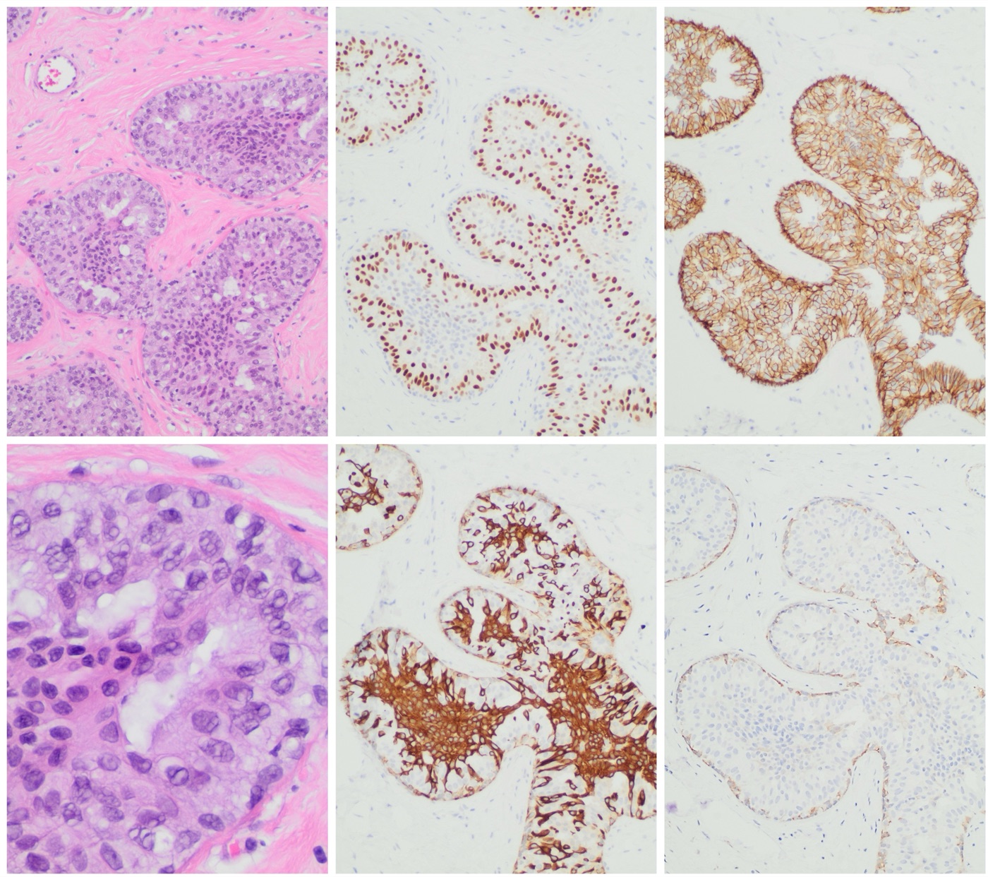
Morphology. UDH consists of a polymorphic population of benign epithelial cells in a haphazard orientation. The cells form irregular secondary lumina and slit-like fenestrations. Solid, streaming and micropapillary patterns occur. Cell borders are indistinct, nuclei vary in size, and intranuclear inclusions and grooves are frequent (Abreu et al. 2023).
Clonality. UDH is morphologically and phenotypically heterogeneous. ADH and DCIS are homogeneous in cell type and marker expression. Loss of heterozygosity is much less frequent in UDH than in ADH and low-grade DCIS (Pinder and Ellis 2003).
Immunophenotype. UDH shows heterogeneous staining for CK5/6, CK14, 34βE12 and estrogen receptor (ER) (Abreu et al. 2023). Luminal cells in ductal hyperplasia predominantly express CK5/6. The neoplastic cells of ADH and atypical lobular hyperplasia lack CK5/6 (Otterbach et al. 2000).
Risk. UDH is associated with a 1.5- to 2-fold relative risk of subsequent breast cancer (Abreu et al. 2023). In an Armed Forces Institute of Pathology series of 117 women with ordinary intraductal hyperplasia, carcinoma developed in 6 (5%) and invasive carcinoma in 3 (2.6%) over an average follow-up of 14 years (Tavassoli and Norris 1990).
Micropapillary UDH. Micropapillary UDH (gynecomastoid hyperplasia) is a differential diagnosis of ADH (Abreu et al. 2023).
Distinguishing usual ductal hyperplasia from atypical ductal hyperplasia
Feature
Usual ductal hyperplasia
Atypical ductal hyperplasia
Source
Cell population
Polymorphic, haphazard, indistinct borders
Monomorphic, evenly spaced, round nuclei
Abreu et al. 2023
Spaces
Irregular slit-like fenestrations
Rigid bridges, arcades and bars; well-developed cribriform secondary spaces
Abreu et al. 2023
CK5/6
Mosaic (heterogeneous) luminal staining
Negative in neoplastic cells
Otterbach et al. 2000; Elfgen et al. 2023
ER
Heterogeneous
Diffuse and strong
Abreu et al. 2023; Elfgen et al. 2023
Clonality
Polyclonal; low frequency of loss of heterozygosity
The WHO 5th edition classifies columnar cell lesions without atypia as columnar cell change (CCC) and columnar cell hyperplasia (CCH). Flat epithelial atypia (FEA) has low-grade (monomorphic) cytologic atypia (Strickland and Turashvili 2022).
Lesion
Architecture
Cytology
Source
Columnar cell change
Dilated acini with smooth contours lined by 1–2 layers of columnar cells
Ovoid nuclei perpendicular to the basement membrane, even chromatin, apical snouts
Strickland and Turashvili 2022
Columnar cell hyperplasia
More than 2 cell layers; mounds, tufts or short micropapillations
As in CCC; crowding can mimic hyperchromasia
Strickland and Turashvili 2022
Flat epithelial atypia
Architecture of CCC or CCH; one to several layers
Round to ovoid nuclei with loss of polarity, mildly raised nuclear-to-cytoplasmic ratio, variably prominent nucleoli
Strickland and Turashvili 2022; Elfgen et al. 2023
Upper limit of FEA. Columnar lesions with rigid bridges, micropapillations or arcades are classified as ADH or low-grade DCIS. High-grade nuclear pleomorphism excludes a columnar cell lesion and indicates high-grade DCIS (Strickland and Turashvili 2022).
Core biopsy category. Columnar cell change and hyperplasia without atypia are B2 lesions on core biopsy. FEA is a B3 lesion (Elfgen et al. 2023).
Immunophenotype. Columnar cell lesions are usually diffusely ER and PR positive and negative for CK5/6, CK14 and HER2 (Strickland and Turashvili 2022). FEA and CCC share this profile, so the distinction is made on H&E morphology (Elfgen et al. 2023).
Proliferation. The Ki-67 index was below 1% in CCC and CCH without atypia, a mean of 8.2% in FEA and a mean of 25.4% in intermediate- to high-grade DCIS (Strickland and Turashvili 2022).
Calcifications. CCC and CCH can contain luminal secretions and calcifications (Strickland and Turashvili 2022). FEA calcifications are mostly amorphous or fine pleomorphic and clustered on mammography (Elfgen et al. 2023).
Reproducibility. Agreement with reference diagnoses of FEA ranged from 17% to 52% among 29–30 pathologists. Agreement improved after tutorials with defined criteria (kappa 0.83 in one study) (Strickland and Turashvili 2022).
The low-grade breast neoplasia family
Associated lesions. Columnar cell lesions often coexist with ADH, lobular neoplasia, low-grade DCIS and tubular carcinoma. They share molecular alterations with these lesions (Strickland and Turashvili 2022; Elfgen et al. 2023).
Genetic changes. Low-grade invasive carcinomas show recurrent loss of 16q and gain of 1q. High-grade tumors are genetically complex (Strickland and Turashvili 2022).
Separate high-grade pathway. Comparative genomic hybridization data indicate that ADH and low-grade DCIS are more closely related to lobular neoplasia than to high-grade DCIS (Pinder and Ellis 2003).
Risk of columnar cell lesions. In a nested case-control study within the Nurses' Health Studies, columnar cell lesions were associated with breast cancer (odds ratio 1.44, 95% CI 1.14–1.83). After adjustment for the category of benign breast disease, the association was not significant (odds ratio 1.20, 95% CI 0.94–1.54) (Aroner et al. 2010).
Risk of FEA. The risk of progression of FEA appears very low compared with ADH and lobular neoplasia. Concomitant ADH or lobular neoplasia may determine the risk (Elfgen et al. 2023).
Definition. Sclerosing adenosis consists of expanded lobular units with whorled, compressed acini (Jensen et al. 1989).
Myoepithelial cells and basement membrane. Sclerosing adenosis retains a myoepithelial cell layer and basement membrane around its tubules. Its epithelial cells are negative for S100 protein (Tavassoli and Bratthauer 1993).
Risk. In the Nashville cohort, the relative risk of invasive breast cancer was 1.7 after exclusion of atypical hyperplasia and 6.7 when sclerosing adenosis and atypical hyperplasia coexisted. Atypical lobular hyperplasia was present 2.7 times more often in biopsies with sclerosing adenosis. Sclerosing adenosis is placed in the proliferative disease without atypia category (Jensen et al. 1989).
Lobular neoplasia in sclerosing adenosis. Lobular carcinoma in situ (LCIS) in sclerosing adenosis distorts the lobules within sclerotic stroma and can mimic invasive carcinoma. Myoepithelial or basement membrane immunohistochemistry resolves the distinction (Simpson et al. 2003).
DCIS in sclerosing adenosis. The WHO 6th edition approach to microinvasion in DCIS involving sclerosing lesions is covered in Ductal Carcinoma In Situ of the Breast.
Apocrine adenosis
Definition. Apocrine adenosis is sclerosing adenosis with superimposed apocrine metaplasia. Atypical apocrine adenosis is apocrine adenosis with enlarged nucleoli and a greater than threefold variation in nuclear area (Seidman et al. 1996).
Immunophenotype. Apocrine cells usually lack estrogen and progesterone receptors and express androgen receptor and GCDFP-15 (Khan et al. 2020).
Differential diagnosis. Atypical apocrine adenosis is distinguished from low-grade apocrine DCIS. Pleomorphic LCIS can also involve sclerosing adenosis (Khan et al. 2020).
Risk. Among 37 women with atypical apocrine adenosis followed for a mean of 8.7 years, the relative risk of carcinoma was 5.5 (95% CI 1.9–16). All women who developed carcinoma were older than 60 years at biopsy (Seidman et al. 1996).
Microglandular adenosis
Morphology. Microglandular adenosis (MGA) is a proliferation of small round glands with open lumina in dense stroma. A single layer of cells with vacuolated or granular cytoplasm lines the glands. Ultrastructurally, a thick multilayered basement membrane surrounds the epithelium (Tavassoli and Norris 1983).
Immunophenotype. MGA lacks a myoepithelial cell layer and retains a basal lamina. Collagen type IV (Tavassoli and Bratthauer 1993) and laminin (Almahari et al. 2023) outline each gland. The epithelial cells are intensely S100 positive (Tavassoli and Bratthauer 1993). MGA cells are ER, PR and HER2 negative (Guerini-Rocco et al. 2016).
Neoplastic nature. All MGAs associated with triple-negative breast cancer (TNBC) carried somatic non-synonymous mutations. Where the TNBC was sequenced, the MGA and the carcinoma shared identical TP53 mutations. Cases without TP53 mutation carried PI3K-pathway mutations (PTEN, PIK3CA, INPP4B). Pure MGAs lacked clonal non-synonymous somatic mutations (Guerini-Rocco et al. 2016). MGA can be a clonal, non-obligate precursor of TNBC (Geyer et al. 2012).
Low-grade triple-negative family.TP53 was mutated in 75% of carcinoma-associated MGAs and 88% of acinic cell carcinomas (acinic cell-like carcinoma in the WHO 6th edition), with loss of the wild-type allele. The two lesions showed no significant difference in mutation repertoire (Geyer et al. 2017).
Acinic cell-like carcinoma. The WHO 6th edition renamed acinic cell carcinoma as acinic cell-like carcinoma because breast tumors lack the t(4;9)(q13;q31) rearrangement and NR4A3 overexpression of salivary acinic cell carcinoma (Quinn et al. 2026). The carcinoma and its relation to MGA are covered in Special-Type Breast Carcinomas.
Myoepithelial cells and basement membrane in gland-forming lesions
Lesion
Myoepithelial layer
Basement membrane
Epithelial S100
Source
Sclerosing adenosis
Present
Present
Negative
Tavassoli and Bratthauer 1993
Secretory adenosis
Present
Present (PAS)
Negative
Tavassoli and Bratthauer 1993
Microglandular adenosis
Absent
Present (collagen IV, laminin)
Intensely positive
Tavassoli and Bratthauer 1993; Almahari et al. 2023
Tubular carcinoma
Absent
Absent
Negative
Tavassoli and Bratthauer 1993
Tubular adenosis
Present
Not assessed in the cited study
Not assessed in the cited study
Lee et al. 1996
Tubular adenosis
Morphology. Tubular adenosis is a haphazard proliferation of elongated, narrow, sometimes branching tubules with no or only vague lobular grouping. Tubules can extend into fat and contain basophilic or granular eosinophilic secretion (Lee et al. 1996).
Myoepithelium. An intact myoepithelial layer surrounds the tubules (Lee et al. 1996).
Differential diagnosis. Tubular adenosis mimics invasive carcinoma, especially at frozen section or when DCIS involves it. It differs from MGA by its myoepithelium. It differs from sclerosing adenosis by the lack of lobular or whorled architecture and the wider spacing of tubules (Lee et al. 1996).
Radial scar and complex sclerosing lesion
Definition. Radial scar shows a central fibroelastotic core with peripherally located compressed glands and cysts. Calcifications and sclerosing adenosis are often associated (Elfgen et al. 2023). A lesion with these features larger than 10 mm is classified as complex sclerosing lesion (Trombadori et al. 2021).
Histology of the core. The center contains fibroelastosis, obliterated ducts and compressed tubules with two cell layers, including CK14-positive myoepithelium (Trombadori et al. 2021).
Associated lesions. A subset of radial scars contains epithelial hyperplasia, atypia or carcinoma (Elfgen et al. 2023).
Imaging. Radial scar is frequently occult. When visible, it forms a stellate lesion with a radiolucent center and radiating spicules, with architectural distortion. It can mimic invasive carcinoma on imaging (Elfgen et al. 2023).
Risk. In the Nurses' Health Study, radial scars were found in 7.1% of benign biopsies and had a median size of 4.0 mm. Women with radial scars had a relative risk of breast cancer of 1.8 (95% CI 1.1–2.9), independent of the histologic category of benign disease (Jacobs et al. 1999).
Radial scar compared with tubular carcinoma. The entrapped tubules of a radial scar retain myoepithelial cells that stain for p63 and CK14 (Elfgen et al. 2023; Trombadori et al. 2021). Tubular carcinoma lacks both a myoepithelial layer and a basement membrane (Tavassoli and Bratthauer 1993). The diagnostic criteria of tubular carcinoma are given in Special-Type Breast Carcinomas.
Collagenous spherulosis
Definition. Collagenous spherulosis consists of intraluminal clusters of eosinophilic spherules about 20–100 µm in diameter within lobular acini and ductules. The spherules contain collagen, acidic mucin and basement membrane-like material. The cells immediately around them stain for actin (Clement et al. 1987).
Setting. Collagenous spherulosis arises within benign proliferations, including intraductal papilloma, sclerosing adenosis and radial scar (Clement et al. 1987). In 59 cases, it was multifocal in 75%. It was associated with benign lesions in 41 cases, ADH in 3 and LCIS in 15. Microcalcifications were present in 15 cases (Resetkova et al. 2006).
Mimics. Collagenous spherulosis can mimic ADH, cribriform DCIS and adenoid cystic carcinoma (Resetkova et al. 2006).
Immunohistochemistry. SMMHC, p63 and calponin show a single layer of myoepithelial cells around the spherules. Adenoid cystic carcinoma is c-kit (CD117) positive. The cells around the spherules in collagenous spherulosis were c-kit negative (Kim et al. 2018).
Collagenous spherulosis involved by LCIS. E-cadherin-negative LCIS cells involve the spherules and replace the luminal cells. E-cadherin with a myoepithelial marker separates this pattern from DCIS (Kim et al. 2018).
Feature
Collagenous spherulosis
Adenoid cystic carcinoma
Cribriform DCIS
Source
Nature
Benign; arises in benign proliferations
Invasive carcinoma; MYB protein overexpressed
Clonal in situ carcinoma
Clement et al. 1987; Neilson et al. 2025; Pinder and Ellis 2003
Luminal contents
Collagen-rich spherules rimmed by myoepithelial cells
Clement et al. 1987; Neilson et al. 2025; Pinder and Ellis 2003
CD117
Negative
Positive
Not used for the distinction
Kim et al. 2018; Neilson et al. 2025
Myoepithelial cells
Single layer around each spherule
Myoepithelial–basal cells line pseudoglandular spaces
Peripheral layer around the involved duct
Kim et al. 2018; Neilson et al. 2025; Pinder and Ellis 2003
Gynecomastia
Definition. Gynecomastia is a benign proliferation of glandular tissue in the male breast (Pozza et al. 2026). It consists of benign ductal and stromal proliferation (Sosnowska-Sienkiewicz et al. 2024).
Pathogenesis. Gynecomastia results from an imbalance between estrogen and androgen action. The imbalance is often a relative shift in the ratio, without an absolute rise in estradiol (Pozza et al. 2026).
Florid and quiescent phases. The early (florid) phase is typically tender and of recent onset (less than 6 months). It appears on ultrasound as nodular subareolar tissue. The later quiescent phase shows a dendritic pattern and responds less to medical therapy (Pozza et al. 2026).
Histology by phase. In the florid (early) phase, ducts increase in number and are elongated and branched, with ductal epithelial proliferation. The periductal stroma is edematous and vascular, with fibroblast proliferation. In the fibrous (late) phase, the periductal stroma is fibrotic and less cellular. Ducts may be fewer, with flattened or atrophic epithelium, and edema and fibroblastic proliferation are absent. In both phases the myoepithelial layer is intact and lobules are generally absent (Sosnowska-Sienkiewicz et al. 2024).
Differential diagnosis of benign epithelial proliferations
Lesion
Main mimic
Distinguishing finding
Source
Usual ductal hyperplasia
Atypical ductal hyperplasia
Mosaic CK5/6 and heterogeneous ER in UDH
Otterbach et al. 2000; Abreu et al. 2023
Columnar cell change
Flat epithelial atypia
Nuclear atypia on H&E; immunophenotype identical
Elfgen et al. 2023
Flat epithelial atypia
ADH or low-grade DCIS
Rigid bridges, arcades or micropapillations exclude FEA
Strickland and Turashvili 2022
Sclerosing adenosis with LCIS
Invasive lobular carcinoma
Myoepithelial cells around distorted acini
Simpson et al. 2003
Microglandular adenosis
Tubular carcinoma
Basement membrane and S100-positive epithelium in MGA
Tavassoli and Bratthauer 1993
Radial scar
Tubular carcinoma
Myoepithelial cells around entrapped tubules
Elfgen et al. 2023; Trombadori et al. 2021
Collagenous spherulosis
Adenoid cystic carcinoma
CD117-negative cells; myoepithelial rim around spherules
Kim et al. 2018
Collagenous spherulosis with LCIS
Cribriform DCIS
E-cadherin loss in the lesional cells
Kim et al. 2018
Usual ductal hyperplasia with the immunostains that separate it from atypical ductal hyperplasia. Top row, left to right: a mixed population of cells with streaming nuclei and irregular, slit-like peripheral lumina (H&E, medium power); heterogeneous estrogen receptor staining (ER); retained membranous E-cadherin. Bottom row, left to right: overlapping, variably sized nuclei (H&E, high power); CK5/6 in a mosaic pattern, strongest centrally; retained membranous p120. In ADH, ER is diffuse and strong and CK5/6 is negative in the lesional cells. Magnification not stated.Image: Mikael Häggström, M.D., Wikimedia Commons, CC0.
Papillary Neoplasms, Adenomyoepithelioma and Nipple Lesions of the Breast
Definition. Papillary lesions of the breast contain papillae, mostly with arborizing fibrovascular stroma. They range from benign intraductal papilloma and papilloma with atypical epithelial proliferation to papillary ductal carcinoma in situ (DCIS), encapsulated papillary carcinoma (EPC), solid papillary carcinoma (SPC) and invasive papillary carcinoma (Kulka et al. 2022). The distribution of myoepithelial cells separates benign, precursor and malignant papillary lesions and identifies invasion (Kulka et al. 2022). Nipple adenoma and syringomatous tumor are benign neoplasms specific to the nipple (Brogi and Scatena 2023; Jones et al. 1989).
Papillae with fibrovascular cores in dilated ducts and microcysts
Absent at the periphery
WHO 5th edition
Kulka et al. 2022
Micropapillae. Micropapillae lack fibrovascular cores. A micropapillary pattern occurs in usual ductal hyperplasia, micropapillary DCIS and invasive micropapillary carcinoma (Kulka et al. 2022).
Biomarker profile. Most carcinomas with papillary features are ER positive and HER2 negative (Kulka et al. 2022).
Quality assurance data. In the UK National Health Service Breast Screening external quality assurance scheme, papillary lesions were among the most frequently misinterpreted breast lesions on H&E sections (Kulka et al. 2022).
Intraductal papilloma, central and peripheral
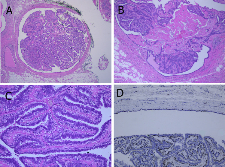
Definition. Intraductal papilloma is a benign intraluminal proliferation of arborizing fibrovascular cores covered by myoepithelial and luminal cells. It is the most common papillary lesion of the breast (Kulka et al. 2022).
Histology. Complex fibrovascular cores are lined by myoepithelial cells and covered by cuboidal or columnar luminal cells. The two cell types are the hallmark of benign papillary lesions. Luminal nuclei can contain intranuclear inclusions (Kulka et al. 2022).
Secondary changes. Hemorrhage and infarction follow needle biopsy or torsion of fibrovascular cores. Sclerosis of the wall entraps epithelial and myoepithelial cells and produces a pseudoinfiltrative pattern. Squamous, apocrine, mucinous and chondroid metaplasia and collagenous spherulosis occur (Kulka et al. 2022).
Apocrine papilloma. Myoepithelial cells can be scant or absent in benign apocrine papillomas and papillary apocrine hyperplasia (Kulka et al. 2022).
Displaced epithelium. Fine-needle aspiration and core biopsy displace papilloma epithelium into the surrounding stroma. Hemosiderin, histiocytes, granulation tissue and cellular scar indicate displacement artifact (Kulka et al. 2022).
Molecular findings. The most common mutation in papillomas is an activating PIK3CA missense mutation. Loss of chromosome X, then losses of 16q and 7q, are the most common copy number changes. Papillomas were clonally related to synchronous DCIS and invasive carcinoma in more than 50% of analyzed cases (Kulka et al. 2022).
Feature
Central papilloma
Peripheral papilloma
Source
Frequency
More common
Less common
Kulka et al. 2022
Site
Large duct, subareolar
Terminal duct–lobular unit region
Kulka et al. 2022
Number
Usually solitary
Smaller and often multiple
Kulka et al. 2022
Presentation
Serous or bloody nipple discharge; filling defect on galactography
Usually asymptomatic; mammographic microcalcifications
Kulka et al. 2022
Associated epithelial proliferation
Less frequent
UDH, ADH, ALH and DCIS more common
Kulka et al. 2022
Age. Most papillomas occur in perimenopausal women between 30 and 50 years (Kulka et al. 2022).
Papilloma with atypical ductal hyperplasia, DCIS or lobular neoplasia
Extent rule (WHO 5th edition). A low-nuclear-grade atypical proliferation covering part of a papilloma is classified by extent. Less than 3 mm is papilloma with ADH. An extent of 3 mm or more is papilloma with DCIS (Kulka et al. 2022).
Grade rule (WHO 5th edition). Intermediate- or high-grade atypia in a papilloma is classified as papilloma with DCIS regardless of size (Kulka et al. 2022).
Terminology. The term "atypical papilloma" was not adopted by the WHO 5th edition (Kulka et al. 2022).
6th edition. The 6th-edition summary does not address atypia in papillomas (Quinn et al. 2026).
Morphology and immunohistochemistry. The atypical cells have uniform hyperchromatic nuclei, often in a cribriform arrangement. They are negative for CK5 and CK14 and strongly and uniformly ER positive (Kulka et al. 2022).
Lobular neoplasia in a papilloma. Lobular neoplasia can involve a papilloma. E-cadherin, p120 or β-catenin highlights the lobular cells. Papilloma with lobular neoplasia on CNB or VAB does not require excision when radiologic and pathologic findings are concordant (Kulka et al. 2022).
Cancer risk. In a nested case-control study, papillomas containing atypical hyperplasia had a relative risk of invasive carcinoma more than 4 times that of papillomas without atypia. Most subsequent carcinomas arose in the same breast near the original papilloma. Usual hyperplasia within a papilloma did not add risk (Page et al. 1996).
Papillary ductal carcinoma in situ
Definition. Papillary DCIS has papillary architecture completely lined by neoplastic ductal epithelium, with no pre-existing benign papilloma. It is a segmental disease of small or large ducts (Kulka et al. 2022).
Morphology. One or several layers of monomorphic, usually columnar cells cover delicate branching fibrovascular cores. Solid, cribriform and micropapillary areas occur. Nuclear grade is usually low or intermediate (Kulka et al. 2022).
Myoepithelial cells. Myoepithelial cells are present only at the periphery of the involved ducts (Kulka et al. 2022).
Dimorphic variant. A dimorphic variant contains "globoid cells" that resemble myoepithelial cells and are negative for all myoepithelial markers (Kulka et al. 2022).
Frequency. Papillary DCIS usually coexists with other DCIS patterns. Pure papillary DCIS is rare (Kulka et al. 2022). DCIS grading is covered in Ductal Carcinoma In Situ of the Breast.
Encapsulated and solid papillary carcinoma: distinguishing features
EPC. EPC presents as a slowly growing retroareolar mass in postmenopausal women. Low- to intermediate-grade cells cover fine fibrovascular cores within a cyst with a thick fibrous capsule. Myoepithelial cells are usually absent in the papillae and at the periphery. The cells are usually ER and PR positive and lack ERBB2 amplification (Kulka et al. 2022). Without frank invasion, EPC is staged and managed as DCIS (Kulka et al. 2022).
EPC staging, edition change. In the 5th edition, EPC with high nuclear grade, high mitotic activity, or a triple-negative or HER2-positive phenotype was graded, staged and managed as invasive carcinoma. The 6th edition accepts that very rare EPC with high grade or ER negativity may still be classified as EPC, with a comment listing the unusual features (Quinn et al. 2026).
SPC. SPC fills large or dilated small ducts with solid growth, delicate fibrovascular septa and nuclear palisading at the stromal–epithelial interface. Cells form rosettes or pseudorosettes. Neuroendocrine differentiation and mucin production are very common, and ER is diffusely and strongly positive (Kulka et al. 2022).
Basal keratins in EPC and SPC. EPC and SPC are clonal proliferations that are strongly ER positive and negative for CK5/6 and CK14. Solid papillary carcinoma can mimic a papilloma with florid UDH, which shows a mosaic CK5/6 pattern (Rakha and Quinn 2025; Quinn et al. 2023). In one series, CK5/6 and CK14 Allred scores were 0–3 in all SPC in situ and 2–5 in all papillomas with UDH, so the ranges overlap at low scores (Maeda et al. 2018).
Basal-like EPC. Rare ER-negative EPC-like tumors with a basal phenotype express CK5/6 and CK14 (Rakha and Quinn 2025).
Immunohistochemistry of papillary lesions
Lesion
Myoepithelial markers (p63, calponin, SMMHC)
CK5/6 and CK14
ER
Source
Intraductal papilloma
Positive along the cores and at the periphery
Positive (mosaic with florid UDH)
Heterogeneous
Kulka et al. 2022; Quinn et al. 2023
Atypical area in a papilloma (ADH or DCIS)
Reduced or absent within the atypical area
Negative
Strong and uniform
Kulka et al. 2022
Papillary DCIS
Peripheral rim only
Negative
Diffuse and strong
Kulka et al. 2022; Quinn et al. 2023
Encapsulated papillary carcinoma
Usually absent in the papillae and at the periphery
Kulka et al. 2022; Rakha and Quinn 2025; Quinn et al. 2023
Solid papillary carcinoma
Can be absent
Negative or low (Allred 0–3 in SPC in situ)
Diffuse and strong
Kulka et al. 2022; Quinn et al. 2026; Maeda et al. 2018
Myoepithelial markers. The staining properties and limits of p63, calponin and SMMHC, and the rule of using at least 2 markers, are given in Ductal Carcinoma In Situ of the Breast.
Juvenile papillomatosis of the breast
Definition. Juvenile papillomatosis is a breast tumor of young women with atypical papillary duct hyperplasia and numerous cysts. It was described as a clinicopathologic entity in 1980 (Rosen et al. 1985).
Registry data. In 180 registry patients, the mean age was 23 years (range 12–48). Twenty-eight percent had one or more relatives with breast cancer. Focal epithelial necrosis was present in 17%, mostly in lesions with the most severe atypia. Eight patients had bilateral disease (Rosen et al. 1985).
Concurrent and subsequent carcinoma. Seven patients (4%) had concurrent breast carcinoma. Two developed carcinoma 8 and 9 years later (Rosen et al. 1985).
Long-term follow-up. In 41 patients with a median follow-up of 14 years, 4 (10%) developed breast carcinoma. All 4 had recurrent and bilateral juvenile papillomatosis and a positive family history. No patient with unilateral, unicentric, nonrecurrent disease developed carcinoma (Rosen and Kimmel 1990).
Treatment. Wide local excision controls the lesion in most cases. Clinical surveillance of the patient and her female relatives is advised (Rosen et al. 1985).
Nipple adenoma, syringomatous tumor and low-grade adenosquamous carcinoma
Nipple adenoma
Definition and site. Nipple adenoma is a benign tumor first described as florid papillomatosis of the nipple. The dermo-epidermal junction is regarded as its site of origin. It occurs in females and males aged 5 months to 89 years and clinically resembles Paget disease (Kulka et al. 2022).
Histologic patterns. In 49 women, the patterns were sclerosing papillomatosis, papillomatosis, adenosis and mixtures of these (Rosen and Caicco 1986). A series of 50 cases described adenosis, large duct, papillary-like and pseudoinfiltrative patterns. Subtyping was unlikely to be clinically relevant (Weigelt et al. 2023).
Features that mimic carcinoma. Focal necrosis in hyperplastic ducts and scattered mitoses were more frequent in the sclerosing papillomatosis pattern than in the other patterns (Rosen and Caicco 1986). Stromal sclerosis distorts ducts into a pseudoinvasive pattern (Kulka et al. 2022).
Immunohistochemistry. CK5, ER and myoepithelial markers separate nipple adenoma from DCIS and invasive carcinoma (Kulka et al. 2022).
Molecular findings.PIK3CA mutations are frequent. KRAS and BRAF mutations also occur (Kulka et al. 2022).
Behavior. Excision was curative in all patterns over a mean follow-up of 8.3 years (Rosen and Caicco 1986). No recurrence occurred in 44 patients followed for a median of 66 months (Weigelt et al. 2023). Incomplete resection is associated with recurrence (Kulka et al. 2022).
Morphology. Tubular duct-like structures and keratinizing cysts resemble syringoma of skin adnexal origin (Rosen 1983). Small ducts and solid strands of epithelial cells are set in desmoplastic stroma (Jones et al. 1989).
Infiltration. Ten of 11 tumors invaded the smooth muscle of the nipple. Four extended into the underlying breast tissue, and 1 showed perineural invasion (Jones et al. 1989).
Behavior. Five of 11 tumors (45%) recurred after local excision, and none metastasized (Jones et al. 1989). Persistent tumor enlarged slowly over 22 years after incomplete excision in one patient. Adequate treatment can require nipple resection (Rosen 1983).
Patients. In a systematic review of 28 cases, the mean age was 44 years and 7% of patients were male. Preoperative biopsy did not show the typical histologic features in 7 of 9 cases (Park et al. 2023).
Immunophenotype. The predominant cell type is myoepithelial and expresses p63, high-molecular-weight keratins and other myoepithelial markers. The tumor is located superficially, can infiltrate the smooth muscle of the areola and rarely extends into the dermis (Quinn et al. 2023).
Overlap with low-grade adenosquamous carcinoma. Syringomatous tumor shows morphologic overlap with low-grade adenosquamous carcinoma, and the two can be difficult to separate on core biopsy. The precise anatomic location helps (Quinn et al. 2023).
Terminology. Similar lesions in the skin, nipple and breast parenchyma were reported under several names. "Infiltrating syringomatous adenoma" was proposed to avoid the term carcinoma (Jones et al. 1989).
Low-grade adenosquamous carcinoma
Classification. Low-grade adenosquamous carcinoma is a variant of metaplastic carcinoma with combined glandular and squamous differentiation (Rosen and Ernsberger 1987). Its low-grade cytomorphology and clinical indolence separate it from other metaplastic carcinomas (Soo and Tan 2013). Metaplastic carcinoma is covered in Special-Type Breast Carcinomas.
Original series. Eleven patients had a mean age of 59 years (range 42–76). Tumors measured 1.5–3.4 cm and were hard, tan-yellow and infiltrative (Rosen and Ernsberger 1987).
Behavior. Four of 8 patients treated by local excision alone had recurrence in the breast within 1–3.5 years. Five axillary dissections showed negative nodes (Rosen and Ernsberger 1987).
Site and p63 pitfall. Low-grade adenosquamous carcinoma arises deeper in the breast than syringomatous tumor, can reach a large size, rarely infiltrates the nipple and can have a neoplastic spindle cell component. Epithelial cells of its small glands and nests show focal p63 staining that can be mistaken for myoepithelial cells (Quinn et al. 2023).
Feature
Nipple adenoma
Syringomatous tumor
Low-grade adenosquamous carcinoma
Source
Site
Nipple; dermo-epidermal junction and subareolar ducts
Nipple and areola, superficial; extension into the breast in some cases
Deeper breast parenchyma; rarely the nipple
Kulka et al. 2022; Jones et al. 1989; Quinn et al. 2023
Focal p63 in epithelial cells of small nests, mimicking myoepithelium
Kulka et al. 2022; Quinn et al. 2023
Recurrence after local excision
Rare when completely excised
45% in one series
4 of 8 in the original series
Weigelt et al. 2023; Jones et al. 1989; Rosen and Ernsberger 1987
Metastasis
None in the cited series
None in the cited series
Negative axillary nodes in the cited series
Rosen and Caicco 1986; Jones et al. 1989; Rosen and Ernsberger 1987
Adenomyoepithelioma of the breast
Classification in the 6th edition. The 6th edition retains the 5th-edition classification. A proposed morphologic classification was not adopted because outcome evidence was insufficient (Quinn et al. 2026).
Patterns. Classic adenomyoepithelioma (AME) shows tubular, spindle cell, lobulated and papillary patterns. Classic tubular AME can have an infiltrative outline, sometimes called adenomyoepithelial adenosis (Quinn et al. 2026).
Distinction from papilloma. AME shows a diffusely expanded myoepithelial compartment. Myoepithelial hyperplasia in a papilloma is focal (Kulka et al. 2022).
DCIS in AME. Epithelial atypia of sufficient extent and degree is diagnosed as DCIS arising in an AME and managed by DCIS protocols (Quinn et al. 2026).
Malignant AME. Malignant AME is an AME with carcinoma arising from the epithelial cells, the myoepithelial cells or both. The underlying AME remains identifiable, and the malignant component shows atypia, increased mitoses and necrosis (Quinn et al. 2026).
Carcinoma types in malignant AME. Epithelial transformation produces invasive carcinoma of no special type or a special type, including lobular carcinoma (Quinn et al. 2026). Carcinomas arising from the myoepithelial component and the reporting of biphasic carcinomas are given in Special-Type Breast Carcinomas.
Immunoprofile. Immunohistochemistry highlights the biphasic epithelial–myoepithelial nature. Malignant AME can lose the distinction between the two cell populations (Quinn et al. 2026).
Category
Atypia
Mitoses
Other features
Source
Classic AME
None to minimal
Up to 1.5/mm² (3 per 10 HPF of 0.5 mm diameter and 0.2 mm² area) in either component
Tubular, spindle cell, lobulated or papillary
Quinn et al. 2026
Atypical AME
Mild to moderate in epithelial or myoepithelial cells
Up to 2.5/mm² (5 per 10 HPF)
Focal necrosis allowed; atypical mitoses raise suspicion of malignant transformation
Quinn et al. 2026
Malignant AME
Carcinoma arising from either or both components
Increased
Necrosis; recognizable underlying AME
Quinn et al. 2026
HRAS and PI3K pathway mutations in adenomyoepithelioma
Genomic groups. ER-positive AMEs have PIK3CA or AKT1 activating mutations. ER-negative AMEs have recurrent HRAS codon Q61 hotspot mutations together with PIK3CA or PIK3R1 mutations (Geyer et al. 2018).
Rarity in other breast cancers.HRAS mutations are very rare in common-type breast cancers (Geyer et al. 2018).
RAS Q61R immunohistochemistry. In 26 sequenced AMEs, 9 ER-negative tumors had HRAS mutations (7 Q61R, 2 Q61K). The RAS Q61R antibody detected 5 of 7 Q61R mutations (sensitivity 71%) and stained no tumor without the mutation (specificity 100%). It did not detect Q61K mutations (Pareja et al. 2020).
Staining pattern. Staining was restricted to the myoepithelium in 4 of 5 positive tumors. Positive tumors were associated with infiltrative borders, necrosis and higher mitotic index (Pareja et al. 2020).
Differential diagnosis of papillary and nipple lesions
Lesion
Main mimic
Distinguishing finding
Source
Sclerosed papilloma
Invasive carcinoma
Myoepithelial cells around entrapped nests; hemosiderin and scar after biopsy
Kulka et al. 2022
Papilloma with ADH
Papilloma with UDH
Atypical cells negative for CK5 and CK14 with uniform strong ER
Kulka et al. 2022
Papillary DCIS
Intraductal papilloma
Myoepithelial cells only at the duct periphery; monotonous cells covering all cores
Kulka et al. 2022
Papillary adenomyoepithelioma
Papilloma with myoepithelial hyperplasia
Diffuse myoepithelial expansion; RAS Q61R staining in ER-negative tumors
Kulka et al. 2022; Pareja et al. 2020
Nipple adenoma
Paget disease, DCIS or invasive carcinoma
Origin at the dermo-epidermal junction; retained myoepithelial cells; CK5 and ER pattern
Kulka et al. 2022
Syringomatous tumor
Nipple adenoma or tubular carcinoma
Small ducts, strands and keratinizing cysts infiltrating nipple smooth muscle
Jones et al. 1989; Rosen 1983
Juvenile papillomatosis
Papilloma or DCIS in a young patient
Numerous cysts with papillary duct hyperplasia; mean age 23 years
Rosen et al. 1985
Intraductal papilloma in a large central duct. A, an arborizing papillary mass within a dilated duct (low power). B, an area of sclerosis within the papilloma. C, fibrovascular cores covered by luminal epithelium with a prominent myoepithelial layer, the two cell types of a benign papillary lesion. D, p63 marks myoepithelial cells along the fibrovascular cores and at the peripheral duct wall. H&E (A–C) and p63 immunostain (D); magnification not stated.Image: Kulka Janina et al., PMC open access (Virchows Arch. 2021 Nov 3;480(1):65–84. doi: 10.1007/s00428-021-03182-7) Fig. 2, CC BY.
Ductal Carcinoma In Situ of the Breast
Definition. Ductal carcinoma in situ (DCIS) is a proliferation of malignant epithelial cells within the breast parenchymal structures with no evidence of invasion across the basement membrane (Pinder and Ellis 2003). DCIS is a non-obligate precursor of invasive breast carcinoma (Abreu et al. 2023). The WHO 6th edition (2026) places DCIS in Part 1 of the epithelial tumor chapter (Quinn et al. 2026). DCIS grading follows the CAP DCIS protocols, which were updated for the 6th edition in June 2026 (CAP DCIS resection protocol 2026).
Incidence. In the United States, DCIS incidence rose from 1.87 per 100,000 in 1973–1975 to 32.5 per 100,000 in 2004. Increased mammographic screening accounts for part of the rise (Virnig et al. 2010).
Share of breast cancers. DCIS forms 20–25% of newly diagnosed breast cancers in the United States (Abreu et al. 2023).
Age and presentation. The mean age at diagnosis is 50–59 years. Mammography detects 80–85% of DCIS, typically as unilateral calcifications. DCIS occasionally presents as a palpable mass, nipple discharge or Paget disease (Abreu et al. 2023).
Risk factors. Older age and family history are risk factors for DCIS (Virnig et al. 2010).
Distribution. DCIS involves one region of the breast in most cases. Two-thirds of tumors involve only one quadrant (Pinder and Ellis 2003).
Invasion found at excision. About 15% of patients with DCIS on core needle biopsy have invasive cancer after excision or mastectomy (Virnig et al. 2010).
Mammographic calcifications in DCIS
Necrosis and calcification. Most areas of necrosis calcify. DCIS that presents as mammographic calcifications often recurs as calcifications (CAP DCIS resection protocol 2026).
Comedo necrosis on imaging. Central (comedo) necrosis often corresponds to linear or branching calcifications on mammography (CAP DCIS resection protocol 2026).
Specimen radiography. The presence of the targeted calcifications in the specimen is confirmed by specimen radiography. The report states the relationship of the calcifications to the DCIS (CAP DCIS biopsy protocol 2026).
Low-grade DCIS. Calcification in low-grade DCIS is usually located in inspissated secretions (Pinder and Ellis 2003).
Architectural patterns of DCIS
Patterns listed in the CAP protocols. Comedo, cribriform, micropapillary, papillary and solid patterns are reported. Solid papillary carcinoma in situ, encapsulated papillary carcinoma and Paget disease are also listed as patterns and are distinct histologic types of DCIS (CAP DCIS resection protocol 2026).
Predictive value. Nuclear grade and necrosis predict clinical outcome better than architectural pattern (CAP DCIS resection protocol 2026).
Pattern and grade. Low-grade DCIS frequently shows cribriform or micropapillary architecture, often both. High-grade DCIS is often solid with central necrotic debris. Intermediate-grade DCIS is often solid and may show some polarization around spaces (Pinder and Ellis 2003).
Reproducibility of the traditional system. Classification by architecture combined with cytology required reclassification of up to 30% of cases in multicenter trials (Pinder and Ellis 2003).
Nuclear grade of DCIS
The nuclear grade of DCIS is assigned from 6 morphologic features (CAP DCIS resection protocol 2026).
Feature
Grade I (low)
Grade II (intermediate)
Grade III (high)
Source
Pleomorphism
Monotonous (monomorphic)
Intermediate
Markedly pleomorphic
CAP DCIS resection protocol 2026
Nuclear size
1.5–2 times a red blood cell or a normal duct epithelial nucleus
Intermediate
More than 2.5 times a red blood cell or a normal duct epithelial nucleus
CAP DCIS resection protocol 2026
Chromatin
Usually diffuse and finely dispersed
Intermediate
Usually vesicular with irregular distribution
CAP DCIS resection protocol 2026
Nucleoli
Only occasional
Intermediate
Prominent, often multiple
CAP DCIS resection protocol 2026
Mitoses
Only occasional
Intermediate
May be frequent
CAP DCIS resection protocol 2026
Orientation
Polarized toward luminal spaces
Intermediate
Usually not polarized toward the luminal space
CAP DCIS resection protocol 2026
Multiple grades. Nuclear grade is a multi-select element in the 2026 protocols, so more than one grade can be reported in one specimen (CAP DCIS resection protocol 2026).
Grade and recurrence. In 79 patients with mammographically detected DCIS up to 25 mm treated by tylectomy alone, high-grade DCIS with comedo necrosis recurred locally in 19% after an average of 26 months. None of 33 patients with low-grade micropapillary or non-necrotic cribriform DCIS recurred during follow-up (Lagios et al. 1989).
Necrosis in DCIS
Type
Definition
Associations
Source
Central (comedo)
Central expansive necrosis replacing the duct center, easily seen at low magnification; ghost cells and karyorrhectic debris
Usually high nuclear grade; also occurs with low or intermediate grade; linear or branching calcifications
CAP DCIS resection protocol 2026
Focal (punctate)
Small foci indistinct at low magnification, or single-cell necrosis (<10%)
Seen in low-grade DCIS as individual apoptotic cells or small foci
CAP DCIS resection protocol 2026; Pinder and Ellis 2003
Necrosis mimics. Secretory material, cytoplasmic blebs and histiocytes can accompany calcifications but lack nuclear debris. They are not classified as necrosis (CAP DCIS resection protocol 2026).
Grading systems for DCIS
Cytonuclear grading. The UK National Coordinating Group for Breast Screening Pathology classifies DCIS as high, intermediate or low grade on cytonuclear features, following the work of Holland and colleagues (Pinder and Ellis 2003).
Van Nuys classification. Silverstein and colleagues grouped DCIS as high grade, non-high grade with necrosis, and non-high grade without necrosis. The groups were associated with local recurrence and disease-free survival (Pinder and Ellis 2003).
Consensus grading. The CAP nuclear grade criteria derive from the 1997 Consensus Conference on the classification of DCIS (CAP DCIS resection protocol 2026; Consensus Conference Committee 1997).
Size (extent) of DCIS
Definition. The size (extent) of DCIS is the greatest dimension estimated from gross and microscopic evaluation. It estimates the volume of breast tissue occupied by DCIS (CAP DCIS resection protocol 2026).
Staging and management. Extent is not required for pT classification. It correlates with residual disease after re-excision, close or positive margins, local recurrence and missed invasion (CAP DCIS resection protocol 2026).
Imaging error. Mammographic assessment based on calcifications frequently underestimates, and sometimes overestimates, the extent of DCIS (CAP DCIS resection protocol 2026).
Extent on a core biopsy. The extent on a core biopsy is the largest span in a single core. Spans in separate cores are not added. The span in a core may guide radiation decisions when no residual DCIS remains in the excision (CAP DCIS biopsy protocol 2026).
Methods for estimating DCIS extent
Method
How the extent is obtained
Limitation or rule
Source
DCIS in one block
Measured on a single slide; the largest distance between separate foci is reported
Underestimates extent when multiple blocks are involved
CAP DCIS resection protocol 2026
Serial sequential sampling
The whole specimen is submitted in a mapped sequence; extent is calculated from the specimen diagram, slice thickness and location of involved blocks
Recommended for all excisions likely to contain DCIS or with DCIS on a prior core biopsy
CAP DCIS resection protocol 2026
Nonsequential sampling (number of involved blocks)
Count of blocks with DCIS
Not recommended as the only method; over- or underestimates
CAP DCIS resection protocol 2026
Edges of the specimen
Distance between 2 opposing involved or close margins
Used when DCIS reaches 2 opposing margins
CAP DCIS resection protocol 2026
Gross lesion
Measurement of a grossly visible lesion, usually high-grade DCIS
The gross size is confirmed microscopically
CAP DCIS resection protocol 2026
Reported value. The largest estimate from any of these methods is reported as the size (extent) of DCIS (CAP DCIS resection protocol 2026).
Extent
Clinical significance
Source
Up to 20 mm
Breast conservation with wide negative margins is achievable in most women
CAP DCIS resection protocol 2026
>20–40 mm
Wide negative margins may be difficult to achieve with breast-conserving surgery
CAP DCIS resection protocol 2026
>40 mm
Breast conservation with wide negative margins may be impossible; undetected invasion is possible when the area is not fully examined; lymph node sampling may be recommended
CAP DCIS resection protocol 2026
Gross handling of DCIS excisions
Target lesion. The clinical or radiologic lesion is examined microscopically. For a nonpalpable lesion, the specimen radiograph or other imaging identifies it (CAP DCIS resection protocol 2026).
Complete submission. When practical, the entire specimen is submitted sequentially. Otherwise, at least the entire targeted region is examined. For a known DCIS, complete serial sequential sampling excludes invasion, evaluates margins and estimates extent. When the whole specimen is not examined, the approximate percentage examined is stated (CAP DCIS resection protocol 2026).
Specimen size. The specimen size is recorded because the volume excised is associated with recurrence (CAP DCIS resection protocol 2026).
Prior biopsy site. The prior biopsy site is sampled and documented. Biopsy changes at a margin of a re-excision can indicate incomplete excision (CAP DCIS resection protocol 2026).
Definition and protocol choice. Microinvasion is invasion measuring ≤1 mm. A case with any invasion or microinvasion is reported with the CAP invasive carcinoma protocol (CAP DCIS resection protocol 2026). Measurement of microinvasion and multifocal microinvasion are covered in Breast Cancer Staging, Margins, Sentinel Lymph Nodes and Post-Neoadjuvant Assessment.
Prior core biopsy. An excision with only residual DCIS after a core biopsy that showed invasion or microinvasion is reported with the invasive protocol and incorporates the core biopsy findings (CAP DCIS resection protocol 2026).
WHO 6th edition emphasis. The 6th edition stresses careful search for small irregular abnormal cell clusters that may represent microinvasion in DCIS involving sclerosing lesions. The search is most important when a prominent lymphocytic infiltrate is present (Quinn et al. 2026).
Inflammation and repair. Inflammatory and reparative changes can distort in situ carcinoma and simulate invasion (Hoda et al. 1999).
Cancerization of lobules. Cancerization of lobules and ADH frequently occur at the edge of DCIS. Cancerization of lobules near the margin was associated with recurrence in high-grade DCIS treated with excision and radiation (Goldstein et al. 1998).
Vascular invasion with DCIS alone. Peritumoral vascular invasion is very rare with DCIS alone and is an indication for additional sampling for invasion. After a prior biopsy, artifactual displacement of epithelium into lymphatics is considered (CAP DCIS resection protocol 2026).
Finding
Supports in situ disease
Supports invasion
Source
Myoepithelial layer
Present with at least 2 markers
Absent around irregular cell clusters in stroma
Quinn et al. 2026; Werling et al. 2003
Configuration
Nests within the duct-lobular system
Small irregular abnormal cell clusters in stroma
Pinder and Ellis 2003; Quinn et al. 2026
DCIS architecture without myoepithelium or basement membrane
Not diagnosed as invasive without a recognizable invasive pattern (WHO 6th edition)
Recognizable invasive pattern
Quinn et al. 2026
Myoepithelial markers in the in situ versus invasive distinction
More than one marker. The 6th edition reiterates the role, limitations and use of more than one myoepithelial cell marker (Quinn et al. 2026).
Smooth muscle actin. Smooth muscle actin (SMA) also stains vessels and spindle-shaped stromal cells, so it can mimic a myoepithelial rim (Moritani et al. 2002). Its expression is reduced around DCIS in only 1% of cases (Hilson et al. 2009).
Reduced expression around DCIS. DCIS-associated myoepithelial cells showed reduced expression of at least one marker in 84.2% of cases. Reduced expression occurred in 76.5% of cases for SMMHC, 34.0% for CD10, 30.2% for CK5/6, 17.4% for calponin, 12.6% for p63, 4.2% for p75 and 1% for smooth muscle actin. Reduced SMMHC was more frequent in high-grade DCIS (84.8% compared with 61.5%) (Hilson et al. 2009).
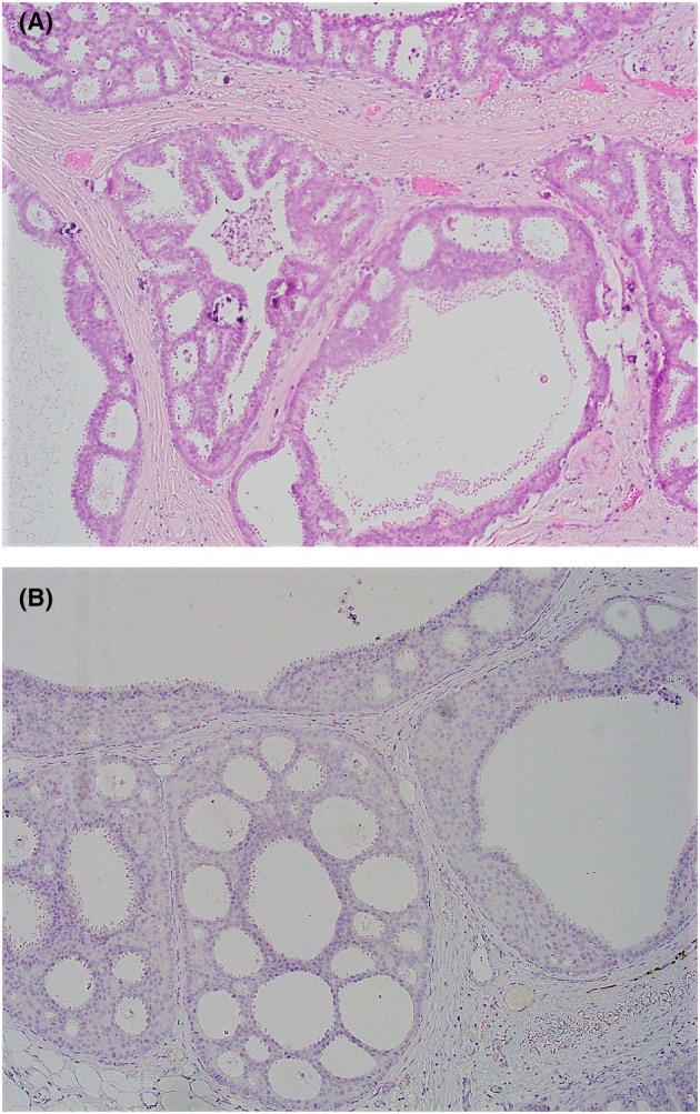
Absent myoepithelium with DCIS architecture. Lesions with DCIS architecture and no demonstrable myoepithelial cells are an unresolved issue in the 6th edition. A surrounding basement membrane supports DCIS. Absence of both myoepithelial markers and basement membrane does not establish invasion without a recognizable invasive pattern. The same rule for encapsulated and solid papillary carcinoma is covered in Special-Type Breast Carcinomas (Quinn et al. 2026).
Grade and genome. Non-high-grade DCIS is generally ER positive and HER2 negative with fewer copy number alterations. High-grade DCIS shows recurrent gains of MYC (8q22–24), CCND1 (11q13) and ERBB2 (17q12). PIK3CA and TP53 are the most frequently mutated genes (Abreu et al. 2023).
CAP reporting of DCIS on biopsy and resection
The CAP DCIS biopsy protocol (version 1.1.0.0) and resection protocol (version 4.5.0.1) were posted in June 2026 with WHO 6th edition updates (CAP DCIS biopsy protocol 2026; CAP DCIS resection protocol 2026).
Element
Biopsy protocol
Resection protocol
Source
Histologic type
Core; multi-select (DCIS, Paget disease, features of encapsulated or solid papillary carcinoma in situ)
Core; multi-select
CAP DCIS biopsy protocol 2026; CAP DCIS resection protocol 2026
Size (extent)
Optional; largest span in a single core
Core; estimated size (extent) of at least a stated value in mm
CAP DCIS biopsy protocol 2026; CAP DCIS resection protocol 2026
Architectural pattern
Optional
Optional
CAP DCIS biopsy protocol 2026; CAP DCIS resection protocol 2026
Nuclear grade
Core; multi-select
Core; multi-select
CAP DCIS biopsy protocol 2026; CAP DCIS resection protocol 2026
Necrosis
Core: not identified, focal or central
Core: not identified, focal or central
CAP DCIS biopsy protocol 2026; CAP DCIS resection protocol 2026
Microcalcifications
Optional
Optional
CAP DCIS biopsy protocol 2026; CAP DCIS resection protocol 2026
Additional lesions (LCIS types, ALH, ADH, FEA)
Optional
Optional; extent of LCIS optional
CAP DCIS biopsy protocol 2026; CAP DCIS resection protocol 2026
Margins
Not applicable
Core
CAP DCIS resection protocol 2026
Regional lymph nodes and pTNM
Not applicable
Core when applicable; AJCC 8th edition
CAP DCIS resection protocol 2026
Changes in 2026. The 2026 revisions made histologic type and nuclear grade multi-select and added an additional lesions question. The biopsy protocol added the extent of DCIS in a limited biopsy (CAP DCIS biopsy protocol 2026; CAP DCIS resection protocol 2026).
Accreditation. The resection protocol is required for CAP accreditation from March 2027. The biopsy protocol is recommended and is not required for accreditation (CAP DCIS resection protocol 2026; CAP DCIS biopsy protocol 2026).
Scope. The DCIS protocols cover DCIS without invasion or microinvasion, Paget disease without invasive carcinoma, and encapsulated and solid papillary carcinoma without invasive carcinoma (CAP DCIS resection protocol 2026).
Incidental DCIS. DCIS found in a biopsy performed for a benign lesion, such as a palpable fibroadenoma, is documented as incidental (CAP DCIS resection protocol 2026).
pT category and lymph nodes in DCIS
pT categories. DCIS, including Paget disease with underlying DCIS, is classified as pTis (DCIS) (CAP DCIS resection protocol 2026). Encapsulated and solid papillary carcinomas without conventional invasive carcinoma are also classified as pTis (DCIS) (CAP DCIS resection protocol 2026; Special-Type Breast Carcinomas). The pTis (Paget) category and the removal of LCIS from AJCC staging are covered in Breast Cancer Staging, Margins, Sentinel Lymph Nodes and Post-Neoadjuvant Assessment.
Multiple and bilateral DCIS. Multiple simultaneous areas of DCIS in one breast remain Tis. Bilateral DCIS is classified separately for each breast (CAP DCIS resection protocol 2026).
Indications for node sampling. Lymph nodes may be sampled in extensive DCIS, after a biopsy that raised concern for invasion, when imaging or clinical findings suggest invasion, and with a planned mastectomy (CAP DCIS resection protocol 2026).
Tumor cells in nodes. Most tumor cells in nodes of patients with DCIS are isolated tumor cells or are displaced by a previous procedure. Epithelium displaced after core biopsy is not classified as isolated tumor cells or micrometastasis. A larger metastasis is an indication for additional sampling of the breast for invasion (CAP DCIS resection protocol 2026).
Distant metastasis. Distant metastasis with DCIS alone is very unusual and indicates an unsampled invasive carcinoma (CAP DCIS resection protocol 2026).
Superficial margins. Breast tissue can remain in skin flaps beyond a superficial margin (CAP DCIS resection protocol 2026).
Extent of DCIS at the margin
Definition
Source
Focal
DCIS at the margin in an area <1 mm in 1 block
CAP DCIS resection protocol 2026
Minimal or moderate
Between focal and extensive
CAP DCIS resection protocol 2026
Extensive
DCIS at the margin over ≥15 mm, in ≥5 low-power fields or in ≥8 blocks
CAP DCIS resection protocol 2026
Significance of the extent at the margin. The extent of margin involvement is associated with the likelihood of residual disease (CAP DCIS resection protocol 2026).
Natural history and recurrence risk of DCIS
Natural history of low-grade DCIS. Among 45 women with low-grade DCIS treated by biopsy only, 16 (36%) developed invasive carcinoma, all in the same breast and quadrant (Sanders et al. 2015). In the earlier report on 28 of these women, invasive carcinoma developed up to 42 years after biopsy (Sanders et al. 2005).
Recurrence after breast-conserving surgery. About 50% of recurrences after breast-conserving surgery are invasive. Radiation reduces ipsilateral recurrence by about 50% (Abreu et al. 2023).
Risk factors. Younger age, positive margins, size, grade and comedo necrosis are consistently related to recurrence (Virnig et al. 2010).
Tool or factor
Components
Result
Source
Van Nuys Prognostic Index (1996)
Size, margin width and pathologic classification, each scored 1–3 (total 3–9)
Scores 3–4: 8-year local recurrence-free survival 97–100% with or without radiation; scores 5–7: 17% benefit from radiation; scores 8–9: recurrence over 60% at 8 years
Silverstein et al. 1996
USC/Van Nuys Prognostic Index (2003)
Adds patient age to size, margin width and pathologic classification (total 4–12)
Scores 4–6: no significant benefit from radiation; scores 7–9: 12–15% benefit; scores 10–12: recurrence almost 50% at 5 years
Silverstein 2003
DCIS Score (12-gene assay)
7 cancer-related and 5 reference genes, ECOG E5194 patients treated by excision without radiation
10-year ipsilateral breast event risk 10.6% (low), 26.7% (intermediate) and 25.9% (high); invasive event risk 3.7%, 12.3% and 19.2%
Solin et al. 2013
DCIS Score in context. In multivariable analysis, the DCIS Score, tumor size and menopausal status were associated with the risk of an ipsilateral breast event (Solin et al. 2013).
Van Nuys Prognostic Index component scores
Predictor
Score 1
Score 2
Score 3
Source
Size (mm)
≤15
16–40
>40
Silverstein et al. 1996; Antonini et al. 2025
Margin width (mm)
≥10
1–9
<1
Silverstein et al. 1996; Antonini et al. 2025
Pathologic classification
Low or intermediate grade without necrosis
Intermediate grade with necrosis
High grade with or without necrosis
Silverstein et al. 1996; Antonini et al. 2025
Age (years), USC/VNPI only
>60
40–60
<40
Silverstein 2003; Antonini et al. 2025
The 1996 index sums the first 3 rows (3–9). The USC/VNPI adds age (4–12) (Silverstein 2003). Published tables differ slightly at the band edges (Antonini et al. 2025).
Differential diagnosis of DCIS
Entity
Main mimic
Distinguishing finding
Source
DCIS
Usual ductal hyperplasia
Homogeneous cell population and marker expression in DCIS
Pinder and Ellis 2003
DCIS in sclerosing adenosis
Invasive carcinoma
Myoepithelial cells around distorted nests; at least 2 markers
Quinn et al. 2026
DCIS
Lymphovascular tumor emboli
Myoepithelium and stromal reaction around DCIS; endothelial lining around emboli
Ductal carcinoma in situ in which the myoepithelial layer cannot be shown. A, cribriform DCIS with rigid, punched-out lumina, focal necrosis and calcifications (H&E). B, p63 shows no myoepithelial rim around the involved ducts; a few positive nuclei at lower right serve as an internal control (p63 immunostain). The duct architecture still supports DCIS rather than invasion. Magnification not stated.Image: Quinn Cecily et al., PMC open access (Histopathology. 2026 Apr 21;89(2):199–218. doi: 10.1111/his.70149) Figure 9, CC BY.
Lobular Neoplasia, Atypical Ductal Hyperplasia and Core Biopsy Upgrade
Definition. Non-invasive lobular neoplasia is a spectrum of atypical epithelial proliferations with cell dyshesion caused by loss or dysfunction of E-cadherin. It comprises atypical lobular hyperplasia (ALH), classic lobular carcinoma in situ (LCIS), florid LCIS and pleomorphic LCIS (WHO 5th edition, as summarized by Abreu et al. 2023). Atypical ductal hyperplasia (ADH) is a clonal proliferation with the cytologic and architectural features of low-grade ductal carcinoma in situ (DCIS) and a limited extent (Abreu et al. 2023). The WHO 6th edition (2026) lists non-invasive lobular neoplasia in Part 1 of the epithelial chapter and adds a new section on small diagnostic samples (Quinn et al. 2026). Entity criteria for lobular neoplasia and ADH follow the WHO 5th edition.
Classic lobular neoplasia: atypical lobular hyperplasia and LCIS
Cytology. Classic LCIS consists of a monomorphic population of small, round, polygonal or cuboidal cells with a thin rim of clear cytoplasm and a high nuclear-to-cytoplasmic ratio. The cells are loosely cohesive and regularly spaced. Intracytoplasmic lumina are often present. Mitoses, calcification and necrosis are uncommon (Simpson et al. 2003).
Type A and type B cells. Type A cells are small with scant cytoplasm and monotonous nuclei. Type B cells are larger, with more cytoplasm and slightly larger nuclei (Abreu et al. 2023). Type B nuclei are up to twice the size of a lymphocyte, and type A nuclei 1–1.5 times (Simpson et al. 2003).
Extent threshold (WHO 5th edition). Classic LCIS fills and distends more than 50% of the acini of a TDLU. ALH has identical cells involving less than 50% of the acini with minimal expansion (Abreu et al. 2023). The B3 consensus states the cut-off as 50% or more (Elfgen et al. 2023). Distension is defined as 8 or more cells across an acinus (Simpson et al. 2003).
Reproducibility. ALH and classic LCIS show no molecular difference, and their separation is poorly reproducible. The B3 consensus favors the combined term classic lobular neoplasia (Elfgen et al. 2023).
Pagetoid spread. Neoplastic cells extend along adjacent ducts between the intact epithelium and the basement membrane (Simpson et al. 2003). ALH and LCIS often coexist and may involve ducts in a pagetoid pattern (Abreu et al. 2023).
Detection. Classic lobular neoplasia has no specific clinical or gross features and is usually an incidental finding (Simpson et al. 2003). It is found in 0.5–2.9% of core needle biopsies (CNB) and vacuum-assisted biopsies (VAB) performed for imaging findings (Elfgen et al. 2023).
Distribution and age. Classic LCIS is multicentric in up to 85% of cases and bilateral in 30–67%. Classic lobular neoplasia has a median age at diagnosis of 50–55 years (Abreu et al. 2023).
E-cadherin, p120 and molecular features of lobular neoplasia
E-cadherin. E-cadherin is encoded by CDH1 (16q22.1). Lobular neoplasia shows loss of membranous E-cadherin and β-catenin and a cytoplasmic distribution of p120 catenin. Benign ductal cells and DCIS show strong uniform membranous staining for all three (Abreu et al. 2023).
Frequency of loss. Loss of the adherens junction proteins (E-cadherin, β-catenin and p120) occurs in more than 80% of lobular neoplasia (Elfgen et al. 2023).
Coexisting LCIS and DCIS. Some lesions show a combination of markers, indicating coexistence of LCIS and low-grade solid DCIS in one duct-lobular unit. Both diagnoses are then given (Simpson et al. 2003).
Clonality. LCIS is a clonal proliferation with recurrent 16q loss and 1q gain. The most commonly mutated genes are CDH1 (81%), PIK3CA (41%) and CBFB (12%) (Abreu et al. 2023).
Relation to invasive lobular carcinoma. LCIS and invasive lobular carcinoma can be clonally related. Lobular neoplasia is therefore a high-risk lesion and a non-obligate precursor (Abreu et al. 2023).
Marker
Classic lobular neoplasia
Low-grade solid DCIS
Source
E-cadherin
Membranous staining lost
Strong uniform membranous
Abreu et al. 2023
β-catenin
Membranous staining lost
Membranous
Abreu et al. 2023
p120 catenin
Cytoplasmic
Membranous
Abreu et al. 2023
34βE12
Usually positive
Usually negative
Simpson et al. 2003
ER
Strong and diffuse
Generally positive in non-high-grade DCIS
Abreu et al. 2023
HER2
Not overexpressed
Generally negative in non-high-grade DCIS
Abreu et al. 2023
Lobular neoplasia as a risk indicator and non-obligate precursor
Relative risk. After classic LCIS, the relative risk of subsequent breast cancer is 8–10 times that of women without the lesion. The absolute risk is 1–2% per year. After ALH, the relative risk is 4–6 times (Abreu et al. 2023).
MSKCC LCIS cohort. Among 1,060 women with LCIS under surveillance, 150 developed 168 breast cancers at a median follow-up of 81 months. The cancers were ipsilateral in 63%, contralateral in 25% and bilateral in 12%. The annual incidence was 2%. Chemoprevention lowered the 10-year cumulative risk from 21% to 7% (King et al. 2015).
Histology of later cancers. The cancers after LCIS were DCIS in 35%, invasive ductal carcinoma in 29% and invasive lobular carcinoma in 27% (King et al. 2015).
Volume of disease. The ratio of slides with LCIS to all slides reviewed was associated with later breast cancer (King et al. 2015).
ALH laterality. Among 252 women with ALH treated by biopsy only, 20% developed invasive cancer. The relative risk was 3.1. Invasive cancer arose in the ipsilateral breast in 68% and the contralateral breast in 24% (Page et al. 2003).
Comedo necrosis in classic LCIS. Comedo necrosis in classic LCIS may also be considered a non-classic variant (CAP DCIS resection protocol 2026).
Pleomorphic LCIS series. In 24 cases of pleomorphic LCIS, the cells were medium to large with moderate to marked pleomorphism. All 23 tested cases were ER positive, and E-cadherin was negative in all 24 (Sneige et al. 2002).
Genome. Pleomorphic and florid LCIS show 16q loss in 94.7% and 1q gain in 81.3% of cases. CDH1 was mutated in 94.3%, ERBB2 in 54.3% and PIK3CA in 42.9% (Ferrucci et al. 2026).
Imaging. Calcifications were present in 78.7% of pooled cases, architectural distortion in 17.2% and a mass in 19.9% (Ferrucci et al. 2026).
Upgrade on core biopsy. In a systematic review of 38 studies, the pooled upgrade rate of pure pleomorphic or florid LCIS on core biopsy was 35.3%, mostly to invasive carcinoma (28.8%). Pleomorphic LCIS upgraded in 35.1% and florid LCIS in 33.3% (Ferrucci et al. 2026).
Margins and recurrence. Among 258 pure cases with follow-up, local recurrence was 38.2% with positive margins, 20.0% with close margins (<2 mm) and 3.0% with negative margins (Ferrucci et al. 2026).
Management. Complete surgical excision with negative margins is advised. The NCCN recommends excision with negative margins for non-classic LCIS on core biopsy (Ferrucci et al. 2026). The role of radiation and endocrine therapy is undefined (Ferrucci et al. 2026).
B category. In Europe, pleomorphic and florid LCIS are usually categorized as B5a. The UK Royal College of Pathologists categorizes florid LCIS as B4 (Ferrucci et al. 2026). The second and third B3 consensus conferences classify non-classic lobular neoplasia, including pleomorphic, florid and apocrine forms, as B5a. The second consensus recommends open excision (Rageth et al. 2019; Elfgen et al. 2023).
Staging. Pleomorphic LCIS is not included in the pTis classification. Evidence is insufficient for definitive treatment recommendations (CAP DCIS resection protocol 2026).
LCIS in AJCC 8th edition staging. LCIS was removed from TNM staging in the AJCC 8th edition. The pT categories for in situ disease in the CAP DCIS resection protocol are pTis (DCIS) and pTis (Paget) (CAP DCIS resection protocol 2026).
Atypical ductal hyperplasia
Cytology and architecture. ADH cells are monomorphic with round nuclei and dense chromatin. They are evenly spaced and form rigid bridges, arcades and bars, bulbous micropapillae or well-developed cribriform spaces (Abreu et al. 2023). Small foci of necrosis may occur in ADH and do not by themselves indicate DCIS (Pinder and Ellis 2003).
Immunophenotype. ADH is almost always negative for basal cytokeratins (CK5, CK5/6, CK14) and uniformly ER positive (Elfgen et al. 2023). The UDH-versus-ADH comparison is tabulated in Benign Epithelial Proliferations of the Breast.
Extent thresholds (WHO 5th edition). Two thresholds separate ADH from low-grade DCIS. Page and colleagues classify a lesion with complete involvement of fewer than 2 separate duct spaces as ADH (Pinder and Ellis 2003; Sapino et al. 2021). Tavassoli and Norris classify a lesion of 2 mm or less in contiguous ducts as ADH (Tavassoli and Norris 1990; Sapino et al. 2021). The WHO 5th edition accepts both cut-offs and describes them as arbitrary (Sapino et al. 2021; Elfgen et al. 2023).
DCIS definition in the Nashville studies. DCIS was defined as complete involvement of at least 2 adjacent spaces, at least one a true duct, measuring at least 2.0 mm (Sanders et al. 2015).
Usual size. In practice, ADH is usually small and focal, measuring less than 2–3 mm (Pinder and Ellis 2003). The upgrade rate after "focal" ADH and after "full" ADH (the upper limit of ADH) does not differ (10% compared with 11%) (Elfgen et al. 2023).
ADH on core biopsy. The size criteria come from excision specimens and are general guidelines. On core biopsy they are applied conservatively because the whole lesion may not be visible (Elfgen et al. 2023). European and UK guidelines use the term atypical intraductal epithelial proliferation (AIDEP) for core biopsies in which ADH and DCIS cannot be separated. The term is not part of the WHO classification (Elfgen et al. 2023).
Risk. ADH is associated with a relative risk of invasive breast carcinoma of 4–5 times, approaching 6 times in premenopausal women and 10 times with a first-degree relative with breast cancer (Pinder and Ellis 2003). The absolute risk is about 1% per year (Abreu et al. 2023).
Frequency. ADH is seen in 4% of symptomatic benign biopsies and in 31% of biopsies for screen-detected benign microcalcifications (Pinder and Ellis 2003).
Atypical ductal hyperplasia compared with low-grade DCIS
Feature
ADH
Low-grade DCIS
Source
Cytology
Same monomorphic cells
Same monomorphic cells; nuclei 1.5–2 times a red blood cell
Abreu et al. 2023; CAP DCIS resection protocol 2026
Extent
Complete involvement of fewer than 2 spaces (Page), or 2 mm or less (Tavassoli) (WHO 5th edition)
Complete involvement of 2 or more spaces (Page; at least 2.0 mm in the Nashville studies), or more than 2 mm (Tavassoli)
Pinder and Ellis 2003; Sapino et al. 2021; Sanders et al. 2015
Involvement of spaces
Partial involvement or uniform involvement of limited extent
Complete involvement
Elfgen et al. 2023; Sanders et al. 2015
Molecular profile
Clonal; loss of heterozygosity similar to low-grade DCIS
Clonal
Pinder and Ellis 2003
Core biopsy category
B3 (AIDEP when DCIS cannot be excluded)
B5a
Elfgen et al. 2023; Thiruvarasu and Kulkarni 2026
Imaging-pathology correlation of ADH and lobular neoplasia
BI-RADS assessment categories. Category 0 is incomplete. Categories 1 and 2 are negative and benign. Category 3 is probably benign (>0% to ≤2% probability), with 6-month follow-up. Category 4 covers >2% to <95% and is divided into 4A (>2% to ≤10%), 4B (>10% to ≤50%) and 4C (>50% to <95%). Category 5 is highly suggestive of malignancy (≥95%). Category 6 is biopsy-proven malignancy (Kaplan and Aydemir 2026).
Calcification morphology. Fine linear or fine linear-branching calcifications have positive predictive values of about 50–70%. Amorphous, coarse heterogeneous and fine pleomorphic calcifications have values of about 7–29% (Kaplan and Aydemir 2026).
ADH on imaging. ADH calcifications are often fine pleomorphic, linear or segmental. Most B3 lesions presenting as calcifications are ADH (Elfgen et al. 2023).
Classic lobular neoplasia on imaging. Classic lobular neoplasia has no typical imaging pattern. Calcifications that lead to its diagnosis are usually located in coexisting lesions (Elfgen et al. 2023).
Concordance and upgrade. Radiologic-pathologic discordance is a risk factor for upgrade of ADH and the strongest predictor of invasive cancer after classic lobular neoplasia (Elfgen et al. 2023). When a different benign lesion explains the imaging target, incidental classic lobular neoplasia has an excisional upgrade rate of about 1–4% (Rageth et al. 2019).
Multidisciplinary review. Management of B3 lesions is decided in a multidisciplinary conference that correlates histologic and radiologic findings (Elfgen et al. 2023). The 6th edition emphasizes triple assessment with correlation of pathologic, clinical and imaging findings (Quinn et al. 2026).
Small diagnostic samples and the B-coding system
WHO 6th edition section. The new 6th-edition section on small diagnostic samples covers fine-needle aspiration biopsy, CNB and VAB, the imaging used to guide them and the evaluation of different imaging abnormalities. It emphasizes standardized reporting with a classification system such as the B-coding system of the UK non-operative guidelines (Quinn et al. 2026).
Frequency of B3. About 7% of screen-detected core biopsies are categorized as B3 (Pinder et al. 2018).
Category
Meaning
Examples
Source
B1
Unsatisfactory or normal tissue only
Normal ducts, lobules, stroma and fat; focal apocrine change or microcysts; lactational change
Thiruvarasu and Kulkarni 2026
B2
Benign
Fibroadenoma, fibrocystic disease, mastitis
Thiruvarasu and Kulkarni 2026
B3
Uncertain malignant potential
ADH or AIDEP, classic lobular neoplasia, FEA, radial scar, papillary lesions, fibroepithelial lesions with cellular stroma, mucocele-like lesions
Thiruvarasu and Kulkarni 2026; Elfgen et al. 2023
B4
Suspicious for malignancy
Atypical cells away from the core, small suspicious foci
Acceptable risk. The B3 consensus conferences set upper limits of 5% upgrade to invasive carcinoma and 10% upgrade to DCIS for radiologic surveillance (Elfgen et al. 2023).
Vacuum-assisted excision. UK guidelines allow vacuum-assisted excision (VAE) with larger needles and at least 12 cores of 7–8 gauge, about 4 g of tissue, as an alternative to open excision (Elfgen et al. 2023).
Groups of B3 lesions. In an Erlangen series of 192 patients with B3 lesions treated by open excision, ADH, FEA and classic lobular neoplasia together upgraded in 17.5%. Fibroepithelial lesions, papillomas and radial scars upgraded less often (Heindl et al. 2025).
Upgrade and management of B3 lesions
The third International Consensus Conference (2022, published 2023) gives the recommendations in the table. Upgrade means DCIS or invasive carcinoma at excision. Each rate depends on the biopsy device, the volume sampled and radiologic-pathologic concordance (Elfgen et al. 2023).
Lesion
Upgrade data (study; biopsy type)
Concordance condition
Management after CNB
Management after VAB
Source
ADH
25.2% after open excision, 5% of upgrades invasive (MIBB database; mainly VAB); 31–78% after CNB and 19–41% after VAB; no more than 19% with 7–8 G VAB
Discordance, residual calcifications, size >15 mm, age >50 years and multifocal ADH favor excision
Open excision
Open excision; VAE or radiologic follow-up in selected cases after multidisciplinary review
Elfgen et al. 2023
FEA
1–8% at excision in major reviews; 16% in the MIBB database; up to 30% after CNB
Discordance, mass lesions and residual calcifications favor open excision
VAE to complete removal of the visible lesion
Radiologic follow-up when >90% of the target is removed
Elfgen et al. 2023
Classic lobular neoplasia
About 20% on average (range 4–67%); 4% after 7 G mammographic VAB; about 1–4% when incidental and concordant
Lower when another lesion explains the target; discordance is the strongest predictor
Open excision or VAE
Radiologic follow-up when the lesion is removed
Elfgen et al. 2023; Rageth et al. 2019
Radial scar
8% after open excision (MIBB); <10% without atypia and 36% with atypia (NHS data); 0.9–1.6% after VAE; 5% after 14 G CNB and 1% after 8–11 G VAB
Correlation with imaging decides management
Open excision or VAE
Radiologic follow-up when completely removed
Elfgen et al. 2023
Papilloma
1–9%; pooled median 2.3% (mostly CNB); median 26.9% with atypia
Size >1 cm, symptoms, peripheral location, calcifications and multiplicity predict upgrade
Open excision or VAE
No further intervention when completely removed
Elfgen et al. 2023
Benign and borderline phyllodes tumor
Upgrade to malignancy uncommon after CNB or VAB
Preoperative multidisciplinary discussion
Open excision (free margins for borderline tumors)
Open excision if screen-detected; follow-up if incidental and removed
Elfgen et al. 2023
Mucocele-like lesion
2 of 25 (8.0%) without atypia and 4 of 23 (17.4%) with atypia upgraded to DCIS (CNB)
All upgraded cases had residual calcifications after biopsy
Not addressed by the consensus
Not addressed by the consensus
Towne et al. 2022
Pleomorphic or florid LCIS
Pooled 35.3% (core biopsy)
Not applicable; classified as B5a in Europe
Surgical excision with negative margins
Surgical excision with negative margins
Ferrucci et al. 2026; Rageth et al. 2019
Follow-up. At least annual radiologic follow-up is recommended after a B3 lesion. Annual mammography is advised for patients with lobular neoplasia or ADH (Elfgen et al. 2023).
ADH risk over time. ADH increases the risk of ipsilateral or contralateral invasive breast cancer by up to 30% over 25 years (Elfgen et al. 2023).
Definition. A mucocele-like lesion consists of dilated mucin-filled ducts or cysts lined by cuboidal epithelium. Rupture expels acellular mucin into the periductal stroma (Dang et al. 2023).
Original description. Rosen described ruptured mucin-containing cysts that discharge secretions and epithelium into the stroma, analogous to mucocele of the minor salivary glands. The lesion mimics mucinous (colloid) carcinoma (Rosen 1986).
Distinction from mucinous carcinoma. Acellular mucin in the stroma separates a mucocele-like lesion from mucinous carcinoma (Dang et al. 2023). Mucinous carcinoma is covered in Special-Type Breast Carcinomas.
Presentation and atypia. Most mucocele-like lesions present as calcifications. Nearly half were associated with atypia, almost always ADH, in a CNB series of 78 lesions (Towne et al. 2022).
Upgrade predictors. All upgraded cases showed coarse calcifications on the core and residual calcifications after biopsy (Towne et al. 2022). In 34 mucocele-like lesions with atypia, 2 of 27 excisions (7.4%) upgraded to carcinoma. Upgraded cases had more ADH foci in the needle biopsy (Sta Ines et al. 2025).
B category. Mucocele-like lesions are included in the B3 category (Thiruvarasu and Kulkarni 2026).
Differential diagnosis of lobular neoplasia and ADH
Entity
Main mimic
Distinguishing finding
Source
Classic LCIS
Low-grade solid DCIS
E-cadherin loss and cytoplasmic p120; DCIS shows cohesion and secondary lumina
Abreu et al. 2023; Simpson et al. 2003
Pleomorphic LCIS
High-grade DCIS
E-cadherin loss in discohesive cells
Abreu et al. 2023; Sneige et al. 2002
Florid LCIS
Low- to intermediate-grade solid DCIS
E-cadherin loss with massive acinar distension
Abreu et al. 2023
ADH
Low-grade DCIS
Extent (2 completely involved spaces or 2 mm, WHO 5th edition)
Pinder and Ellis 2003
Mucocele-like lesion
Mucinous carcinoma
Acellular extravasated mucin
Dang et al. 2023; Rosen 1986
Fibroadenoma, Phyllodes Tumor and Other Fibroepithelial Lesions of the Breast
Definition. Fibroepithelial lesions of the breast are biphasic tumors of benign epithelium and a proliferating stroma. They range from fibroadenoma, the most common breast tumor in women under 30, to malignant phyllodes tumor (Lim et al. 2014; Tan et al. 2016). The WHO 6th edition (2026) classifies phyllodes tumors as benign, borderline or malignant and lowers the threshold for malignancy to at least 4 of 5 adverse stromal features. The 4th and 5th editions required all 5 (Quinn et al. 2026). Both lesion types share MED12 exon 2 mutations in the stromal cells. TERT promoter mutations and mutations in cancer genes accumulate with increasing phyllodes grade (Lim et al. 2014; Piscuoglio et al. 2016).
Edition status. The 6th-edition summary does not address fibroadenoma types, periductal stromal tumor, hamartoma or the adenomas (Quinn et al. 2026).
Fibroadenoma of the breast
Histology (WHO 6th edition). Fibroadenoma has a well-defined border, variable stromal cellularity from scant to uncommonly cellular, no stromal atypia, no stromal overgrowth and usually no mitoses (Quinn et al. 2026).
Growth patterns. All but one usual-type fibroadenoma in an adolescent series had an intracanalicular growth pattern. Juvenile fibroadenomas had a pericanalicular pattern with a well-developed glandular component (Ross et al. 2017).
Young patients. Fibroadenomas in young people can show increased stromal cellularity and mitoses (Quinn et al. 2026). In adolescents, mean stromal mitoses were 1.3 per 10 HPF in usual fibroadenomas (range 0–6) and 1.8 per 10 HPF in juvenile fibroadenomas (range 0–7) (Ross et al. 2017).
Lactational change. Lactational changes occur within fibroadenomas, and foci of tubular adenomatous change are present in otherwise typical fibroadenomas (O'Hara and Page 1985).
Recurrence. In 48 adolescents, none of the 24 fibroadenomas with follow-up recurred after lumpectomy, although 18 fibroadenomas had a positive final margin. Two phyllodes tumors recurred, both excised with margins under 1 mm (Ross et al. 2017). After ultrasound-guided vacuum-assisted excision, 8 of 110 fibroadenomas (7%) showed a residual or recurrent lesion on imaging at 6 months (Carriero et al. 2023). In a systematic review of multiple fibroadenomas, recurrence was noted in 15.4% of patients, and 72% of the included studies were low-level evidence (Math et al. 2026).
Complex fibroadenoma. Fibroadenomas with cysts, sclerosing adenosis, epithelial calcifications or papillary apocrine change are classified as complex (Dupont et al. 1994).
Breast cancer risk. In 1835 patients, the relative risk of invasive breast cancer was 2.17 for fibroadenoma and 3.10 for complex fibroadenoma. Proliferative disease in the adjacent parenchyma raised the relative risk to 3.88. Two thirds of patients had noncomplex fibroadenomas without a family history and no increased risk (Dupont et al. 1994).
Juvenile fibroadenoma
Morphology. Juvenile fibroadenomas had circumscribed borders, a pericanalicular pattern, uniform stromal cellularity with short collagen bundles and no distinction between intralobular and extralobular stroma (Ross et al. 2017).
Epithelium. Micropapillary epithelial hyperplasia was present in 30% (Ross et al. 2017).
Absence of phyllodes features. No juvenile fibroadenoma showed stromal overgrowth or nuclear atypia (Ross et al. 2017).
Proportion. Juvenile fibroadenomas formed 43% of fibroepithelial lesions in patients 18 years old or younger (Ross et al. 2017).
MED12 mutations in fibroadenoma
Frequency.MED12 exon 2 mutations were present in 58 of 98 fibroadenomas (59%), and 71% of mutations involved codon 44 (Lim et al. 2014).
Cell of origin. Laser capture microdissection placed the mutations in stromal cells. Epithelial cells were wild type (Lim et al. 2014).
Uterine leiomyoma. The MED12 mutation spectrum in fibroadenoma is nearly identical to that of uterine leiomyoma (Lim et al. 2014).
RARA.RARA mutations, like MED12 mutations, occur in both fibroadenomas and phyllodes tumors (Tan J et al. 2015).
Phyllodes tumor of the breast
Definition. Phyllodes tumors show an exaggerated intracanalicular pattern with leaf-like fronds protruding into cystically dilated spaces and stromal hypercellularity. Elongated branching cleft-like ducts in cellular stroma (a staghorn pattern) favor phyllodes tumor when fronds are poorly developed (Tan et al. 2016).
Distribution by grade. Benign phyllodes tumors form 60–75% of phyllodes tumors, borderline 15–26% and malignant 8–20% (Quinn et al. 2026). In a series of 605 tumors, 72.7% were benign, 18.4% borderline and 8.9% malignant (Tan et al. 2012).
Grading of phyllodes tumor (WHO 6th edition)
Feature
Fibroadenoma
Benign phyllodes tumor
Borderline phyllodes tumor
Malignant phyllodes tumor
Border
Well defined
Well defined
Well defined; focal permeation allowed
Permeative
Stromal cellularity
Scant to uncommonly cellular; usually uniform
Usually mild; can be uneven or diffuse
Usually moderate; can be uneven or diffuse
Usually marked and diffuse
Stromal atypia
None
None or mild
Mild or moderate
Marked
Stromal mitoses
Usually none
Usually under 2.5/mm² (under 5 per 10 HPF)
Usually 2.5 to under 5/mm² (5–9 per 10 HPF)
Usually 5/mm² or more (10 or more per 10 HPF)
Stromal overgrowth
Absent
Absent
Absent or very focal
Often present
Malignant heterologous elements
Absent
Absent
Absent
Can be present
Source: Quinn et al. 2026 (Table 1). HPF of 0.5 mm diameter and 0.2 mm² area.
Malignant phyllodes tumor, 6th edition. Malignant phyllodes tumor requires at least 4 of the 5 adverse features: permeative border, marked and diffuse stromal cellularity, marked stromal atypia, at least 10 stromal mitoses per 10 HPF (5/mm²) and stromal overgrowth. The criteria used to support the malignant grade are documented in the report (Quinn et al. 2026).
Malignant phyllodes tumor, 4th and 5th editions. Without malignant heterologous elements, all 5 adverse features were required. A tumor lacking any one was classified as borderline (Quinn et al. 2026; Tan et al. 2025).
Reason for the change. In 2 studies with 140 malignant phyllodes tumors, rare tumors with only 2 or 3 adverse features metastasized (Quinn et al. 2026). An international survey found that 85% of breast pathologists regarded the all-5 rule as too rigid (Tan et al. 2025).
Stromal overgrowth. Stromal overgrowth is one low-power field (4× objective with 10× eyepiece) containing only stroma without epithelium (Quinn et al. 2026).
Mitotic count. WHO does not state it explicitly, but the threshold of at least 10 per 10 HPF is based on the highest count in the tumor (Tan et al. 2025).
Grading cellularity and atypia. Cellularity is assessed in the most cellular zones. Mild cellularity has evenly spaced nuclei that do not touch. Marked cellularity has confluent areas of overlapping nuclei. Marked atypia has marked variation in nuclear size, coarse chromatin, irregular membranes and nucleoli (Tan et al. 2016).
Carcinoma within a phyllodes tumor. In situ or invasive carcinoma within a phyllodes tumor does not change its grade and is reported separately (Tan et al. 2025).
Malignant heterologous elements in phyllodes tumor
Rule in both editions. Malignant heterologous elements, except well-differentiated liposarcoma, allow a malignant classification without the other adverse features (Quinn et al. 2026; Tan et al. 2025).
Types. Osteosarcoma, chondrosarcoma, rhabdomyosarcoma, and pleomorphic and myxoid or round cell liposarcoma qualify (Tan et al. 2025).
Well-differentiated liposarcoma. Well-differentiated liposarcoma in phyllodes tumors lacks CDK4 and MDM2 amplification. It does not establish malignancy alone, and such tumors often meet borderline criteria (Tan et al. 2025).
Gray-zone phyllodes tumors between grades
Benign–borderline zone. A well-sampled tumor with mostly benign features and slight deviation into borderline criteria can be classified as favoring benign phyllodes tumor. Examples are mitoses above 5 per 10 HPF or focal border irregularity. A comment lists the parameters outside the benign range (Quinn et al. 2026).
Borderline category. A tumor with most features in the borderline range is classified as borderline (Quinn et al. 2026).
Borderline–malignant zone. A tumor with mostly borderline features and fewer than 4 adverse criteria can be classified as borderline with an explanatory comment (Quinn et al. 2026).
Sampling, 6th edition. Large tumors and tumors with features suggesting malignancy are sampled with at least 2 blocks per 1 cm of the greatest tumor dimension. The malignant component can be very focal (Quinn et al. 2026).
Sampling, 2016 consensus. A 2016 consensus review recommended at least 1 section per cm of maximal tumor size, with additional sections of grossly heterogeneous areas (Tan et al. 2016).
Molecular findings in fibroepithelial tumors
Alteration
Fibroadenoma
Benign phyllodes tumor
Borderline phyllodes tumor
Malignant phyllodes tumor
Source
MED12 exon 2 mutation
59%
65.1%
65.6%
42.8%
Lim et al. 2014; Tan et al. 2016
TERT promoter mutation or TERT amplification
0%
18%
57%
68%
Piscuoglio et al. 2016
TP53, RB1, SETD2 and EGFR mutations
Not tested
Not found (6 tumors sequenced)
Present
Present
Piscuoglio et al. 2016
MED12 rates in phyllodes tumors vary between series; one series found 80%, 64% and 23% in benign, borderline and malignant tumors (Piscuoglio et al. 2016).
TERT as a diagnostic marker.TERT alterations were restricted to the stromal component. Their presence separated phyllodes tumors from fibroadenomas with a positive predictive value of 100%. Sensitivity was 39% because many benign phyllodes tumors lack them (Piscuoglio et al. 2016).
Other phyllodes genes.FLNA, SETD2 and KMT2D mutations occur in phyllodes tumors. Borderline and malignant tumors have additional mutations in cancer-associated genes (Tan J et al. 2015).
Malignant phyllodes tumor series. In 24 malignant phyllodes tumors, TP53 mutations were found in 58.3%, NF1 mutations in 45.8% and CDKN2A/B alterations in 33.3% (Nozad et al. 2017).
Targetable alterations.KIAA1549::BRAF or FGFR3::TACC3 fusions were found in 2 of 24 malignant phyllodes tumors (Nozad et al. 2017).
MED12 and outcome.MED12-mutated phyllodes tumors were associated with longer disease-free survival (Tan et al. 2016).
Recurrence, metastasis and margins of phyllodes tumor
Recurrence by grade. Reported local recurrence rates are 10–17% for benign, 14–25% for borderline and 23–30% for malignant phyllodes tumors (Tan et al. 2016).
Metastasis. Borderline phyllodes tumors have a metastatic rate of 1.6% and malignant tumors 16.7% (Tan et al. 2025). Metastases consist of malignant stroma without epithelium and involve mostly lung and skeleton (Tan et al. 2016).
Nomogram. In 605 phyllodes tumors, stromal atypia, stromal overgrowth and surgical margins independently predicted outcome, and mitoses approached significance. The AMOS nomogram (atypia, mitoses, overgrowth, surgical margins) predicts recurrence-free survival at 1, 3, 5 and 10 years. Stromal hypercellularity and tumor border were not independently predictive (Tan et al. 2012).
Margin width. Evidence supporting a 10-mm margin is insufficient. Tumor on ink or a margin under 1 mm is a pragmatic definition of a positive margin (Tan et al. 2016).
Management by grade. Benign phyllodes tumors enucleated without margins can be managed conservatively. Recurrent and malignant tumors are excised with negative margins, and most borderline tumors are completely excised (Tan et al. 2016).
Axilla. Routine axillary dissection is not recommended because nodal disease is infrequent (Tan et al. 2016).
Cellular fibroadenoma versus benign phyllodes tumor on core biopsy
Interobserver agreement. Ten specialist breast pathologists agreed uniformly on fibroadenoma versus phyllodes tumor in only 2 of 21 cellular fibroepithelial lesions (Tan et al. 2016).
Core biopsy concordance. Core biopsy and excision agreed in 60% of phyllodes tumors in one study, and larger tumors were more often discordant (Tan et al. 2016).
Discriminating features. Leaf-like architecture and increased stromal cellularity separate benign phyllodes tumor from cellular fibroadenoma. Stromal multinucleated giant cells in fibroadenomas can be mistaken for phyllodes tumor (Tan et al. 2016).
Overlapping lesions. Cellular fibroadenoma, heterogeneous benign phyllodes tumor and myoid hamartoma overlap on core biopsy. A 2025 consensus proposes descriptive reporting with the UK/European B categories (Rakha et al. 2025). B codes and management after core biopsy are covered in Lobular Neoplasia, Atypical Ductal Hyperplasia and Core Biopsy Upgrade.
Term for equivocal cases. "Benign fibroepithelial neoplasm" can be used sparingly when the distinction cannot be made. It is not a separate diagnostic category (Tan et al. 2016).
Adolescents. Mitotic activity in fibroadenomas of adolescents can be substantial and is interpreted cautiously to avoid overdiagnosis of phyllodes tumor (Ross et al. 2017).
Periductal stromal tumor of the breast
Definition. Periductal stromal tumor is a biphasic tumor with a spindle cell stromal proliferation around open tubules and ducts and no phyllodes architecture (Burga and Tavassoli 2003). Burga and Tavassoli used the term periductal stromal sarcoma (Burga and Tavassoli 2003).
Criteria. The tumor forms one or more nodules separated by fat. Stromal mitoses are at least 3 per 10 HPF, and the stroma infiltrates the surrounding breast (Burga and Tavassoli 2003).
Periductal stromal hyperplasia. Bland nodular stroma cuffing ducts with no or minimal atypia and 0–2 mitoses per 10 HPF was classified as periductal stromal hyperplasia (Burga and Tavassoli 2003).
Immunohistochemistry. The stromal cells were at least focally CD34 positive (13 of 15) and CD117 positive (6 of 15), and negative for ER and PR (Burga and Tavassoli 2003).
Behavior. Two of 20 patients (10%) had recurrence or probable metastasis. A phyllodes pattern developed in a recurrence, which suggests evolution to phyllodes tumor. Excision with a rim of uninvolved tissue is required (Burga and Tavassoli 2003).
Hamartoma of the breast
Histology. Hamartomas are well demarcated with condensed fibrous tissue at the edge. Adipose tissue was present in all 25 cases (5–90%, mean 31%) and interlobular fibrosis in 21 (Tse et al. 2002).
Other findings. Benign epithelial hyperplasia was present in 10 cases and PASH or cystic ducts in 8 each. Two cases had DCIS confined to the hamartoma (Tse et al. 2002).
Small samples. Hamartomas have no specific histologic features. Core biopsy and fine-needle aspiration were largely insufficient or nonspecific, so clinical and radiologic correlation is required (Tse et al. 2002).
Tubular and lactating adenomas of the breast
Spectrum. Tubular adenomas, lactating adenomas and fibroadenomas with lactational change show overlapping histologic and clinical features (O'Hara and Page 1985).
Pregnancy association. The overlapping morphology and the strong association with pregnancy or lactation suggest a common pathogenesis (O'Hara and Page 1985).
Ectopic sites. Ectopic lactating adenomas occurred in the axilla, chest wall and vulva (O'Hara and Page 1985).
Differential diagnosis of fibroepithelial and spindle cell breast lesions
Lesion
Main mimic
Distinguishing finding
Source
Benign phyllodes tumor
Cellular fibroadenoma
Leaf-like fronds and increased stromal cellularity; TERT alteration when present
Tan et al. 2016; Piscuoglio et al. 2016
Juvenile fibroadenoma
Benign phyllodes tumor
Pericanalicular pattern, uniform stroma, no atypia or overgrowth
Benign Mesenchymal and Inflammatory Lesions of the Breast
Definition. Benign mesenchymal lesions of the breast include pseudoangiomatous stromal hyperplasia (PASH), myofibroblastoma, fibromatosis, granular cell tumor and benign vascular lesions. Inflammatory lesions include fat necrosis, acute mastitis and abscess, periductal mastitis and duct ectasia, granulomatous lobular mastitis, IgG4-related mastitis, diabetic mastopathy and the reaction to silicone. Many of these lesions form a firm mass that mimics carcinoma clinically or on imaging (Brown et al. 2011; Vasei et al. 2019). Spindle cell lesions are separated from metaplastic carcinoma and phyllodes tumor with an immunohistochemical panel (Tan et al. 2016).
The 6th-edition mesenchymal chapter is restricted to tumors occurring more commonly or principally in the breast. It focuses on fibromatosis, myofibroblastoma, granular cell tumor, PASH and vascular lesions classified by the ISSVA system. Entities that occur more commonly at other sites are covered in the WHO Soft Tissue and Bone volume (Quinn et al. 2026). The 5th-edition scope is given in Angiosarcoma, Lymphoma and Metastases in the Breast.
Pseudoangiomatous stromal hyperplasia (PASH) of the breast
Morphology. PASH consists of complex anastomosing slit-like spaces lined by slender spindle cells in the mammary stroma. Ultrastructurally, the spaces are separations of stromal collagen. Vascular channels are absent (Vuitch et al. 1986).
Frequency. Small foci are common in hyperplastic breast tissue of premenopausal women. A discrete mass with this pattern is regarded as exaggerated stromal hyperplasia (Vuitch et al. 1986).
Tumorous PASH. In 40 women aged 14–67 years (mean 37), PASH formed a palpable, well-circumscribed mass with a firm white-gray cut surface. It was usually diagnosed clinically as fibroadenoma (Powell et al. 1995).
Spectrum. Lesions range from classic slit-like spaces to fascicles of plump spindle cells that obscure the pseudoangiomatous pattern (Powell et al. 1995).
Immunohistochemistry. The spindle cells are vimentin and CD34 positive and factor VIII negative. Cellular fascicular lesions express desmin and actin. Progesterone receptor staining exceeds ER staining, and most stromal nuclei are ER negative (Powell et al. 1995).
Behavior. Five of 40 patients had ipsilateral recurrences. Simple excision is adequate initial treatment (Powell et al. 1995). There is no evidence that PASH is a precursor of angiosarcoma (Vuitch et al. 1986).
Feature
PASH
Angiosarcoma
Source
Spaces
Slit-like separations of collagen without true vascular channels
Vascular channels
Vuitch et al. 1986
Lining cells
Slender, bland spindle cells; CD34 positive, factor VIII negative
Original series. Sixteen tumors occurred in 11 men and 5 women with an average age of 63 years. They were nodular, well demarcated and did not engulf ducts or lobules (Wargotz et al. 1987).
Morphology. Uniform, slender, bipolar spindle cells form fascicular clusters separated by broad bands of hyalinized collagen. S100 and cytokeratin were negative (Wargotz et al. 1987).
Mammary-type myofibroblastoma. In 143 tumors, 66% occurred in males and only 10% in the breast. The inguinal region was the most common site (45%). Mean size was 6.6 cm (Howitt and Fletcher 2016).
Immunohistochemistry. CD34 was positive in 89% and desmin in 91%. Both were negative in 3% (Howitt and Fletcher 2016).
13q14 and Rb. Myofibroblastoma has rearrangement or deletion of 13q14, and Rb expression was lost in 92% of tested cases (Howitt and Fletcher 2016). Rb was deficient in 89% of mammary-type myofibroblastomas and intact in solitary fibrous tumor, myxoid liposarcoma and other mimics (Chen et al. 2012).
Epithelioid variant. Epithelioid myofibroblastoma consists of dyscohesive ER-positive epithelioid cells and can mimic invasive lobular carcinoma (Quinn et al. 2023).
Related tumors. Spindle cell lipoma, pleomorphic lipoma and cellular angiofibroma share 13q losses and Rb deficiency (Chen et al. 2012).
Behavior. No tumor with follow-up recurred, including tumors with positive margins (Howitt and Fletcher 2016).
Fibromatosis versus metaplastic spindle cell carcinoma
Fibromatosis. Breast fibromatoses are infiltrative and locally recurrent and do not metastasize (Abraham et al. 2002).
Wnt pathway. Nuclear β-catenin was present in 82% of 33 breast fibromatoses. Somatic APC/β-catenin pathway alterations were found in 79%: activating CTNNB1 mutations in 15 and APC alterations in 11 (Abraham et al. 2002).
Familial adenomatous polyposis. Breast fibromatosis occurs in some patients with familial adenomatous polyposis (Abraham et al. 2002).
Specificity of β-catenin. Nuclear β-catenin was found in all 8 fibromatoses, 23% of metaplastic carcinomas and the stroma of 94% of benign and 57% of malignant phyllodes tumors. No CTNNB1 mutation was found in 21 metaplastic carcinomas. β-catenin is not used as a single marker (Lacroix-Triki et al. 2010).
Keratin and p63. Diffuse cytokeratin or p63 expression in malignant spindle cells supports metaplastic carcinoma. Focal staining is interpreted with caution because phyllodes stromal cells can show it (Tan et al. 2016). Fibromatosis-like metaplastic carcinoma is the most important differential of a bland spindle cell lesion on core biopsy, and a broad-spectrum keratin such as MNF116 supports it (Quinn et al. 2023). Metaplastic carcinoma marker frequencies are given in Special-Type Breast Carcinomas.
Lesion
Cytokeratin and p63
Nuclear β-catenin
CD34
Desmin
Other
Source
Fibromatosis
Not reported
Positive (82–100%)
Not reported
Not reported
CTNNB1 or APC alteration
Abraham et al. 2002; Lacroix-Triki et al. 2010
Metaplastic spindle cell carcinoma
Diffuse positivity supports the diagnosis
Positive in 23%
Not reported
Not reported
No CTNNB1 mutation in 21 tested
Tan et al. 2016; Lacroix-Triki et al. 2010
Phyllodes tumor stroma
Focal staining possible
Positive in 57–94%
Positive; lower in high grade
Not reported
Leaf-like fronds
Tan et al. 2016; Lacroix-Triki et al. 2010
Myofibroblastoma
Cytokeratin negative
Not reported
Positive (89%)
Positive (91%)
Rb loss
Wargotz et al. 1987; Howitt and Fletcher 2016
Nodular fasciitis
Negative
Negative
Negative
Negative
SMA positive; USP6 fusion
Cloutier et al. 2021
PASH
Not reported
Not reported
Positive
Positive in cellular lesions
PR positive
Powell et al. 1995
Granular cell tumor of the breast
Origin and presentation. Granular cell tumor of the breast arises from Schwann cells. It mimics breast carcinoma clinically, radiologically and grossly (Brown et al. 2011).
Morphology. Tumor cells have distinctive granular eosinophilic cytoplasm with abundant lysosomes (Brown et al. 2011).
Immunohistochemistry. Tumor cells are positive for S100, CD68 and neuron-specific enolase (Brown et al. 2011). SOX10 expression was reported in 26 granular cell tumors (Karamchandani et al. 2012).
Malignancy. Malignant granular cell tumor is extremely rare (Brown et al. 2011).
Margins. In 13 patients, no tumor recurred over an average of 77 months, including tumors excised with positive margins. One patient had coexistent invasive ductal carcinoma (Papalas et al. 2011).
Benign vascular lesions of the breast
Perilobular hemangioma. A microscopic meshwork of thin-walled, dilated vascular channels is present within a lobule or in the extralobular stroma. It was found in 1.2% of 555 mastectomies and showed no significant atypia (Rosen and Ridolfi 1977).
Size and atypia of perilobular hemangiomas. Perilobular hemangiomas measured 2 mm or less. Five of 11 had nuclear hyperchromasia or focal anastomoses, and all patients remained well (Jozefczyk and Rosen 1985).
Macroscopic hemangiomas. Fourteen grossly apparent hemangiomas measured 0.3–2.5 cm. They were well circumscribed and lobulated, and more than half had large feeding vessels. None recurred (Jozefczyk and Rosen 1985).
Size and angiosarcoma. Few hemangiomas exceed 2 cm, and angiosarcomas are rarely smaller than 2 cm. Vascular tumors of the breast require microscopic study of the entire lesion (Jozefczyk and Rosen 1985). Angiosarcoma and atypical vascular lesion are covered in Angiosarcoma, Lymphoma and Metastases in the Breast.
Nodular fasciitis of the breast
In 12 breast or axillary cases of nodular fasciitis, bland myofibroblastic spindle cells in myxoid stroma were SMA positive and negative for keratin, p63, desmin, CD34 and nuclear β-catenin. USP6 rearrangement was found in 9 cases: fusions in 8 of 11 cases by RNA sequencing and 1 more case by break-apart FISH. Three lesions resolved without excision (Cloutier et al. 2021).
Fat necrosis of the breast
Causes. Fat necrosis follows trauma, surgery, radiation therapy, biopsy, cyst aspiration and anticoagulation. Some cases are idiopathic (Vasei et al. 2019).
Patients. It affects mostly perimenopausal women and mimics breast cancer clinically and radiologically (Tan et al. 2006).
Pathogenesis. Lipase releases fatty acids from triglycerides, and the fatty acids combine with calcium to form soaps (Vasei et al. 2019).
Histology. Disrupted fat cells form vacuoles. Fibroblasts, multinucleated giant cells and lipid-laden histiocytes accumulate between cyst-like spaces (Vasei et al. 2019).
Evolution. Lesions become firm and fibrotic. Older lesions form oil cysts with ring-like calcification of the wall (Vasei et al. 2019).
Acute mastitis and breast abscess
Setting. Most breast abscesses complicate lactational mastitis. Abscess develops in 0.4–11% of lactating mothers (Kataria et al. 2013).
Organisms.Staphylococcus aureus predominates (Kataria et al. 2013). In 127 hospitalized patients with puerperal mastitis, 64.2% of S aureus isolates were oxacillin resistant (Lee et al. 2010).
Treatment. Ultrasound-guided aspiration has replaced open surgery as first-line treatment of abscess (Kataria et al. 2013).
Periductal mastitis, duct ectasia and squamous metaplasia of lactiferous ducts
Separate conditions. In 14,225 patients, periductal mastitis affected younger women than duct ectasia. Smokers formed 89% of patients with clinical periductal mastitis and 91% with pathologic periductal mastitis. Patients with duct ectasia had no excess of smokers (Dixon et al. 1996).
Subareolar abscess. Recurrent subareolar abscess is caused by keratin plugging of a lactiferous duct within the nipple. Pathologic findings range from squamous metaplasia with keratinization of lactiferous ducts to chronic abscess (Meguid et al. 1995).
Fistula and recurrence. A lactiferous duct fistula was present in 11 of 24 patients. Every abscess treated by antibiotics or incision and drainage recurred. Excision of the abscess with the plugged duct prevented recurrence in that duct (Meguid et al. 1995).
Organisms. First abscesses usually grew staphylococci, and recurrences grew mixed flora (Meguid et al. 1995).
Granulomatous lobular mastitis and cystic neutrophilic granulomatous mastitis
Granulomatous lobular mastitis. Granulomatous mastitis has a lobule-centered distribution, affects younger women than duct ectasia and periductal mastitis, and tends to persist or recur. All 9 patients in one series were parous, 5 within 3 years of pregnancy (Going et al. 1987).
Corynebacteria. Corynebacteria were isolated from mastitis in 24 women, most often Corynebacterium kroppenstedtii. Nine of 12 women with adequate histology met criteria for granulomatous lobular mastitis (Paviour et al. 2002).
Location of bacilli. In 34 cases with corynebacterial infection, gram-positive bacilli were present within empty spaces consistent with dissolved lipid, surrounded by neutrophils and suppurative granulomas. Fourteen of 24 speciated isolates were C kroppenstedtii (Taylor et al. 2003).
Cystic neutrophilic granulomatous mastitis. Suppurative inflammation with distinct enlarged cystic spaces contains rare gram-positive bacilli within the vacuoles. Cultures can be negative. Gram-positive bacilli are sought in tissue sections when this pattern is present (Renshaw et al. 2011).
Treatment. Going et al. (1987) found surgery unsatisfactory for recurrent granulomatous mastitis and recommended trials of drug treatment, including corticosteroids. Patients with cystic neutrophilic granulomatous mastitis recovered after tetracycline-based therapy (Renshaw et al. 2011).
IgG4-related mastitis
Original description. Four women with a mean age of 47.5 years had painless masses in one or both breasts. Dense lymphoplasmacytic infiltrates, prominent stromal sclerosis and loss of lobules were present. Phlebitis was present in only 1 of the 4 cases (Cheuk et al. 2009).
IgG4 counts in the breast series. IgG4-positive plasma cells numbered 272–495 per HPF and formed 49–85% of IgG-positive cells (Cheuk et al. 2009).
Controls. IgG4-positive cells were scarce in 9 of 9 cases of lymphocytic mastitis and 6 of 7 cases of granulomatous mastitis (Cheuk et al. 2009).
Consensus criteria (2012). The diagnosis requires the characteristic histology and increased IgG4-positive plasma cells. The 3 major features are a dense lymphoplasmacytic infiltrate, fibrosis arranged at least focally in a storiform pattern and obliterative phlebitis. Two of the 3 are usually required (Deshpande et al. 2012).
Counts and ratio. Required IgG4 counts differ by organ, from more than 10 to more than 200 per HPF. An IgG4:IgG ratio above 40% is mandatory, and above 50% for the aorta. The consensus sets no breast-specific count; the breast is not among the organs with a stated cutoff. A ratio above 40% alone is insufficient, particularly with a low IgG4 count (Deshpande et al. 2012). In the original breast series, counts were 272–495 per HPF (Cheuk et al. 2009).
Outcome. All patients were well without recurrence after excision or biopsy (Cheuk et al. 2009).
Diabetic mastopathy
Patients. Eight patients with longstanding insulin-dependent diabetes presented with breast masses of 2–6 cm. Six had nephropathy, retinopathy or neuropathy (Tomaszewski et al. 1992).
Histology. The lesions showed lymphocytic lobulitis and ductitis, lymphocytic vasculitis composed mainly of B cells, and dense keloid-like fibrosis (Tomaszewski et al. 1992).
Epithelioid fibroblasts. Six of 8 lesions contained epithelioid fibroblasts embedded in dense fibrous stroma. These cells appeared unique to diabetic patients. Controls with chronic mastitis lacked the complete constellation of findings (Tomaszewski et al. 1992).
Silicone reaction and implant capsule pathology
Normal capsule. The fibrous capsule around a breast implant consists of dense fibrosis with an inner lining of histiocytes (pseudosynovium) (Fleury et al. 2018).
Silicone-induced granuloma. Silicone particles that reach the capsule induce a granulomatous reaction. The ratio of giant cells to lymphocytes varies between patients. Silicone is present inside and outside cells. Intracellular silicone appears as clear spheres of varied size within CD68-positive histiocytes (Fleury et al. 2018).
Differential diagnosis of inflammatory breast lesions
Lesion
Typical patient
Key histologic finding
Ancillary finding
Source
Fat necrosis
Perimenopausal; prior trauma, surgery or radiation
Lipid-laden histiocytes, giant cells and oil cysts
Ring calcification on imaging
Vasei et al. 2019; Tan et al. 2006
Lactational abscess
Lactating patient
Abscess
S aureus, often oxacillin resistant
Kataria et al. 2013; Lee et al. 2010
Periductal mastitis with squamous metaplasia
Younger smoker
Keratinizing squamous metaplasia of lactiferous ducts, abscess, fistula
Staphylococci, then mixed flora
Dixon et al. 1996; Meguid et al. 1995
Granulomatous lobular mastitis
Young parous patient, often within 3 years of pregnancy
Lobule-centered granulomas
Corynebacteria in some cases
Going et al. 1987; Paviour et al. 2002
Cystic neutrophilic granulomatous mastitis
Young parous patient
Neutrophils around clear vacuoles within granulomas
Gram-positive bacilli in vacuoles; C kroppenstedtii
Renshaw et al. 2011; Taylor et al. 2003
IgG4-related mastitis
Middle-aged patient with painless masses
Lymphoplasmacytic infiltrate, sclerosis, lobular loss
Many IgG4-positive plasma cells, IgG4:IgG ratio above 40%
Cheuk et al. 2009; Deshpande et al. 2012
Diabetic mastopathy
Longstanding insulin-dependent diabetes
Lymphocytic lobulitis, ductitis and vasculitis; keloid-like fibrosis
Epithelioid fibroblasts
Tomaszewski et al. 1992
Silicone granuloma
Breast implant
Histiocytes with clear silicone vacuoles, giant cells
CD68-positive histiocytes; no CD30-positive T cells
Fleury et al. 2018
References
Abraham SC, Reynolds C, Lee JH, et al. Fibromatosis of the breast and mutations involving the APC/beta-catenin pathway. Hum Pathol. 2002;33(1):39–46. doi:10.1053/hupa.2002.30196. PMID 11823972
Abreu RF, Gobbi H, Brot MD. An update on intraductal and intralobular proliferative lesions of the breast. Rev Assoc Med Bras. 2023;69(Suppl 1):e2023S121. doi:10.1590/1806-9282.2023S121. PMID 37556640
Almahari SAI, Chandran N, Maki RJ. Deceptive triple-negative breast cancer of intermediate grade: a case of rare microglandular adenosis-associated carcinoma. Cureus. 2023;15(5):e39531. doi:10.7759/cureus.39531. PMID 37250605
Antonini M, Barros Vasconcelos R, Mattar A, et al. Comparative analyses of Van Nuys Prognostic Index and NCCN guidelines in ductal carcinoma in situ treatment in a Brazilian hospital. Life (Basel). 2025;15(3):432. doi:10.3390/life15030432. PMID 40141777
Aroner SA, Collins LC, Schnitt SJ, Connolly JL, Colditz GA, Tamimi RM. Columnar cell lesions and subsequent breast cancer risk: a nested case-control study. Breast Cancer Res. 2010;12(4):R61. doi:10.1186/bcr2624. PMID 20691043
Brogi E, Scatena C. Nipple lesions of the breast: an update on morphologic features, immunohistochemical findings and differential diagnosis. Adv Anat Pathol. 2023;30(6):397–414. doi:10.1097/PAP.0000000000000413. PMID 37750594
Brown AC, Audisio RA, Regitnig P. Granular cell tumour of the breast. Surg Oncol. 2011;20(2):97–105. doi:10.1016/j.suronc.2009.12.001. PMID 20074934
Burga AM, Tavassoli FA. Periductal stromal tumor: a rare lesion with low-grade sarcomatous behavior. Am J Surg Pathol. 2003;27(3):343–348. doi:10.1097/00000478-200303000-00007. PMID 12604890
Carriero S, Depretto C, Cozzi A, et al. Efficacy and safety of vacuum-assisted excision (VAE) of fibroadenomas: experience in a tertiary centre. Radiol Med. 2023;128(10):1199–1205. doi:10.1007/s11547-023-01684-9. PMID 37530965
Chen BJ, Mariño-Enríquez A, Fletcher CD, Hornick JL. Loss of retinoblastoma protein expression in spindle cell/pleomorphic lipomas and cytogenetically related tumors: an immunohistochemical study with diagnostic implications. Am J Surg Pathol. 2012;36(8):1119–1128. doi:10.1097/PAS.0b013e31825d532d. PMID 22790852
Cheuk W, Chan AC, Lam WL, et al. IgG4-related sclerosing mastitis: description of a new member of the IgG4-related sclerosing diseases. Am J Surg Pathol. 2009;33(7):1058–1064. doi:10.1097/PAS.0b013e3181998cbe. PMID 19384187
Clement PB, Young RH, Azzopardi JG. Collagenous spherulosis of the breast. Am J Surg Pathol. 1987;11(6):411–417. doi:10.1097/00000478-198706000-00001. PMID 3035952
Cloutier JM, Kunder CA, Charville GW, et al. Nodular fasciitis of the breast: clinicopathologic and molecular characterization with identification of novel USP6 fusion partners. Mod Pathol. 2021;34(10):1865–1875. doi:10.1038/s41379-021-00844-4. PMID 34099872
Dang BQ, Miles B, Young P, He J. Mucocele-like lesion of the breast. Cureus. 2023;15(4):e37829. doi:10.7759/cureus.37829. PMID 37214075
de Faria Castro Fleury E, D'Alessandro GS, Lordelo Wludarski SC. Silicone-induced granuloma of breast implant capsule (SIGBIC): histopathology and radiological correlation. J Immunol Res. 2018;2018:6784971. doi:10.1155/2018/6784971. PMID 30327786
Deshpande V, Zen Y, Chan JK, et al. Consensus statement on the pathology of IgG4-related disease. Mod Pathol. 2012;25(9):1181–1192. doi:10.1038/modpathol.2012.72. PMID 22596100
Dixon JM, Ravisekar O, Chetty U, Anderson TJ. Periductal mastitis and duct ectasia: different conditions with different aetiologies. Br J Surg. 1996;83(6):820–822. doi:10.1002/bjs.1800830630. PMID 8696751
Dupont WD, Page DL, Parl FF, et al. Long-term risk of breast cancer in women with fibroadenoma. N Engl J Med. 1994;331(1):10–15. doi:10.1056/NEJM199407073310103. PMID 8202095
Dupont WD, Page DL. Risk factors for breast cancer in women with proliferative breast disease. N Engl J Med. 1985;312(3):146–151. doi:10.1056/NEJM198501173120303. PMID 3965932
Elfgen C, Leo C, Kubik-Huch RA, et al. Third International Consensus Conference on lesions of uncertain malignant potential in the breast (B3 lesions). Virchows Arch. 2023;483(1):5–20. doi:10.1007/s00428-023-03566-x. PMID 37330436
Ferrucci M, Passeri D, Milardi F, et al. Pleomorphic and florid lobular carcinoma in situ of the breast: a systematic review of current evidence and knowledge gaps. Breast. 2026;86:104711. doi:10.1016/j.breast.2026.104711. PMID 41643481
Geyer FC, Berman SH, Marchiò C, et al. Genetic analysis of microglandular adenosis and acinic cell carcinomas of the breast provides evidence for the existence of a low-grade triple-negative breast neoplasia family. Mod Pathol. 2017;30(1):69–84. doi:10.1038/modpathol.2016.161. PMID 27713419
Geyer FC, Lacroix-Triki M, Colombo PE, et al. Molecular evidence in support of the neoplastic and precursor nature of microglandular adenosis. Histopathology. 2012;60(6B):E115–E130. doi:10.1111/j.1365-2559.2012.04207.x. PMID 22486256
Geyer FC, Li A, Papanastasiou AD, et al. Recurrent hotspot mutations in HRAS Q61 and PI3K-AKT pathway genes as drivers of breast adenomyoepitheliomas. Nat Commun. 2018;9(1):1816. doi:10.1038/s41467-018-04128-5. PMID 29739933
Going JJ, Anderson TJ, Wilkinson S, Chetty U. Granulomatous lobular mastitis. J Clin Pathol. 1987;40(5):535–540. doi:10.1136/jcp.40.5.535. PMID 3584506
Goldstein NS, Lacerna M, Vicini F. Cancerization of lobules and atypical ductal hyperplasia adjacent to ductal carcinoma in situ of the breast. Am J Clin Pathol. 1998;110(3):357–367. doi:10.1093/ajcp/110.3.357. PMID 9728611
Guerini-Rocco E, Piscuoglio S, Ng CK, et al. Microglandular adenosis associated with triple-negative breast cancer is a neoplastic lesion of triple-negative phenotype harbouring TP53 somatic mutations. J Pathol. 2016;238(5):677–688. doi:10.1002/path.4691. PMID 26806567
Hartmann LC, Sellers TA, Frost MH, et al. Benign breast disease and the risk of breast cancer. N Engl J Med. 2005;353(3):229–237. doi:10.1056/NEJMoa044383. PMID 16034008
Heindl F, Schiel J, Hack CC, et al. Malignant upgrade in lesions of uncertain malignant potential in the breast (B3 lesions) – is open excision always necessary? Breast Cancer Res Treat. 2025;211(1):173–183. doi:10.1007/s10549-025-07632-7. PMID 39960605
Hilson JB, Schnitt SJ, Collins LC. Phenotypic alterations in ductal carcinoma in situ-associated myoepithelial cells: biologic and diagnostic implications. Am J Surg Pathol. 2009;33(2):227–232. doi:10.1097/PAS.0b013e318180431d. PMID 18936688
Hoda SA, Prasad ML, Moore A, Hoda RS, Giri D. Microinvasive carcinoma of the breast: can it be diagnosed reliably and is it clinically significant? Histopathology. 1999;35(5):468–470. doi:10.1046/j.1365-2559.1999.0820a.x. PMID 10583563
Howitt BE, Fletcher CD. Mammary-type myofibroblastoma: clinicopathologic characterization in a series of 143 cases. Am J Surg Pathol. 2016;40(3):361–367. doi:10.1097/PAS.0000000000000540. PMID 26523539
Jacobs TW, Byrne C, Colditz G, Connolly JL, Schnitt SJ. Radial scars in benign breast-biopsy specimens and the risk of breast cancer. N Engl J Med. 1999;340(6):430–436. doi:10.1056/NEJM199902113400604. PMID 9971867
Jones MW, Norris HJ, Snyder RC. Infiltrating syringomatous adenoma of the nipple. A clinical and pathological study of 11 cases. Am J Surg Pathol. 1989;13(3):197–201. doi:10.1097/00000478-198903000-00003. PMID 2919717
Jozefczyk MA, Rosen PP. Vascular tumors of the breast. II. Perilobular hemangiomas and hemangiomas. Am J Surg Pathol. 1985;9(7):491–503. doi:10.1097/00000478-198507000-00004. PMID 4091183
Kaplan E, Aydemir AB. How does ACR BI-RADS v2025 change the radiologist's approach? A practical guide across mammography, ultrasound, and MRI: a narrative review. Diagnostics (Basel). 2026;16(13):2135. doi:10.3390/diagnostics16132135. PMID 42449916
Karamchandani JR, Nielsen TO, van de Rijn M, West RB. Sox10 and S100 in the diagnosis of soft-tissue neoplasms. Appl Immunohistochem Mol Morphol. 2012;20(5):445–450. doi:10.1097/PAI.0b013e318244ff4b. PMID 22495377
Kataria K, Srivastava A, Dhar A. Management of lactational mastitis and breast abscesses: review of current knowledge and practice. Indian J Surg. 2013;75(6):430–435. doi:10.1007/s12262-012-0776-1. PMID 24465097
Khan KA, Alkistawi F, Idaewor P, Barron M, Saad Abdalla Al-Zawi A. Breast atypical apocrine adenosis: a case report and literature review. Cureus. 2020;12(6):e8624. doi:10.7759/cureus.8624. PMID 32685294
Kim G, Kim NI, Lee JS, et al. Collagenous spherulosis associated with lobular carcinoma in situ of the breast: two case reports. J Pathol Transl Med. 2018;52(6):420–424. doi:10.4132/jptm.2018.03.29. PMID 29590747
King TA, Pilewskie M, Muhsen S, et al. Lobular carcinoma in situ: a 29-year longitudinal experience evaluating clinicopathologic features and breast cancer risk. J Clin Oncol. 2015;33(33):3945–3952. doi:10.1200/JCO.2015.61.4743. PMID 26371145
Kulka J, Madaras L, Floris G, Lax SF. Papillary lesions of the breast. Virchows Arch. 2022;480(1):65–84. doi:10.1007/s00428-021-03182-7. PMID 34734332
Lacroix-Triki M, Geyer FC, Lambros MB, et al. β-catenin/Wnt signalling pathway in fibromatosis, metaplastic carcinomas and phyllodes tumours of the breast. Mod Pathol. 2010;23(11):1438–1448. doi:10.1038/modpathol.2010.141. PMID 20693983
Lagios MD, Margolin FR, Westdahl PR, Rose MR. Mammographically detected duct carcinoma in situ. Frequency of local recurrence following tylectomy and prognostic effect of nuclear grade on local recurrence. Cancer. 1989;63(4):618–624. doi:10.1002/1097-0142(19890215)63:4<618::aid-cncr2820630403>3.0.co;2-j. PMID 2536582
Lee IW, Kang L, Hsu HP, et al. Puerperal mastitis requiring hospitalization during a nine-year period. Am J Obstet Gynecol. 2010;203(4):332.e1–6. doi:10.1016/j.ajog.2010.05.012. PMID 20599181
Lee KC, Chan JK, Gwi E. Tubular adenosis of the breast. A distinctive benign lesion mimicking invasive carcinoma. Am J Surg Pathol. 1996;20(1):46–54. doi:10.1097/00000478-199601000-00005. PMID 8540608
Lim WK, Ong CK, Tan J, et al. Exome sequencing identifies highly recurrent MED12 somatic mutations in breast fibroadenoma. Nat Genet. 2014;46(8):877–880. doi:10.1038/ng.3037. PMID 25038752
Maeda I, Tajima S, Kanemaki Y, et al. Use of immunohistochemical analysis of CK5/6, CK14, and CK34betaE12 in the differential diagnosis of solid papillary carcinoma in situ from intraductal papilloma with usual ductal hyperplasia of the breast. SAGE Open Med. 2018;6:2050312118811542. doi:10.1177/2050312118811542. PMID 30455948
Math P, Sajith G, Mahadevappa K, Hegde SG. Defining multiple fibroadenomas: a systematic review of clinical characteristics and management. Breast J. 2026;2026:e5572826. doi:10.1155/tbj/5572826. PMID 41919577
Meguid MM, Oler A, Numann PJ, Khan S. Pathogenesis-based treatment of recurring subareolar breast abscesses. Surgery. 1995;118(4):775–782. doi:10.1016/s0039-6060(05)80049-2. PMID 7570336
Moritani S, Kushima R, Sugihara H, et al. Availability of CD10 immunohistochemistry as a marker of breast myoepithelial cells on paraffin sections. Mod Pathol. 2002;15(4):397–405. doi:10.1038/modpathol.3880536. PMID 11950913
Neilson T, Li Z, Minami C, et al. Adenoid cystic carcinoma of the breast: a narrative review and update on management. Cancers (Basel). 2025;17(7):1079. doi:10.3390/cancers17071079. PMID 40227583
Nozad S, Sheehan CE, Gay LM, et al. Comprehensive genomic profiling of malignant phyllodes tumors of the breast. Breast Cancer Res Treat. 2017;162(3):597–602. doi:10.1007/s10549-017-4156-1. PMID 28210881
O'Hara MF, Page DL. Adenomas of the breast and ectopic breast under lactational influences. Hum Pathol. 1985;16(7):707–712. doi:10.1016/s0046-8177(85)80156-8. PMID 4007846
Otterbach F, Bànkfalvi A, Bergner S, Decker T, Krech R, Boecker W. Cytokeratin 5/6 immunohistochemistry assists the differential diagnosis of atypical proliferations of the breast. Histopathology. 2000;37(3):232–240. doi:10.1046/j.1365-2559.2000.00882.x. PMID 10971699
Page DL, Schuyler PA, Dupont WD, Jensen RA, Plummer WD Jr, Simpson JF. Atypical lobular hyperplasia as a unilateral predictor of breast cancer risk: a retrospective cohort study. Lancet. 2003;361(9352):125–129. doi:10.1016/S0140-6736(03)12230-1. PMID 12531579
Papalas JA, Wylie JD, Dash RC. Recurrence risk and margin status in granular cell tumors of the breast: a clinicopathologic study of 13 patients. Arch Pathol Lab Med. 2011;135(7):890–895. doi:10.5858/2010-0430-OAR.1. PMID 21732779
Pareja F, Toss MS, Geyer FC, et al. Immunohistochemical assessment of HRAS Q61R mutations in breast adenomyoepitheliomas. Histopathology. 2020;76(6):865–874. doi:10.1111/his.14057. PMID 31887226
Park SK, Samat SH, Whitelock CM, Fortes T. Syringomatous adenoma of the nipple: a case series and systematic review. Clin Case Rep. 2023;11(6):e7521. doi:10.1002/ccr3.7521. PMID 37346881
Paviour S, Musaad S, Roberts S, et al. Corynebacterium species isolated from patients with mastitis. Clin Infect Dis. 2002;35(11):1434–1440. doi:10.1086/344463. PMID 12439810
Pinder SE, Ellis IO. The diagnosis and management of pre-invasive breast disease: ductal carcinoma in situ (DCIS) and atypical ductal hyperplasia (ADH) – current definitions and classification. Breast Cancer Res. 2003;5(5):254–257. doi:10.1186/bcr623. PMID 12927035
Pinder SE, Shaaban A, Deb R, et al. NHS Breast Screening multidisciplinary working group guidelines for the diagnosis and management of breast lesions of uncertain malignant potential on core biopsy (B3 lesions). Clin Radiol. 2018;73(8):682–692. doi:10.1016/j.crad.2018.04.004. PMID 29773220
Piscuoglio S, Ng CK, Murray M, et al. Massively parallel sequencing of phyllodes tumours of the breast reveals actionable mutations, and TERT promoter hotspot mutations and TERT gene amplification as likely drivers of progression. J Pathol. 2016;238(4):508–518. doi:10.1002/path.4672. PMID 26832993
Powell CM, Cranor ML, Rosen PP. Pseudoangiomatous stromal hyperplasia (PASH). A mammary stromal tumor with myofibroblastic differentiation. Am J Surg Pathol. 1995;19(3):270–277. doi:10.1097/00000478-199503000-00004. PMID 7872425
Pozza C, Selice R, Barbonetti A, et al. Management of gynecomastia in adolescence and adults: the clinical practice guidelines from the Italian Society of Andrology and Sexual Medicine (SIAMS). J Endocrinol Invest. 2026;49(10):2583. doi:10.1007/s40618-026-02915-2. PMID 42258023
Quinn C, Maguire A, Rakha E. Pitfalls in breast pathology. Histopathology. 2023;82(1):140–161. doi:10.1111/his.14799. PMID 36482276
Quinn C, Tan PH, Allison KH, et al. World Health Organization classification of tumours of the breast 6th edition 2026. Histopathology. 2026;89(2):199–218. doi:10.1111/his.70149. PMID 42011085
Rageth CJ, O'Flynn EAM, Pinker K, et al. Second International Consensus Conference on lesions of uncertain malignant potential in the breast (B3 lesions). Breast Cancer Res Treat. 2019;174(2):279–296. doi:10.1007/s10549-018-05071-1. PMID 30506111
Rakha EA, Quinn C, Raymond W, et al. Classification of fibroepithelial lesions of the breast in core needle biopsy with implications for further management. Mod Pathol. 2025;38(5):100734. doi:10.1016/j.modpat.2025.100734. PMID 39947480
Rakha EA, Quinn C. Controversies regarding encapsulated papillary carcinoma of the breast: an approach to evaluation and categorisation. Histopathology. 2025;86(4):497–513. doi:10.1111/his.15310. PMID 39209705
Renshaw AA, Derhagopian RP, Gould EW. Cystic neutrophilic granulomatous mastitis: an underappreciated pattern strongly associated with gram-positive bacilli. Am J Clin Pathol. 2011;136(3):424–427. doi:10.1309/AJCP1W9JBRYOQSNZ. PMID 21846918
Resetkova E, Albarracin C, Sneige N. Collagenous spherulosis of breast: morphologic study of 59 cases and review of the literature. Am J Surg Pathol. 2006;30(1):20–27. doi:10.1097/01.pas.0000179237.91515.81. PMID 16330938
Rosen PP, Caicco JA. Florid papillomatosis of the nipple. A study of 51 patients, including nine with mammary carcinoma. Am J Surg Pathol. 1986;10(2):87–101. PMID 3006526
Rosen PP, Ernsberger D. Low-grade adenosquamous carcinoma. A variant of metaplastic mammary carcinoma. Am J Surg Pathol. 1987;11(5):351–358. doi:10.1097/00000478-198705000-00003. PMID 3578645
Rosen PP, Kimmel M. Juvenile papillomatosis of the breast. A follow-up study of 41 patients having biopsies before 1979. Am J Clin Pathol. 1990;93(5):599–603. doi:10.1093/ajcp/93.5.599. PMID 2158224
Rosen PP, Ridolfi RL. The perilobular hemangioma. A benign microscopic vascular lesion of the breast. Am J Clin Pathol. 1977;68(1):21–23. doi:10.1093/ajcp/68.1.21. PMID 868802
Rosen PP. Syringomatous adenoma of the nipple. Am J Surg Pathol. 1983;7(8):739–745. PMID 6660349
Ross DS, Giri DD, Akram MM, et al. Fibroepithelial lesions in the breast of adolescent females: a clinicopathological study of 54 cases. Breast J. 2017;23(2):182–192. doi:10.1111/tbj.12706. PMID 28299887
Sanders ME, Schuyler PA, Dupont WD, Page DL. The natural history of low-grade ductal carcinoma in situ of the breast in women treated by biopsy only revealed over 30 years of long-term follow-up. Cancer. 2005;103(12):2481–2484. doi:10.1002/cncr.21069. PMID 15884091
Sanders ME, Schuyler PA, Simpson JF, Page DL, Dupont WD. Continued observation of the natural history of low-grade ductal carcinoma in situ reaffirms proclivity for local recurrence even after more than 30 years of follow up. Mod Pathol. 2015;28(5):662–669. doi:10.1038/modpathol.2014.141. PMID 25502729
Sapino A, Marchiò C, Kulka J. "Borderline" epithelial lesions of the breast: what have we learned in the past three decades? Pathologica. 2021;113(5):354–359. doi:10.32074/1591-951X-374. PMID 34837093
Silverstein MJ. The University of Southern California/Van Nuys prognostic index for ductal carcinoma in situ of the breast. Am J Surg. 2003;186(4):337–343. doi:10.1016/s0002-9610(03)00265-4. PMID 14553846
Simpson PT, Gale T, Fulford LG, Reis-Filho JS, Lakhani SR. The diagnosis and management of pre-invasive breast disease: pathology of atypical lobular hyperplasia and lobular carcinoma in situ. Breast Cancer Res. 2003;5(5):258–262. doi:10.1186/bcr624. PMID 12927036
Sneige N, Wang J, Baker BA, Krishnamurthy S, Middleton LP. Clinical, histopathologic, and biologic features of pleomorphic lobular (ductal-lobular) carcinoma in situ of the breast: a report of 24 cases. Mod Pathol. 2002;15(10):1044–1050. doi:10.1097/01.MP.0000027624.08159.19. PMID 12379750
Solin LJ, Gray R, Baehner FL, et al. A multigene expression assay to predict local recurrence risk for ductal carcinoma in situ of the breast. J Natl Cancer Inst. 2013;105(10):701–710. doi:10.1093/jnci/djt067. PMID 23641039
Sosnowska-Sienkiewicz P, Januszkiewicz-Lewandowska D, Mańkowski P. Benign and malignant breast lesions in children and adolescents – diagnostic and therapeutic approach. Front Pediatr. 2024;12:1417050. doi:10.3389/fped.2024.1417050. PMID 39507494
Sta Ines FM, Eadara A, Quddus MR, Sung CJ, Singh K. Re-appraisal of mucocele-like lesion of the breast with atypia: study of 34 cases with focus on impact of type and extent of atypia on upgrade. Histopathology. 2025;87(6):943–950. doi:10.1111/his.15534. PMID 40823858
Strickland S, Turashvili G. Are columnar cell lesions the earliest non-obligate precursor in the low-grade breast neoplasia pathway? Curr Oncol. 2022;29(8):5664–5681. doi:10.3390/curroncol29080447. PMID 36005185
Tan BY, Acs G, Apple SK, et al. Phyllodes tumours of the breast: a consensus review. Histopathology. 2016;68(1):5–21. doi:10.1111/his.12876. PMID 26768026
Tan J, Ong CK, Lim WK, et al. Genomic landscapes of breast fibroepithelial tumors. Nat Genet. 2015;47(11):1341–1345. doi:10.1038/ng.3409. PMID 26437033
Tan PH, Ellis IO, Allison KH, et al. Malignant phyllodes tumours of the breast: the case for revising WHO's 'full house' diagnostic criteria. Histopathology. 2025;87(2):169–182. doi:10.1111/his.15455. PMID 40223225
Tan PH, Lai LM, Carrington EV, et al. Fat necrosis of the breast—a review. Breast. 2006;15(3):313–318. doi:10.1016/j.breast.2005.07.003. PMID 16198567
Tan PH, Thike AA, Tan WJ, et al. Predicting clinical behaviour of breast phyllodes tumours: a nomogram based on histological criteria and surgical margins. J Clin Pathol. 2012;65(1):69–76. doi:10.1136/jclinpath-2011-200368. PMID 22049216
Tavassoli FA, Bratthauer GL. Immunohistochemical profile and differential diagnosis of microglandular adenosis. Mod Pathol. 1993;6(3):318–322. PMID 8346180
Tavassoli FA, Norris HJ. Microglandular adenosis of the breast. A clinicopathologic study of 11 cases with ultrastructural observations. Am J Surg Pathol. 1983;7(8):731–737. PMID 6660348
Taylor GB, Paviour SD, Musaad S, Jones WO, Holland DJ. A clinicopathological review of 34 cases of inflammatory breast disease showing an association between corynebacteria infection and granulomatous mastitis. Pathology. 2003;35(2):109–119. PMID 12745457
Thiruvarasu PB, Kulkarni S. Categorization of the histopathological diagnosis of breast core needle biopsies and the correlation of the risk of malignancy with diagnostic accuracy. Cureus. 2026;18(2):e103866. doi:10.7759/cureus.103866. PMID 41869202
Tomaszewski JE, Brooks JS, Hicks D, Livolsi VA. Diabetic mastopathy: a distinctive clinicopathologic entity. Hum Pathol. 1992;23(7):780–786. doi:10.1016/0046-8177(92)90348-7. PMID 1612578
Towne WS, Michaels AY, Ginter PS. Mucocele-like lesion of the breast diagnosed on core biopsy. Arch Pathol Lab Med. 2022;146(2):213–219. doi:10.5858/arpa.2020-0497-OA. PMID 33929495
Trombadori CML, D'Angelo A, Ferrara F, et al. Radial scar: a management dilemma. Radiol Med. 2021;126(6):774–785. doi:10.1007/s11547-021-01344-w. PMID 33743143
Tse GM, Law BK, Ma TK, et al. Hamartoma of the breast: a clinicopathological review. J Clin Pathol. 2002;55(12):951–954. doi:10.1136/jcp.55.12.951. PMID 12461066
Vasei N, Shishegar A, Ghalkhani F, Darvishi M. Fat necrosis in the breast: a systematic review of clinical. Lipids Health Dis. 2019;18(1):139. doi:10.1186/s12944-019-1078-4. PMID 31185981
Virnig BA, Tuttle TM, Shamliyan T, Kane RL. Ductal carcinoma in situ of the breast: a systematic review of incidence, treatment, and outcomes. J Natl Cancer Inst. 2010;102(3):170–178. doi:10.1093/jnci/djp482. PMID 20071685
Wargotz ES, Weiss SW, Norris HJ. Myofibroblastoma of the breast. Sixteen cases of a distinctive benign mesenchymal tumor. Am J Surg Pathol. 1987;11(7):493–502. doi:10.1097/00000478-198707000-00001. PMID 3037930
Weigelt MA, Sciallis AP, McIntire PJ, Ko JS, Billings SD, Ronen S. Nipple adenoma: clinicopathologic characterization of 50 cases. Am J Surg Pathol. 2023;47(8):926–932. doi:10.1097/PAS.0000000000002063. PMID 37272622
Werling RW, Hwang H, Yaziji H, Gown AM. Immunohistochemical distinction of invasive from noninvasive breast lesions: a comparative study of p63 versus calponin and smooth muscle myosin heavy chain. Am J Surg Pathol. 2003;27(1):82–90. doi:10.1097/00000478-200301000-00009. PMID 12502930
Yang XR, Figueroa JD, Falk RT, et al. Analysis of terminal duct lobular unit involution in luminal A and basal breast cancers. Breast Cancer Res. 2012;14(2):R64. doi:10.1186/bcr3170. PMID 22513288
Image credits
Usual ductal hyperplasia with the immunostains that separate it from atypical ductal hyperplasia. Top row, left to right. Image: Mikael Häggström, M.D., Wikimedia Commons, CC0.
Intraductal papilloma in a large central duct. A, an arborizing papillary mass within a dilated duct (low power). B, an area of sclerosis within the papilloma. C, fibrovascular cores covered by luminal epithelium with a prominent myoepithelial layer, the two cell types of a benign papillary lesion. D, p63 marks myoepithelial cells along the fibrovascular cores and at the peripheral duct wall. H&E (A–C) and p63 immunostain (D); magnification not stated. Image: Kulka Janina et al., PMC open access (Virchows Arch. 2021 Nov 3;480(1):65–84. doi: 10.1007/s00428-021-03182-7) Fig. 2, CC BY.
Ductal carcinoma in situ in which the myoepithelial layer cannot be shown. A, cribriform DCIS with rigid, punched-out lumina, focal necrosis and calcifications (H&E). B, p63 shows no myoepithelial rim around the involved ducts; a few positive nuclei at lower right serve as an internal control (p63 immunostain). The duct architecture still supports DCIS rather than invasion. Magnification not stated. Image: Quinn Cecily et al., PMC open access (Histopathology. 2026 Apr 21;89(2):199–218. doi: 10.1111/his.70149) Figure 9, CC BY.