Library › Urine cytology and the Paris System
Urine cytology and the Paris System
Last reviewed 2026-11-04 by a board-certified pathologist. 146 references.
The Paris System for Reporting Urinary Cytology (TPS)
Definition. The Paris System (TPS) is a standard system for reporting urine cytology. Its main goal is the detection of high-grade urothelial carcinoma (HGUC), the clinically important lesion. Low-grade lesions are usually not recognizable in cytology. The current version is the 2nd edition (2022), often called TPS 2.0.
Development of the Paris System for Reporting Urinary Cytology
- Origin. The Paris System Working Group formed at the 2013 International Congress of Cytology in Paris. It included cytopathologists, surgical pathologists and urologists. Its stated purpose was a standardized platform with specific categories and cytomorphologic criteria for the reliable diagnosis of HGUC (Barkan 2016).
- First edition (2016). The first edition was published as a Springer atlas (Rosenthal 2016). It had seven categories: nondiagnostic, NHGUC, AUC, SHGUC, HGUC, low-grade urothelial neoplasm (LGUN) and other malignancies (Nikas 2022). TPS 2.0 renamed the last category non-urothelial malignancies (Wojcik 2026).
- Second edition (2022). TPS 2.0 removed LGUN as a separate category and dropped the fixed cell count for SHGUC and HGUC. (Wojcik 2022; Vlajnic and Bubendorf 2024).
- Adoption. In a 2020 College of American Pathologists survey of laboratories in its nongynecologic cytology program, 53.9% (397 of 736) had adopted the Paris System. Only the Bethesda System for thyroid cytopathology had a higher adoption rate (74.1%). The most common reason for not adopting a system was satisfaction with the laboratory's current system (VandenBussche 2024).
- Clinical integration. Urologists combine the Paris category with cystoscopic and radiologic findings. No urologic guideline specifies management for each Paris category (Khorsandi 2025). Each category is linked with a specific risk of high-grade malignancy, which guides clinical management (Nikas 2022).
Related page: Ancillary Tests, Indications and Management in Urine Cytology.
Clinical use of the Paris System for Reporting Urinary Cytology
- Low-grade papillary tumors closely resemble benign urothelium in cytology and rarely progress. Cystoscopy detects them.
- HGUC and carcinoma in situ (CIS) progress, and they are difficult to detect on cystoscopy, especially CIS. Cytology is most sensitive for these lesions.
- Upper-tract specimens (TPS 2.0 chapter): HGUC cells in upper-tract washings have smaller nuclei and a higher nuclear-to-cytoplasmic (N/C) ratio than lower-tract HGUC cells, and they are easily underdiagnosed.
Specimen adequacy in the Paris System for Reporting Urinary Cytology
- Voided urine: adequacy is essentially unchanged from the 1st edition. The suggested minimum volume is >25 mL for ThinPrep and >30 mL for SurePath.
- Instrumented specimens: adequacy is judged on cellularity. >20 urothelial cells per 10 high-power fields is adequate, and 10–20 per 10 high-power fields is satisfactory but limited.
- Any atypical cell makes the specimen adequate, regardless of volume.
- Evidence for the volume threshold. In 15,731 voided urine specimens from 8,594 patients, adequacy rose linearly with each increment of volume up to 30 mL. Specimens below this volume also had lower rates of suspicious and malignant diagnoses (VandenBussche 2016).
Specimen types in the Paris System for Reporting Urinary Cytology
The Paris categories apply to every urinary specimen type. Cellularity, background and expected benign findings differ by collection method (Wojcik 2022).
| Specimen type | Expected cellularity and background | Main interpretive issue | Evidence |
|---|---|---|---|
| Voided urine | Variable; scant urothelial cells, squamous cells in women, degenerated cells after prolonged dwell time | Degeneration and low cellularity; volume affects adequacy | Adequacy rises with volume up to 30 mL (VandenBussche 2016) |
| Catheterized urine | Higher cellularity; fresher cells; some instrumentation groups | Clusters of benign cells from catheter trauma | Adequacy judged on cellularity for instrumented specimens (Wojcik 2022) |
| Bladder washing (barbotage) | High cellularity; well-preserved cells; large benign sheets and papillary-like groups | Instrumentation clusters read as papillary neoplasm | Instrumented specimens had sensitivity 88.7% and specificity 94.3% for HGUC in a TPS 2.0 series of 371 cases with histology (Uyar 2026) |
| Upper-tract washing or brushing | High cellularity; marked instrumentation artifact; small HGUC cells with high N/C ratio | High AUC rates and low positive predictive value of AUC | AUC was 53.3% of 122 upper-tract samples; ROHM of AUC 47% and of HGUC 77.7% (Mansour 2025) |
| Ileal conduit or neobladder urine | Degenerative changes and a necroinflammatory background | Lack of preserved urothelial cells; degenerated cells read as atypical | In 442 neobladder specimens, 60.5% lacked preserved urothelial cells; TPS sensitivity for HGUC was 61.5% and specificity 96.8% (Ordobazari 2026). In 45 diversion cases with histology, TPS reduced atypical and suspicious rates (Kim 2023) |
Upper-tract specimens are the weakest area of TPS performance. A review of TPS studies published since 2022 found AUC rates of up to 50%, above the suggested 15% ceiling, and identified few studies focused on upper-tract urothelial carcinoma (Chukwudebe 2025). In one TPS 2.0 series, cytology was 75% accurate for upper-tract carcinoma, with 62.5% sensitivity and 100% specificity (Jangir 2025).
Cytomorphology of high-grade urothelial carcinoma in urine
- N/C ratio ≥0.7.
- Moderate to severe hyperchromasia in most cases. About 15% of HGUC are hypochromatic, a recognized pitfall (Vlajnic and Bubendorf 2024).
- Irregular nuclear membranes and coarse, clumped chromatin.
- Supporting features: pleomorphism, single cells, necrotic background, cell-in-cell arrangements.
- Size reference: HGUC nuclei are compared with the nucleus of an intermediate squamous cell.
Related page: Low-Grade Urothelial Neoplasia and High-Grade Urothelial Carcinoma Variants in Urine Cytology.
Ancillary testing within the Paris System for Reporting Urinary Cytology
- UroVysion FISH: detects gains of chromosomes 3, 7 and 17 and homozygous loss of 9p21 (CDKN2A).
- Positive result (manufacturer criteria): ≥4 of 25 cells with gains of ≥2 of chromosomes 3, 7 and 17, or ≥12 of 25 cells with homozygous 9p21 loss.
- It stratifies the risk of AUC, especially during surveillance.
- Immunocytochemistry: uroplakin II and GATA3 indicate urothelial origin. SV40 immunostaining identifies BK polyomavirus.
- FISH in daily practice: in 3,900 prospectively reported cases, 4.6% were AUC and 1.6% SHGUC. UroVysion FISH was positive in 40 of 90 (44%) AUC cases and 16 of 21 (76%) SHGUC cases (Vlajnic 2020).
FISH performance, other urinary biomarkers and image analysis: Ancillary Tests, Indications and Management in Urine Cytology.
Common misclassifications in the Paris System for Reporting Urinary Cytology
| Mimic | Key clue |
|---|---|
| BK polyomavirus decoy cells | Enlarged nucleus with a glassy, homogeneous ("ground-glass") inclusion, sometimes with a comet tail; the cells are often degenerated. SV40 IHC is positive. Reported as NHGUC |
| Umbrella cells | Large and often multinucleated, with a low N/C ratio |
| Degenerated cells | Hyperchromatic but smudgy, without crisp membrane irregularity. TPS 2.0 raises the threshold for AUC when only degenerated cells are present |
| Instrumentation or stones | Large three-dimensional groups of benign cells. Clusters alone do not constitute atypia |
| Treatment effect (BCG, mitomycin, radiation) | Large cells with a low N/C ratio and vacuoles. Granulomas after BCG |
| Ileal conduit | Degenerated columnar enteric cells in a dirty background |
| Seminal vesicle cells | Lipofuscin pigment, degenerated hyperchromatic nuclei |
Related page: Normal and Benign Findings in Urine Cytology.
Reporting categories of the Paris System, 2nd edition
| Category | Key criteria | Risk of high-grade malignancy (ROHM) |
|---|---|---|
| Nondiagnostic or unsatisfactory | Too few cells, obscured, or poorly preserved | 0–16% |
| Negative for HGUC (NHGUC) | Benign urothelial, squamous and glandular cells; reactive changes; BK polyomavirus effect; low-grade urothelial neoplasms, with an optional comment when a low-grade neoplasm is suspected | 8–24% |
| Atypical urothelial cells (AUC) | N/C ratio >0.5 (required) plus one of: nuclear hyperchromasia, irregular nuclear membranes, irregular coarse or clumped chromatin | 24–53% |
| Suspicious for HGUC (SHGUC) | N/C ratio ≥0.7 plus two of: moderate to severe hyperchromasia, irregular clumpy chromatin, irregular nuclear membranes; few diagnostic cells | 59–94% |
| HGUC | The same cytologic criteria in many diagnostic cells | 76–100% |
| Non-urothelial malignancies | Squamous cell carcinoma, adenocarcinoma, small cell carcinoma, metastases | Not given in TPS 2.0 |
- Low-grade urothelial neoplasm (LGUN) is no longer a separate category in the 2nd edition. Low-grade lesions are reported as NHGUC.
- No fixed cell count separates SHGUC from HGUC. The 1st edition used 5–10 cells for lower-tract and ≥10 cells for upper-tract specimens. TPS 2.0 states that the data do not support a strict numeric cutoff. The distinction is based on the number of diagnostic cells ("few" or "many"), the severity of atypia, the clinical context and the specimen type.
- Expected category frequencies for laboratory quality assurance: unsatisfactory 0–5%, NHGUC 70–90%, AUC 5–15%, SHGUC 0.5–3% and HGUC 0.1–3% (Wojcik 2026).
- ROHM values are from the 2nd edition (Vlajnic and Bubendorf 2024, Table 2).
- Non-urothelial malignancies category: Non-Urothelial and Secondary Malignancies in Urine Cytology.
Report wording in the Paris System for Reporting Urinary Cytology, 2nd edition
The report states the category as its heading line. TPS 2.0 adds a second-line diagnosis for low-grade urothelial neoplasm and separates atypical squamous cells from AUC (Wojcik 2026).
| Category wording in TPS 2.0 | Second line or note | Expected frequency |
|---|---|---|
| Unsatisfactory | Volume guidance for voided urine; cell counts for instrumented specimens | 0–5% |
| Negative for high grade urothelial carcinoma (NHGUC) | Low-grade urothelial neoplasm as a second-line diagnosis | 70–90% |
| Atypical urothelial cells (AUC) | Atypical squamous cells are reported as a separate entity | 5–15% |
| Suspicious for high grade urothelial carcinoma (SHGUC) | Few diagnostic cells | 0.5–3% |
| Positive for high grade urothelial carcinoma (HGUC) | Many diagnostic cells | 0.1–3% |
| Non-urothelial malignancies | Primary and secondary non-urothelial tumors | Not given |
Source: Wojcik 2026, Tables 2 and 3.
Risk of high-grade malignancy in published Paris System series
Pooled and single-institution data show a stepwise rise in risk of high-grade malignancy (ROHM) across the categories. An NHGUC result lowers the likelihood of high-grade malignancy but does not exclude it (Farahani 2024).
| Study | Design | Nondiagnostic | NHGUC | AUC | SHGUC | HGUC |
|---|---|---|---|---|---|---|
| Nikas 2022 | Meta-analysis, TPS 1.0 | 17.7% | 13.0% | 38.7% | 76.9% | 91.8% (with non-urothelial malignancies) |
| Farahani 2024 | Meta-analysis, 31 TPS cohorts | 20.4% | 15.5% | 40.2% | 80.8% | 91.4% (high-grade malignancy) |
| Jangir 2025 | 240 patients, TPS 2.0 | 71.4% | 12.1% | 29.7% | 79.2% | 89.4% |
| Uyar 2026 | 371 cases with histology, TPS 2.0 | – | 4.4% | 55% | 72.7% | 96% |
| Gupta 2026 | 170 prospective samples, TPS 2.0 | – | 0.8% | 57.1% | 100% | 100% |
- Pooled accuracy. The Nikas meta-analysis reported a pooled sensitivity of 0.669, a false-positive rate of 0.101 and an area under the summary ROC curve of 0.849 (Nikas 2022).
- Category distribution. Among satisfactory specimens in TPS cohorts, the pooled distribution was NHGUC 83.8%, AUC 8.0%, SHGUC 2.2% and high-grade malignancy 4.2% (Farahani 2024).
- Effect of adopting TPS 1.0. The ratio of AUC to high-grade malignancy fell from 4.1 in non-TPS cohorts to 2.0 in TPS cohorts. The proportion of low-grade neoplasms reported as AUC fell from 34.1% to 20.8% (Farahani 2024).
- Likelihood ratios. The pooled high-grade malignancy likelihood ratio was 0.2 for NHGUC, 0.9 for AUC, 6.9 for SHGUC and 16.8 for high-grade malignancy (Farahani 2024).
- Threshold choice. In the Uyar series, HGUC alone as the positive threshold gave 51.6% sensitivity and 99.3% specificity. Including SHGUC raised sensitivity to 68.8%. Including AUC raised it to 80.6%, with specificity of 93.9% (Uyar 2026).
Interobserver agreement in the Paris System for Reporting Urinary Cytology
Agreement is highest for NHGUC and HGUC and lowest for the indeterminate categories in most studies.
| Study | Design | Main result |
|---|---|---|
| Long 2017 | 357 specimens, 4 cytopathologists, TPS 1.0 | Mean absolute agreement 65%; kappa 0.32; adequate precision only for NHGUC; about 15% of disagreements of high clinical impact |
| Kurtycz 2018 (PIRST) | Web survey; 1,313 qualified participants; 85 atlas images | Agreement with the author consensus: NHGUC 71%, LGUN 62%, HGUC 57%; low concordance in indeterminate categories; board-certified cytopathologists and specialist cytotechnologists performed best |
| Bakkar 2019 | 100 specimens, 6 cytopathologists | Overall kappa 0.362; kappa 0.483 for NHGUC, 0.178 for AUC, 0.258 for SHGUC and 0.520 for HGUC; AUC rate 23% with TPS against 44% with the traditional method |
| Sahai 2020 | 149 cytocentrifuged voided urines, 5 cytopathologists | Gwet AC2 0.67–0.89; 72.5% of cases differed by one category or less; adequacy thresholds differed between raters |
| Gupta 2026 | 170 prospective samples, 2 cytopathologists, TPS 2.0 | Kappa 0.985 across categories; 1.000 after binary classification |
The Paris Interobserver Reproducibility Study (PIRST) was cosponsored by the American Society of Cytopathology and the International Academy of Cytology. It serves as a baseline for later comparisons (Kurtycz 2018).
Paris System for Reporting Urinary Cytology, 2nd edition category lookup
Paris System 2nd edition criteria checker for urothelial atypia
Normal and Benign Findings in Urine Cytology
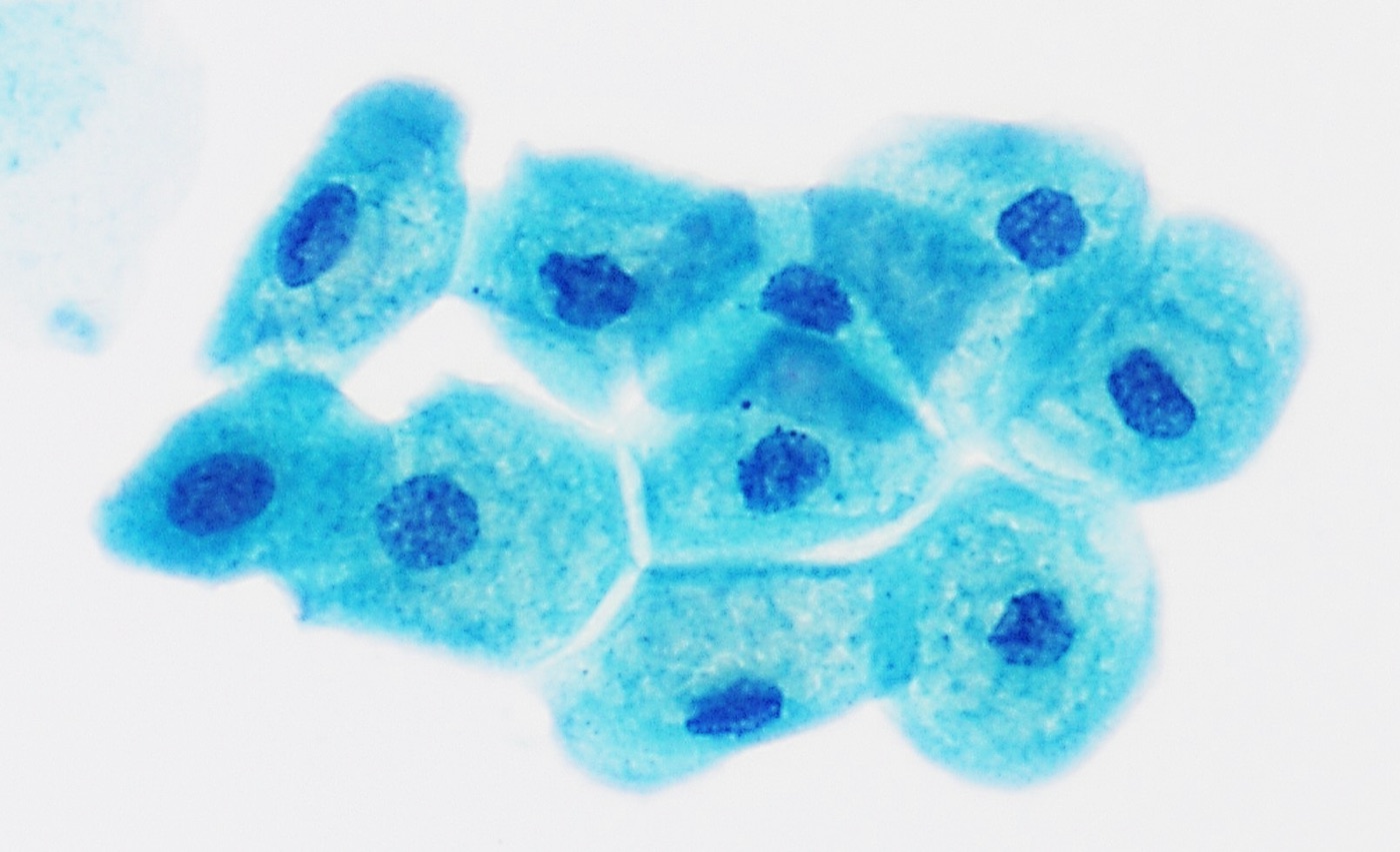
Definition. Benign urine cytology comprises the normal cells of the urinary tract, cells shed from adjacent organs, non-cellular elements, organisms, and reactive, iatrogenic and viral changes of urothelial cells. The Paris System for Reporting Urinary Cytology, 2nd edition (TPS 2.0), reports all of these findings as negative for high-grade urothelial carcinoma (NHGUC). NHGUC is the largest category. In a meta-analysis of 31 cohorts reported with the first edition, it accounted for 83.8% of satisfactory specimens (Farahani 2024). The diagnostic problem is the separation of benign cells with a high nuclear-to-cytoplasmic (N/C) ratio, dark chromatin or large clusters from high-grade urothelial carcinoma (HGUC) (Wojcik 2022).
Related page: The Paris System for Reporting Urinary Cytology (categories, risk of high-grade malignancy, adequacy and the specimen-type comparison).
Urothelial cell types in urine cytology
The urothelium has three layers. Cells from all three layers shed into urine. Instrumented specimens contain more cells and more deep-layer cells than voided urine (Wojcik 2022).
| Cell type | Size and shape | Cytoplasm | Nucleus | Specimen and significance |
|---|---|---|---|---|
| Superficial (umbrella) cell | Large; flat or dome-shaped | Abundant, dense to vacuolated | Single or multiple (several to many); low N/C ratio; nucleoli can be visible | Any specimen; multinucleation and nuclear enlargement are normal |
| Intermediate cell | Medium; polygonal or "tadpole" | Moderate, homogeneous | Round to oval, central, fine chromatin | Voided and instrumented specimens |
| Basal cell | Small; cuboidal | Scant | Round, N/C ratio up to about 0.5, fine even chromatin | Mostly instrumented specimens and washings; mimic of high-grade cells |
- Umbrella cells are the main source of large, multinucleated benign cells. Their N/C ratio remains low regardless of the number of nuclei. TPS 2.0 does not classify them as atypical (Wojcik 2022).
- Intermediate and basal cells in sheets show uniform nuclear spacing and smooth membranes. Single basal cells in a washing can have an N/C ratio near 0.5. Fine chromatin and smooth membranes separate them from atypical urothelial cells (Wojcik 2022).
- Urothelial tissue fragments. Benign urothelium, low-grade neoplasms and, less often, HGUC can shed tissue fragments. Overlap and three-dimensionality limit the assessment of nuclear detail in these fragments (Allison 2022).
Non-urothelial cells in urine cytology
Squamous cells in urine
- Sources: contamination from the vulva, vagina or distal urethra, and squamous metaplasia of the trigone. Trigonal metaplasia is common in women and has a glycogenated, nonkeratinizing pattern (Wojcik 2022).
- Benign appearance: superficial and intermediate squamous cells with small nuclei. The intermediate squamous nucleus is the reference size for comparison with urothelial nuclei.
- Atypical and malignant squamous cells: Non-Urothelial and Secondary Malignancies in Urine Cytology.
Glandular cells in urine
- Cystitis glandularis and cystitis cystica produce columnar or cuboidal cells with mucin vacuoles. Florid lesions mimic urothelial carcinoma on cystoscopy (Wang 2023).
- Ileal conduit and neobladder specimens contain intestinal epithelium, described under urine from urinary diversions in urine cytology.
- Prostatic and seminal vesicle cells appear after prostatic massage or ejaculation. Seminal vesicle cells contain golden-brown lipofuscin granules and enlarged, hyperchromatic, degenerated nuclei. Their DNA content can be abnormal. Hyperploid cells were found in 5 of 6 urine specimens with seminal vesicle cells, and 48% of seminal vesicle epithelia in prostate biopsies had an aneuploid peak (Wojcik 1999).
Renal tubular cells in urine
- Cytomorphology: small round or cuboidal cells, single or in small groups, with granular eosinophilic or vacuolated cytoplasm and round nuclei. They are numerous in glomerular disease, acute tubular injury and rejection.
- p53 expression in reactive tubular cells: repair or reactive renal tubular cells were present in 68 of 80 (85%) voided urine samples from patients with glomerular disease. All 68 contained p53-positive tubular cells, with a mean positivity rate of 47.7% per sample (Enomoto 2021). p53 immunocytochemistry therefore does not separate these cells from urothelial carcinoma.
- Mimic of decoy cells: damaged tubular cells have been misidentified as polyomavirus decoy cells in a kidney transplant recipient without BK viremia (Malvica 2022).
Inflammatory cells and histiocytes in urine
Neutrophils indicate infection, stones, catheterization or necrosis. Lymphocytes are common after BCG therapy. Eosinophils suggest eosinophilic or drug-related cystitis. Histiocytes are single cells with eccentric bean-shaped nuclei and foamy cytoplasm. Multinucleated histiocytes and epithelioid histiocytes indicate granulomatous inflammation, described under treatment effect in urine cytology.
Non-cellular elements in urine cytology
Crystals in urine cytology
Crystals form in urine according to its pH and solute concentration. Most are incidental. Some indicate a metabolic disorder, a drug or a stone (Torous 2022).
| Crystal | Appearance | Urine pH | Significance |
|---|---|---|---|
| Uric acid | Yellow to brown rhomboid plates, rosettes, barrels | Acidic | Hyperuricosuria, gout, tumor lysis; stone formation |
| Calcium oxalate dihydrate | Colorless envelope (octahedral) shapes | Acidic or neutral | Common; diet; stone formation |
| Calcium oxalate monohydrate | Ovoid or dumbbell shapes | Acidic or neutral | Ethylene glycol ingestion when numerous |
| Triple phosphate (struvite) | Colorless "coffin-lid" prisms | Alkaline | Infection with urease-producing bacteria; staghorn calculi |
| Cystine | Colorless hexagonal plates | Acidic | Cystinuria; always pathologic |
| Drug crystals | Needles, sheaves or fans (sulfonamides, acyclovir, indinavir and others) | Variable | Drug precipitation; crystal nephropathy |
Source: Torous 2022.
Casts, mucus and corpora amylacea are the other non-cellular elements.
- Casts are cylindrical molds of the renal tubules. Hyaline casts are nonspecific. Red cell casts indicate glomerular bleeding. Granular and renal tubular cell casts indicate tubular injury.
- Mucus and Tamm-Horsfall protein form amorphous pale strands.
- Corpora amylacea from the prostate are laminated round bodies.
Contaminants in urine cytology
- Lubricant from catheterization or cystoscopy forms granular purple to pink amorphous material that can coat cells.
- Starch granules from gloves are round, refractile bodies with a central cross on polarization.
- Pollen grains and plant cells enter from containers or the environment.
- Fungal contaminants. In more than 11,000 cytology specimens from one institution, Fusarium species were the most common nonpathogenic fungal contaminant and Alternaria the second most common, in urine and other fluids (Madan 2025).
- Spermatozoa are a normal finding in male urine and in female urine after intercourse.
Organisms in urine cytology
Schistosoma haematobium in urine cytology
- Morphology: large oval eggs with a terminal spine. The eggs of S. mansoni have a lateral spine. The background shows hematuria, eosinophils and granulomatous inflammation.
- Detection: eggs are identified on voided urine cytology, mainly from immigrants from and travelers to endemic areas (Laser 2009; Bellafiore 2018).
- Cancer risk: in a case-control study in Alexandria, Egypt, a history of urinary schistosomiasis carried a multivariate odds ratio of 1.72 for invasive bladder cancer. The odds ratio was 15.8 for male ever-smokers with urinary schistosomiasis, compared with never-smokers without it (Bedwani 1998). Chronic S. haematobium infection is associated with squamous cell carcinoma of the bladder (WHO 2022).
Trichomonas vaginalis and inflammation in urine cytology
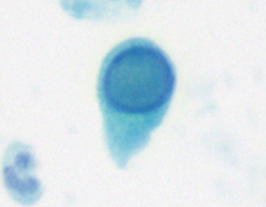
- Morphology: pear-shaped, pale gray-green organisms with an eccentric, faint nucleus and red cytoplasmic granules. Flagella are rarely visible. A neutrophil-rich background is typical.
- Specimens: trichomonads appear in voided urine from women and, less often, from men (Loo 2000).
- Ancillary testing: p16 immunocytochemistry stained trichomonads and supported the diagnosis in a urine specimen from a male patient (Pantanowitz 2005).
- Reactive urothelial cells in infection show nuclear enlargement, small nucleoli and perinuclear halos. Their chromatin remains fine.
Other infections in urine cytology
Fungi (Candida yeasts and pseudohyphae), bacteria, adenovirus, herpes simplex virus and cytomegalovirus are identified in urine. Adenoviral inclusions have been described in the intestinal cells of an ileal conduit (Zaharopoulos 2004). Malakoplakia produces histiocytes with Michaelis-Gutmann bodies. These bodies have been found in voided urine together with decoy cells (Mizutamari 2026).
BK polyomavirus effect in urine cytology
Cytomorphology of decoy cells
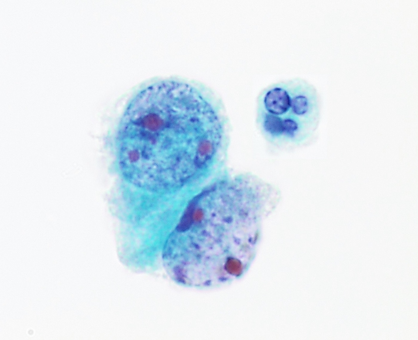
- Decoy cells are urothelial or renal tubular cells infected by BK polyomavirus. The nucleus is enlarged and filled by a homogeneous, glassy, basophilic inclusion. The chromatin is marginated against the nuclear membrane. Degenerated forms show a "comet" shape or a net-like ("spider-web") chromatin pattern (Wojcik 2022).
- Reporting: typical polyomavirus effect is reported as NHGUC (Wojcik 2022).
- SV40 immunostaining detects the large T antigen of BK polyomavirus in infected cells (Nankivell 2015).
Polyomavirus-like changes and HGUC
Degenerated HGUC cells can resemble decoy cells. In 284 urine specimens with polyomavirus or polyomavirus-like changes, four cell types were defined (Li 2025):
- Cell A was the typical decoy cell. Its presence was associated with HGUC on follow-up in only 1.2% of cases.
- Cells B, C and D had a spider-web chromatin pattern, clumpy chromatin, or homogeneous severely hyperchromatic nuclei, respectively. The positive predictive values for HGUC were 8%, 15.5% and 44%.
- When only cells B, C or D were present, HGUC followed in 22.4%, 33.3% and 61.5% of cases.
| Feature | Polyomavirus decoy cell | HGUC cell | Degenerated benign urothelial cell |
|---|---|---|---|
| Distribution | Single cells | Single cells and loose clusters | Single cells and groups |
| Nucleus | Enlarged; round; eccentric | Enlarged; irregular | Normal size or shrunken |
| Chromatin | Homogeneous, glassy inclusion; or net-like when degenerated | Coarse, clumped, usually hyperchromatic | Smudged or pyknotic |
| Nuclear membrane | Smooth, thin | Irregular, notched | Smooth or wrinkled |
| Nucleolus | Absent | Variable; can be prominent | Absent |
| SV40 | Positive | Negative | Negative |
| Report | NHGUC | SHGUC or HGUC | NHGUC |
Sources: Wojcik 2022; Li 2025; Nankivell 2015.
Decoy cells, BK viremia and nephropathy in transplant recipients
- Natural history: in 78 renal transplant recipients, the Kaplan-Meier probabilities of decoy-cell shedding, BK viremia and BK nephropathy were 30%, 13% and 8%. Shedding started at a median of 16 weeks after transplantation (Hirsch 2002).
- Test performance: in 704 samples from kidney and kidney-pancreas recipients, decoy cells had a sensitivity of 66.7%, a specificity of 88.6%, a positive predictive value of 11.7% and a negative predictive value of 98.5% for biopsy-proven BK nephropathy. A threshold of 10 or more decoy cells raised the positive predictive value to 32.1%. Quantitative viremia performed better (area under the curve 0.95 against 0.79) (Nankivell 2015).
- Duration of shedding: decoy cells present for 6 or more consecutive months had the best predictive performance for BK nephropathy (area under the curve 0.832). Nephropathy developed in 5 of 44 recipients with 6 months or more of shedding and in none of 77 with shorter shedding (Sekito 2021).
- Quantity: the maximum decoy-cell count was higher in recipients with BK viremia and correlated with urine BK DNA load (r = 0.74) (Yamada 2018).
- Guideline: the Second International Consensus Guidelines screen kidney recipients by plasma BK DNA. In resource-limited settings, urine cytology at the same time points can exclude nephropathy, and plasma testing follows when decoy cells are detected. Screening is monthly until month 9 and then every 3 months until 2 years after transplant (3 years in children). Immunosuppression is reduced when plasma BK DNA persists above 1,000 copies/mL, exceeds 10,000 copies/mL, or when nephropathy is biopsy-proven (Kotton 2024).
- Decoy cells outside transplantation: in 30 patients with decoy cells among 22,867 urine specimens, 14 had urothelial carcinoma and 9 of them had received intravesical BCG or mitomycin. BK polyomavirus was detected in urine and was absent from the carcinoma tissue (Klufah 2020).
Reactive urothelial changes, instrumentation and lithiasis in urine cytology
Reactive urothelial cells
- Causes: infection, stones, catheters, instrumentation, intravesical therapy and radiation.
- Cytomorphology: enlarged nuclei with a proportional increase in cytoplasm, so the N/C ratio remains low. Nucleoli are often prominent. The chromatin is fine and evenly distributed. The nuclear membranes are smooth. Cytoplasmic vacuoles and neutrophils are common (Wojcik 2022).
- Reporting: reactive cells are NHGUC. TPS 2.0 does not list a prominent nucleolus among the criteria for atypia (Wojcik 2022).
Instrumentation artifact
- Cytomorphology: instrumented specimens and washings are cellular. They contain cohesive, ball-shaped and papillary clusters with smooth borders and a dense cytoplasmic collar (Chu 2002).
- Contrast with low-grade neoplasia: low-grade tumors in washings form papillary and irregular groups with ragged borders, cytoplasmic homogeneity and subtle nuclear changes (Chu 2002). Low-grade urothelial neoplasia in urine: Low-Grade Urothelial Neoplasia and High-Grade Urothelial Carcinoma Variants in Urine Cytology.
Urinary tract calculi
- Frequency of changes: in voided urine from 154 patients with calculi, 52.6% showed no abnormality. Smooth-bordered clusters of urothelial cells with central, normal nuclei were present in 40.9%. In routine urine, such clusters predicted a calculus in 62.5% of cases (Highman 1982).
- Atypia: 6.5% of urines from patients with calculi showed features suspicious for well-differentiated carcinoma. They contained fewer single cells and clusters with abnormal nuclei than urines from proven carcinoma (Highman 1982).
- Background: crystals, red cells and neutrophils.
Treatment effect in urine cytology
Intravesical BCG therapy
- Cytomorphology: at the start of BCG therapy, inflammatory cells increase greatly. Urothelial nuclei enlarge and can show hyperchromasia, prominent nucleoli and anisonucleosis. These changes persisted through the first year of maintenance therapy and diminished slowly in the second year (Mack 1994).
- Granulomas: epithelioid histiocytes and multinucleated giant cells appear after BCG. The same findings occur in urinary tuberculosis, which acid-fast stains and culture confirm (Arora 2010).
- Accuracy after BCG: in 389 matched cases from patients with prior HGUC, sensitivity was 61.9% within 100 days of the last BCG instillation and 82.1% at longer intervals. Reactive atypia and degeneration were associated with false-positive results. Of patients with a false-positive result, 44.7% developed HGUC on follow-up (Hermans 2022).
- Sensitivity and specificity: after BCG induction, cytology detected high-grade disease with a sensitivity of 53% and a specificity of 95% compared with systematic biopsies (Gupta 2019).
- Erythematous lesions after BCG: for these lesions, voided cytology had a sensitivity of 59% and a negative predictive value of 94%. Most erythematous lesions were chronic nonspecific or granulomatous inflammation, and 10% were malignant (Nurminen 2019).
Intravesical and systemic chemotherapy
- Intravesical mitomycin C and thiotepa act on the superficial urothelium. Umbrella cells become large, multinucleated and vacuolated. The N/C ratio remains low (Wojcik 2022; Lopez-Beltran 2018).
- Degeneration after device-assisted mitomycin: after chemohyperthermia or electromotive mitomycin, AUC without degenerated suspicious cells was associated with negative biopsies. SHGUC rendered on degenerated cells was associated with HGUC or carcinoma in situ (Pierconti 2018).
- Systemic cyclophosphamide and the recreational drug ketamine also cause urothelial atypia that mimics carcinoma in situ (Lopez-Beltran 2018).
- Positive cytology after treatment remains prognostically significant. After intravesical mitomycin, a positive cytology at the 3-month visit without visible tumor carried a prognosis as poor as endoscopic or biopsy evidence of cancer in high-grade carcinoma (Cant 1986).
Radiation effect and pseudocarcinomatous hyperplasia
- Radiation change: cytomegaly with proportional nuclear enlargement, cytoplasmic vacuoles, multinucleation and smudged chromatin. The N/C ratio remains low (Wojcik 2022).
- Pseudocarcinomatous urothelial hyperplasia occurs after pelvic irradiation. In 39 patients, 25 urine specimens (64%) showed its features: nucleomegaly in all, nucleoli in 23, and an N/C ratio above 0.5 in 11 (44%). All cases first read as AUC or HGUC contained these cells (Velez Torres 2023).
| Feature | Reactive or treatment effect | Degenerated cells | HGUC |
|---|---|---|---|
| N/C ratio | Low; nucleus and cytoplasm enlarge together | Variable; often low | ≥0.7 in diagnostic cells |
| Chromatin | Fine, even; can be smudged after radiation | Smudged, pyknotic or glassy | Coarse, clumped, hyperchromatic (some hypochromatic) |
| Nuclear membrane | Smooth | Smooth or wrinkled, without sharp notches | Irregular, notched, indented |
| Nucleoli | Often prominent | Absent | Variable; can be prominent |
| Cytoplasm | Vacuolated; abundant | Degenerated, frayed, with vacuoles | Scant, dense or vacuolated |
| Background | Inflammation; granulomas after BCG | Debris; long transport time; diversions | Necrosis, blood, single atypical cells |
| TPS 2.0 report | NHGUC | NHGUC or AUC | SHGUC or HGUC |
Sources: Wojcik 2022; Hermans 2022; Velez Torres 2023.
Urine from urinary diversions in urine cytology
- Ileal conduit urine contains well-preserved columnar cells and degenerated, small round cells of intestinal origin, in a background of mucus, bacteria and debris. The columnar cells can be mistaken for carcinoma. Indiana pouch urine contains degenerated small round cells without columnar cells (Watarai 2000).
- Neobladder washings: in 45 diversion specimens with histologic correlation, application of TPS reduced atypical and suspicious interpretations by 13.4% and 11.1%. Sensitivity and negative predictive value were each 93.75% (Kim 2023).
- Adequacy in neobladder urine: in 442 neobladder specimens, 60.5% contained no preserved urothelial cells. Requiring at least one preserved urothelial cell for adequacy raised sensitivity for HGUC to 100%. A volume of 45 mL or more was the optimal adequacy cutoff (Ordobazari 2026).
Differential diagnosis of benign findings that mimic HGUC in urine cytology
| Benign finding | Feature that mimics HGUC | Separating features |
|---|---|---|
| Multinucleated umbrella cell | Large size, many nuclei | Low N/C ratio; fine chromatin; smooth membranes |
| Basal cells in washings | N/C ratio near 0.5 | Fine even chromatin; sheets with even spacing |
| Polyomavirus decoy cell | Large dark nucleus | Glassy homogeneous inclusion; smooth membrane; SV40 positive |
| Degenerated urothelial cell | Hyperchromasia | Smudged chromatin; no sharp membrane irregularity |
| Seminal vesicle cell | Enlarged hyperchromatic nucleus; aneuploidy | Lipofuscin granules; male patient |
| Renal tubular cell | Small cell with high N/C ratio; p53 positive | Granular cytoplasm; renal disease; casts |
| Instrumentation cluster | Large papillary groups | Smooth borders; cytoplasmic collar; benign single cells |
| Calculus-associated cluster | Three-dimensional clusters | Smooth borders; central normal nuclei; crystals |
| BCG effect | Nuclear enlargement and nucleoli | Granulomas; lymphocytes; low N/C ratio |
| Radiation and pseudocarcinomatous hyperplasia | Nucleomegaly; N/C ratio >0.5 in some | Pelvic irradiation history; nucleoli; vacuolated cytoplasm |
| Ileal conduit columnar cells | Columnar cells with dark degenerated nuclei | Mucin; diversion history; degenerated background |
Low-Grade Urothelial Neoplasia and High-Grade Urothelial Carcinoma Variants in Urine Cytology
Definition. Low-grade urothelial neoplasia (LGUN) is the cytologic term for papillary urothelial neoplasms with low-grade cytology: urothelial papilloma, papillary urothelial neoplasm of low malignant potential (PUNLMP) and low-grade non-invasive papillary urothelial carcinoma, together with flat low-grade lesions. The first edition of the Paris System (TPS) reported LGUN as a separate category. The second edition (TPS 2.0) reports these lesions as negative for high-grade urothelial carcinoma (NHGUC) (Wojcik 2022). High-grade urothelial carcinoma (HGUC) comprises high-grade papillary carcinoma, urothelial carcinoma in situ (CIS) and invasive carcinoma. Invasive urothelial carcinoma includes more than 10 subtypes and divergent differentiation in the 2022 WHO classification, and several of them do not meet the TPS criteria for HGUC in urine (Arshia 2024; WHO 2022).
Related page: The Paris System for Reporting Urinary Cytology (category definitions, risk of high-grade malignancy and adequacy).
WHO 2022 urothelial tumors and their appearance in urine cytology
The 5th edition WHO classification of urinary and male genital tumours keeps the two-tier grading (low grade and high grade) for non-invasive papillary urothelial carcinoma. It retains PUNLMP and urothelial papilloma. It uses the term "subtype" for morphologic variants of invasive urothelial carcinoma and "divergent differentiation" for squamous, glandular and other lineages (WHO 2022).
| WHO 2022 lesion | Usual urine cytology | TPS 2.0 category |
|---|---|---|
| Urothelial papilloma, PUNLMP | Benign-appearing cells; occasional papillary groups | NHGUC |
| Non-invasive papillary urothelial carcinoma, low grade | Clusters with cytoplasmic homogeneity and subtle nuclear changes | NHGUC, optional comment |
| Non-invasive papillary urothelial carcinoma, high grade | Single cells and clusters with high-grade nuclei | SHGUC or HGUC |
| Urothelial carcinoma in situ | Numerous loosely cohesive single high-grade cells | SHGUC or HGUC |
| Invasive urothelial carcinoma, conventional | High-grade cells; necrotic background in some | SHGUC or HGUC |
| Invasive subtypes (plasmacytoid, micropapillary, nested and others) | Variable; some below TPS thresholds | AUC to HGUC |
| Divergent squamous or glandular differentiation | Keratinized or columnar malignant cells with high-grade urothelial cells | HGUC; pure non-urothelial tumors under non-urothelial malignancies |
Sources: WHO 2022; Wojcik 2022; Arshia 2024.
Related page: Non-Urothelial and Secondary Malignancies in Urine Cytology (pure squamous cell carcinoma, adenocarcinoma, small cell carcinoma and metastases).
Cytomorphology of low-grade urothelial neoplasia in urine
- Architecture: papillary and irregular groups with ragged borders. Bladder washings from low-grade carcinomas showed these groups with cytoplasmic homogeneity and subtle nuclear changes, including a higher N/C ratio and irregular nuclear borders (Chu 2002).
- Fibrovascular cores: three-dimensional papillary fragments with a fibrovascular core are diagnostic of a papillary neoplasm. They were present in only 2 of 98 urine specimens from biopsy-proven low-grade carcinoma (McCroskey 2015b).
- Other proposed features: paired cells, clumpy chromatin and cytoplasmic homogeneity were more frequent in low-grade carcinoma in univariate analysis. None remained a significant predictor in multivariate analysis, and no single feature separated low-grade carcinoma from negative controls (McCroskey 2015b).
- Nuclear features: nuclei are mildly enlarged, slightly eccentric and evenly spaced, with fine chromatin. The N/C ratio and chromatin remain below the thresholds for atypical urothelial cells (Wojcik 2022).
| Feature | Instrumentation artifact | Low-grade urothelial neoplasia | HGUC |
|---|---|---|---|
| Groups | Cohesive balls and papillary clusters with smooth borders | Papillary and irregular groups with ragged borders | Loose clusters; many single cells |
| Cytoplasmic collar | Dense collar at the group edge | Absent | Absent |
| Cytoplasm | Variable, including umbrella cells | Homogeneous | Scant; dense or vacuolated |
| Fibrovascular core | Absent | Present in a minority | Rare |
| N/C ratio | Low | Slightly increased | ≥0.7 in diagnostic cells |
| Chromatin and membranes | Fine; smooth | Fine; subtle irregularity | Coarse, hyperchromatic; irregular |
| TPS 2.0 category | NHGUC | NHGUC, optional comment | SHGUC or HGUC |
Sources: Chu 2002; McCroskey 2015b; Wojcik 2022.
Performance of urine cytology for low-grade urothelial tumors
- Sensitivity by grade: in 3,251 specimens from 1,114 consecutive patients, sensitivity was 10% for low-grade and 51% for high-grade tumors. Specificity was 83–88%. The anticipatory positive rate was 44% after a median of 15 months (Yafi 2014).
- Prospective comparison: in 109 patients with bladder cancer, cytology had a sensitivity of 16% for low-grade and 84% for high-grade tumors, with a specificity of 86% (Yafi 2015).
- Instrumented specimens: six cytopathologists diagnosed low-grade carcinoma in instrumented ThinPrep specimens with a sensitivity of 21–53% and a specificity of 81–95%. Overall agreement was fair (κ = 0.30) (McCroskey 2015a).
- Meta-analytic risk: the pooled risk of high-grade malignancy for the first-edition LGUN category was 12.45% in one meta-analysis (Nikas 2022) and 15.1% in another (Farahani 2024).
- Effect of TPS: the proportion of low-grade neoplasms called atypical on cytology fell from 34.1% in non-TPS cohorts to 20.8% in TPS cohorts (Farahani 2024). In a single-institution reclassification of 299 urine samples, the AUC rate fell from 43% to 31% (Koh 2020).
- Evidence for folding LGUN into NHGUC: in a 3-year review of histologically diagnosed low-grade carcinomas, every cytologic LGUN diagnosis was concordant with histology (positive predictive value 100%), and sensitivity was only 21.7%. In the correlation of all cytology cases of the period, LGUN had a sensitivity of 21.7%, a specificity of 97.2% and a positive predictive value of 87.5%. The risk of high-grade malignancy of the LGUN category was 12.5%. In the same series, sensitivity for HGUC was 90.1% and specificity 70.8% (Christofidis 2023).
Reporting of low-grade urothelial neoplasia in TPS 2.0
- Category: low-grade lesions are reported as NHGUC, because cytology cannot reliably detect them and they rarely progress (Wojcik 2022).
- Optional comment: when cytomorphology suggests a low-grade neoplasm, for example a papillary fragment with a fibrovascular core, the report can add a comment that a low-grade urothelial neoplasm is suspected (Wojcik 2022).
Detection of HGUC and carcinoma in situ by urine cytology
- Carcinoma in situ: in 61 patients with pathologically confirmed bladder CIS, urine cytology had a sensitivity of 77% (Shigehisa 2026).
- Bladder diverticula: sensitivity for HGUC was 80% for tumors arising within a diverticulum and 82% for tumors outside it. Voided and cystoscopic samples performed equally (Alhamar 2024).
- TPS 2.0 in practice: in 180 samples classified with the 2nd edition, the distribution was nondiagnostic 3.9%, NHGUC 65.6%, AUC 10%, SHGUC 11.7% and HGUC 8.9%. In the 30.6% with histologic correlation, the risk of malignancy was 66.7% for AUC, 94.1% for SHGUC and 100% for HGUC (Singh 2024).
Management by category: Ancillary Tests, Indications and Management in Urine Cytology.
Cytomorphology of HGUC and carcinoma in situ in urine
- Single cells and loose clusters: HGUC and CIS cells are loosely cohesive and exfoliate as single cells. (Allison 2022)
- Criteria: N/C ratio ≥0.7 (required) plus two of three: moderate to severe hyperchromasia, irregular clumpy chromatin, irregular nuclear membranes. TPS 2.0 recognizes a hypochromatic subset (Wojcik 2022; Lin 2024).
- Supporting features: pleomorphism, necrosis, mitoses and a hemorrhagic background.
- Chromasia varies within and between cases. In 69 HGUC cases measured by digital image analysis, 11.6% had extremely dark ("India ink") nuclei, 74.0% had hyperchromatic nuclei and 14.5% had hypochromatic nuclei. Heterogeneity within a single case was universal (McIntire 2022).
Hypochromatic HGUC in urine
- Frequency: malignant cells with hypochromatic chromatin were present in 16 of 93 (17.2%) HGUC cases with biopsy follow-up. They were the only malignant cells in 4 of 101 malignant cases (4.0%) (Renshaw 2021).
- Criteria: all cases had cells with high N/C ratios and markedly indented, irregular nuclear membranes. There were no false-positive diagnoses (Renshaw 2021).
- Outcome of AUC with hypochromatic cells: in a series of 67 specimens with large hypochromatic malignant-appearing urothelial cells, 12 were classified as AUC because hyperchromasia was absent. All 12 had HGUC on subsequent biopsy (Pierconti 2018). The cells had very large nuclei, an N/C ratio above 0.7, irregular nuclear outlines and coarse, often peripheral chromatin.
Nuclear-to-cytoplasmic ratio estimation in urine cytology
The N/C ratio is the only required criterion of the AUC category and a major criterion of SHGUC and HGUC. It is estimated by eye as the proportion of the cell area occupied by the nucleus. An N/C ratio of 0.5 means that the nuclear area equals the cytoplasmic area. An N/C ratio of 0.7 corresponds to a narrow rim of cytoplasm (Wojcik 2022).
| Study | Material | Main finding |
|---|---|---|
| Hang 2017 | 48 AUC cases with follow-up | Maximum N/C ratio 0.53 with HGUC on follow-up against 0.43 without; optimal threshold 0.486 (sensitivity 73.3%, specificity 84.8%) |
| McIntire 2019 | 64 HGUC and SHGUC cases, 640 cells | Average N/C ratio 0.57 in HGUC and 0.53 in SHGUC; maximum average 0.73 and 0.68 |
| Richardson 2020 | 19 split samples, ThinPrep and cytospin | Average N/C ratio 0.5465 in HGUC and 0.2846 in NHGUC cells; TPS criteria applicable to both preparations |
| McIntire 2021 | 59 HGUC ThinPrep specimens | Upper-tract N/C ratio 0.58 against 0.54 in lower tract; smaller nuclei and less cytoplasm in upper tract |
| Wang 2020 | Survey, 137 participants, 10 atypical cases | N/C ratio had the highest κ (0.386) of the TPS criteria; overestimation near 0.5 |
| Yang 2025 | 106 slides with biopsy-confirmed HGUC or CIS, AI measurement | Suspicious cells averaged an N/C ratio of 0.66 and atypical cells 0.58; a cutoff of 0.66 proposed |
- Measured N/C ratios and the TPS 2.0 threshold: image-analysis studies measure lower average N/C ratios in HGUC than the visual threshold of 0.7. Two studies proposed lowering the threshold for SHGUC and HGUC (McIntire 2019; Yang 2025). TPS 2.0 keeps 0.7 (Wojcik 2022).
- Automation: a hybrid deep-learning and morphometric model calculated the N/C ratio for every cell in whole-slide images and tabulated the Paris System criteria (Vaickus 2019). Related page: Ancillary Tests, Indications and Management in Urine Cytology (digital image analysis and artificial intelligence).
Atypical urothelial cells: frequency and risk of high-grade malignancy
- Criteria: an N/C ratio above 0.5 plus one of hyperchromasia, irregular nuclear membranes or irregular coarse, clumped chromatin (Wojcik 2022).
- Frequency: AUC accounted for 8.0% of satisfactory specimens in TPS cohorts and 11.3% in non-TPS cohorts. The ratio of atypical to high-grade diagnoses fell from 4.1 to 2.0 with TPS (Farahani 2024).
- Risk: the pooled risk of high-grade malignancy for AUC was 38.65% (Nikas 2022) and 40.2% (Farahani 2024). The likelihood ratio of AUC for high-grade malignancy was 0.9, so AUC alone neither rules in nor rules out a high-grade lesion (Farahani 2024).
- Reproducibility: mean diagnostic agreement on 10 atypical cases was 51.2%, and agreement was below 50% for 6 of them (Wang 2020).
HGUC subtypes and divergent differentiation in urine cytology
In 72 urine specimens from patients with high-grade carcinoma at cystectomy, 24 (33%) had a histologic subtype or divergent differentiation in the resection. Cytology reflected the subtype in 13 (54%) of these 24: squamous differentiation in 11 of 13, micropapillary features in 1 of 3 and sarcomatoid features in 1 of 3. Detection depended on mucosal involvement by the subtype and was independent of its amount (Yoo 2021).
| Subtype or differentiation | Urine cytomorphology | Pitfall under TPS criteria | Source |
|---|---|---|---|
| Plasmacytoid | Large discohesive single cells; abundant dense or vacuolated cytoplasm; eccentric hyperchromatic nuclei; inconspicuous nucleoli | N/C ratio below 0.5 in many cells; read as NHGUC or AUC | Straccia 2020; Suo 2021 |
| CIS with plasmacytoid features | Eccentric, enlarged, hyperchromatic nuclei | Only 7 of 74 specimens showed the proposed features; κ poor to fair | Nichols 2021 |
| Micropapillary | Micropapillae without fibrovascular cores; single high-grade cells | Abundant cytoplasm and weak hyperchromasia in some cases | Suo 2021; Straccia 2020 |
| Nested | Medium-sized round or polygonal cells; abundant dense basophilic cytoplasm; increased N/C ratio; irregular membranes; occasional nucleoli | Subtle features; primary diagnosis in urine not recommended | Cardillo 2003 |
| Sarcomatoid | Spindle cells with high-grade nuclei | Detected in 1 of 3 cases | Yoo 2021 |
| Squamous differentiation | Keratinized malignant cells with high-grade urothelial cells | Detected in 11 of 13 cases | Yoo 2021 |
| Glandular differentiation | Columnar or vacuolated malignant cells with high-grade urothelial cells | Rare; separated from adenocarcinoma by the urothelial component | Arshia 2024 |
- Plasmacytoid HGUC in TPS: in 74 urine specimens from 18 patients with plasmacytoid CIS, TPS diagnoses were NHGUC in 26, AUC in 26, SHGUC in 12 and HGUC in 10. Plasmacytoid CIS cytology that does not meet HGUC criteria is best classified as AUC (Nichols 2021).
- Nested subtype: cells resembling the tumor nests were found in all 13 urine specimens from 7 patients. Their presence in a patient with a history of the nested subtype supports cystoscopy and biopsy (Cardillo 2003).
- Cell block and immunohistochemistry on residual urine improve the recognition of divergent differentiation (Arshia 2024).
- Clinical significance: micropapillary and plasmacytoid carcinomas are clinically aggressive (Straccia 2020; Suo 2021). The nested subtype has bland cytology and aggressive behavior (Cardillo 2003). Many subtype cases are recognized in urine only in retrospect, after the resection shows the subtype (Arshia 2024).
Reporting of HGUC subtypes and tissue fragments in urine cytology
- Category first: a urothelial carcinoma subtype is reported under the TPS category that its cells meet. Subtype cells that meet the HGUC criteria are reported as HGUC. Plasmacytoid cells below the criteria are best reported as AUC (Nichols 2021).
- Descriptive comment: a comment describing plasmacytoid, micropapillary or nested features supports correlation with histology. (Cardillo 2003).
- Divergent differentiation: keratinized malignant cells accompanied by conventional high-grade urothelial cells indicate urothelial carcinoma with squamous differentiation (Arshia 2024).
- Tissue fragments: fragments in urine arise from benign reactive urothelium, low-grade neoplasms and, less often, HGUC or CIS. Their significance is often unclear because cell overlap obscures nuclear detail. HGUC and CIS cells usually exfoliate singly, so loose single cells provide most of the diagnostic information (Allison 2022).
Upper urinary tract HGUC in urine cytology
- Cell features: upper-tract HGUC cells are smaller than lower-tract HGUC cells. Average nuclear area was 126.3 µm² in upper-tract and 158.2 µm² in lower-tract specimens. Cytoplasmic area was 219.1 µm² and 296.2 µm². The N/C ratio was higher in the upper tract (McIntire 2021).
- Washings and voided urine: among patients with upper-tract HGUC, 50% of voided urine specimens showed high-grade features, against almost 90% of upper-tract washings (Zhang 2017).
- TPS in the upper tract: studies of upper-tract cytology before and after TPS remain few, and the most predictive cytologic features in upper-tract specimens are uncertain (Zhang 2021a; Zhang 2021b).
- N/C ratio across sites: an AI-based study found no significant difference in N/C ratio between upper-tract and lower-tract cases and supported consistent criteria across both. Nuclear area differed by lesion type. Abnormal cells averaged 101.5 µm² in CIS and 83.5 µm² in HGUC (Yang 2025).
- First-edition cell-count rules for upper-tract specimens, removed in TPS 2.0: The Paris System for Reporting Urinary Cytology.
Differential diagnosis of LGUN and HGUC variants in urine cytology
| Finding | Main mimic | Separating features |
|---|---|---|
| Papillary groups in a washing | Instrumentation artifact | Ragged borders and cytoplasmic homogeneity in LGUN; smooth borders and a cytoplasmic collar in artifact |
| Papillary groups in voided urine | Calculus-associated clusters | Stone history; central normal nuclei; crystals |
| Hypochromatic HGUC | Degenerated or reactive cells | High N/C ratio; markedly indented membranes; coarse peripheral chromatin |
| Plasmacytoid HGUC | Histiocytes, plasma cells, signet-ring adenocarcinoma | Single cells with eccentric hyperchromatic nuclei; keratin and GATA3 on cell block |
| Micropapillary HGUC | Low-grade papillary groups | No fibrovascular cores; high-grade single cells in background |
| Nested subtype | Reactive urothelium | History of nested carcinoma; irregular membranes; dense basophilic cytoplasm |
| Upper-tract HGUC | Benign upper-tract cells in washings | High N/C ratio; hyperchromasia; irregular membranes in small cells |
Related page: Normal and Benign Findings in Urine Cytology (benign mimics, polyomavirus effect and treatment effect).
Non-Urothelial and Secondary Malignancies in Urine Cytology
Definition. The Paris System for Reporting Urinary Cytology, 2nd edition (TPS 2.0), reports malignant cells that are not high-grade urothelial carcinoma (HGUC) under the category "non-urothelial malignancies" (Wojcik 2026). The category includes primary non-urothelial carcinomas of the urinary tract (squamous cell carcinoma, adenocarcinoma, small cell carcinoma), tumors of neighboring organs that invade the urinary tract (prostate, kidney, colorectum, uterus and cervix), distant metastases, hematologic malignancies and rare sarcomas. Most malignancies in urine are urothelial carcinomas. Non-urothelial elements raise the possibility of a urothelial carcinoma subtype, a non-urothelial bladder primary or a primary outside the bladder. Each has a different management pathway (Lew 2022).
Related page: The Paris System for Reporting Urinary Cytology (category definitions and risk of high-grade malignancy). Related page: Low-Grade Urothelial Neoplasia and High-Grade Urothelial Carcinoma Variants in Urine Cytology (HGUC subtypes, including squamous and glandular divergent differentiation). Related page: Normal and Benign Findings in Urine Cytology (benign squamous, glandular and seminal vesicle cells).
Clinical setting of non-urothelial malignancies in urine cytology
- Frequency. In a prospective series of 3,900 urine specimens reported with TPS (TPS 1.0), the category for non-urothelial malignancies accounted for 4 cases (0.1%). NHGUC accounted for 89.7%, AUC for 4.6%, SHGUC for 1.6% and HGUC for 4% and LGUN for 0.1% (Vlajnic 2020).
- Predictive value. In 149 urine specimens reported as positive for HGUC, the positive predictive value for high-grade urothelial carcinoma, carcinoma in situ or non-urothelial carcinoma on biopsy within 12 months was 85% (Kuan 2020).
- Divergent differentiation. Squamous, glandular or small cell differentiation within urothelial carcinoma is more common than a pure non-urothelial carcinoma. The WHO 2022 classification recognizes more than 10 urothelial carcinoma subtypes and divergent differentiation. Cell block preparations and immunohistochemistry on residual urine improve diagnostic accuracy (Arshia 2024).
- Clinical clues. A history of a primary tumor elsewhere, a mass in an adjacent organ, a renal mass or a rising serum prostate-specific antigen (PSA) changes the differential diagnosis. The clinical and radiologic history is part of the interpretation of any non-urothelial cell population in urine (Lew 2022).
Squamous cell carcinoma and atypical squamous cells in urine cytology
Squamous cell carcinoma of the bladder
- Etiology. Schistosoma haematobium infection is a proven carcinogen. It causes mainly squamous cell carcinoma of the bladder, with distinct epidemiologic and histologic features and a poor prognosis (Efared 2022). In an endemic community in Ghana, squamous metaplastic cells were found in 48% of participants with past infection and 47.1% of participants with current infection. They were absent in participants without exposure (Smith-Togobo 2023). S. haematobium ova: Normal and Benign Findings in Urine Cytology.
- Cytomorphology. Keratinized malignant cells with dense orangeophilic cytoplasm, bizarre shapes (spindle, tadpole and caudate cells), keratin pearls, hyperchromatic or pyknotic nuclei, and a background of necrotic keratin debris (Wojcik 2022).
- Pure squamous cell carcinoma versus urothelial carcinoma with squamous differentiation. Cytology alone does not separate the two. The distinction requires histology, because the WHO diagnosis of pure squamous cell carcinoma requires the absence of a conventional urothelial component (Netto 2022).
Atypical squamous cells in urine
- Frequency. Atypical squamous cells (ASC), defined as keratinized squamous cells, were present in 0.15% of urine specimens (123 of 81,018) in a 10-year period (Ho 2024).
- Risk of high-grade malignancy (ROHM). Low-grade and high-grade squamous atypia carried a ROHM of 70% and 92%. ASC without an accompanying urothelial cell abnormality carried a ROHM of 37%. ASC with an accompanying urothelial cell abnormality carried a ROHM of 94%. Reactive squamous changes and koilocytosis carried a ROHM of 0% (Ho 2024).
- Rare atypical squamous cells in malignancy. In 51% of malignancies (34 of 67), only rare ASC were present in the urine (Ho 2024).
- Surrogate of high-grade urothelial carcinoma. In 68 patients with histologic high-grade urothelial carcinoma, 17 (25%) had dysplastic squamous cells in urine. In 4 of these 17 patients, isolated dysplastic squamous cells were the only abnormal finding (Sojitra 2012).
- Size of squamous nuclei against neutrophils. Keratinized ASC with nuclei larger than an adjacent neutrophil were found in 15 of 17 specimens from urothelial carcinoma with squamous differentiation and in none of 79 benign specimens with squamous cells. The sensitivity was 88% and the specificity 100% (Furuhata 2025).
- Condyloma acuminatum. Urinary tract condylomas showed low-grade squamous intraepithelial lesion features in urine in 57% of cases. A smaller group (28%) showed atypical keratinized cells that mimic keratinizing squamous cell carcinoma. All specimens were reclassified as NHGUC (Velez Torres 2022).
- Contamination. Cells from outside the urinary tract can enter a voided specimen during collection (Betancourt 2021). In women, a squamous lesion of the lower genital tract is part of the differential diagnosis of ASC in a voided specimen.
| Squamous finding in urine | Cytomorphology | Reported significance |
|---|---|---|
| Benign squamous cells and keratinizing metaplasia | Mature superficial or intermediate squames with small nuclei | NHGUC; ROHM 0% for reactive changes (Ho 2024) |
| Koilocytes (condyloma) | Perinuclear halo, binucleation, nuclear enlargement | NHGUC; ROHM 0% (Ho 2024; Velez Torres 2022) |
| Low-grade squamous atypia | Keratinized cells with mild nuclear enlargement and hyperchromasia | ROHM 70% (Ho 2024) |
| High-grade squamous atypia | Keratinized cells with large, dark, irregular nuclei | ROHM 92% (Ho 2024) |
| Squamous cell carcinoma | Pleomorphic keratinized cells, pearls, necrotic keratin debris | Non-urothelial malignancies, or HGUC with squamous differentiation when urothelial cells are malignant (Wojcik 2022) |
Adenocarcinoma in urine cytology
Primary adenocarcinoma of the bladder and urachus
- Incidence. Primary adenocarcinoma of the urinary bladder accounts for up to 2% of bladder cancers and occurs predominantly in men (Santa 2025).
- Histologic patterns. Enteric, mucinous, signet ring cell and mixed types. Urachal adenocarcinoma shares the morphology but arises in younger patients and has a distinct genomic and immunohistochemical profile. Villous adenoma, cystitis glandularis and intestinal metaplasia are potential precursors (Santa 2025).
- Cytomorphology. Columnar cells in three-dimensional clusters, acinar or palisaded groups, with vacuolated or mucinous cytoplasm, eccentric nuclei, vesicular chromatin and prominent nucleoli (Wojcik 2022).
- Signet ring cell carcinoma. In 6 urine specimens from 5 primary bladder carcinomas and 1 metastatic colonic carcinoma, the tumor cells had distinct cell borders, a single large mucin vacuole and eccentric irregular nuclei with prominent nucleoli. Primary tumors also shed intact glandular fragments. The metastatic tumor shed mostly single cells. Distended histiocytes and degenerated urothelial cells are the benign mimics (Guan 2012).
Clear cell adenocarcinoma of the urinary tract
- Clinical features. Clear cell adenocarcinoma of the urinary tract shows a female predominance (Zheng 2025). In 33 cases, 78% arose in the bladder, most often at the trigone, and 22% in the urethra (Akgul 2022).
- Cytomorphology. Urine specimens from 3 cases contained scattered aggregates and single enlarged cells with abundant clear, wispy, vacuolated cytoplasm, hobnail and signet ring forms, and nuclei with granular to vesicular chromatin and prominent, often multiple nucleoli (Doria 1996).
- Immunohistochemistry. GATA3 was expressed in 54.5% of urinary clear cell adenocarcinomas and in 11.7% of gynecologic clear cell carcinomas, a potential pitfall for a urothelial diagnosis (Akgul 2022). High SOX17 expression occurred in 29% of urinary clear cell adenocarcinomas, in all gynecologic clear cell carcinomas and in none of 18 nephrogenic adenomas (Zheng 2025).
Colorectal and other secondary adenocarcinomas
- Colorectal adenocarcinoma. In 30 urine specimens with colorectal adenocarcinoma invading the urinary tract, tall columnar cells were present in 12, large cells that resembled HGUC cells in 27, and small cells with irregular or crescentic nuclei in 27. CDX2 was positive in all 4 cases tested. The small cells corresponded to desquamated, degenerated cells on the tumor surface (Matsuyama 2026). All 30 specimens had been classified as HGUC by TPS criteria before review (Matsuyama 2026).
- Endometrial adenocarcinoma. Cohesive glandular clusters in a voided urine, positive for PAX8 and negative for GATA3 on cell block, led to a diagnosis of endometrial adenocarcinoma (Betancourt 2021).
- Cervical adenocarcinoma. Gastric-type endocervical adenocarcinoma was first recognized as a non-urothelial malignancy in urine cytology after invasion of the bladder (Lee 2024).
| Feature | High-grade urothelial carcinoma | Adenocarcinoma (primary or secondary) |
|---|---|---|
| Cell shape | Round to polygonal; single cells and loose groups | Columnar or cuboidal; palisades, acini, three-dimensional clusters |
| Cytoplasm | Dense, cyanophilic or squamoid | Vacuolated, mucinous or clear; signet ring forms |
| Nucleus | Central or eccentric; N/C ratio ≥0.7 | Eccentric, basal; N/C ratio variable |
| Chromatin | Coarse, clumped; hyperchromatic | Vesicular or finely granular |
| Nucleoli | Variable; can be prominent | Prominent, often macronucleoli |
| Background | Necrosis, inflammation | Mucin; "dirty" necrosis in colorectal carcinoma |
| Immunohistochemistry | GATA3, p63, uroplakin positive | CDX2 and SATB2 (enteric), PAX8 (Müllerian), NKX3.1 (prostate) |
Sources: Wojcik 2022; Lew 2022; Matsuyama 2026; Betancourt 2021.
Small cell carcinoma and other neuroendocrine carcinomas in urine cytology
- Cytomorphology. Small malignant cells with scant cytoplasm, a high N/C ratio, nuclear molding, finely granular chromatin and inconspicuous nucleoli, in small clusters or single cells. The background is inflammatory and sometimes necrotic (Yoshida 2014). Apoptotic debris is common (Corbin 2026).
- Immunophenotype. In 6 primary bladder small cell carcinomas, synaptophysin and CD56 were positive in all cases and chromogranin in 3. The Ki-67 labeling index ranged from 30% to 100% (Sharma 2016).
- Mixed tumors. Small cell carcinoma of the bladder is often admixed with conventional high-grade urothelial carcinoma. Voided urine cytology can be the first evidence of the tumor, and immunohistochemistry on the urine cell block supports the diagnosis (Corbin 2026).
- Prostatic origin. Small cell carcinoma of the prostate can also shed into urine. The cytologic features match those of bladder primaries (Yoshida 2014).
- Therapeutic significance and TPS 2.0 category. The distinction from HGUC has therapeutic and prognostic consequences (Sharma 2016). Small cell carcinoma is classified under non-urothelial malignancies in TPS 2.0 (Wojcik 2022).
Prostatic adenocarcinoma in urine cytology
- Sensitivity. In 2,115 urine specimens from 1,119 patients with histologically proven prostatic adenocarcinoma, the rate of atypia or above was 19.48% and the rate of suspicious or above was 3.36% (Tang 2025).
- Correlates of detection. Bilobar and extracapsular involvement, lymphovascular invasion, Gleason score and ISUP grade were associated with a positive urine diagnosis. Lesion size and multifocality were not (Tang 2025).
- Cytomorphology of acinar adenocarcinoma. Single cells or small clusters of cuboidal glandular cells with large, eccentric, round to oval, uniform nuclei, conspicuous nucleoli, and scant to moderate delicate or granular cytoplasm (Lin 2018).
- Ductal adenocarcinoma. Columnar glandular cells in flat nests or three-dimensional clusters with prominent nucleoli. Urine cytology was the first evidence of disease in 4 of 5 patients with ductal adenocarcinoma and in 6 of 23 (26%) with acinar adenocarcinoma (Lin 2018).
- Separation from HGUC. Prostatic carcinoma cells have uniform nuclei with prominent nucleoli and lack the coarse chromatin and pleomorphism of HGUC. NKX3.1, PSA and PSAP support prostatic origin, and GATA3 supports urothelial origin (Wojcik 2022; Netto 2022).
- Sarcoma of the prostate. A high-grade prostatic sarcoma was detected in a catheterized urine as fragments of uniform spindle cells with ovoid nuclei and finely dispersed chromatin (Stoll 2011).
Renal cell carcinoma in urine cytology
- Detection rate. In 1,147 urine specimens matched to nephrectomy-proven renal cell carcinoma, the rate of atypia or above was 13.1% and the rate of suspicious or above was 1.5%. The corresponding rates for renal urothelial and squamous carcinomas were 63.1% and 24.0% (Li 2024).
- Factors that increase detection. Upper-tract sampling (atypia or above in 45.0%), male sex, collecting duct carcinoma, high nuclear grade, involvement of the renal sinus, sarcomatoid cells, lymphovascular invasion and perinephric fat involvement. Tumor size and urine volume did not improve detection (Li 2024).
- Cytomorphology. Compared with upper-tract urothelial carcinoma, renal cell carcinoma showed lower cellularity, no sheets, tubules or papillae, a clean background without necrosis or neutrophils, smaller nuclei and a lower N/C ratio (Ng 2024).
- Clear cell renal cell carcinoma sheds cells with abundant clear or finely vacuolated cytoplasm and round nuclei with nucleoli. Foamy histiocytes are the main benign mimic. PAX8 positivity and GATA3 negativity support renal origin (Wojcik 2022).
| Entity | Key cytomorphology in urine | Supporting immunostains | Detection by urine cytology |
|---|---|---|---|
| Squamous cell carcinoma | Keratinized pleomorphic cells, pearls, necrotic keratin | p40, p63, CK5/6 (also positive in urothelial carcinoma) | ASC ROHM 37–94% by context (Ho 2024) |
| Primary or urachal adenocarcinoma | Columnar cells, mucin, prominent nucleoli | CDX2, CK20 | Rare; signet ring forms (Guan 2012) |
| Clear cell adenocarcinoma | Clear wispy cytoplasm, hobnail cells, multiple nucleoli | PAX8; GATA3 in 54.5% (Akgul 2022) | Case series (Doria 1996) |
| Small cell carcinoma | Molding, finely granular chromatin, apoptosis | Synaptophysin, CD56; chromogranin in half (Sharma 2016) | Often first detected in urine (Corbin 2026) |
| Prostatic adenocarcinoma | Cuboidal cells, uniform nuclei, prominent nucleoli | NKX3.1, PSA, PSAP | Atypia or above 19.48% (Tang 2025) |
| Renal cell carcinoma | Clean background, lower N/C ratio, clear cytoplasm | PAX8 | Atypia or above 13.1% (Li 2024) |
| Colorectal adenocarcinoma | Tall columnar cells, crescentic small cells, necrosis | CDX2, SATB2, CK20 | Mimics HGUC (Matsuyama 2026) |
| Melanoma | Single cells, pigment, prominent nucleoli | SOX10, HMB45, Melan-A | Case reports (Kim 2018) |
| Lymphoma and leukemia | Discohesive atypical lymphoid cells or blasts | CD45 and lineage markers; CD30 in anaplastic large cell lymphoma | Rare (Murro 2015) |
Sources for immunostains without a cited study: Wojcik 2022; Netto 2022.
Metastases and direct extension in urine cytology
- Routes. Non-urothelial tumor cells reach urine by direct invasion of the bladder from an adjacent organ, by metastasis from a distant primary, or by contamination during specimen collection (Betancourt 2021).
- Melanoma. A metastatic blue nevus-like melanoma in a catheterized urine showed singly scattered oval to spindle pigmented cells with mostly small nuclei and inconspicuous nucleoli. HMB45 immunostaining confirmed the diagnosis. The bland nuclei were a diagnostic pitfall (Kim 2018).
- Reporting of metastatic tumors. The report names the tumor type and, when ancillary studies allow, the likely primary site (Wojcik 2022).
Lymphoma and leukemia in urine cytology
- Clinical presentation. In 8 hematologic lesions in urine, the most common symptom was hematuria. Leukemia cases had echogenic debris or an adjacent soft tissue mass on imaging, and lymphoma cases had bladder wall thickening (Murro 2015).
- Leukemia. Acute myeloid leukemia showed numerous myeloid blasts with a high N/C ratio, central nuclei, nuclear indentations and inconspicuous nucleoli (Murro 2015).
- Lymphoma. Diffuse large B-cell lymphoma showed a discohesive population of large atypical lymphoid cells with irregular nuclear borders and prominent nucleoli (Murro 2015). Anaplastic large cell lymphoma, ALK-negative, was diagnosed in a bladder washing by CD30 membrane and Golgi positivity (Lobo 2017). Mantle cell lymphoma of the bladder has also been diagnosed by urine cytology with immunocytochemistry (Hamele-Bena 2017).
- Follicular cystitis. The benign mimic shows a maturation spectrum from small lymphocytes to immunoblasts with tingible body macrophages. Imaging is normal (Murro 2015).
- Ancillary studies. Flow cytometry and immunocytochemistry on a cell block establish the lineage. A fresh specimen is required for flow cytometry (Wojcik 2022).
Sarcomas and other rare malignancies in urine cytology
- Leiomyosarcoma and other sarcomas. Exfoliated mesenchymal tumor cells have no established cytologic criteria in urine. Discohesive atypical cells that lack epithelial features raise the possibility of a sarcoma (Hemachandran 2004). Inflammatory myofibroblastic tumor, gastrointestinal stromal tumor and leiomyosarcoma occur in the bladder and are rarely identified in urine (Stoll 2011).
- Positive FISH in non-urothelial tumors. A primary undifferentiated pleomorphic sarcoma of the renal pelvis produced a positive urine FISH result. Positive FISH results occur in non-urothelial malignancies and do not establish a urothelial origin (Li 2026).
Differential diagnosis of non-urothelial cells in urine cytology
| Finding | Malignant consideration | Benign or urothelial alternative | Separating features |
|---|---|---|---|
| Keratinized atypical cells | Squamous cell carcinoma; HGUC with squamous differentiation | Genital tract contamination; condyloma; keratinizing metaplasia | Grade of nuclear atypia; accompanying urothelial abnormality (Ho 2024) |
| Columnar mucinous cells | Primary, urachal or colorectal adenocarcinoma | Cystitis glandularis; intestinal metaplasia; conduit or neobladder cells | Nucleoli, necrosis, cell number, surgical history (Santa 2025) |
| Signet ring cells | Signet ring cell carcinoma | Distended histiocytes; degenerated urothelial cells | Single sharp mucin vacuole; irregular nucleus with nucleolus (Guan 2012) |
| Clear vacuolated cells | Clear cell adenocarcinoma; renal cell carcinoma | Foamy histiocytes; nephrogenic adenoma | PAX8, SOX17, clinical site (Zheng 2025) |
| Cells with molding | Small cell carcinoma | Lymphocytes; degenerated urothelial cells | Molding, granular chromatin, neuroendocrine markers (Sharma 2016) |
| Discohesive lymphoid cells | Lymphoma; leukemia | Follicular cystitis | Maturation spectrum, tingible body macrophages (Murro 2015) |
| Uniform cells with prominent nucleoli | Prostatic adenocarcinoma | Seminal vesicle cells; reactive urothelial cells | Lipofuscin in seminal vesicle cells; NKX3.1 (Lin 2018) |
Ancillary Tests, Indications and Management in Urine Cytology
Definition. Urine cytology is a noninvasive test for the detection of high-grade urothelial carcinoma (HGUC) and carcinoma in situ in patients with hematuria and in patients under surveillance for urothelial carcinoma. Its clinical use is defined by the American Urological Association (AUA) and Society of Urodynamics, Female Pelvic Medicine and Urogenital Reconstruction (SUFU) microhematuria guideline (Barocas 2020; Barocas 2025) and by the AUA and Society of Urologic Oncology (SUO) guideline on non-muscle invasive bladder cancer (NMIBC) (Chang 2016; Holzbeierlein 2024). The Paris System for Reporting Urinary Cytology, 2nd edition (TPS 2.0), links each diagnostic category to a risk of high-grade malignancy and a management pathway. It also describes ancillary tests, including UroVysion fluorescence in situ hybridization (FISH), protein and molecular urine markers, and digital image analysis (Wojcik 2022).
Related page: The Paris System for Reporting Urinary Cytology (category criteria, risk of high-grade malignancy, adequacy and UroVysion positivity criteria). Related page: Normal and Benign Findings in Urine Cytology (benign mimics and treatment effect). Related page: Low-Grade Urothelial Neoplasia and High-Grade Urothelial Carcinoma Variants in Urine Cytology (low-grade urothelial neoplasm and HGUC cytomorphology).
Indications for urine cytology in the evaluation of microhematuria
- Benign and gynecologic causes. When a gynecologic or non-malignant genitourinary cause is found, urinalysis is repeated after that cause resolves (Barocas 2020).
- Risk classification. The 2020 guideline classifies patients as low, intermediate or high risk for genitourinary malignancy. The factors are age, sex, smoking and other urothelial cancer risk factors, the degree and persistence of microhematuria, and prior gross hematuria. Cystoscopy and upper-tract imaging are recommended according to risk, with shared decision-making (Barocas 2020).
- Urine markers in the 2020 classification. Urine cytology and urine-based tumor markers were not part of the 2020 risk classification (Woldu 2021b). The 2020 guideline advises against urine cytology and urine-based tumor markers in the initial evaluation of microhematuria (Barocas 2020).
- Effect of a urine test on risk. Pooled sensitivity was 68% for cytology and 58–95% for commercial urine markers. Pooled specificity was 91% for cytology and 34–90% for the markers. A positive test raised the post-test probability of cancer to 1.1–3.7% in low-risk, 2.1–7.8% in intermediate-risk and 4.2–19.2% in high-risk patients (Woldu 2021b).
- 2025 amendment. The 2025 update revised the risk stratification system and updated the statements on urine-based tumor markers and cytology, diagnosis and surveillance (Barocas 2025).
| AUA/SUFU microhematuria element | Content | Source |
|---|---|---|
| Definition | ≥3 red blood cells per high-power field on a single specimen | Barocas 2020 |
| Risk factors used for stratification | Age, sex, smoking and other urothelial cancer risk factors, degree and persistence of microhematuria, prior gross hematuria | Barocas 2020 |
| Evaluation | Cystoscopy and upper-tract imaging by risk group, with shared decision-making | Barocas 2020 |
| Observed cancer incidence by stratum (15,779 patients) | Low 0.4%; intermediate 1.0%; high 6.3% | Woldu 2021a |
| Urine markers and cytology | Not part of the 2020 classification; not used in the initial evaluation; statements updated in 2025 | Barocas 2020; Woldu 2021b; Barocas 2025 |
Role of urine cytology in surveillance of non-muscle invasive bladder cancer
- Risk-stratified care. The AUA/SUO guideline groups NMIBC into low-, intermediate- and high-risk categories. Evaluation, treatment and surveillance follow the risk group and the response to therapy (Chang 2016).
- 2024 amendment. The update revised the statements on urine markers after a diagnosis of bladder cancer, intravesical therapy, BCG maintenance and enhanced cystoscopy (Holzbeierlein 2024).
- Cytology in surveillance. Cytology is most useful for high-grade disease and carcinoma in situ, which are difficult to see at cystoscopy. Low-grade papillary tumors are detected by cystoscopy (Wojcik 2022).
Sensitivity and specificity of urine cytology by tumor grade
- Grade dependence. In a prospective study of 109 patients with bladder cancer, cytology had an overall sensitivity of 48%, 16% for low-grade and 84% for high-grade tumors, with a specificity of 86% (Yafi 2015).
- Systematic review. In 71 studies, pooled sensitivity was 44% for cytology and 84% for ImmunoCyt. Pooled specificity was 96% for cytology and 75% for ImmunoCyt (Mowatt 2010).
- Earlier meta-analysis. All commercial urine markers had better sensitivity than cytology, especially for low-grade and low-stage tumors, but none matched its specificity. Sensitivity for carcinoma in situ was poor for every marker (Lotan 2003).
- TPS meta-analysis. Pooled sensitivity of TPS-reported cytology was 66.9%, with a false-positive rate of 10.1% and an area under the summary receiver operating characteristic curve of 0.849 (Nikas 2022).
- Pooled risk of high-grade malignancy by TPS category (first-edition categories): nondiagnostic 17.70%, NHGUC 13.04%, AUC 38.65%, LGUN 12.45%, SHGUC 76.89%, and HGUC with non-urothelial malignancies 91.79% (Nikas 2022). TPS 2.0 risk ranges: The Paris System for Reporting Urinary Cytology.
- Upper tract. In 83 upper-tract specimens with histologic follow-up, TPS criteria detected HGUC with a sensitivity of 87%, specificity of 92%, positive predictive value of 96.4% and negative predictive value of 73% (Jia 2024).
- AUC without a history of carcinoma. Of 51 voided-urine AUC cases with tissue follow-up in patients without prior urothelial carcinoma, 64.7% had a positive follow-up and 11.8% had a non-urothelial malignancy (Van Tassel 2025).
| Population and test | Sensitivity | Specificity | Source |
|---|---|---|---|
| Cytology, low-grade tumors | 16% | 86% (all grades) | Yafi 2015 |
| Cytology, high-grade tumors | 84% | 86% (all grades) | Yafi 2015 |
| Cytology, 71 pooled studies | 44% | 96% | Mowatt 2010 |
| Cytology reported with TPS, pooled | 66.9% | False-positive rate 10.1% | Nikas 2022 |
| Cytology, upper tract, HGUC | 87% | 92% | Jia 2024 |
| Cytology, NMIBC surveillance | 23.2% (47.6% for high grade) | 88.3% | Cowan 2021 |
Diagnostic algorithm of the Paris System for Reporting Urinary Cytology
TPS 2.0 applies its criteria in a fixed order (Wojcik 2022). The thresholds are defined in The Paris System for Reporting Urinary Cytology.
| Step | Question | Result |
|---|---|---|
| 1 | Is the specimen adequate for its type and volume, or are atypical cells present? | No adequate material and no atypical cells: nondiagnostic or unsatisfactory |
| 2 | Are the malignant cells of non-urothelial type (squamous, glandular, neuroendocrine, hematologic, metastatic)? | Yes: non-urothelial malignancies |
| 3 | Do urothelial cells show an increased N/C ratio? | No: NHGUC, including reactive, instrumented, treatment-related, polyomavirus-infected and low-grade lesions |
| 4 | Is the N/C ratio >0.5 with one minor criterion (hyperchromasia, irregular membranes or coarse chromatin)? | Yes, without the full high-grade criteria: AUC |
| 5 | Is the N/C ratio ≥0.7 plus two of three: moderate to severe hyperchromasia, irregular clumpy chromatin, irregular nuclear membranes? | Yes in few cells: SHGUC. Yes in many cells: HGUC |
| 6 | Do degeneration, specimen type or clinical history explain the change? | Yes: the lower category; TPS 2.0 raises the threshold for AUC when only degenerated cells are present |
- Specimen type. Upper-tract HGUC cells are smaller and are underdiagnosed. Instrumented specimens contain benign three-dimensional clusters that do not count as atypia (Wojcik 2022).
- Ancillary testing is applied after the morphologic category, chiefly to AUC and SHGUC (Wojcik 2022).
Management by Paris System category
| Category | Usual clinical response | Ancillary testing | Source |
|---|---|---|---|
| Nondiagnostic or unsatisfactory | Repeat sampling, with clinical correlation | Not applicable | Wojcik 2022 |
| NHGUC | Clinical follow-up according to risk group or surveillance schedule | Not routinely indicated | Wojcik 2022; Chang 2016 |
| AUC | Clinical correlation, repeat cytology and cystoscopy according to risk | UroVysion FISH or another marker can stratify risk | Wojcik 2022; Virk 2017 |
| SHGUC | Cystoscopy with biopsy; upper-tract and prostatic urethra evaluation if the bladder is negative | FISH or molecular markers support the diagnosis | Wojcik 2022; Montalbo 2020 |
| HGUC | Cystoscopy, biopsy and upper-tract evaluation | Not needed for diagnosis | Wojcik 2022 |
| Non-urothelial malignancies | Work-up of the specific tumor type and primary site | Immunocytochemistry on a cell block | Wojcik 2022 |
- SHGUC with negative cystoscopy. In 83 patients with suspicious cytology and a negative cystoscopy, a tumor was found in 41%. Of these tumors, 82% were found at the second evaluation, 76% of them high grade (Montalbo 2020).
- Positive predictive value. For biopsy-proven high-grade or non-urothelial carcinoma within 12 months, the positive predictive value was 72% for SHGUC and 85% for HGUC. Both categories therefore lead to cystoscopic evaluation (Kuan 2020).
UroVysion fluorescence in situ hybridization in urine cytology
UroVysion probe set and positive-result criteria: The Paris System for Reporting Urinary Cytology.
Performance of UroVysion FISH
- In the AUC category. In 377 patients with AUC, FISH was positive in 16.71%, negative in 71.35% and uninformative in 11.94%. Urothelial carcinoma followed in 49.21% of FISH-positive and 9.29% of FISH-negative patients. For HGUC, sensitivity was 48.39%, specificity 78.77% and negative predictive value 89.81% (Virk 2017).
- In AUC and SHGUC prospectively. FISH was positive in 44% of AUC cases and 76% of SHGUC cases (Vlajnic 2020).
- In indeterminate cytology with negative cystoscopy. FISH detected high-grade tumors at the second evaluation with a sensitivity of 83% in AUC cases. Cytology reached 33% (Montalbo 2020).
- Discordant results. Sensitivity and specificity were 54.8% and 92% for cytology and 50% and 88% for FISH. Up to 24% of high-grade urothelial carcinomas were FISH negative because the tumor cells lacked FISH-detectable changes (McHale 2019).
- Upper tract. In 21 patients with atypical upper-tract cytology, FISH detected HGUC with a sensitivity of 75% and specificity of 86% (Jia 2024).
Anticipatory positive UroVysion results
- Definition. An anticipatory positive result is a positive urine test in a patient with no tumor at concurrent cystoscopy who later develops a tumor (Yoder 2007).
- Frequency. Of 211 surveillance patients with negative cystoscopy, 56 (26.5%) had a positive FISH result. Recurrent urothelial carcinoma developed in 35 (62.5%) of them, within 29 months in about 65% of the group (Yoder 2007).
- Comparison with cytology. After a positive test with an initially negative cystoscopy, 76% of patients with positive cytology and 40% of patients with positive FISH developed a tumor within 1 year (Gopalakrishna 2017).
- Contrary data. In the AUC setting, the false-positive rate of FISH was 52.83% and remained high after extended follow-up (Virk 2017). Only 1 of 7 false-positive FISH results proved anticipatory in another series (McHale 2019).
UroVysion FISH during BCG therapy
- Prospective trial. In 126 patients receiving BCG, a positive FISH result during therapy carried a 3- to 5-fold risk of recurrence and a 5- to 13-fold risk of progression. Patients negative at baseline, 6 weeks and 3 months had a recurrence rate of 8.3%. Patients positive at all 3 points had a rate of 48.1% (Kamat 2012).
- Individual patient data meta-analysis. In 408 patients, the hazard ratio for recurrence with a positive FISH result was 1.20 before BCG, 2.23 at 6 weeks, 3.70 at 3 months and 23.44 at 6 months. Three months after transurethral resection was the preferred time point (Liem 2020).
- Non-urothelial tumors. Positive FISH results also occur in non-urothelial malignancies (Non-Urothelial and Secondary Malignancies in Urine Cytology).
Other urine biomarkers for urothelial carcinoma
| Marker | Analyte | Setting | Sensitivity | Specificity or NPV | Source |
|---|---|---|---|---|---|
| NMP22 | Nuclear matrix protein | Diagnosis; meta-analysis of 12 studies | 79% (cytology 55%) | 59% (cytology 91%) | Wang 2022 |
| BTA stat | Complement factor H-related protein | Diagnosis; meta-analysis of 13 studies | 67% (cytology 43%) | Lower than cytology | Guo 2014 |
| ImmunoCyt (uCyt+) | Immunofluorescence for tumor-associated antigens on exfoliated cells | Meta-analysis of 15 studies | 75% (cytology 45%) | 73% (cytology 97%) | Yang 2014 |
| Cxbladder Triage | mRNA gene expression | Microhematuria; randomized trial | 90% | 56%; NPV 99% | Lotan 2024 |
| Xpert Bladder Cancer Monitor | mRNA | NMIBC surveillance | 60.3%; 87% for high grade | 76.5%; NPV 99% for high grade | Cowan 2021 |
| Bladder EpiCheck | 15 DNA methylation markers | NMIBC surveillance | 68.2%; 91.7% without low-grade Ta | 88.0%; NPV 99.3% without low-grade Ta | Witjes 2018 |
| Bladder EpiCheck | DNA methylation | Meta-analysis of 7 studies | 82%; 91% for high grade | 85%; NPV 98% for high grade | Fleshner 2024 |
| UroSEEK | Mutations in 11 genes and copy number of 39 chromosome arms | At-risk and surveillance patients | 83%; 95% combined with cytology | Not given in the abstract | Springer 2018 |
- Combined testing. For high-grade tumors, cytology combined with NMP22 BladderChek reached a sensitivity of 94% and a specificity of 84% (Yafi 2015).
- Low-grade tumors. UroSEEK detected 67% of low-grade bladder cancers, and cytology detected none (Springer 2018).
- Upper-tract tumors. UroSEEK was positive in 75% of 56 patients with upper-tract urothelial cancer (Springer 2018).
- Cystoscopy reduction. In low-risk microhematuria, use of Cxbladder Triage reduced cystoscopy use by 59% (Lotan 2024).
- Positive marker with negative cystoscopy. Patients with a positive Xpert result and a negative cystoscopy had a hazard ratio of 6.84 for high-grade recurrence (Cowan 2021). A positive Bladder EpiCheck result with negative cystoscopy and cytology raised the 12-month recurrence risk 5.3-fold (Fleshner 2024).
Factors that affect the performance of urine cytology and urine markers
- Paired testing against cystoscopy. In 6,729 urine tests paired with cystoscopy (4,729 cytology and 2,040 UroVysion FISH), sensitivity and specificity in one institution were 63% and 41% for cytology and 37% and 84% for FISH. A 1-year lag for cancer anticipation did not improve either test (Gopalakrishna 2017).
- Grade and stage. Higher stage and high-grade disease were independent predictors of better performance for cytology and for every marker tested (Yafi 2015). Sensitivity of both ImmunoCyt and cytology rose with tumor grade and stage (Yang 2014).
- Gene expression after indeterminate cytology. An 8-gene expression classifier detected high-grade tumors in AUC cases with a sensitivity of 75% at the second evaluation (Montalbo 2020).
- Low-grade Ta recurrences. The area under the receiver operating characteristic curve of Bladder EpiCheck was 0.82 with low-grade Ta lesions and 0.94 without them (Witjes 2018). Most urine tests, like cytology, are most sensitive for high-grade lesions (Yafi 2015).
- Surveillance urines with recurrence. UroSEEK detected genetic abnormalities in 68% of urines from surveillance patients with clinical evidence of recurrence (Springer 2018).
- Category frequencies. When TPS was applied prospectively to 3,900 specimens, inconclusive categories were uncommon: AUC accounted for 4.6% and SHGUC for 1.6% (Vlajnic 2020). Ancillary tests are therefore needed in a small fraction of specimens.
- Urine markers and enhanced cystoscopy compared with white-light cystoscopy. Photodynamic diagnosis had higher sensitivity and lower specificity than white-light cystoscopy in pooled patient-level analysis (92% and 57% against 71% and 72%) (Mowatt 2010). Urine markers can be used in follow-up but do not replace cystoscopy (Lotan 2003).
Digital image analysis and artificial intelligence in urine cytology
TPS 2.0 includes a chapter on computer-assisted and digital image analysis of urine cytology (Wojcik 2022).
| System | Design | Main result | Source |
|---|---|---|---|
| VisioCyt (development phase) | Prospective, 14 centers, 598 patients | Sensitivity 84.9% (cytology 43%); low-grade tumors 77% (cytology 26.3%) | Lebret 2022 |
| VisioCyt (validation phase) | Prospective, 391 patients | Sensitivity 80.9%, specificity 61.8%; high grade 93.7%, low grade 66.7% | Lebret 2023 |
| AutoParis-X | Retrospective large-scale validation; open-source | Atypia burden score that predicts TPS categories; N/C ratio estimation in overlapping clusters | Levy 2023 |
| AIxURO | 116 slides read by microscopy, whole-slide image and AI | Sensitivity for AUC cases 63.9% (25.0–30.6% without AI); screening time reduced by 52.3–83.2% | Liu 2024 |
- Reader agreement with artificial intelligence. Interobserver agreement was moderate (κ = 0.57–0.61) across microscopy, whole-slide image review and AI-assisted review. The cytopathologist had higher intraobserver agreement (κ = 0.75–0.88) than the cytotechnologists (Liu 2024).
The developers of AutoParis-X describe these systems as approaching clinical readiness and requiring head-to-head clinical trials (Levy 2023).
Discordant urine cytology and ancillary test results: explanations and management
| Discordance | Explanations | Response |
|---|---|---|
| Positive FISH, negative cystoscopy and cytology | Anticipatory positive result; false positive; upper-tract tumor | Closer surveillance and upper-tract evaluation (Yoder 2007; Gopalakrishna 2017) |
| Negative FISH, HGUC on cytology | Tumor without FISH-detectable changes; few tumor cells | Cytology result governs management (McHale 2019) |
| AUC with negative FISH | Reactive change; low-grade lesion | Lower risk of urothelial carcinoma, 9.29% (Virk 2017) |
| Positive marker in low-risk microhematuria | False positive; true early cancer | Cystoscopy by shared decision-making (Woldu 2021b) |
| Positive FISH in a non-urothelial tumor | Aneuploid sarcoma or carcinoma | Morphology and immunocytochemistry establish the tumor type |
References
- Akgul M, Humble R, Osme A, et al. GATA3 expression in clear cell adenocarcinoma of the lower urinary tract: a potential diagnostic pitfall. Diagn Pathol. 2022;17(1):87. PMID 36320040. doi:10.1186/s13000-022-01269-6
- Alhamar M, Al-Ahmadie HA, Feratovic R, et al. Sensitivity of urine cytology in detecting high-grade urothelial carcinoma in patients with neoplastic urinary bladder diverticula: a major cancer center experience. Cancer Cytopathol. 2024;132(3):144–151. PMID 38054371. doi:10.1002/cncy.22782
- Allison DB, Zhang ML, Vohra P, et al. The diagnostic dilemma of urothelial tissue fragments in urinary tract cytology specimens. Diagnostics (Basel). 2022;12(4):931. PMID 35453979. doi:10.3390/diagnostics12040931
- Arora SK, Gupta N, Nijhawan R, et al. Epithelioid cell granulomas in urine cytology smears: same cause, different implications. Diagn Cytopathol. 2010;38(10):765–767. PMID 20187110. doi:10.1002/dc.21327
- Arshia A, Hassan FA, Hensley PJ, et al. Urinary tract cytology showing variant morphology and divergent differentiation. Cytopathology. 2024;35(2):199–212. PMID 37919868. doi:10.1111/cyt.13322
- Bakkar R, Mirocha J, Fan X, et al. Impact of the Paris system for reporting urine cytopathology on predictive values of the equivocal diagnostic categories and interobserver agreement. Cytojournal. 2019;16:21. PMID 31741668. doi:10.4103/cytojournal.cytojournal_30_19
- Barkan GA, Wojcik EM, Nayar R, et al. The Paris System for Reporting Urinary Cytology: the quest to develop a standardized terminology. Acta Cytol. 2016;60(3):185–197. PMID 27318895. doi:10.1159/000446270
- Barocas DA, Boorjian SA, Alvarez RD, et al. Microhematuria: AUA/SUFU guideline. J Urol. 2020;204(4):778-786. PMID 32698717. doi:10.1097/JU.0000000000001297
- Barocas DA, Lotan Y, Matulewicz RS, et al. Updates to Microhematuria: AUA/SUFU Guideline (2025). J Urol. 2025;213(5):547-557. PMID 40013563. doi:10.1097/JU.0000000000004490
- Bedwani R, Renganathan E, El Kwhsky F, et al. Schistosomiasis and the risk of bladder cancer in Alexandria, Egypt. Br J Cancer. 1998;77(7):1186–1189. PMID 9569060. doi:10.1038/bjc.1998.197
- Bellafiore S, Zanichelli M, Piana S. Schistosoma haematobium in urine cytology: diagnosis is possible. Am J Med. 2018;131(3):e87–e88. PMID 29032230. doi:10.1016/j.amjmed.2017.09.041
- Betancourt RL, Samulski DT. Endometrial adenocarcinoma presenting in a voided urine cytology specimen: case report. Diagn Cytopathol. 2021;49(11):E419-E422. PMID 34412168. doi:10.1002/dc.24838
- Cant JD, Murphy WM, Soloway MS. Prognostic significance of urine cytology on initial follow-up after intravesical mitomycin C for superficial bladder cancer. Cancer. 1986;57(11):2119–2122. PMID 3084057. doi:10.1002/1097-0142(19860601)57:11<2119::aid-cncr2820571105>3.0.co;2-p
- Cardillo M, Reuter VE, Lin O. Cytologic features of the nested variant of urothelial carcinoma: a study of seven cases. Cancer. 2003;99(1):23–27. PMID 12589642. doi:10.1002/cncr.10955
- Chang SS, Boorjian SA, Chou R, et al. Diagnosis and treatment of non-muscle invasive bladder cancer: AUA/SUO guideline. J Urol. 2016;196(4):1021-1029. PMID 27317986. doi:10.1016/j.juro.2016.06.049
- Christofidis K, Moulavasilis N, Fragkiadis E, et al. Diagnosis of low-grade urothelial neoplasm in the era of the second edition of the Paris System for Reporting Urinary Cytology. Diagnostics (Basel). 2023;13(16):2625. PMID 37627884. doi:10.3390/diagnostics13162625
- Chu YC, Han JY, Han HS, et al. Cytologic evaluation of low grade transitional cell carcinoma and instrument artifact in bladder washings. Acta Cytol. 2002;46(2):341–348. PMID 11917583. doi:10.1159/000326732
- Chukwudebe O, Lynch E, Vira M, et al. A review of the performance of urinary cytology with a focus on atypia, upper tract and updates on novel ancillary testing. J Am Soc Cytopathol. 2025;14(1):23–35. PMID 39505676. doi:10.1016/j.jasc.2024.09.001
- Corbin H, Khader S, Quiroga-Garza G, et al. When small cells matter: diagnosing bladder small cell carcinoma on urine cytology. Cytopathology. 2026;37(6):667-672. PMID 42003828. doi:10.1111/cyt.70081
- Cowan B, Klein E, Jansz K, et al. Longitudinal follow-up and performance validation of an mRNA-based urine test (Xpert Bladder Cancer Monitor) for surveillance in patients with non-muscle-invasive bladder cancer. BJU Int. 2021;128(6):713-721. PMID 33793062. doi:10.1111/bju.15418
- Doria MI, Saint Martin G, Wang HH, et al. Cytologic features of clear cell carcinoma of the urethra and urinary bladder. Diagn Cytopathol. 1996;14(2):150-154. PMID 8964172. doi:10.1002/(SICI)1097-0339(199603)14:2<150::AID-DC9>3.0.CO;2-N
- Efared B, Bako ABA, Idrissa B, et al. Urinary bladder Schistosoma haematobium-related squamous cell carcinoma: a report of two fatal cases and literature review. Trop Dis Travel Med Vaccines. 2022;8(1):3. PMID 35164874. doi:10.1186/s40794-022-00161-x
- Enomoto K, Matsunaga T, Sofue T, et al. p53 expression in repair/reactive renal tubular cells: a potential pitfall leading to a false-positive diagnosis of urine cytology. Cancer Med. 2021;10(24):8846–8853. PMID 34783171. doi:10.1002/cam4.4389
- Farahani SJ, Li J, Minder B, et al. Impact of implementing the first edition of the Paris system for reporting: a systematic review and meta-analysis. Cytopathology. 2024;35(5):616–633. PMID 38934101. doi:10.1111/cyt.13407
- Fleshner N, Grossman HB, Berglund R, et al. North American study and meta-analysis evaluating performance of Bladder EpiCheck, a FDA cleared test, in non-muscle invasive bladder cancer recurrence. Bladder Cancer. 2024;10(4):278-289. PMID 40035078. doi:10.1177/23523735241304348
- Furuhata A, Teramoto Y, Minamiguchi S, et al. Neutrophil-calibrated atypical squamous cells in ThinPrep urine cytology for detecting squamous differentiation in urothelial carcinoma. Cancer Cytopathol. 2025;133(11):e70055. PMID 41081342. doi:10.1002/cncy.70055
- Gopalakrishna A, Fantony JJ, Longo TA, et al. Anticipatory positive urine tests for bladder cancer. Ann Surg Oncol. 2017;24(6):1747-1753. PMID 28074325. doi:10.1245/s10434-016-5763-5
- Guan H, Tatsas AD, Ali SZ. Signet ring cell carcinoma in urine cytology: cytomorphologic findings and differential diagnosis. Acta Cytol. 2012;56(2):177-182. PMID 22378081. doi:10.1159/000335118
- Guo A, Wang X, Gao L, et al. Bladder tumour antigen (BTA stat) test compared to the urine cytology in the diagnosis of bladder cancer: a meta-analysis. Can Urol Assoc J. 2014;8(5-6):E347-E352. PMID 24940462. doi:10.5489/cuaj.1668
- Gupta A, Chandra S, Verma T, et al. The Paris System for urine cytology: prospective assessment of reproducibility, diagnostic accuracy, and malignancy risk. Diagn Cytopathol. 2026;54(7):506–518. PMID 42023644. doi:10.1002/dc.70128
- Gupta M, Milbar N, Tema G, et al. Impact of intravesical therapy for non-muscle invasive bladder cancer on the accuracy of urine cytology. World J Urol. 2019;37(10):2051–2058. PMID 30671639. doi:10.1007/s00345-018-02624-3
- Hamele-Bena D, Gonzalez AA. Urinary tract involvement of mantle cell lymphoma diagnosed by urine cytology. Diagn Cytopathol. 2017;45(12):1125-1127. PMID 28664604. doi:10.1002/dc.23776
- Hang JF, Charu V, Zhang ML, et al. Digital image analysis supports a nuclear-to-cytoplasmic ratio cutoff value of 0.5 for atypical urothelial cells. Cancer Cytopathol. 2017;125(9):710–716. PMID 28581671. doi:10.1002/cncy.21883
- Hemachandran M, Nada R, Rajwanshi A. Leiomyosarcoma of the urinary bladder: a diagnostic challenge in urine cytology. Diagn Cytopathol. 2004;31(4):281-282. PMID 15452910. doi:10.1002/dc.20053
- Hermans J, Jokisch F, Volz Y, et al. Impact of bacillus Calmette-Guerin intravesical therapy on the diagnostic efficacy of The Paris System for Reporting Urinary Cytology in patients with high-grade bladder cancer. Cancer Cytopathol. 2022;130(4):294–302. PMID 34919338. doi:10.1002/cncy.22539
- Highman W, Wilson E. Urine cytology in patients with calculi. J Clin Pathol. 1982;35(3):350–356. PMID 7068927. doi:10.1136/jcp.35.3.350
- Hirsch HH, Knowles W, Dickenmann M, et al. Prospective study of polyomavirus type BK replication and nephropathy in renal-transplant recipients. N Engl J Med. 2002;347(7):488–496. PMID 12181403. doi:10.1056/NEJMoa020439
- Ho L, Elsheikh TM. Atypical squamous cells in urine cytology are associated with a significant risk of high-grade malignancy. Cancer Cytopathol. 2024;132(8):499-509. PMID 38613788. doi:10.1002/cncy.22816
- Holzbeierlein JM, Bixler BR, Buckley DI, et al. Diagnosis and treatment of non-muscle invasive bladder cancer: AUA/SUO guideline: 2024 amendment. J Urol. 2024;211(4):533-538. PMID 38265030. doi:10.1097/JU.0000000000003846
- Jangir H, Narwal A, Adhikari SS, et al. Evaluation of diagnostic accuracy of the Paris System (TPS 2.0) in urine cytology specimens: an institutional experience from a large cohort of a tertiary care centre. Cytopathology. 2025;36(2):140–149. PMID 39562500. doi:10.1111/cyt.13458
- Jia L, Rood T, Kirkpatrick J, et al. Utility of The Paris System (TPS) for upper urinary tract cytopathology: correlation with histology follow-up and UroVysion fluorescence in situ hybridization (FISH) analysis. J Am Soc Cytopathol. 2024;13(2):149-155. PMID 38341300. doi:10.1016/j.jasc.2023.12.003
- Kamat AM, Dickstein RJ, Messetti F, et al. Use of fluorescence in situ hybridization to predict response to bacillus Calmette-Guérin therapy for bladder cancer: results of a prospective trial. J Urol. 2012;187(3):862-867. PMID 22245325. doi:10.1016/j.juro.2011.10.144
- Khorsandi N, Porten S, VandenBussche CJ, et al. Bridging perspectives: integrating urologist and cytopathologist insights of urine cytology. Semin Diagn Pathol. 2025;42(6):150960. PMID 41124709. doi:10.1016/j.semdp.2025.150960
- Kim JM, Lee J, Sung SH. Application of The Paris System in neobladder washing cytology: comparison between the original diagnosis and correlation with histopathology. Diagn Cytopathol. 2023;51(12):744–750. PMID 37610033. doi:10.1002/dc.25215
- Kim SK, Yang JY, Han JH, et al. Metastatic blue nevus-like melanoma detected by liquid-based catheterized urine cytology. Ann Dermatol. 2018;30(3):356-360. PMID 29853754. doi:10.5021/ad.2018.30.3.356
- Klufah F, Mobaraki G, Hausen AZ, et al. Reactivation of BK polyomavirus in urine cytology is not associated with urothelial cell carcinoma. Viruses. 2020;12(12):1412. PMID 33302606. doi:10.3390/v12121412
- Koh HH, Lee MJ, Park NJ, et al. Impact of implementing the Paris System for Reporting Urinary Cytology: a single-institutional experience with emphasis on diagnostic yield of high-grade urothelial carcinoma and low-grade urothelial neoplasm. Anticancer Res. 2020;40(6):3477–3484. PMID 32487647. doi:10.21873/anticanres.14334
- Kotton CN, Kamar N, Wojciechowski D, et al. The Second International Consensus Guidelines on the Management of BK Polyomavirus in Kidney Transplantation. Transplantation. 2024;108(9):1834–1866. PMID 38605438. doi:10.1097/TP.0000000000004976
- Kuan KC, Segura SE, Ahlstedt J, et al. The predictive value of positive and suspicious urine cytology: Are they different? Diagn Cytopathol. 2020;48(11):998-1002. PMID 32558388. doi:10.1002/dc.24531
- Kurtycz DFI, Barkan GA, Pavelec DM, et al. Paris Interobserver Reproducibility Study (PIRST). J Am Soc Cytopathol. 2018;7(4):174–184. PMID 31043274. doi:10.1016/j.jasc.2018.02.005
- Laser A, Kacher J, Amodio J, et al. Diagnosis of Schistosoma haematobium on voided urine cytology: a case report with radiologic correlation. Acta Cytol. 2009;53(1):98–100. PMID 19248562. doi:10.1159/000325092
- Lebret T, Paoletti X, Pignot G, et al. Artificial intelligence to improve cytology performance in urothelial carcinoma diagnosis: results from validation phase of the French, multicenter, prospective VISIOCYT1 trial. World J Urol. 2023;41(9):2381-2388. PMID 37480491. doi:10.1007/s00345-023-04519-4
- Lebret T, Pignot G, Colombel M, et al. Artificial intelligence to improve cytology performances in bladder carcinoma detection: results of the VisioCyt test. BJU Int. 2022;129(3):356-363. PMID 33751774. doi:10.1111/bju.15382
- Lee KH, Kim NI, Nam JH, et al. Gastric-type adenocarcinoma of the uterine cervix presenting in urine cytology specimens: a case report and literature review. Diagn Cytopathol. 2024;52(4):E80-E83. PMID 38140795. doi:10.1002/dc.25268
- Levy JJ, Chan N, Marotti JD, et al. Large-scale validation study of an improved semiautonomous urine cytology assessment tool: AutoParis-X. Cancer Cytopathol. 2023;131(10):637-654. PMID 37377320. doi:10.1002/cncy.22732
- Lew M. An Approach to Nonurothelial Malignancies of the Urinary Bladder in Urine Cytology. Adv Anat Pathol. 2022;29(6):412-422. PMID 35687328. doi:10.1097/PAP.0000000000000352
- Li J, Wang X, Zhen J, et al. Case report: a rare case of primary undifferentiated pleomorphic sarcoma of the renal pelvis with high PD-L1 expression and a misleading positive urine FISH. Front Immunol. 2026;17:1769769. PMID 41853282. doi:10.3389/fimmu.2026.1769769
- Li JJX, Ng JKM, Tang CY, et al. Urine cytology in the detection of renal cell carcinomas: a territory-wide multi-institutional retrospective review of more than 2 decades. Cancer Cytopathol. 2024;132(3):186-192. PMID 38329359. doi:10.1002/cncy.22789
- Li Y, Kanber Y, Caglar D, et al. The spectrum of polyoma virus and polyoma virus-like changes in urine cytology: when should an atypical diagnosis be considered. Diagn Cytopathol. 2025;53(9):447–453. PMID 40488217. doi:10.1002/dc.25494
- Liem EIML, Oddens JR, Vernooij RWM, et al. The role of fluorescence in situ hybridization for predicting recurrence after adjuvant bacillus Calmette-Guérin in patients with intermediate and high risk nonmuscle invasive bladder cancer: a systematic review and meta-analysis of individual patient data. J Urol. 2020;203(2):283-291. PMID 31549936. doi:10.1097/JU.0000000000000566
- Lin X, Allison DB, Magers MJ. Reporting urine cytology: how Paris 2.0 differs from 1.0. CAP TODAY. January 2024.
- Lin X, Jordan BJ, Zhang Y. Importance of identification of prostatic adenocarcinoma in urine cytology. J Am Soc Cytopathol. 2018;7(5):268-273. PMID 31043286. doi:10.1016/j.jasc.2018.05.001
- Liu TJ, Yang WC, Huang SM, et al. Evaluating artificial intelligence-enhanced digital urine cytology for bladder cancer diagnosis. Cancer Cytopathol. 2024;132(11):686-695. PMID 39003588. doi:10.1002/cncy.22884
- Lobo J, Henrique R, Monteiro P, et al. ALK-negative anaplastic large cell lymphoma with urinary bladder involvement diagnosed in urine cytology: a case report and literature review. Diagn Cytopathol. 2017;45(4):354-358. PMID 28139895. doi:10.1002/dc.23669
- Long T, Layfield LJ, Esebua M, et al. Interobserver reproducibility of The Paris System for Reporting Urinary Cytology. Cytojournal. 2017;14:17. PMID 28828030. doi:10.4103/cytojournal.cytojournal_12_17
- Loo CK, Guné S. Trichomonas vaginalis in urine cytology. Acta Cytol. 2000;44(3):484–485. PMID 10834019
- Lopez-Beltran A, Montironi R, Raspollini MR, et al. Iatrogenic pathology of the urinary bladder. Semin Diagn Pathol. 2018;35(4):218–227. PMID 29576423. doi:10.1053/j.semdp.2018.03.001
- Lotan Y, Daneshmand S, Shore N, et al. A multicenter prospective randomized controlled trial comparing Cxbladder Triage to cystoscopy in patients with microhematuria: the Safe Testing of Risk for Asymptomatic Microhematuria trial. J Urol. 2024;212(1):41-51. PMID 38700731. doi:10.1097/JU.0000000000003991
- Lotan Y, Roehrborn CG. Sensitivity and specificity of commonly available bladder tumor markers versus cytology: results of a comprehensive literature review and meta-analyses. Urology. 2003;61(1):109-118. PMID 12559279. doi:10.1016/s0090-4295(02)02136-2
- Mack D, Frick J. Diagnostic problems of urine cytology on initial follow-up after intravesical immunotherapy with Calmette-Guérin bacillus for superficial bladder cancer. Urol Int. 1994;52(4):204–207. PMID 8030167. doi:10.1159/000282609
- Madan NK, Agrawal M, Teckchandani V, et al. Non pathogenic fungal contaminants in cytological specimens: beware the imitation. J Cytol. 2025;42(4):199–203. PMID 41341477. doi:10.4103/joc.joc_45_25
- Malvica S, Mateus C, Garigali G, et al. Misidentification of epithelial renal tubular cells as decoy cells in the urinary sediment of a kidney transplant recipient: the importance of adequate clinical information. Clin Chim Acta. 2022;531:273–276. PMID 35469798. doi:10.1016/j.cca.2022.04.015
- Mansour MA, Ozretić L, El Sheikh S. The diagnostic accuracy of the Paris System for reporting upper urinary tract cytology: the atypical urothelial cell conundrum. Cancers (Basel). 2025;17(7):1097. PMID 40227574. doi:10.3390/cancers17071097
- Matsuyama A, Ochi S, Yamada H, et al. Cytological appearances of colorectal adenocarcinoma cells in urine. Cytopathology. 2026;37(3):240-246. PMID 41367082. doi:10.1111/cyt.70042
- McCroskey Z, Kliethermes S, Bahar B, et al. Is a consistent cytologic diagnosis of low-grade urothelial carcinoma in instrumented urinary tract cytologic specimens possible? A comparison between cytomorphologic features of low-grade urothelial carcinoma and non-neoplastic changes shows extensive overlap, making a reliable diagnosis impossible. J Am Soc Cytopathol. 2015;4(2):90–97. PMID 31051715. doi:10.1016/j.jasc.2014.10.006 (McCroskey 2015b)
- McCroskey Z, Pambuccian SE, Kleitherms S, et al. Accuracy and interobserver variability of the cytologic diagnosis of low-grade urothelial carcinoma in instrumented urinary tract cytology specimens. Am J Clin Pathol. 2015;144(6):902–908. PMID 26572997. doi:10.1309/AJCPE1O9YKMRSQKG (McCroskey 2015a)
- McHale T, Ohori NP, Cieply KM, et al. Comparison of urinary cytology and fluorescence in situ hybridization in the detection of urothelial neoplasia: an analysis of discordant results. Diagn Cytopathol. 2019;47(4):282-288. PMID 30417563. doi:10.1002/dc.24108
- McIntire PJ, Aragao A, Burns BL, et al. Digital image analysis of high-grade urothelial carcinoma in urine cytology confirms chromasia heterogeneity and reveals a subset with hypochromatic nuclei and another with extremely dark or "India ink" nuclei. Cancer Cytopathol. 2022;130(5):363–369. PMID 35104393. doi:10.1002/cncy.22554
- McIntire PJ, Elsoukkary SS, Robinson BD, et al. High-grade urothelial carcinoma in urine cytology: different spaces - different faces, highlighting morphologic variance. J Am Soc Cytopathol. 2021;10(1):36–40. PMID 32958411. doi:10.1016/j.jasc.2020.08.001
- McIntire PJ, Snow JT, Elsoukkary SS, et al. Digital image analysis supports a nuclear-to-cytoplasmic ratio cutoff value below 0.7 for positive for high-grade urothelial carcinoma and suspicious for high-grade urothelial carcinoma in urine cytology specimens. Cancer Cytopathol. 2019;127(2):120–124. PMID 30395388. doi:10.1002/cncy.22061
- Mizutamari M, Tsuji K, Mizoguchi K, et al. Hiding behind the decoy: Michaelis-Gutmann bodies overlooked among BK decoy cells in voided urine from a patient with vaginal malakoplakia. Diagn Cytopathol. 2026 (online ahead of print). PMID 42860836. doi:10.1002/dc.70219
- Montalbo R, Izquierdo L, Ingelmo-Torres M, et al. Urine cytology suspicious for urothelial carcinoma: prospective follow-up of cases using cytology and urine biomarker-based ancillary techniques. Cancer Cytopathol. 2020;128(7):460-469. PMID 32083810. doi:10.1002/cncy.22252
- Mowatt G, Zhu S, Kilonzo M, et al. Systematic review of the clinical effectiveness and cost-effectiveness of photodynamic diagnosis and urine biomarkers (FISH, ImmunoCyt, NMP22) and cytology for the detection and follow-up of bladder cancer. Health Technol Assess. 2010;14(4):1-331. PMID 20082749. doi:10.3310/hta14040
- Murro D, Slade JM, Lamzabi I, et al. Hematologic lesions in urine cytology. J Am Soc Cytopathol. 2015;4(3):107-112. PMID 31051690. doi:10.1016/j.jasc.2014.08.005
- Nankivell BJ, Renthawa J, Jeoffreys N, et al. Clinical utility of urinary cytology to detect BK viral nephropathy. Transplantation. 2015;99(8):1715–1722. PMID 25769077. doi:10.1097/TP.0000000000000642
- Netto GJ, Amin MB, Berney DM, et al. The 2022 World Health Organization Classification of Tumors of the Urinary System and Male Genital Organs-Part B: Prostate and Urinary Tract Tumors. Eur Urol. 2022;82(5):469-482. PMID 35965208. doi:10.1016/j.eururo.2022.07.002
- Ng JKM, Li JJX. Cytomorphologic comparison of upper urinary tract urothelial carcinomas and renal cell carcinomas on urine cytology. Diagn Cytopathol. 2024;52(11):671-678. PMID 38975643. doi:10.1002/dc.25378
- Nichols MM, Reynolds JP, McKenney JK, et al. Urine cytology findings in patients with biopsy-confirmed urothelial carcinoma in situ with plasmacytoid features. Cancer Cytopathol. 2021;129(10):798–804. PMID 33900681. doi:10.1002/cncy.22445
- Nikas IP, Seide S, Proctor T, et al. The Paris System for Reporting Urinary Cytology: a meta-analysis. J Pers Med. 2022;12(2):170. PMID 35207658. doi:10.3390/jpm12020170
- Nurminen P, Ettala O, Seppänen M, et al. Urine cytology is a feasible tool for assessing erythematous bladder lesions after bacille Calmette-Guérin (BCG) treatment. BJU Int. 2019;123(2):246–251. PMID 29959824. doi:10.1111/bju.14470
- Ordobazari A, Nikfar R, Zhao X, et al. Evaluating the performance of the Paris System in neobladder cytology: the role of cellularity and volume adequacy criteria. Diagn Cytopathol. 2026;54(4):299–306. PMID 41567090. doi:10.1002/dc.70087
- Pantanowitz L, Cao QJ, Goulart RA, et al. Diagnostic utility of p16 immunocytochemistry for Trichomonas in urine cytology. Cytojournal. 2005;2(1):11. PMID 15985185. doi:10.1186/1742-6413-2-11
- Pierconti F, Martini M, Straccia P, et al. Hypochromatic large urothelial cells in urine cytology are indicative of high grade urothelial carcinoma. APMIS. 2018;126(9):705–709. PMID 30160022. doi:10.1111/apm.12877
- Pierconti F, Rossi ED, Straccia P, et al. The risk of malignancy of atypical urothelial cells of undetermined significance in patients treated with chemohyperthermia or electromotive drug administration. Cancer Cytopathol. 2018;126(3):200–206. PMID 29360198. doi:10.1002/cncy.21957
- Renshaw AA, Gould EW. High-grade urothelial carcinoma with hypochromatic chromatin in urine cytology. J Am Soc Cytopathol. 2021;10(1):25–28. PMID 33132055. doi:10.1016/j.jasc.2020.10.003
- Richardson CJ, Pambuccian SE, Barkan GA. Split-sample comparison of urothelial cells in ThinPrep and cytospin preparations in urinary cytology: do we need to adjust The Paris System for Reporting Urinary Cytology criteria? Cancer Cytopathol. 2020;128(2):119–125. PMID 31774630. doi:10.1002/cncy.22218
- Rosenthal DL, Wojcik EM, Kurtycz DFI, eds. The Paris System for Reporting Urinary Cytology. Springer; 2016. doi:10.1007/978-3-319-22864-8
- Sahai R, Rajkumar B, Joshi P, et al. Interobserver reproducibility of The Paris System of Reporting Urine Cytology on cytocentrifuged samples. Diagn Cytopathol. 2020;48(11):979–985. PMID 32543091. doi:10.1002/dc.24476
- Santa F, Akgul M, Tannous E, et al. Primary adenocarcinoma of the urinary tract and its precursors: diagnostic criteria and classification. Hum Pathol. 2025;155:105734. PMID 39988060. doi:10.1016/j.humpath.2025.105734
- Sekito T, Araki M, Yoshinaga K, et al. Presence of decoy cells for 6 months on urine cytology efficiently predicts BK virus nephropathy in renal transplant recipients. Int J Urol. 2021;28(12):1240–1246. PMID 34467590. doi:10.1111/iju.14679
- Sharma A, Sharma S, Patnaik N, et al. Cytomorphologic and immunophenotypic profile of a cohort of small cell carcinoma of the urinary bladder. Acta Cytol. 2016;60(5):475-480. PMID 27698324. doi:10.1159/000449399
- Shigehisa R, Fukata S, Sugimoto K, et al. Comparison of automated urinalysis parameter Atyp.C and urine cytology for detecting bladder carcinoma in situ. Cancer Diagn Progn. 2026;6(4):715–722. PMID 42388592. doi:10.21873/cdp.10571
- Singh A, Khan AA, Ahluwalia C, et al. Diagnostic accuracy of the second edition of the Paris System for reporting high-grade urothelial carcinoma in urinary cytology. Acta Cytol. 2024;68(6):525–531. PMID 39293409. doi:10.1159/000541504
- Smith-Togobo C, Mprah R, Yeboah EA, et al. Detection of cytological abnormalities in urothelial cells from individuals previously exposed or currently infected with Schistosoma haematobium. PLoS One. 2023;18(3):e0278202. PMID 36996097. doi:10.1371/journal.pone.0278202
- Sojitra P, Venkataraman G, Masoom S, et al. Dysplastic squamous cells are frequently present in urine cytology specimens of patients with high-grade urothelial carcinoma. Acta Cytol. 2012;56(4):408-412. PMID 22846853. doi:10.1159/000337644
- Sokolova IA, et al. The development of a multitarget, multicolor fluorescence in situ hybridization assay for the detection of urothelial carcinoma in urine. J Mol Diagn. 2000;2:116–123. doi:10.1016/S1525-1578(10)60625-3
- Springer SU, Chen CH, Rodriguez Pena MDC, et al. Non-invasive detection of urothelial cancer through the analysis of driver gene mutations and aneuploidy. eLife. 2018;7:e32143. PMID 29557778. doi:10.7554/eLife.32143
- Stoll LM, Johnson MW, Rosenthal DL. High-grade prostatic sarcoma seen in a catheterized urine specimen: case report and differential diagnosis. Diagn Cytopathol. 2011;39(10):762-766. PMID 20890999. doi:10.1002/dc.21545
- Straccia P, Martini M, Sacco E, et al. Cytological features of micropapillary and plasmacytoid variants of urothelial carcinoma. Diagn Cytopathol. 2020;48(2):111–117. PMID 31650714. doi:10.1002/dc.24331
- Suo L, Vega I, Thrall M. Cyto-histo correlations of plasmacytoid and micropapillary variants of high-grade urothelial carcinoma: do they fit well in The Paris System for reporting urinary cytology? J Am Soc Cytopathol. 2021;10(1):20–24. PMID 32620533. doi:10.1016/j.jasc.2020.05.013
- Tang CY, Li JJX, Leung KL, et al. Is prostatic adenocarcinoma detectable by urine cytology? A multicenter retrospective review. Prostate. 2025;85(1):97-104. PMID 39400384. doi:10.1002/pros.24805
- Torous VF, Dodd LG, McIntire PJ, et al. Crystals and crystalloids in cytopathology: incidence and importance. Cancer Cytopathol. 2022;130(10):759–770. PMID 35666580. doi:10.1002/cncy.22602
- Uyar Göçün P, Şimşek B, Öztürk E, et al. Diagnostic performance of The Paris System 2022 in urinary cytology: a single-institution retrospective study comparing voided and instrumented specimens. Acta Cytol. 2026:1–11 (online ahead of print). PMID 41926490. doi:10.1159/000551809
- Vaickus LJ, Suriawinata AA, Wei JW, et al. Automating the Paris System for urine cytopathology: a hybrid deep-learning and morphometric approach. Cancer Cytopathol. 2019;127(2):98–115. PMID 30702803. doi:10.1002/cncy.22099
- Van Tassel E, Howell J, Walls K, et al. Outcome of atypical urothelial cells (AUC) in patients without prior history of urothelial carcinoma. J Am Soc Cytopathol. 2025;14(5):334-340. PMID 40783378. doi:10.1016/j.jasc.2025.06.001
- VandenBussche CJ, Nwosu A, Souers R, et al. The implementation of nongynecologic reporting systems in cytopathology laboratories is highly variable: analysis of data from a 2020 supplemental survey of participants in the College of American Pathologists Interlaboratory Comparison Program in Nongynecologic Cytology. Arch Pathol Lab Med. 2024;148(5):531–537. PMID 37603681. doi:10.5858/arpa.2023-0010-CP
- VandenBussche CJ, Rosenthal DL, Olson MT. Adequacy in voided urine cytology specimens: the role of volume and a repeat void upon predictive values for high-grade urothelial carcinoma. Cancer Cytopathol. 2016;124(3):174–180. PMID 26524350. doi:10.1002/cncy.21634
- Velez Torres JM, Gonzalez ML, Duarte EM, et al. Urine cytology findings in cases of pseudocarcinomatous urothelial hyperplasia of the bladder often represent a diagnostic challenge. Arch Pathol Lab Med. 2023;147(6):716–721. PMID 36191340. doi:10.5858/arpa.2022-0125-OA
- Velez Torres JM, Zhao J, Epstein JI, et al. Condyloma acuminatum of the urinary tract demonstrates atypical squamous cells in urine cytology. Hum Pathol. 2022;130:110-116. PMID 36244465. doi:10.1016/j.humpath.2022.10.006
- Virk RK, Abro S, de Ubago JMM, et al. The value of the UroVysion FISH assay in the risk-stratification of patients with "atypical urothelial cells" in urinary cytology specimens. Diagn Cytopathol. 2017;45(6):481-500. PMID 28397365. doi:10.1002/dc.23686
- Vlajnic T, Bubendorf L. New edition of the Paris classification 2022: what is new? Pathologie (Heidelb). 2024;45:389–396. doi:10.1007/s00292-024-01340-7
- Vlajnic T, Gut A, Savic S, et al. The Paris System for reporting urinary cytology in daily practice with emphasis on ancillary testing by multiprobe FISH. J Clin Pathol. 2020;73(2):90–95. PMID 31467041. doi:10.1136/jclinpath-2019-206109
- Wang J, Zhao X, Jiang XL, et al. Diagnostic performance of nuclear matrix protein 22 and urine cytology for bladder cancer: a meta-analysis. Diagn Cytopathol. 2022;50(6):300-312. PMID 35322590. doi:10.1002/dc.24954
- Wang KR, Simhal RK, Dinatale GG, et al. Recurring cystitis cystica and cystitis glandularis masquerading as urothelial carcinoma. Can J Urol. 2023;30(6):11752–11755. PMID 38104334
- Wang YH, Hang JF, Wen CH, et al. Diagnostic agreement for high-grade urothelial cell carcinoma in atypical urine cytology: a nationwide survey reveals a tendency for overestimation in specimens with an N/C ratio approaching 0.5. Cancers (Basel). 2020;12(2):272. PMID 31979119. doi:10.3390/cancers12020272
- Watarai Y, Satoh H, Matubara M, et al. Comparison of urine cytology between the ileal conduit and Indiana pouch. Acta Cytol. 2000;44(5):748–751. PMID 11015974. doi:10.1159/000328556
- WHO Classification of Tumours Editorial Board. Urinary and Male Genital Tumours. WHO Classification of Tumours, 5th ed., vol. 8. IARC; 2022.
- Witjes JA, Morote J, Cornel EB, et al. Performance of the Bladder EpiCheck methylation test for patients under surveillance for non-muscle-invasive bladder cancer: results of a multicenter, prospective, blinded clinical trial. Eur Urol Oncol. 2018;1(4):307-313. PMID 31100252. doi:10.1016/j.euo.2018.06.011
- Wojcik EM, Bassler TJ, Orozco R. DNA ploidy in seminal vesicle cells. A potential diagnostic pitfall in urine cytology. Anal Quant Cytol Histol. 1999;21(1):29–34. PMID 10068772
- Wojcik EM, Kurtycz DFI, Rosenthal DL, eds. The Paris System for Reporting Urinary Cytology, 2nd ed. Springer; 2022. doi:10.1007/978-3-030-88686-8
- Wojcik EM. The Paris system for reporting urinary cytology: what worked and what still needs to be improved. Histopathology. 2026;88(1):131–136. PMID 41384697. doi:10.1111/his.70038
- Woldu SL, Ng CK, Loo RK, et al. Evaluation of the new American Urological Association guidelines risk classification for hematuria. J Urol. 2021;205(5):1387-1393. PMID 33356483. doi:10.1097/JU.0000000000001550
- Woldu SL, Souter L, Boorjian SA, et al. Urinary-based tumor markers enhance microhematuria risk stratification according to baseline bladder cancer prevalence. Urol Oncol. 2021;39(11):787.e1-787.e7. PMID 33858747. doi:10.1016/j.urolonc.2021.03.022
- Yafi FA, Brimo F, Auger M, et al. Is the performance of urinary cytology as high as reported historically? A contemporary analysis in the detection and surveillance of bladder cancer. Urol Oncol. 2014;32(1):27.e1–6. PMID 23410943. doi:10.1016/j.urolonc.2012.09.011
- Yafi FA, Brimo F, Steinberg J, et al. Prospective analysis of sensitivity and specificity of urinary cytology and other urinary biomarkers for bladder cancer. Urol Oncol. 2015;33(2):66.e25–31. PMID 25037483. doi:10.1016/j.urolonc.2014.06.008
- Yamada Y, Tsuchiya T, Inagaki I, et al. Prediction of early BK virus infection in kidney transplant recipients by the number of cells with intranuclear inclusion bodies (decoy cells). Transplant Direct. 2018;4(2):e340. PMID 29464201. doi:10.1097/TXD.0000000000000759
- Yang M, Zheng Z, Zhuang Z, et al. ImmunoCyt and cytology for diagnosis of bladder carcinoma: a meta analysis. Chin Med J (Engl). 2014;127(4):758-764. PMID 24534237
- Yang WL, Crothers BA, Liu TJ, et al. Does artificial intelligence redefine nuclear-to-cytoplasmic ratio threshold for diagnosing high-grade urothelial carcinoma? Cancer Cytopathol. 2025;133(5):e70017. PMID 40317981. doi:10.1002/cncy.70017
- Yoder BJ, Skacel M, Hedgepeth R, et al. Reflex UroVysion testing of bladder cancer surveillance patients with equivocal or negative urine cytology: a prospective study with focus on the natural history of anticipatory positive findings. Am J Clin Pathol. 2007;127(2):295-301. PMID 17210520. doi:10.1309/ADJL7E810U1H42BJ
- Yoo Y, Choi E, Lee DH, et al. Diagnostic value of urine cytology in detecting histologic variants of urothelial carcinomas in the urinary bladder: cytopathologic correlation of 72 cases. Diagn Cytopathol. 2021;49(3):367–373. PMID 33331144. doi:10.1002/dc.24660
- Yoshida K, Ishida M, Kagotani A, et al. Small cell carcinoma of the urinary bladder and prostate: cytological analyses of four cases with emphasis on the usefulness of cytological examination. Oncol Lett. 2014;7(2):369-372. PMID 24396449. doi:10.3892/ol.2013.1715
- Zaharopoulos P. Viral cytopathic changes in urine cytology of ileal conduit attributed to adenovirus: report of a case. Diagn Cytopathol. 2004;30(4):284–287. PMID 15048968. doi:10.1002/dc.20025
- Zhang ML, Miki Y, Hang JF, et al. A review of upper urinary tract cytology performance before and after the implementation of The Paris System. Cancer Cytopathol. 2021;129(4):264–274. PMID 32897658. doi:10.1002/cncy.22343 (Zhang 2021a)
- Zhang ML, Rosenthal DL, VandenBussche CJ. Upper urinary tract washings outperform voided urine specimens to detect upper tract high-grade urothelial carcinoma. Diagn Cytopathol. 2017;45(8):700–704. PMID 28556525. doi:10.1002/dc.23746
- Zhang ML, VandenBussche CJ, Hang JF, et al. A review of urinary cytology in the setting of upper tract urothelial carcinoma. J Am Soc Cytopathol. 2021;10(1):29–35. PMID 32792229. doi:10.1016/j.jasc.2020.06.011 (Zhang 2021b)
- Zheng L, Tabet G, Guo CC, et al. Clinicopathological study of 34 cases of clear cell adenocarcinoma of the urinary tract and diagnostic value of SOX17 in its differential diagnosis. Hum Pathol. 2025;157:105767. PMID 40189026. doi:10.1016/j.humpath.2025.105767
Image credits
- Negative for high-grade urothelial carcinoma (NHGUC). Image: Mikael Häggström, M.D., Wikimedia Commons, CC0.
- BK polyomavirus decoy cell in the centre. Image: Huang Y, Chen XT, Yang SC, Yang HF, Hou XT, Chen WF, Wikimedia Commons, CC BY 4.0.
- Atypical urothelial cells (AUC). Image: Mikael Häggström, M.D., Wikimedia Commons, CC0.


