Library › Thyroid fine-needle aspiration and the Bethesda System
Thyroid fine-needle aspiration and the Bethesda System
Last reviewed 2026-11-04 by a board-certified pathologist. 107 references.
The Bethesda System for Reporting Thyroid Cytopathology (TBSRTC)
Definition. The Bethesda System for Reporting Thyroid Cytopathology (TBSRTC) is a six-category system for reporting thyroid fine-needle aspiration (FNA). Each category has an implied risk of malignancy (ROM) and a recommended next step. The current version is the 3rd edition (2023).
Clinical use of the Bethesda System for Reporting Thyroid Cytopathology
- Indication: FNA is performed on thyroid nodules selected by size and ultrasound risk pattern (Haugen 2016). The ATA 2015 and ACR TI-RADS thresholds, aspiration technique and rapid on-site evaluation are described in Thyroid Nodule Selection for FNA and Aspiration Technique.
- Purpose: the category assigns each nodule to follow-up, repeat FNA, molecular testing or surgery.
History of the Bethesda System for Reporting Thyroid Cytopathology
| Year | Milestone | Main features |
|---|---|---|
| 2007 | National Cancer Institute (NCI) Thyroid FNA State of the Science Conference, Bethesda, Maryland | Agreement on diagnostic terminology and morphologic criteria (Baloch 2008) |
| 2009 | 1st edition and the Bethesda Thyroid Atlas Project | Six general diagnostic categories, each with an implied cancer risk linked to management (Cibas 2009) |
| 2017 | 2nd edition | Category names unchanged; two alternative names for some categories (AUS or FLUS; follicular neoplasm or suspicious for a follicular neoplasm), with one name used exclusively by each laboratory; risks updated with post-2010 data; reclassification of noninvasive follicular thyroid neoplasm with papillary-like nuclear features (NIFTP) incorporated (Cibas 2017) |
| 2023 | 3rd edition | One name per category; AUS subtypes; oncocytic terminology aligned with WHO 2022; mean ROM with range; pediatric, imaging and molecular chapters (Ali 2023) |
Specimen adequacy in the Bethesda System for thyroid FNA
- Benign diagnosis requires: at least 6 groups of well-preserved follicular cells, each with at least 10 cells.
- Exceptions (no minimum cell count):
- solid nodules with cytologic atypia: the atypia is reported
- colloid nodules with abundant thick colloid: benign
- thyroiditis with abundant inflammatory cells: benign
- Cyst fluid only (macrophages without follicular cells) is reported as Nondiagnostic, cyst fluid only. The result is correlated with the ultrasound findings.
Reporting categories of the Bethesda System, 3rd edition
| Category | Name | Mean ROM (range) | Usual management |
|---|---|---|---|
| I | Nondiagnostic | 13% (5–20%) | Repeat FNA under ultrasound |
| II | Benign | 4% (2–7%) | Clinical and ultrasound follow-up |
| III | Atypia of undetermined significance (AUS) | 22% (13–30%) | Repeat FNA, molecular testing, lobectomy, or surveillance |
| IV | Follicular neoplasm (FN) | 30% (23–34%) | Molecular testing, diagnostic lobectomy |
| V | Suspicious for malignancy (SFM) | 74% (67–83%) | Molecular testing, lobectomy or near-total thyroidectomy |
| VI | Malignant | 97% (97–100%) | Lobectomy or near-total thyroidectomy |
- The 3rd edition gives a mean ROM and range for each category (Ali 2023).
- Pediatric mean ROMs are higher: I 14%, II 6%, III 28%, IV 50%, V 81%, VI 98% (Ali 2023). Pediatric category IV and V nodules are usually managed with surgery; molecular testing has a smaller role.
Changes in the 3rd edition of the Bethesda System
- One term per category. "AUS/FLUS" becomes AUS only, and "Follicular neoplasm" replaces "suspicious for a follicular neoplasm".
- AUS has two subtypes: AUS-nuclear atypia (higher ROM) and AUS-other (architectural atypia, oncocytic atypia and other non-nuclear atypia). "Hürthle cell atypia" is renamed oncocytic atypia, to match WHO 2022.
- Follicular neoplasm, oncocytic type replaces "Hürthle cell" terminology.
- Category VI includes the WHO 2022 entities cribriform-morular thyroid carcinoma and high-grade follicular cell-derived non-anaplastic carcinoma (poorly differentiated carcinoma and differentiated high-grade carcinoma).
- New chapters on pediatric thyroid FNA, clinical and imaging perspectives (TI-RADS), and molecular testing.
Normal thyroid cytology
- Follicular cells form flat monolayer sheets and small follicles. The nuclei are round, uniform and evenly spaced, with dense, finely granular chromatin. The cytoplasm is scant to moderate and pale. Blue-black paravacuolar granules (lipofuscin or hemosiderin) are seen in Romanowsky-stained smears (Ali 2023).
- Colloid: thin, watery colloid forms a film that cracks and folds on drying. Thick colloid forms dense, sharp-edged fragments. Colloid stains blue-violet with Romanowsky stains and green to pink with Papanicolaou stain (Ali 2023).
- Oncocytes (Hürthle cells): large polygonal cells with abundant granular cytoplasm, an enlarged round nucleus and a prominent nucleolus. They occur in follicular nodular disease, chronic lymphocytic thyroiditis and oncocytic neoplasms (Ali 2023).
- Macrophages: foamy or hemosiderin-laden cells that indicate cystic change or prior hemorrhage (Ali 2023).
- Contaminants and incidental cells: respiratory epithelium from tracheal puncture, skeletal muscle, adipose tissue and blood. Parathyroid cells can be sampled and mimic follicular cells (Ali 2023). C cells are not recognized in routine aspirates.
| Component | Romanowsky (Diff-Quik) | Papanicolaou |
|---|---|---|
| Colloid | Blue-violet to magenta; cracked film or dense fragments | Green to pink; thin film or dense fragments |
| Follicular cell cytoplasm | Pale gray-blue; paravacuolar granules | Pale, finely granular |
| Oncocyte cytoplasm | Abundant, blue-gray, granular | Abundant, granular, green to orange |
| Nuclear detail | Less precise; air-drying artifact | Chromatin, grooves and pseudoinclusions well shown |
Source: Ali 2023.
Cytomorphology of thyroid FNA by Bethesda category
- II Benign:
- follicular nodular disease: colloid, macrofollicles, flat sheets, macrophages
- lymphocytic (Hashimoto) thyroiditis: polymorphous lymphocytes, oncocytes
- granulomatous (subacute) thyroiditis and Graves disease, described in Graves Disease and Thyroiditis in Thyroid FNA
- III AUS:
- focal nuclear atypia insufficient for SFM (for example, enlarged nuclei with pale chromatin in a few cells)
- microfollicles in a sparse sample
- a sparse oncocytic-only sample
- atypical lymphoid cells that require flow cytometry
- IV FN:
- cellular sample of crowded microfollicles or trabeculae, with scant or absent colloid
- mild nuclear changes are permitted when they do not meet the criteria for papillary thyroid carcinoma (PTC)
- follicular neoplasm, oncocytic type: almost all oncocytes, with little or no colloid and few lymphocytes
- V SFM:
- features suspicious for PTC, medullary carcinoma, lymphoma or metastasis, below the threshold for a malignant diagnosis; the four patterns of suspicious for PTC are described in Suspicious for Malignancy (Bethesda V) in Thyroid FNA
- VI Malignant:
- PTC: papillae, nuclear grooves, intranuclear pseudoinclusions, powdery chromatin, psammoma bodies (full criteria in Papillary Thyroid Carcinoma (Cytology))
- medullary thyroid carcinoma (MTC): plasmacytoid or spindle cells, salt-and-pepper chromatin, amyloid; calcitonin positive (full criteria in Medullary Thyroid Carcinoma (Cytology))
- high-grade follicular cell-derived non-anaplastic carcinoma: cellular aspirates with solid, trabecular or insular groups, necrosis or mitoses (Baloch 2022)
- anaplastic carcinoma, lymphoma and metastases, described in Lymphoma, Metastases and Undifferentiated Malignancies in Thyroid FNA
Validation of the Bethesda risk of malignancy estimates
| Study | Population | Main finding |
|---|---|---|
| Bongiovanni 2012 | Meta-analysis; 25,445 FNAs, 6,362 (25%) resected | Sensitivity 97%, specificity 50.7%, accuracy 68.8%; positive predictive value 55.9%, negative predictive value 96.3%; false-negative rate 3%, false-positive rate 0.5% |
| Vuong 2020 | Meta-analysis; 38 studies, 145,066 FNAs | Western series had lower ROMs in most categories. For indeterminate nodules, resection rate 51.3% (Western) against 37.6% (Asian), and ROM 25.4% against 41.9% |
| Vuong 2019 | Meta-analysis; 14,153 resected nodules | NIFTP reclassification lowered ROM in all categories except I; largest absolute fall in category V |
| Vuong 2021 | Meta-analysis; 17 studies, 3,687 pediatric nodules | Category frequencies and ROMs similar to adults; resection rates higher in children (for example, AUS 62.6% against 36.2%) |
| Wang 2019 | 302 pediatric FNAs | Indeterminate categories less frequent and less often malignant than in earlier pediatric series |
- ROM depends on practice. Resection rates, NIFTP diagnosis and regional practice change the observed risk. A laboratory compares its own category rates and outcomes with the published ranges (Vuong 2020; Słowińska-Klencka 2021).
- Laboratory quality targets: the 3rd edition sets an AUS rate of no more than 10% of thyroid aspirates and an AUS-to-malignant ratio of no more than 3. AUS-nuclear carries a higher ROM than AUS-other. Both are described in Atypia of Undetermined Significance (Thyroid) (Ali 2023).
Ancillary testing within the Bethesda System for Reporting Thyroid Cytopathology
- Molecular testing (for example, ThyroSeq v3, Afirma GSC/XA, or ThyGeNEXT/ThyraMIR) is used mainly in AUS and FN to refine the risk of malignancy. Test performance is given in Thyroid Molecular Testing.
- BRAF V600E strongly predicts PTC.
- RAS mutations suggest a follicular-patterned neoplasm (benign or malignant, including NIFTP).
- Cell-block IHC:
- calcitonin, CEA and chromogranin for MTC
- TTF-1, PAX8 and thyroglobulin to confirm thyroid origin of a metastasis
- PTH and GATA3 for parathyroid tissue (TTF-1 negative)
- Flow cytometry for suspected lymphoma.
- FNA washout: thyroglobulin in a lymph-node aspirate confirms metastatic thyroid carcinoma.
Sample reports in the Bethesda System for Reporting Thyroid Cytopathology
The report states the general category first, then a subcategory or specific interpretation, then optional explanatory notes and recommendations (Ali 2023).
| Category | Recommended interpretation wording | Example explanatory note |
|---|---|---|
| I. Nondiagnostic | Cyst fluid only; virtually acellular specimen; other (obscuring blood, clotting artifact, drying artifact) | Correlation with ultrasound is recommended. Repeat aspiration targeting any solid component is suggested. |
| II. Benign | Consistent with follicular nodular disease; consistent with chronic lymphocytic (Hashimoto) thyroiditis in the proper clinical context; consistent with granulomatous (subacute) thyroiditis | – |
| III. Atypia of undetermined significance | AUS-nuclear; AUS-other (architectural, oncocytic, atypical lymphoid cells, other) | The atypia is insufficient for a follicular neoplasm or suspicious for malignancy. Repeat FNA or molecular testing is suggested. |
| IV. Follicular neoplasm | Follicular neoplasm; follicular neoplasm, oncocytic type | Capsular or vascular invasion cannot be assessed on cytology. Molecular testing or diagnostic lobectomy is suggested. |
| V. Suspicious for malignancy | Suspicious for papillary thyroid carcinoma; for medullary thyroid carcinoma; for metastatic malignancy; for lymphoma; other | The features are insufficient for a conclusive diagnosis. A follicular-patterned neoplasm, including NIFTP, is in the differential diagnosis. |
| VI. Malignant | Papillary thyroid carcinoma; high-grade follicular cell-derived carcinoma; medullary thyroid carcinoma; undifferentiated (anaplastic) carcinoma; metastatic malignancy; non-Hodgkin lymphoma; other | Immunostains on the cell block support the diagnosis (for example, calcitonin and CEA for medullary carcinoma). |
Sources: Ali 2023; Cibas 2017.
- Example of a complete report: "Thyroid, right lobe, 2.1-cm nodule, ultrasound-guided FNA. Atypia of undetermined significance (Bethesda III), AUS-nuclear. Rare follicular cells show nuclear enlargement and chromatin clearing. Molecular testing or repeat FNA is suggested."
- Risk statement: the report may add the category's risk of malignancy and its source, either the 3rd edition values or the laboratory's own data (Ali 2023).
Common misclassifications in the Bethesda System for Reporting Thyroid Cytopathology
- Follicular adenoma vs follicular carcinoma: cannot be distinguished on FNA, because the diagnosis requires capsular or vascular invasion. Category IV exists for this reason.
- Lymphocytic thyroiditis: reactive oncocytes can show nuclear enlargement and mild clearing. These changes alone do not indicate PTC.
- Cyst-lining cells: elongated, with grooves and pale chromatin, which mimic PTC. Their repair-like, streaming arrangement distinguishes them.
- Hyalinizing trabecular tumor: grooves and pseudoinclusions, which mimic PTC. It shows hyaline material and membranous MIB-1 staining on cell block.
- Parathyroid tissue: mimics FN or AUS. It is PTH and GATA3 positive and TTF-1 negative.
Bethesda System for Reporting Thyroid Cytopathology, 3rd edition category lookup
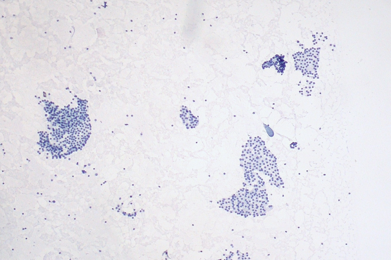
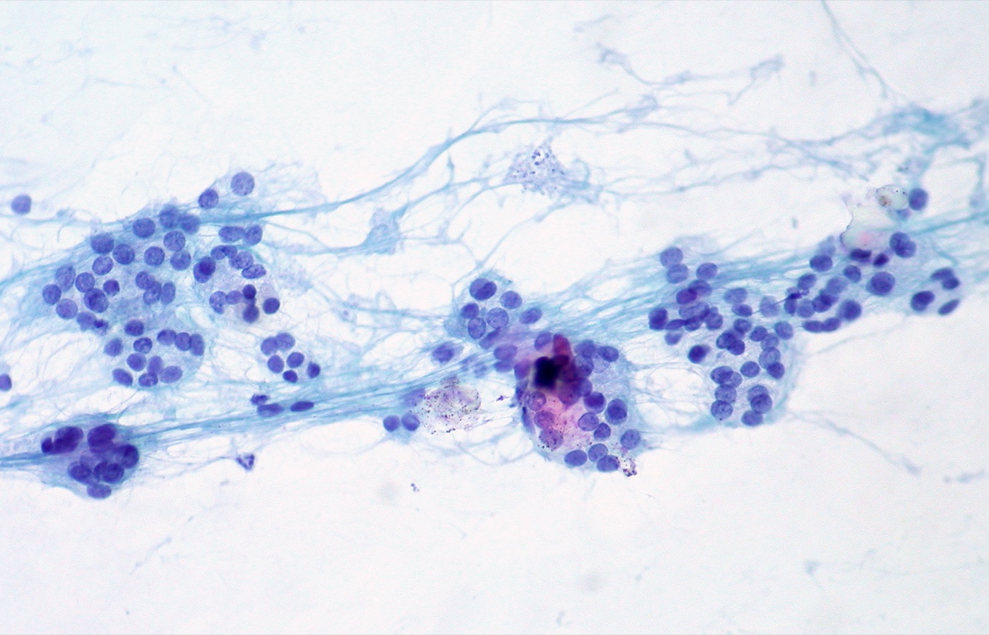
Atypia of Undetermined Significance (AUS), Thyroid
Definition. Bethesda category III. The aspirate contains cells with nuclear or architectural atypia that is insufficient for follicular neoplasm (FN), suspicious for malignancy (SFM) or malignant, and more marked than benign changes. The 3rd edition (2023) uses the single term AUS and removes "follicular lesion of undetermined significance" (FLUS). Related topic: The Bethesda System for Reporting Thyroid Cytopathology (categories and risks of malignancy).
Clinical features of atypia of undetermined significance in thyroid cytology
- Risk of malignancy (ROM): mean 22% (range 13–30%) in adults and 28% in children (Bethesda 3rd edition).
- Subtype and risk: AUS-nuclear has a higher ROM than AUS-other (Ali 2023; Bagıs 2024).
- Management options (Bethesda 3rd edition): repeat FNA, molecular testing, diagnostic lobectomy or surveillance.
- ATA 2015: after review of clinical and ultrasound features, repeat FNA or molecular testing may supplement malignancy risk assessment for AUS nodules. If these are not performed or are inconclusive, surveillance or diagnostic surgery is chosen by clinical risk, ultrasound pattern and patient preference.
Subtypes (3rd edition)
AUS-nuclear
- Focal nuclear atypia: a few cells with enlarged, pale nuclei, grooves or irregular membranes, in an otherwise benign sample.
- Extensive but mild nuclear atypia: widespread mild nuclear enlargement and chromatin pallor without pseudoinclusions.
- Nuclear and architectural atypia together.
- Atypical cyst-lining cells: elongated cells with nuclear grooves, prominent nucleoli and, rarely, intranuclear pseudoinclusions, in a generally benign cystic sample.
- "Histiocytoid" cells: rounded nuclei, a higher nuclear-to-cytoplasmic ratio, glassy cytoplasm and large discrete vacuoles, without the hemosiderin or fine vacuoles of macrophages.
AUS-other
- Architectural atypia:
- a scantly cellular sample with rare groups, almost all microfollicles or crowded three-dimensional groups, and scant colloid
- a moderately or markedly cellular sample with architectural atypia in 50–70% of follicular cells, below the ≥70% predominance required for FN
- focally prominent microfollicles without nuclear atypia, for example in multinodular goiter
- Oncocytic atypia:
- a sparsely cellular aspirate of almost exclusively oncocytic cells with minimal colloid
- a moderately or markedly cellular oncocytic aspirate (≥70% oncocytic cells) in a clinical setting that favors a benign oncocytic nodule, such as lymphocytic thyroiditis or multinodular goiter
- Atypia, not otherwise specified:
- a minor population of follicular cells with nuclear enlargement and prominent nucleoli
- psammomatous calcifications without cells showing papillary carcinoma nuclear features
- rare atypia not described elsewhere
- Atypical lymphoid cells: an atypical lymphoid infiltrate below the threshold for SFM. A repeat aspirate for flow cytometry is desirable.
Source for both lists: Bethesda 3rd edition, as tabulated by Bagıs et al. (2024).
Laboratory quality measures
- AUS rate: the 3rd edition keeps the upper limit of AUS at ≤10% of all thyroid FNAs. Reported rates range from 1% to 20% (Bagıs 2024).
- AUS-to-malignant ratio: the 3rd edition suggests a ratio of ≤3.0 as a laboratory quality measure (Bagıs 2024).
- Subtype reporting: the AUS-nuclear or AUS-other subtype is stated in the report, because the implied risk differs.
Ancillary testing in atypia of undetermined significance in thyroid cytology
- Molecular testing refines risk in AUS. BRAF V600E strongly predicts papillary thyroid carcinoma (PTC). RAS-like alterations indicate a follicular-patterned neoplasm. Details are in Thyroid Molecular Testing.
- Flow cytometry on a repeat aspirate evaluates atypical lymphoid cells.
- Cell-block immunohistochemistry identifies parathyroid tissue (PTH and GATA3 positive, TTF-1 negative) or a non-follicular tumor.
Differential diagnosis of atypia of undetermined significance in thyroid cytology
- Benign: benign follicular nodules contain colloid, macrofollicles and flat sheets. Reactive oncocytes in lymphocytic thyroiditis are mixed with polymorphous lymphocytes.
- Follicular neoplasm: a moderately or markedly cellular sample with a marked predominance (≥70%) of microfollicles or crowded groups.
- Suspicious for malignancy: nuclear features of PTC that are more extensive or better developed than in AUS-nuclear and insufficient for a malignant diagnosis.
- Cyst-lining cells: benign reactive cells with grooves and elongated nuclei. Marked atypia in these cells is reported as AUS-nuclear.
Follicular Neoplasm (Bethesda IV)
Definition. Bethesda category IV. A cellular thyroid aspirate of follicular cells in which most cells show a significant architectural abnormality: microfollicles, crowding, trabeculae or single cells. The 3rd edition (2023) uses the single term "follicular neoplasm" (FN) and replaces "suspicious for a follicular neoplasm". The oncocytic form is reported as follicular neoplasm, oncocytic type. Related topic: The Bethesda System for Reporting Thyroid Cytopathology (categories and risks of malignancy).
Clinical features of follicular neoplasm (Bethesda IV)
- Risk of malignancy (ROM): mean 30% (range 23–34%); 50% in children (Bethesda 3rd edition).
- Reason for the category: follicular adenoma and follicular carcinoma have the same cytology. Carcinoma requires capsular or vascular invasion, which only histology shows.
- Management (Bethesda 3rd edition): molecular testing or diagnostic lobectomy. Pediatric FN nodules are usually managed with surgery.
Cytomorphology of follicular neoplasm (Bethesda IV)
- Cellularity: at least moderately cellular.
- Architecture: a marked predominance of microfollicles or crowded three-dimensional groups, trabeculae or single cells. Architectural atypia in 50–70% of follicular cells is AUS; FN requires ≥70% (Bagıs 2024).
- Colloid: scant or absent.
- Nuclei: usually round and uniform. Mild nuclear changes (enlargement, contour irregularity, chromatin clearing) are permitted when true papillae are absent and intranuclear pseudoinclusions are absent or very rare. The differential diagnosis of these cases includes NIFTP and follicular variant papillary thyroid carcinoma (PTC).
- FN, oncocytic type: a cellular sample composed almost entirely of oncocytic cells, with little or no colloid and few or no lymphocytes. A sparse oncocytic sample is AUS.
Histologic outcomes after lobectomy
- follicular adenoma
- follicular carcinoma (minimally invasive, encapsulated angioinvasive or widely invasive)
- NIFTP and invasive encapsulated follicular variant PTC
- oncocytic adenoma and oncocytic carcinoma (WHO 2022 terms; "Hürthle cell" is no longer used)
- cellular nodules of follicular nodular disease
Ancillary testing in follicular neoplasm (Bethesda IV)
- Molecular testing: RAS mutations and PAX8::PPARG fusion indicate a follicular-patterned neoplasm (RAS-like group). They occur in benign and malignant tumors. BRAF V600E indicates PTC. A negative result on a high-NPV test supports surveillance. Details are in Thyroid Molecular Testing.
- Cell-block immunohistochemistry:
- thyroglobulin, TTF-1 and PAX8 confirm follicular-cell origin
- calcitonin and CEA identify medullary thyroid carcinoma (MTC)
- GATA3 and PTH identify parathyroid tissue, which is TTF-1 negative
Differential diagnosis of follicular neoplasm (Bethesda IV)
- Follicular nodular disease: colloid, macrofollicles and flat sheets. A focal microfollicular population without the ≥70% predominance is benign or AUS.
- Follicular variant PTC and NIFTP: well-developed nuclear features of PTC are reported as SFM or malignant. Aspirates with only mild nuclear changes are reported as FN.
- Parathyroid adenoma: microfollicles and crowded groups that mimic FN. GATA3 and PTH positive, TTF-1 negative.
- MTC: dispersed plasmacytoid or spindle cells with salt-and-pepper chromatin. Calcitonin and CEA positive.
- Hyalinizing trabecular tumor: trabeculae around hyaline material, nuclear grooves and pseudoinclusions, and membranous MIB-1 staining.
Thyroid Molecular Testing (ThyroSeq, Afirma, ThyGeNEXT/ThyraMIR)
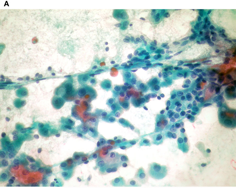
Definition. Molecular tests performed on thyroid fine-needle aspiration (FNA) material to refine the risk of malignancy in nodules with indeterminate cytology. The commercial platforms detect gene variants, gene fusions, expression profiles or microRNA patterns. Related topic: The Bethesda System for Reporting Thyroid Cytopathology (categories and risks of malignancy).
Clinical features of thyroid molecular testing
- Frequency of indeterminate cytology: about 20% of thyroid FNAs are indeterminate, most often Bethesda III or IV (Steward 2019).
- Purpose: a negative result allows surveillance and avoids diagnostic surgery for many benign nodules. A positive result supports surgery and can indicate the likely tumor type.
- Guidelines:
- ATA 2015 states that repeat FNA or molecular testing may supplement malignancy risk assessment for AUS nodules, after review of clinical and ultrasound features.
- The Bethesda 3rd edition (2023) lists molecular testing as a management option for AUS, follicular neoplasm (FN) and suspicious for malignancy (SFM), and adds a chapter on molecular and ancillary testing.
Test platforms
| Test | Method | Validation cohort (Bethesda III and IV) | Sensitivity | Specificity | NPV | PPV |
|---|---|---|---|---|---|---|
| ThyroSeq v3 | Multigene genomic classifier (DNA and RNA sequencing) | Prospective, blinded, 10 centers; cancer/NIFTP prevalence 28% | 94% | 82% | 97% | 66% |
| Afirma Genomic Sequencing Classifier (GSC) | RNA sequencing with machine-learning classifiers | Blinded, 49 centers; cancer prevalence 24% | 91% | 68% | 96% | 47% |
| ThyGeNEXT plus ThyraMIR | Mutation and fusion panel plus microRNA risk classifier | Blinded, multicenter; NPV and PPV at 30% prevalence | 95% | 90% | 97% | 75% |
Sources: Steward 2019; Patel 2018; Lupo 2020.
- Afirma Xpression Atlas (XA): reports variants and fusions from a curated panel of 511 genes by whole-transcriptome RNA sequencing. Its intended use is GSC-suspicious nodules, Bethesda V and VI nodules, and known thyroid metastases (Angell 2019).
- Predictive values depend on prevalence. The negative predictive value (NPV) and positive predictive value (PPV) in each study apply to the stated cancer prevalence. A laboratory with a different Bethesda III or IV malignancy rate has different predictive values.
- Rule-out and rule-in: all three tests have a high NPV and a lower PPV. A negative result has the greater effect on management.
Molecular alterations and their meaning
| Alteration | Usual association |
|---|---|
| BRAF V600E | Papillary thyroid carcinoma (PTC); highly specific |
| RAS (HRAS, KRAS, NRAS) | Follicular-patterned neoplasms: follicular adenoma, follicular carcinoma, NIFTP, invasive encapsulated follicular variant PTC |
| PAX8::PPARG fusion | Follicular-patterned neoplasms (RAS-like group) |
| RET and other kinase fusions | PTC |
| TERT promoter mutation | With BRAF V600E, the PTC group with the highest recurrence rate |
| RET point mutation (somatic or germline) | Medullary thyroid carcinoma (MTC) |
| PAX8::GLIS3 fusion | Hyalinizing trabecular tumor |
- Two molecular groups in PTC: The Cancer Genome Atlas (2014) divided PTC into BRAF V600E-like and RAS-like groups. The groups differ in signaling and differentiation. RAS-like tumors usually have a follicular growth pattern (TCGA 2014; Baloch 2022).
- RAS is not specific for malignancy. In the ThyGeNEXT/ThyraMIR validation, RAS mutations in benign adenomas accounted for 90% of the false-positive mutation-panel results (Lupo 2020).
- BRAF V600E plus TERT promoter mutation: recurrence was 68.6% in PTC with both mutations and 8.7% in PTC with neither (Xing 2014).
- MTC: about 50% of sporadic MTCs carry a somatic RET mutation. HRAS, KRAS or, rarely, NRAS mutations occur in 18% to 80% of sporadic MTCs without a somatic RET mutation (Wells 2015).
Interpretation in practice
- Negative (benign) result in Bethesda III or IV: the residual risk of cancer is low. In the ThyroSeq v3 study, the false-negative rate was 3%, similar to benign cytology, and the missed cancers were low-risk tumors (Steward 2019).
- Positive result: the probability of cancer depends on the specific alteration. In the ThyroSeq v3 study, cancer probability among positive nodules ranged from 59% to 100% by alteration group (Steward 2019).
- Isolated RAS or RAS-like alteration: indicates a neoplasm. Histology separates follicular adenoma, NIFTP and carcinoma by capsular and vascular invasion and by nuclear features.
- Germline relevance: a RET variant in an FNA from an MTC is followed by germline RET testing. ATA 2015 recommends germline RET testing for every patient with presumed sporadic MTC, because 1% to 7% of these patients have hereditary disease (Wells 2015).
Differential diagnosis of thyroid molecular testing
- Follicular adenoma with a RAS mutation is the main source of false-positive results.
- NIFTP has RAS-like alterations. The ThyroSeq v3 study counted NIFTP together with cancer as a positive outcome (Steward 2019).
Thyroid Nodule Selection for FNA and Aspiration Technique
Definition. Thyroid fine-needle aspiration (FNA) is the sampling of a thyroid nodule or cervical lymph node with a thin needle, usually under ultrasound guidance, for cytologic diagnosis. The decision to aspirate is based on nodule size and the ultrasound risk pattern. The American Thyroid Association (ATA) 2015 guidelines and the American College of Radiology Thyroid Imaging, Reporting and Data System (ACR TI-RADS, 2017) are the two frameworks used most often in the United States (Haugen 2016; Tessler 2017). The cytologic result is reported with the Bethesda System for Reporting Thyroid Cytopathology, 3rd edition (Ali 2023), described in The Bethesda System for Reporting Thyroid Cytopathology.
Epidemiology of thyroid nodules and the purpose of selection
- Prevalence: a meta-analysis of 102 studies estimated a pooled thyroid nodule prevalence of 24.83% in the general population. Prevalence was higher in women (36.51%) than in men (23.47%) and higher in 2012–2022 than in 2000–2011 (Mu 2022).
- Most nodules are benign. Selection criteria aim to aspirate the small fraction that is malignant or needs surgery (Tessler 2017).
- Incidental nodules on FDG PET-CT: focal thyroid uptake was found in 1.3% of 10,118 patients scanned for other reasons. Ten malignant lesions were found among the 60 patients who had both ultrasound and FNA (Thuillier 2017).
- AACE/ACE/AME 2016: nodules ≤10 mm are aspirated only when suspicious ultrasound signs are present, and nodules ≤5 mm are monitored without biopsy (Gharib 2016).
Clinical evaluation before thyroid FNA
- Serum TSH is measured in every patient with a thyroid nodule. A subnormal TSH leads to radionuclide scintigraphy. A hyperfunctioning ("hot") nodule has a low risk of malignancy and does not require FNA (Haugen 2016).
- Neck ultrasound characterizes the nodule and the cervical lymph nodes. A sonographically suspicious lymph node is aspirated, with thyroglobulin washout; a positive node establishes diagnosis and stage (Haugen 2016).
- History: prior radioactive iodine, radiation exposure, antithyroid drug therapy and known malignancy elsewhere change the interpretation of the aspirate. These factors are described in Graves Disease and Thyroiditis in Thyroid FNA and Lymphoma, Metastases and Undifferentiated Malignancies in Thyroid FNA.
ATA 2015 sonographic patterns and FNA size thresholds
| ATA pattern | Ultrasound features | Estimated risk of malignancy | FNA recommendation |
|---|---|---|---|
| High suspicion | Solid hypoechoic nodule, or solid hypoechoic component of a partially cystic nodule, with ≥1 of: irregular margins, microcalcifications, taller-than-wide shape, rim calcification with an extrusive soft-tissue component, extrathyroidal extension | >70–90% | ≥1 cm |
| Intermediate suspicion | Hypoechoic solid nodule with smooth margins and none of the high-suspicion features | 10–20% | ≥1 cm |
| Low suspicion | Isoechoic or hyperechoic solid nodule, or partially cystic nodule with eccentric solid areas, without high-suspicion features | 5–10% | ≥1.5 cm |
| Very low suspicion | Spongiform or partially cystic nodule without the features above | <3% | ≥2 cm, or observation without FNA |
| Benign | Purely cystic nodule | <1% | No diagnostic FNA |
Source: Haugen 2016.
- Sub-centimeter nodules are generally not aspirated under ATA 2015, including those with a high-suspicion pattern. Suspicious cervical lymph nodes or extrathyroidal extension change this approach (Haugen 2016).
- FDG-avid nodules: ATA 2015 recommends ultrasound evaluation of focal thyroid FDG uptake and FNA of a corresponding nodule ≥1 cm (Haugen 2016).
ACR TI-RADS point system for thyroid nodules
ACR TI-RADS assigns points in five ultrasound feature categories. The sum determines the TI-RADS level, and the level and the maximum diameter together determine the recommendation (Tessler 2017; Tessler 2018). The terms come from the ACR thyroid ultrasound lexicon (Grant 2015).
| Feature category | 0 points | 1 point | 2 points | 3 points |
|---|---|---|---|---|
| Composition | Cystic or almost completely cystic; spongiform | Mixed cystic and solid | Solid or almost completely solid | – |
| Echogenicity | Anechoic | Hyperechoic or isoechoic | Hypoechoic | Very hypoechoic |
| Shape (transverse plane) | Wider-than-tall | – | – | Taller-than-wide |
| Margin | Smooth; ill-defined | – | Lobulated or irregular | Extrathyroidal extension |
| Echogenic foci (each type scored) | None; large comet-tail artifacts | Macrocalcifications | Peripheral (rim) calcifications | Punctate echogenic foci |
Source: Tessler 2017.
| ACR TI-RADS level | Points | FNA if maximum diameter is | Ultrasound follow-up if maximum diameter is |
|---|---|---|---|
| TR1 Benign | 0 | No FNA | No follow-up |
| TR2 Not suspicious | 2 | No FNA | No follow-up |
| TR3 Mildly suspicious | 3 | ≥2.5 cm | ≥1.5 cm |
| TR4 Moderately suspicious | 4–6 | ≥1.5 cm | ≥1.0 cm |
| TR5 Highly suspicious | ≥7 | ≥1.0 cm | ≥0.5 cm |
Source: Tessler 2017.
- Follow-up schedule: TR5 nodules are imaged every year for up to 5 years, TR4 nodules at 1, 2, 3 and 5 years, and TR3 nodules at 1, 3 and 5 years (Tessler 2017).
- Significant growth is defined as an increase of ≥20% in at least two dimensions with a minimal increase of 2 mm, or an increase of ≥50% in volume (Tessler 2017).
- Multiple nodules: no more than two nodules are aspirated. The nodules with the highest TI-RADS levels are chosen, and size alone does not decide. No more than four nodules are followed with ultrasound (Tessler 2017).
- Children: the ATA pediatric guidelines select nodules for FNA by ultrasound features and clinical context, because size alone is a poorer guide in a gland whose volume changes with age (Francis 2015).
Other ultrasound risk systems for thyroid nodules
- EU-TIRADS (European Thyroid Association, 2017) defines benign, low-, intermediate- and high-risk categories, each with an estimated risk of malignancy and an FNA indication (Russ 2017).
| EU-TIRADS category | Estimated risk of malignancy | FNA threshold |
|---|---|---|
| 2 Benign | About 0% | No FNA, except for therapeutic aspiration |
| 3 Low risk | 2–4% | >20 mm |
| 4 Intermediate risk | 6–17% | >15 mm |
| 5 High risk | 26–87% | >10 mm |
Source: Russ 2017.
- ETA 2023 nodule guidelines update the European workup and favor risk-adapted, conservative management of the many benign and small indolent lesions (Durante 2023).
Thyroid FNA technique
Ultrasound guidance in thyroid FNA
- Ultrasound guidance improves sampling. In 9,683 patients, inadequate specimens fell from 8.7% with palpation-guided FNA to 3.5% with sonography-guided FNA. Sensitivity rose from 91.8% to 97.1% (Danese 1998).
- Cystic and small nodules yield more nondiagnostic samples. In 1,493 nodules, a cystic portion >50% (odds ratio 2.82) and hypoechogenicity (odds ratio 1.88) independently predicted a nondiagnostic result. Mixed nodules >10 mm and solid nodules ≤5 mm carried higher odds (Moon 2011). The solid or wall component of a partially cystic nodule is therefore targeted.
Needle gauge and aspiration versus capillary sampling
- Capillary (non-aspiration) technique: a meta-analysis of 24 studies (4,428 nodules) found a lower relative risk of nondiagnostic cytology with capillary action (relative risk 0.57) and a nonsignificant trend in favor of 24- to 27-gauge needles (Moss 2018). An earlier meta-analysis of 16 studies found no difference in adequacy or accuracy between aspiration and non-aspiration (Song 2015).
- Needle gauge: a 2025 meta-analysis of 16 studies (3,438 nodules) found no significant difference in adequacy between larger and smaller needles (Mohebbi 2025).
- In a blinded randomized trial, 25-gauge needles gave a nondiagnostic rate of 25% and 27-gauge needles 11%, with more blood and fibrin in the 25-gauge samples (Shumrick 2021).
- 21-gauge needles gave more cellular samples than 25-gauge needles, without a higher rate of sufficient samples (Tangpricha 2001).
Specimen preparation for thyroid FNA
- Direct smears: air-dried smears are stained with a Romanowsky stain (Diff-Quik). Alcohol-fixed smears are stained with Papanicolaou stain. Air-dried smears show colloid, cytoplasmic granules and lymphoid cells well. Fixed smears show nuclear detail (Ali 2023).
- Liquid-based preparations (ThinPrep, SurePath): a meta-analysis of 17 studies (15,861 samples) found no significant difference in diagnostic accuracy or inadequacy rate between conventional smears and liquid-based preparations. SurePath had a higher area under the curve than ThinPrep (Kang 2024).
- Cell block material from a dedicated pass or the needle rinse supports immunohistochemistry for parathyroid, medullary, lymphoid and metastatic lesions.
- Dedicated passes are collected for flow cytometry when a lymphoproliferative process is suspected, and for molecular testing in indeterminate nodules (Ali 2023). Molecular testing is described in Thyroid Molecular Testing.
Needle washout assays in thyroid and neck FNA
- Thyroglobulin washout of a lymph node aspirate: pooled sensitivity 0.94 and specificity 0.92 for metastatic papillary thyroid carcinoma (Yu 2025).
- Calcitonin washout of a thyroid aspirate: 93 of 95 medullary carcinomas (97.9%) were detected in a systematic review (Trimboli 2016). ATA 2015 recommends washout calcitonin when FNA findings are inconclusive or suggest medullary carcinoma (Haugen 2016). Cytology of medullary carcinoma is described in Medullary Thyroid Carcinoma (Cytology).
Rapid on-site evaluation in thyroid FNA
Rapid on-site evaluation (ROSE) is a stained-smear assessment at the time of the procedure. It confirms that lesional material is present and directs the collection of additional passes for ancillary studies.
| Study | Design | Main finding |
|---|---|---|
| Muri 2022 | 5,030 FNAs; ROSE in 1,304 | Combined Bethesda I and III rate 4.3% with ROSE against 39.9% without; higher area under the curve with ROSE (0.895 against 0.811) |
| Fawcett 2022 | 410 FNAs before and after ROSE | Nondiagnostic rate fell from 41.1% to 23.8% (odds ratio 0.42) |
| Poller 2020 | Systematic review; UK Thy terminology | Thy1 rate 6.0% with ROSE against 18.5% without |
FNA technique and Bethesda specimen adequacy in thyroid FNA
- Adequacy threshold: a benign interpretation requires at least six groups of well-preserved follicular cells, each with at least 10 cells. Exceptions for atypia, abundant colloid and thyroiditis are given in The Bethesda System for Reporting Thyroid Cytopathology (Ali 2023).
- Nondiagnostic subtypes follow technical causes: cyst fluid only, virtually acellular specimen, and other causes such as obscuring blood, clotting artifact or drying artifact (Ali 2023). Ultrasound targeting of the solid component and ROSE address the first two. Smaller needles and prompt fixation address the third.
- Timing of repeat FNA: the 2009 Bethesda System advised an interval of at least 3 months to avoid reparative atypia (Singh 2011). In 966 nodules, the adequacy and AUS rates on repeat FNA did not vary with intervals from 1 to 6 months (Dellal Kahramanca 2024). An earlier study found the same yield and accuracy at early and late intervals (Singh 2011).
Complications of thyroid FNA and core needle biopsy
- FNA: local pain and minor hematoma are the most common complications after thyroid FNA. Serious complications are rare, and most complications resolve without treatment (Polyzos 2009).
Core needle biopsy of thyroid nodules
- Role: the Korean Society of Thyroid Radiology regards core needle biopsy (CNB) as complementary to FNA. Its use is mainly considered after nondiagnostic or indeterminate FNA results (Na 2017).
- Safety: a meta-analysis of 39 studies (14,818 patients) found an overall CNB complication rate of 1.11% and a major complication rate of 0.06% (Ha 2018).
Causes of nondiagnostic thyroid FNA and corrective steps
| Cause | Typical setting | Corrective step |
|---|---|---|
| Cyst fluid only | Partially cystic nodule, cystic portion >50% | Aspiration of the solid or wall component under ultrasound |
| Virtually acellular specimen | Fibrotic, calcified or very small nodule | Capillary technique; additional passes; CNB after repeated failure |
| Obscuring blood | Vascular nodule; large-bore needle; prolonged aspiration | Smaller needle; capillary technique; short dwell time |
| Air-drying or crush artifact | Delayed fixation; heavy smearing pressure | Immediate fixation; gentle smearing |
| Clotting artifact | Delay before smearing | Immediate smearing or needle rinse into preservative |
Sources: Ali 2023; Moon 2011; Moss 2018.
Suspicious for Malignancy (Bethesda V) in Thyroid FNA
Definition. Suspicious for malignancy (SFM) is category V of the Bethesda System for Reporting Thyroid Cytopathology. It applies when some features of a malignancy are present but are insufficient in number, extent or quality for a conclusive diagnosis (Ali 2023). Most SFM aspirates are suspicious for papillary thyroid carcinoma (PTC) (Mahajan 2013). The report names the suspected malignancy: PTC, medullary thyroid carcinoma, metastatic carcinoma, lymphoma, or another malignancy (Ali 2023). The full cytomorphology of the malignant entities is given in Papillary Thyroid Carcinoma (Cytology), Medullary Thyroid Carcinoma (Cytology) and Lymphoma, Metastases and Undifferentiated Malignancies in Thyroid FNA.
Risk of malignancy and management of Bethesda V thyroid aspirates
- Risk of malignancy (3rd edition): mean 74% (range 67–83%) (Ali 2023).
- Recommended management: molecular testing, lobectomy or near-total thyroidectomy (Ali 2023).
- ATA 2015: surgical management of a nodule suspicious for PTC is similar to that of a malignant nodule. Mutational testing is considered when the result would change the surgical decision (Haugen 2016).
- Pediatric risk: the 3rd edition gives a mean pediatric ROM of 81% for category V, described in The Bethesda System for Reporting Thyroid Cytopathology (Ali 2023).
- Center-specific risk: five experienced cytopathologists from three centers reclassified 213 equivocal aspirates with poor agreement (Krippendorff alpha 0.34). The frequency of malignancy in the categories that led to surgery differed by center (Słowińska-Klencka 2021). Each laboratory therefore monitors its own SFM risk.
Patterns of suspicious for papillary thyroid carcinoma
The Bethesda System describes four patterns. In each pattern the findings are too sparse, incomplete or patchy for the malignant category (Ali 2023).
| Pattern | Cytomorphology | Reason the sample is not malignant |
|---|---|---|
| A. Patchy nuclear changes | Enlarged nuclei, pale chromatin, grooves, membrane irregularity and molding in some groups; the rest are benign follicular cells | Changes are focal; a benign nodule with focal reactive change remains possible |
| B. Incomplete nuclear changes | Mild to moderate enlargement, mild pallor and grooves throughout; pseudoinclusions absent or rare | Changes are diffuse but not fully developed; noninvasive follicular thyroid neoplasm with papillary-like nuclear features (NIFTP) and follicular-patterned neoplasms remain possible |
| C. Sparsely cellular specimen | Many features of PTC, including pseudoinclusions, in very few cells | Too few cells for a conclusive diagnosis |
| D. Cystic degeneration | Histiocytoid cells, cyst-lining cells, hemosiderin-laden macrophages and scattered groups with grooves or pseudoinclusions | Cyst-lining and reparative cells share some features with PTC |
Source: Ali 2023.
- Histiocytoid cells are large, vacuolated, keratin-positive cells without classic PTC nuclei. They were present in about 6% of aspirates with features of PTC (Renshaw 2002).
- Undercalls of PTC as suspicious occur in cystic aspirates with minor features, such as histiocytoid cells, "bubblegum" colloid, syncytial sheets and cellular swirls (Mahajan 2013).
Restricted criteria for a malignant diagnosis of papillary thyroid carcinoma
After NIFTP was introduced in 2016, some authors required papillae, psammoma bodies or three or more pseudoinclusions for a malignant PTC diagnosis. Aspirates with PTC-type nuclei and none of these features were placed in category V (Elsheikh 2024).
- Effect on classification: in 207 aspirates with resection, the restricted criteria were met in 83% of cases originally diagnosed as PTC and in 31% of cases originally diagnosed as suspicious (Elsheikh 2024).
- Role of restricted criteria: restricted criteria are not required when the PTC nuclear features are diffuse and overt. They support the reclassification of suspicious cases as PTC (Elsheikh 2024).
NIFTP and the suspicious for malignancy category
NIFTP shares the nuclear features of PTC and lacks invasion, so it cannot be separated from invasive follicular-patterned PTC on FNA (Maletta 2016).
| Study | Design | Main finding |
|---|---|---|
| Maletta 2016 | 96 histologically proven NIFTPs, blind cytology review | Bethesda IV in 56%, V in 27%, III in 15%, VI in 2% |
| Strickland 2016 | Prospective scoring of 56 resected nodules with Bethesda V or VI cytology | 8 of 9 nodules classified as NIFTP/follicular variant pattern on FNA were follicular-patterned on histology; classic PTC identified in 95% (38 of 40) |
| Ohori 2017 | 139 SFM and 338 malignant aspirates with resection | SFM risk fell from 75.5% to 66.9% with NIFTP reclassification; malignant category 99.4% to 99.1% |
| Bongiovanni 2019 | Meta-analysis, 13,752 thyroidectomies | Pooled NIFTP prevalence 14% in Bethesda V and 3% in Bethesda VI |
| Vuong 2019 | Meta-analysis, 14,153 resected nodules | Largest absolute fall in risk in category V (16%); larger falls in Western than in Asian cohorts |
- Features that favor a follicular-patterned lesion: microfollicles, absent papillae, absent psammoma bodies and rare pseudoinclusions. Papillae, psammomatous calcification, sheet-like architecture and pseudoinclusions favor classic PTC (Strickland 2016).
- Clinical consequence: a lesion with incomplete PTC nuclear features in microfollicles is often NIFTP or an invasive follicular-patterned neoplasm. Category V allows lobectomy as the initial operation (Ali 2023).
Other suspicious categories in thyroid FNA
- Suspicious for medullary thyroid carcinoma: a monomorphic population of dispersed plasmacytoid, round or spindle cells with granular chromatin, without amyloid or without enough cells for immunostains. Calcitonin immunostaining or washout calcitonin resolves most cases (Ali 2023). The full criteria are in Medullary Thyroid Carcinoma (Cytology).
- Suspicious for lymphoma: a monotonous lymphoid population, or atypical large lymphoid cells, without flow cytometry or immunostains. A repeat aspirate for flow cytometry is the usual next step (Ali 2023). Lymphoma criteria are in Lymphoma, Metastases and Undifferentiated Malignancies in Thyroid FNA.
- Suspicious for metastatic carcinoma or other malignancy: cells foreign to the thyroid that are too few for immunocytochemistry, or a history that raises metastasis without confirmatory material (Ali 2023).
Benign mimics of suspicious for malignancy in thyroid FNA
| Mimic | Feature that raises suspicion | Distinguishing finding |
|---|---|---|
| Chronic lymphocytic (Hashimoto) thyroiditis | Powdery chromatin, occasional grooves, nuclear holes; microfollicles with few lymphocytes | Lymphocytes infiltrating follicular groups; features scattered in few cells (Harvey 2012) |
| Oncocytic (Hürthle cell) lesions | Pseudopapillae, syncytial sheets, grooves, pinpoint nucleoli | Granular oncocytic cytoplasm throughout; chromatin not powdery (Mahajan 2013) |
| Hyalinizing trabecular tumor | Elongated nuclei, grooves, many pseudoinclusions, hyaline material | Hyaline stroma within cell groups; PAX8::GLIS3 or GLIS1 fusion (Nikiforova 2019a) |
| Parathyroid tissue | Crowded groups, microfollicles, occasional pseudoinclusions | Stippled chromatin, naked nuclei; PTH and GATA3 positive, TTF-1 negative (Domingo 2017) |
| Cyst-lining and reparative cells | Elongated nuclei, grooves, pale chromatin | Streaming sheets, abundant cytoplasm, macrophage background (Mahajan 2013) |
| Radioactive iodine effect | Nuclear enlargement and atypia | History of Graves disease treated with radioactive iodine (Centeno 1996) |
| Langerhans cell histiocytosis | Dispersed cells with prominent nuclear grooves | Eosinophils; CD1a and S100 positive (Pusztaszeri 2013) |
- Hyalinizing trabecular tumor: in a literature review, 60% of reported cases had been misdiagnosed as suggestive, suspicious or positive for PTC. Only 8% were diagnosed correctly on cytology (Saglietti 2017). PAX8::GLIS3 was present in 13 of 14 tumors and PAX8::GLIS1 in 1 (Nikiforova 2019b). A GLIS fusion supports lobectomy (Nikiforova 2019a).
- Parathyroid tissue: in 60 thyroid FNAs that contained parathyroid tissue, 13 (22%) were initially classified as Bethesda III or IV (Domingo 2017).
- Radioactive iodine: in six patients with Graves disease treated with radioactive iodine, the atypia was severe enough to cause a false-positive diagnosis of PTC in one patient and a suspicious diagnosis in another. None had malignancy at resection (Centeno 1996).
Molecular testing in suspicious for malignancy thyroid aspirates
| Test and study | Population | Result |
|---|---|---|
| BRAF V600E (Trimboli 2020) | Meta-analysis, 1,428 SFM nodules | Pooled prevalence 47%; positive predictive value 99%; negative predictive value 24% |
| BRAF V600E (Fnais 2015) | Meta-analysis, 9,924 FNA samples | Pooled sensitivity 69% for PTC overall and 52% in nodules suspicious for PTC |
| ThyroSeq v3 (Skaugen 2022) | 128 SFM samples with surgery | Sensitivity 89.6%; specificity 77.3%; 78.1% test positive; 95% of positive samples malignant |
| ThyGeNEXT or ThyroSeq v3 (Hier 2021) | 122 Bethesda V and VI nodules | Optimal extent of surgery in 91.9% with molecular testing against 61.1% without |
- BRAF V600E confirms malignancy with high specificity. Its absence does not exclude PTC, so routine BRAF testing in SFM is not recommended. A positive result supports a more extensive operation in selected cases (Trimboli 2020).
- A RAS-like profile in an SFM nodule favors a follicular-patterned tumor or NIFTP (Skaugen 2022). Molecular profiles are described in Thyroid Molecular Testing.
Report wording for suspicious for malignancy in thyroid FNA
The report begins with the category name and then states the suspected entity. A short explanatory note gives the reason for the qualified interpretation and, when relevant, the main alternative (Ali 2023).
| Suspected entity | Example interpretation | Example note |
|---|---|---|
| Papillary thyroid carcinoma, follicular pattern | Suspicious for malignancy. Suspicious for papillary thyroid carcinoma. | The nuclear changes are incomplete and the architecture is microfollicular. A follicular-patterned neoplasm, including NIFTP, is in the differential diagnosis (Cibas 2017). |
| Papillary thyroid carcinoma, cystic | Suspicious for malignancy. Suspicious for papillary thyroid carcinoma. | Rare groups with nuclear grooves and pseudoinclusions are present in a cystic background with histiocytoid cells. |
| Medullary thyroid carcinoma | Suspicious for malignancy. Suspicious for medullary thyroid carcinoma. | Dispersed plasmacytoid cells with granular chromatin are present. Material is insufficient for immunocytochemistry. Serum and washout calcitonin are suggested. |
| Lymphoma | Suspicious for malignancy. Suspicious for lymphoma. | A monotonous population of atypical lymphoid cells is present. A repeat aspirate for flow cytometry is suggested. |
| Metastatic carcinoma | Suspicious for malignancy. Suspicious for metastatic carcinoma. | Rare atypical epithelial cells of non-thyroid appearance are present in a patient with a history of carcinoma. |
Sources: Ali 2023; Cibas 2017.
- Optional NIFTP note: since the 2017 edition, a note may state that a suspicious or malignant follicular-patterned aspirate can represent NIFTP. The note informs the choice of a conservative first operation (Cibas 2017).
- Effect on surgery: the NIFTP note and the molecular result guide the extent of the initial operation (Cibas 2017; Hier 2021).
Differential diagnosis of suspicious for malignancy against adjacent Bethesda categories
| Category | Defining finding | Example |
|---|---|---|
| III. AUS-nuclear | Focal or mild nuclear atypia, below the SFM threshold | Few cells with enlarged, pale nuclei without grooves or pseudoinclusions |
| IV. Follicular neoplasm | Crowded microfollicles; at most mild nuclear changes | Cellular microfollicular aspirate without PTC nuclei |
| V. Suspicious for malignancy | Features of a specific malignancy, insufficient in quantity or quality | Patchy or incomplete PTC nuclear changes |
| VI. Malignant | Conclusive features of a specific malignancy | Diffuse PTC nuclei with papillae or pseudoinclusions |
Sources: Ali 2023; Atypia of Undetermined Significance (Thyroid); Follicular Neoplasm (Bethesda IV).
Lymphoma, Metastases and Undifferentiated Malignancies in Thyroid FNA
Definition. Non-follicular, non-C-cell malignancies in thyroid FNA are aspirates whose malignant cells are not of differentiated follicular or C-cell type. The group comprises primary thyroid lymphoma, secondary (metastatic) tumors, anaplastic (undifferentiated) thyroid carcinoma and rare primary tumors such as intrathyroid thymic carcinoma. Under the Bethesda System, conclusive cases are reported as malignant (category VI) with the specific entity, and cases with insufficient material are reported as suspicious for malignancy (category V) (Ali 2023). Papillary and medullary carcinoma are described in Papillary Thyroid Carcinoma (Cytology) and Medullary Thyroid Carcinoma (Cytology).
Primary thyroid lymphoma in FNA
Epidemiology and clinical features of primary thyroid lymphoma
- Frequency: primary thyroid lymphoma accounts for 1–5% of thyroid malignancies (Adhikari 2016).
- Population data: in 1,408 cases from the SEER database, the mean age was 66 years and 75% of patients were female. Non-Hodgkin lymphoma made up 98%: diffuse large B-cell lymphoma (DLBCL) 68%, follicular lymphoma 10%, marginal zone lymphoma 10% and small lymphocytic lymphoma 3%. Stage I–II disease was present in 88% (Graff-Baker 2009).
- Background thyroiditis: most thyroid lymphomas arise in chronic lymphocytic (Hashimoto) thyroiditis (WHO 2022). In a 40-case series, 42.1% of patients had a history of Hashimoto thyroiditis (Harahap 2025).
- Presentation: a rapidly enlarging thyroid mass is the most common presentation (Morgen 2010).
Cytomorphology of thyroid lymphoma
- DLBCL: a monotonous population of dispersed large lymphoid cells with vesicular chromatin, prominent nucleoli and lymphoglandular bodies in the background. Follicular cells are scant (Morgen 2010).
- Extranodal marginal zone (MALT) lymphoma: small to intermediate lymphoid cells, including centrocyte-like, monocytoid and plasmacytoid cells. Lymphoid cells infiltrating follicular groups form lymphoepithelial lesions. Separation from the polymorphous infiltrate of Hashimoto thyroiditis is the main difficulty (Ali 2023; Morgen 2010; Hirokawa 2017).
- Liquid-based preparations: large nuclei in more than 10% of lymphoid cells were present in 62.9% of MALT lymphomas and in all DLBCLs. Swollen naked nuclei with thin nuclear membranes were found in 92.1% of lymphomas and in 20.5% of non-neoplastic lymphoid infiltrates. Lymphoglandular bodies were less frequent in liquid-based than in conventional preparations (Suzuki 2018).
| Feature | Chronic lymphocytic thyroiditis | MALT lymphoma | DLBCL |
|---|---|---|---|
| Lymphoid population | Polymorphous: small lymphocytes, centroblasts, plasma cells, tingible-body macrophages | Small to intermediate centrocyte-like and monocytoid cells; plasma cells | Monotonous large cells |
| Oncocytes and follicular cells | Present, often numerous | Present; may be scant | Scant or absent |
| Flow cytometry | Polyclonal B cells; T cells prominent | Light chain-restricted B cells | Light chain-restricted large B cells, or nonviable sample |
| Usual Bethesda category | II, benign | III (atypical lymphoid cells), V or VI | VI, malignant |
Sources: Ali 2023; Hirokawa 2017; Zeppa 2009.
Flow cytometry and diagnostic accuracy in thyroid lymphoma
- Multi-institutional series: of 64 patients with thyroid lymphoma, 47 aspirates were diagnosed as lymphoma or suspicious for lymphoma (sensitivity 73%). DLBCL was the most frequent type (43 cases), followed by classic Hodgkin lymphoma and chronic lymphocytic leukemia (5 each). Light chain restriction was detected in 34 specimens (Adhikari 2016).
- Small clones in thyroiditis: two cases of lymphocytic thyroiditis on resection had a small monoclonal population by flow cytometry (Adhikari 2016). When flow cytometry is inconclusive or the sample is nonviable, immunoglobulin gene rearrangement by PCR can show clonality; PCR detected light chain restriction in 2 cases in the same series (Adhikari 2016; Zeppa 2009). In Hashimoto thyroiditis, flow cytometry shows T cells in all cases and B cells in most (Zeppa 2009).
- Diagnostic algorithm: in 75 patients, flow cytometry had a sensitivity of 75.0% and a specificity of 88.4% for lymphoma. A κ/λ ratio ≤0.33 or ≥3 was scored as abnormal. A combined score of ultrasound, cytology and flow cytometry ≥4 was proposed as the threshold for resection. About one-fifth of MALT lymphomas could be missed by this approach (Hirokawa 2017).
- Collection for flow cytometry: a dedicated pass for flow cytometry is collected whenever a monotonous lymphoid population is seen at rapid on-site evaluation (Ali 2023).
Secondary tumors in thyroid FNA
Frequency and sources of thyroid metastases
- Cytology series: secondary tumors made up 0.16% of thyroid FNAs in a seven-institution study of 62 cases. The mean interval from the primary diagnosis was 45 months. FNA was diagnostic of malignancy in 87% and gave a specific diagnosis in 93% of patients (Pusztaszeri 2015).
| Secondary tumor type | Share in Pusztaszeri 2015 (62 cases) | Most frequent sources |
|---|---|---|
| Adenocarcinoma | 37% | Kidney 39%, lung 26%, breast 22%, colon 13% |
| Squamous cell carcinoma | 35.5% | Head and neck 59%, lung, esophagus |
| Lymphoma | 8% | Systemic lymphoma |
| Melanoma | 8% | Skin |
| Adenoid cystic carcinoma | 5% | Salivary and upper aerodigestive tract |
| Sarcoma | 5% | Various |
- Surgical series: metastases are reported in 0.02–1.4% of resected thyroid specimens. Renal cell carcinoma was the source in 10 of 21 patients in one series (Nixon 2011). In 31 patients from a Swedish center, clear cell renal cell carcinoma made up 36% of metastases. Its median interval from the primary was 107 months, the longest of all sources (Stergianos 2021).
- Late presentation of renal cell carcinoma: in eight thyroid metastases of clear cell renal cell carcinoma, the median interval from the renal primary was 84 months. In one patient, the thyroid metastasis was the first sign of the tumor (Queipo 2019).
Cytomorphology of metastases in thyroid FNA
- Clear cell renal cell carcinoma: cohesive groups of cells with abundant clear or finely vacuolated cytoplasm and prominent nucleoli in a bloody background. It mimics follicular and oncocytic neoplasms and parathyroid tissue.
- Squamous cell carcinoma: keratinized cells, necrosis and inflammation. Most squamous cell carcinomas in the thyroid come from the head and neck (Pusztaszeri 2015).
- Melanoma: dispersed epithelioid or spindle cells with eccentric nuclei, macronucleoli, intranuclear pseudoinclusions and sometimes pigment. The pseudoinclusions can lead to a misdiagnosis of PTC.
- Lung and breast adenocarcinoma: three-dimensional clusters without colloid; signet-ring or single-file cells in lobular breast carcinoma.
Sources: Ali 2023; Pusztaszeri 2015.
Anaplastic thyroid carcinoma in FNA
- Clinical setting: a rapidly growing neck mass, usually in an older patient. The ATA 2021 guidelines call for a rapid diagnosis and staging because treatment decisions are urgent (Bible 2021).
- Stage: every anaplastic carcinoma is stage IV under AJCC 8th edition: IVA (intrathyroidal), IVB (extrathyroidal extension or nodal metastasis) and IVC (distant metastasis) (Bible 2021).
- Cytomorphology: in 18 aspirates, cellularity was moderate to high in 83% and the cells were epithelioid in 83%. Nuclear enlargement and membrane irregularity were present in all cases, multinucleated tumor cells in 33% and acute inflammation in 50%. Pleomorphism, coarse chromatin, macronucleoli, apoptosis and necrosis separated anaplastic carcinoma from differentiated carcinoma (Podany 2022).
- Morphologic patterns: spindle (sarcomatoid), giant cell (pleomorphic) and squamoid. WHO 2022 classifies squamous cell carcinoma of the thyroid as a morphologic pattern of anaplastic carcinoma (Baloch 2022).
- PAX8: retained in 26 of 34 anaplastic carcinomas (76%): 16 of 16 squamoid, 7 of 12 giant cell and 3 of 6 spindle cell tumors. All 118 head and neck squamous cell carcinomas were PAX8 negative (Bishop 2011).
- BRAF V600E immunostaining (clone VE1): on cell block sections, sensitivity was 94.4% and specificity 100%. On direct smears, sensitivity was 80% and specificity 63.6% (Smith 2018). BRAF status determines eligibility for targeted therapy and is assessed early in anaplastic carcinoma (Bible 2021).
- Primary thyroid squamous cell carcinoma: a review of 117 cases found PAX8 positivity useful to confirm thyroid origin. The tumors were positive for p63, p40 and cytokeratins 5/6, 7 and 19, and negative for cytokeratin 20. Median survival was 8 months (Lam 2020).
- High-grade follicular cell-derived non-anaplastic carcinoma (poorly differentiated and differentiated high-grade carcinoma) is a separate WHO 2022 category reported as malignant on FNA (Baloch 2022). It is described in The Bethesda System for Reporting Thyroid Cytopathology.
Rare primary tumors in the differential of thyroid metastases
- Intrathyroid thymic carcinoma (CASTLE): all seven tumors in a comparative study showed diffuse p63 and high-molecular-weight keratin staining and were positive for CEA and CD5. None of the other thyroid neoplasms examined stained consistently for all four markers (Reimann 2006). Polyclonal PAX8 stained 4 of 10 intrathyroid thymic carcinomas and monoclonal PAX8 none, whereas all poorly differentiated carcinomas and 92.9% of squamous cell carcinomas were positive with both. CD5 stained 80% of thymic carcinomas (Suzuki, Endocr J 2018).
- Small-cell malignancies: the differential of a small round cell aspirate includes medullary carcinoma, poorly differentiated carcinoma, lymphoma, Ewing family tumors and metastatic small cell carcinoma (Canberk 2022).
- Parathyroid tissue and parathyroid carcinoma: in 81 parathyroid aspirates, the main features were high cellularity, naked nuclei, cribriform and three-dimensional clusters, stippled chromatin and oxyphilic cytoplasm. Diagnosis was supported by washout PTH and GATA3, PTH and chromogranin A immunostaining (Bakuła-Zalewska 2024).
Immunohistochemical panel for non-follicular malignancies in thyroid FNA
| Tumor | TTF-1 | PAX8 | Thyroglobulin | Other positive markers | Key negative markers |
|---|---|---|---|---|---|
| Follicular cell-derived carcinoma (PTC, follicular, oncocytic) | + | + | + | Keratins | Calcitonin |
| Anaplastic carcinoma | Usually − | + in about 76% | Usually − | Keratins (variable); BRAF V600E (VE1) in a subset | CD45 |
| Medullary carcinoma | + | Variable | − | Calcitonin, CEA, chromogranin, synaptophysin | – |
| Primary thyroid squamous cell carcinoma | Variable | + | − | p40, p63, CK5/6 | CK20 |
| Head and neck squamous cell carcinoma (extension or metastasis) | − | − | − | p40, p63, CK5/6 | PAX8 |
| Clear cell renal cell carcinoma | − | + | − | CA-IX, CD10 | TTF-1, thyroglobulin |
| Lung adenocarcinoma | + | − | − | Napsin A | PAX8, thyroglobulin |
| Breast carcinoma | − | − | − | GATA3, ER | TTF-1, PAX8 |
| Colorectal adenocarcinoma | − | − | − | CDX2, SATB2, CK20 | CK7 |
| Melanoma | − | − | − | SOX10, S100, Melan-A, HMB-45 | Keratins |
| Lymphoma | − | − | − | CD45; CD20 (B cell); CD3 (T cell) | Keratins |
| Intrathyroid thymic carcinoma | − | Polyclonal + in about 40%; monoclonal − | − | CD5, p63, CEA, high-molecular-weight keratin | Thyroglobulin, calcitonin |
| Parathyroid | − | – | − | PTH, GATA3, chromogranin A | TTF-1, thyroglobulin |
Sources: Ali 2023; Bishop 2011; Ye 2012; Queipo 2019; Lam 2020; Reimann 2006; Suzuki, Endocr J 2018; Bakuła-Zalewska 2024.
- Thyroid metastasis to and from the lung: all 120 primary lung adenocarcinomas were PAX8 negative. Metastatic carcinomas of kidney (29 of 33) and thyroid (4 of 5) were PAX8 positive. All 103 metastatic carcinomas to the lung were napsin A negative, and 14 (13.6%) showed TTF-1 staining, including four thyroid carcinomas (Ye 2012).
- Renal cell carcinoma to the thyroid was PAX8 and CA-IX positive and TTF-1 and thyroglobulin negative in a clinicopathologic series (Queipo 2019). PAX8 alone does not separate renal from thyroid primaries; thyroglobulin and TTF-1 complete the panel.
Differential diagnosis of high-grade malignancies in thyroid FNA
| Entity | Cytologic clues | Confirmatory test |
|---|---|---|
| Anaplastic carcinoma | Pleomorphic, giant or spindle cells; necrosis; neutrophils; history of goiter or differentiated carcinoma | PAX8; keratin; BRAF V600E |
| DLBCL | Dispersed large lymphoid cells; lymphoglandular bodies; no cohesion | Flow cytometry; CD45, CD20 |
| Metastatic renal cell carcinoma | Clear cytoplasm; prominent nucleoli; blood | PAX8+, CA-IX+, thyroglobulin−, TTF-1− |
| Metastatic melanoma | Dispersed cells; pseudoinclusions; macronucleoli; pigment | SOX10, S100, Melan-A |
| Squamous cell carcinoma, extension or metastasis | Keratinized cells; necrosis | p40+; PAX8− |
| Medullary carcinoma | Plasmacytoid or spindle cells; granular chromatin; amyloid | Calcitonin, CEA; washout calcitonin |
| Intrathyroid thymic carcinoma | Cohesive squamoid groups; lymphocytes | CD5, p63 |
Sources: Ali 2023; Podany 2022; Pusztaszeri 2015; Reimann 2006.
Graves Disease and Thyroiditis in Thyroid FNA
Definition. Graves disease and thyroiditis in thyroid FNA comprise the non-neoplastic inflammatory and hyperfunctional thyroid diseases that are sampled by fine-needle aspiration (FNA): Graves disease, subacute granulomatous (de Quervain) thyroiditis, acute suppurative thyroiditis, Riedel thyroiditis and related fibrosing disease, and minor findings such as palpation thyroiditis and black thyroid. Most are diagnosed clinically. FNA is performed when a nodule develops, when the presentation is atypical, or when lymphoma or carcinoma must be excluded (García Solano 1997; Hang 2017). They are reported as benign (Bethesda category II) when the findings are characteristic (Ali 2023). Chronic lymphocytic (Hashimoto) thyroiditis is described in The Bethesda System for Reporting Thyroid Cytopathology, and its lymphoma differential in Lymphoma, Metastases and Undifferentiated Malignancies in Thyroid FNA.
Graves disease in thyroid FNA
Diffuse Graves disease is not aspirated. FNA is performed for a discrete nodule in a patient with Graves disease (Hang 2017). Antithyroid drugs and radioactive iodine alter follicular cells, so the clinical history is part of the request (Centeno 1996; Hang 2017).
Cytomorphology of Graves disease
- Untreated Graves disease: cellular smears with scant, watery colloid. Follicular cells form flat sheets and loose microfollicles and have abundant, pale, finely vacuolated cytoplasm. Nuclei are enlarged, round and vesicular, sometimes with small nucleoli. Lymphocytes and oncocytes are present in variable numbers (Ali 2023).
- Marginal vacuoles ("fire-flares"): pink to red frayed vacuoles at the edges of follicular cell groups in Romanowsky-stained smears. They were first described as a distinctive feature of thyrotoxic goiter. They also occur in many non-toxic lesions, including 85.7% of follicular neoplasms in one series, and are therefore not specific (Das 2006).
- Treated Graves disease: nuclear enlargement, hyperchromasia, nucleoli and oncocytic change. These changes can mimic papillary thyroid carcinoma (PTC) and follicular neoplasm (Hang 2017).
Diagnostic accuracy of FNA in Graves disease
| Study | Population | Main finding |
|---|---|---|
| Hang 2017 | 55 FNAs from 43 patients with Graves disease and thyroidectomy | 18 of 46 non-neoplastic nodules (39.1%) were reported as indeterminate. Sensitivity for PTC 60%, specificity 100% |
| Centeno 1996 | 6 patients with Graves disease treated with radioactive iodine | Radiation atypia caused one false-positive PTC diagnosis and one suspicious diagnosis. No malignancy at resection |
- Neoplasms in Graves disease: of five patients with PTC, FNA was read as PTC in three, AUS in one and benign in one with a 0.5-cm incidental microcarcinoma. Of four follicular adenomas, two were read as follicular neoplasm, one as AUS and one as benign (Hang 2017).
- Effect of the treatment history on interpretation: atypia in a treated Graves nodule is interpreted with the treatment history. In six patients treated with radioactive iodine, the two aspirates submitted without the treatment history were reported as suspicious for PTC or descriptively. Three of the four aspirates submitted with the history were interpreted correctly. In the fourth, the atypia was severe enough to cause a false-positive PTC diagnosis despite the history (Centeno 1996).
Subacute granulomatous (de Quervain) thyroiditis in FNA
Clinical features of subacute granulomatous thyroiditis
- Presentation: anterior neck pain, tenderness and transient thyrotoxicosis, usually after a viral infection and most often in adult women. Treatment is with non-steroidal anti-inflammatory drugs, and with corticosteroids in severe cases (Toschetti 2025).
- Ultrasound: hypoechoic, heterogeneous areas with irregular or indistinct margins. A hypoechoic nodule with irregular margins was present in 12 of 20 cases with imaging. Some cases form a painless, hypoechoic, solid nodule that mimics carcinoma (Vural 2015).
- Role of FNA: the diagnosis is usually clinical. FNA is used in atypical cases and to exclude a coexisting neoplasm (García Solano 1997; Shabb 2006).
Cytomorphology of subacute granulomatous thyroiditis
- Diagnostic combination: the most reliable diagnosis is based on the following findings in the same aspirate (García Solano 1997):
- follicular cells with intravacuolar granules or plump transformed follicular cells
- epithelioid granulomas
- multinucleated giant cells
- an acute and chronic inflammatory, "dirty" background
- absence of fire-flare cells, hypertrophic follicular cells, oncocytes and transformed lymphocytes
- Multinucleated giant cells were present in all 14 cases presenting as nodules, some containing colloid or neutrophils. Fibrous fragments with inflammatory cells were constant. Granulomas were rare, and follicular cells were scant. Colloid, when present, was thick with central cracks and frayed edges. One of the 14 aspirates was reported as suspicious for malignancy (Shabb 2006).
- Concordance across series: in 69 cases, lymphocytes, giant cells, loose epithelioid histiocyte groups, granulomas, colloid and neutrophils were the diagnostic features. Giant cells were the finding most consistently reported in the literature, and microfollicles the least (Sahin 2019).
- Evolution with time: in 61 serial examinations of nine patients, early aspirates showed enlarged, degenerated follicular cells with prominent nucleoli, neutrophils and giant cells. Neutrophils and degenerated cells disappeared and cohesive follicles appeared as the sonographic hypoechogenicity regressed. Cytologic recovery lagged behind sonographic recovery (Lu 1997).
- Sources of error: reactive atypia in the early phase and microfollicles in the late phase can lead to a false diagnosis and an unnecessary resection (Sahin 2019).
Acute suppurative thyroiditis in FNA
- Clinical features: a rare bacterial infection, under 1% of thyroid diseases, more frequent in children (Toschetti 2025). A pyriform sinus fistula is the main route of infection, especially in recurrent left-sided disease in children (Masuoka 2011). Fever, painful neck swelling, abscess and airway compromise occur (Toschetti 2025).
- Pyriform sinus fistula: all 60 patients in one imaging series had a fistula. Early ultrasound and CT findings were often nonspecific and led to an erroneous diagnosis of subacute thyroiditis (Masuoka 2011).
- FNA: ultrasound-guided aspiration provides material for culture and drains the abscess (Toschetti 2025). Smears show neutrophils, necrotic debris, macrophages and sometimes bacteria or fungi (Ali 2023).
- Management: broad-spectrum antibiotics, aspiration or surgical drainage, and treatment of the fistula (Toschetti 2025).
Riedel thyroiditis and fibrosing thyroiditis in FNA
- Definition: a fibroinflammatory process that involves the thyroid and extends beyond the thyroid capsule into adjacent neck tissues (Weidner 2015).
- IgG4-related disease: all three thyroidectomy specimens in one series contained IgG4-positive plasma cells, and one patient had cholangitis, lung and lacrimal pseudotumors and an IgG4-rich perithyroidal lymph node. Riedel thyroiditis is regarded as part of the IgG4-related disease spectrum (Dahlgren 2010).
- IgG4 thyroiditis: IgG4 immunostaining divided 13 cases of Hashimoto thyroiditis into IgG4-rich and IgG4-poor groups. The IgG4-rich group showed severe lymphoplasmacytic infiltration, dense fibrosis, marked follicular degeneration, oxyphilic change and lymphoid follicles (Li 2009). Extrathyroidal extension is the feature that defines Riedel thyroiditis (Weidner 2015).
- Cytomorphology: aspirates are often scant and nondiagnostic because of fibrosis (Weidner 2015). Fragments of fibrous tissue with bland spindle cells and myofibroblasts are the main finding (Harigopal 2004). Lymphocytes and plasma cells are present, and oncocytes are absent (Kumar 2019).
- Clinical and imaging mimicry: a hard gland with extrathyroidal extension around the trachea suggests malignancy on imaging. Multiple ultrasound-guided passes with a thin needle improved the yield in one case (Kumar 2019).
- Differential diagnosis: fibrosing variant of Hashimoto thyroiditis, subacute granulomatous thyroiditis, paucicellular anaplastic carcinoma and other spindle cell lesions (Harigopal 2004). Core or open biopsy is often needed.
Other inflammatory and pigmentary findings in thyroid FNA
- Palpation thyroiditis: multifocal granulomatous folliculitis from traumatic rupture of single follicles. It was found in more than 83% of surgically removed thyroids and has little clinical significance (Carney 1975). Isolated histiocytes and giant cells in an otherwise benign aspirate can reflect this change.
- Black thyroid (minocycline): pigment in follicular cells and macrophages after long-term tetracycline use. Degenerative hyperchromasia can mimic neoplasia, and the pigment stains with Fontana-Masson and bleaches with potassium permanganate (Keyhani-Rofagha 1991). In four patients with black thyroid at surgery, the prior aspirates showed no characteristic pigment (Oertel 2006).
Comparison of thyroiditis patterns in thyroid FNA
| Feature | Chronic lymphocytic (Hashimoto) | Graves disease | Subacute granulomatous | Acute suppurative | Riedel |
|---|---|---|---|---|---|
| Clinical setting | Hypothyroidism; TPO antibodies | Hyperthyroidism; TSH receptor antibodies | Painful gland after viral illness; transient thyrotoxicosis | Fever; painful swelling; children; pyriform sinus fistula | Hard gland; extrathyroidal extension |
| Cellularity | Moderate to high | High | Variable | High (inflammatory) | Low |
| Colloid | Scant | Scant, watery | Scant, thick or cracked | Absent | Absent |
| Follicular cells | Oncocytes; reactive groups permeated by lymphocytes | Flat sheets; abundant pale cytoplasm; marginal vacuoles | Scant; degenerated; intravacuolar granules | Scant | Scant or absent |
| Inflammatory cells | Polymorphous lymphocytes; plasma cells; germinal center cells | Lymphocytes in some cases | Giant cells; epithelioid granulomas; neutrophils; dirty background | Neutrophils; necrosis; bacteria | Lymphocytes; plasma cells |
| Other | – | Treatment atypia after drugs or radioactive iodine | Fibrous fragments | Culture positive | Fibrous fragments with spindle cells |
| Usual Bethesda category | II | II (III if atypia persists) | II | II (benign, consistent with acute thyroiditis); culture | I or II |
Sources: Ali 2023; García Solano 1997; Shabb 2006; Hang 2017; Harigopal 2004; MacDonald 1999.
- Hashimoto thyroiditis as a source of error: in 39 aspirates with histologic follow-up, hyperplastic follicular cells in Hashimoto thyroiditis mimicked follicular neoplasm. Five Hürthle cell adenomas and one lymphoma were missed when the aspirate was read as thyroiditis (MacDonald 1999).
Differential diagnosis of giant cells and granulomas in thyroid FNA
| Finding | Entity | Separating features |
|---|---|---|
| Giant cells, granulomas, dirty background, painful gland | Subacute granulomatous thyroiditis | Clinical setting; degenerated follicular cells; regression on follow-up |
| Giant cells with PTC nuclei | Papillary thyroid carcinoma | Nuclear grooves and pseudoinclusions in follicular groups; papillae |
| Isolated histiocytes and giant cells in a benign nodule | Palpation thyroiditis | No neck pain; incidental |
| Necrotizing granulomas | Tuberculosis or fungal infection | Caseous necrosis; acid-fast or fungal stains; culture |
| Pleomorphic tumor giant cells | Anaplastic carcinoma | Malignant nuclei; necrosis; rapid growth |
Sources: García Solano 1997; Carney 1975; Sahin 2019; Ali 2023.
Report wording for thyroiditis in thyroid FNA
- Benign, consistent with granulomatous (subacute) thyroiditis: used when giant cells, granulomas and degenerated follicular cells are present in the appropriate clinical setting (Ali 2023).
- Benign, consistent with chronic lymphocytic (Hashimoto) thyroiditis in the proper clinical context (Ali 2023).
- Benign, consistent with Graves disease is reserved for a known clinical diagnosis. Treatment-related atypia that cannot be resolved is reported as atypia of undetermined significance (Ali 2023; Hang 2017).
References
- Adhikari LJ, Reynolds JP, Wakely PE Jr. Multi-institutional study of fine-needle aspiration for thyroid lymphoma. J Am Soc Cytopathol. 2016;5(3):170–176. PMID 31042520. doi:10.1016/j.jasc.2015.11.002
- Ali SZ, Baloch ZW, Cochand-Priollet B, Schmitt FC, Vielh P, VanderLaan PA. The 2023 Bethesda System for Reporting Thyroid Cytopathology. Thyroid. 2023;33(9):1039–1044. PMID 37427847. doi:10.1089/thy.2023.0141
- Ali SZ, VanderLaan PA, eds. The Bethesda System for Reporting Thyroid Cytopathology, 3rd ed. Springer; 2023. doi:10.1007/978-3-031-28046-7
- Angell TE, et al. Analytical and clinical validation of expressed variants and fusions from the whole transcriptome of thyroid FNA samples. Front Endocrinol (Lausanne). 2019;10:612. doi:10.3389/fendo.2019.00612. PMID 31572297
- Bagıs M, et al. A comprehensive approach to the thyroid Bethesda category III (AUS) in the transition zone between 2nd edition and 3rd edition of The Bethesda System for Reporting Thyroid Cytopathology: subcategorization, nuclear scoring, and more. Endocr Pathol. 2024;35(1):51–76. doi:10.1007/s12022-024-09797-1. PMID 38280141
- Bakuła-Zalewska E, Długosińska J, Stanek-Widera A, et al. Fine needle aspiration biopsy of parathyroid; is it meaningful? A cytologic study of 81 cases with histological and clinical correlations. Cytopathology. 2024;35(3):362–370. PMID 38213192. doi:10.1111/cyt.13356
- Baloch ZW, et al. Overview of the 2022 WHO Classification of Thyroid Neoplasms. Endocr Pathol. 2022;33(1):27–63. PMID 35288841. doi:10.1007/s12022-022-09707-3
- Baloch ZW, LiVolsi VA, Asa SL, et al. Diagnostic terminology and morphologic criteria for cytologic diagnosis of thyroid lesions: a synopsis of the National Cancer Institute Thyroid Fine-Needle Aspiration State of the Science Conference. Diagn Cytopathol. 2008;36(6):425–437. PMID 18478609. doi:10.1002/dc.20830
- Bible KC, Kebebew E, Brierley J, et al. 2021 American Thyroid Association guidelines for management of patients with anaplastic thyroid cancer. Thyroid. 2021;31(3):337–386. PMID 33728999. doi:10.1089/thy.2020.0944
- Bishop JA, Sharma R, Westra WH. PAX8 immunostaining of anaplastic thyroid carcinoma: a reliable means of discerning thyroid origin for undifferentiated tumors of the head and neck. Hum Pathol. 2011;42(12):1873–1877. PMID 21663937. doi:10.1016/j.humpath.2011.02.004
- Bongiovanni M, Faquin WC, Giovanella L, et al. Impact of non-invasive follicular thyroid neoplasms with papillary-like nuclear features (NIFTP) on risk of malignancy in patients undergoing lobectomy/thyroidectomy for suspected malignancy or malignant fine-needle aspiration cytology findings: a systematic review and meta-analysis. Eur J Endocrinol. 2019;181(4):389–396. PMID 31340203. doi:10.1530/EJE-19-0223
- Bongiovanni M, Spitale A, Faquin WC, et al. The Bethesda System for Reporting Thyroid Cytopathology: a meta-analysis. Acta Cytol. 2012;56(4):333–339. PMID 22846422. doi:10.1159/000339959
- Canberk S, Thodou E, Bongiovanni M. Small-cell malignancies of thyroid: challenge solved? Acta Cytol. 2022;66(4):307–318. PMID 35016187. doi:10.1159/000520876
- Cancer Genome Atlas Research Network. Integrated genomic characterization of papillary thyroid carcinoma. Cell. 2014;159(3):676–690. doi:10.1016/j.cell.2014.09.050. PMID 25417114
- Carney JA, Moore SB, Northcutt RC, et al. Palpation thyroiditis (multifocal granulomatous folliculitis). Am J Clin Pathol. 1975;64(5):639–647. PMID 1242618. doi:10.1093/ajcp/64.5.639
- Centeno BA, Szyfelbein WM, Daniels GH, et al. Fine needle aspiration biopsy of the thyroid gland in patients with prior Graves' disease treated with radioactive iodine: morphologic findings and potential pitfalls. Acta Cytol. 1996;40(6):1189–1197. PMID 8960027. doi:10.1159/000333979
- Cibas ES, Ali SZ. The 2017 Bethesda System for Reporting Thyroid Cytopathology. Thyroid. 2017;27(11):1341–1346. PMID 29091573. doi:10.1089/thy.2017.0500
- Cibas ES, Ali SZ. The Bethesda System for Reporting Thyroid Cytopathology. Thyroid. 2009;19(11):1159–1165. PMID 19888858. doi:10.1089/thy.2009.0274
- Dahlgren M, Khosroshahi A, Nielsen GP, et al. Riedel's thyroiditis and multifocal fibrosclerosis are part of the IgG4-related systemic disease spectrum. Arthritis Care Res (Hoboken). 2010;62(9):1312–1318. PMID 20506114. doi:10.1002/acr.20215
- Danese D, Sciacchitano S, Farsetti A, et al. Diagnostic accuracy of conventional versus sonography-guided fine-needle aspiration biopsy of thyroid nodules. Thyroid. 1998;8(1):15–21. PMID 9492148. doi:10.1089/thy.1998.8.15
- Das DK. Marginal vacuoles (fire-flare appearance) in fine needle aspiration smears of thyroid lesions: does it represent diffusing out of thyroid hormones at the base of follicular cells? Diagn Cytopathol. 2006;34(4):277–283. PMID 16544335. doi:10.1002/dc.20416
- Dellal Kahramanca FD, Sacikara M, Kilicarslan A, et al. Timing of the repeat thyroid fine-needle aspiration biopsy: does early repeat biopsy change the rate of nondiagnostic or atypia of undetermined significance cytology result? Endocrine. 2024;86(3):1065–1072. PMID 38987433. doi:10.1007/s12020-024-03953-7
- Domingo RP, Ogden LL, Been LC, et al. Identification of parathyroid tissue in thyroid fine-needle aspiration: a combined approach using cytology, immunohistochemical, and molecular methods. Diagn Cytopathol. 2017;45(6):526–532. PMID 28371486. doi:10.1002/dc.23711
- Durante C, Hegedüs L, Czarniecka A, et al. 2023 European Thyroid Association clinical practice guidelines for thyroid nodule management. Eur Thyroid J. 2023;12(5):e230067. PMID 37358008. doi:10.1530/ETJ-23-0067
- Elsheikh TM, Thomas M, Brainard J, et al. Papillae, psammoma bodies, and/or many nuclear pseudoinclusions are helpful criteria but should not be required for a definitive cytologic diagnosis of papillary thyroid carcinoma: an institutional experience of 207 cases with surgical follow up. Cancer Cytopathol. 2024;132(6):348–358. PMID 38563725. doi:10.1002/cncy.22817
- Fawcett C, Eppenberger-Castori S, Zechmann S, et al. Effects of rapid on-site evaluation on diagnostic accuracy of thyroid fine-needle aspiration. Acta Cytol. 2022;66(5):371–378. PMID 35512664. doi:10.1159/000522662
- Fnais N, Soobiah C, Al-Qahtani K, et al. Diagnostic value of fine needle aspiration BRAF(V600E) mutation analysis in papillary thyroid cancer: a systematic review and meta-analysis. Hum Pathol. 2015;46(10):1443–1454. PMID 26232865. doi:10.1016/j.humpath.2015.06.001
- Francis GL, Waguespack SG, Bauer AJ, et al. Management guidelines for children with thyroid nodules and differentiated thyroid cancer. Thyroid. 2015;25(7):716–759. PMID 25900731. doi:10.1089/thy.2014.0460
- García Solano J, Giménez Bascuñana A, Sola Pérez J, et al. Fine-needle aspiration of subacute granulomatous thyroiditis (De Quervain's thyroiditis): a clinico-cytologic review of 36 cases. Diagn Cytopathol. 1997;16(3):214–220. PMID 9099540. doi:10.1002/(sici)1097-0339(199703)16:3<214::aid-dc4>3.0.co;2-f
- Gharib H, Papini E, Garber JR, et al. American Association of Clinical Endocrinologists, American College of Endocrinology, and Associazione Medici Endocrinologi medical guidelines for clinical practice for the diagnosis and management of thyroid nodules: 2016 update. Endocr Pract. 2016;22(5):622–639. PMID 27167915. doi:10.4158/EP161208.GL
- Graff-Baker A, Roman SA, Thomas DC, et al. Prognosis of primary thyroid lymphoma: demographic, clinical, and pathologic predictors of survival in 1,408 cases. Surgery. 2009;146(6):1105–1115. PMID 19958938. doi:10.1016/j.surg.2009.09.020
- Grant EG, Tessler FN, Hoang JK, et al. Thyroid ultrasound reporting lexicon: white paper of the ACR Thyroid Imaging, Reporting and Data System (TIRADS) Committee. J Am Coll Radiol. 2015;12(12 Pt A):1272–1279. PMID 26419308. doi:10.1016/j.jacr.2015.07.011
- Ha EJ, Suh CH, Baek JH. Complications following ultrasound-guided core needle biopsy of thyroid nodules: a systematic review and meta-analysis. Eur Radiol. 2018;28(9):3848–3860. PMID 29589112. doi:10.1007/s00330-018-5367-5
- Hang JF, Lilo MT, Bishop JA, et al. Diagnostic accuracy of fine needle aspiration in thyroid nodules arising in patients with Graves disease. Acta Cytol. 2017;61(2):117–124. PMID 28259878. doi:10.1159/000464094
- Harahap AS, Santoso I, Charles S, et al. Variability in primary thyroid lymphoma: a clinicopathological exploration of diffuse large B-cell, marginal zone, and follicular lymphoma. Ann Diagn Pathol. 2025;75:152444. PMID 39893967. doi:10.1016/j.anndiagpath.2025.152444
- Harigopal M, Sahoo S, Recant WM, et al. Fine-needle aspiration of Riedel's disease: report of a case and review of the literature. Diagn Cytopathol. 2004;30(3):193–197. PMID 14986301. doi:10.1002/dc.10420
- Harvey AM, Truong LD, Mody DR. Diagnostic pitfalls of Hashimoto's/lymphocytic thyroiditis on fine-needle aspirations and strategies to avoid overdiagnosis. Acta Cytol. 2012;56(4):352–360. PMID 22846513. doi:10.1159/000338738
- Haugen BR, et al. 2015 American Thyroid Association Management Guidelines for Adult Patients with Thyroid Nodules and Differentiated Thyroid Cancer. Thyroid. 2016;26(1):1–133. PMID 26462967. doi:10.1089/thy.2015.0020
- Hier J, Avior G, Pusztaszeri M, et al. Molecular testing for cytologically suspicious and malignant (Bethesda V and VI) thyroid nodules to optimize the extent of surgical intervention: a retrospective chart review. J Otolaryngol Head Neck Surg. 2021;50(1):29. PMID 33910629. doi:10.1186/s40463-021-00500-6
- Hirokawa M, Kudo T, Ota H, et al. Preoperative diagnostic algorithm of primary thyroid lymphoma using ultrasound, aspiration cytology, and flow cytometry. Endocr J. 2017;64(9):859–865. PMID 28690277. doi:10.1507/endocrj.EJ17-0111
- Kang YJ, Lee HW, Stybayeva G, et al. Comparison of liquid-based preparations with conventional smears in thyroid fine-needle aspirates: a systematic review and meta-analysis. Cancers (Basel). 2024;16(4):751. PMID 38398142. doi:10.3390/cancers16040751
- Keyhani-Rofagha S, Kooner DS, Landas SK, et al. Black thyroid: a pitfall for aspiration cytology. Diagn Cytopathol. 1991;7(6):640–643. PMID 1769297. doi:10.1002/dc.2840070621
- Kumar N, Gupta R, Sayed S, et al. Difficulties in diagnosis of Riedel's thyroiditis on aspiration cytology: a case report and brief review of the literature. Diagn Cytopathol. 2019;47(5):512–516. PMID 30589235. doi:10.1002/dc.24130
- Lam AK. Squamous cell carcinoma of thyroid: a unique type of cancer in World Health Organization classification. Endocr Relat Cancer. 2020;27(6):R177–R192. PMID 32252028. doi:10.1530/ERC-20-0045
- Li Y, Bai Y, Liu Z, et al. Immunohistochemistry of IgG4 can help subclassify Hashimoto's autoimmune thyroiditis. Pathol Int. 2009;59(9):636–641. PMID 19712131. doi:10.1111/j.1440-1827.2009.02419.x
- Lu CP, Chang TC, Wang CY, et al. Serial changes in ultrasound-guided fine needle aspiration cytology in subacute thyroiditis. Acta Cytol. 1997;41(2):238–243. PMID 9100749. doi:10.1159/000332449
- Lupo MA, et al. Multiplatform molecular test performance in indeterminate thyroid nodules. Diagn Cytopathol. 2020;48(12):1254–1264. doi:10.1002/dc.24564. PMID 32767735
- MacDonald L, Yazdi HM. Fine needle aspiration biopsy of Hashimoto's thyroiditis: sources of diagnostic error. Acta Cytol. 1999;43(3):400–406. PMID 10349369. doi:10.1159/000331088
- Mahajan A, Lin X, Nayar R. Thyroid Bethesda reporting category, 'suspicious for papillary thyroid carcinoma', pitfalls and clues to optimize the use of this category. Cytopathology. 2013;24(2):85–91. PMID 22356185. doi:10.1111/j.1365-2303.2012.00966.x
- Maletta F, Massa F, Torregrossa L, et al. Cytological features of "noninvasive follicular thyroid neoplasm with papillary-like nuclear features" and their correlation with tumor histology. Hum Pathol. 2016;54:134–142. PMID 27085556. doi:10.1016/j.humpath.2016.03.014
- Masuoka H, Miyauchi A, Tomoda C, et al. Imaging studies in sixty patients with acute suppurative thyroiditis. Thyroid. 2011;21(10):1075–1080. PMID 21875365. doi:10.1089/thy.2010.0366
- Mohebbi A, Abdi A, Mohammadzadeh S, et al. Impact of needle gauge selection on sample adequacy in ultrasound-guided thyroid fine-needle aspiration: a systematic review and meta-analysis. Acad Radiol. 2025;32(12):7309–7319. PMID 40883178. doi:10.1016/j.acra.2025.08.022
- Moon HJ, Kwak JY, Kim EK, et al. Ultrasonographic characteristics predictive of nondiagnostic results for fine-needle aspiration biopsies of thyroid nodules. Ultrasound Med Biol. 2011;37(4):549–555. PMID 21420581. doi:10.1016/j.ultrasmedbio.2011.01.015
- Morgen EK, Geddie W, Boerner S, et al. The role of fine-needle aspiration in the diagnosis of thyroid lymphoma: a retrospective study of nine cases and review of published series. J Clin Pathol. 2010;63(2):129–133. PMID 20154034. doi:10.1136/jcp.2009.071423
- Moss WJ, Finegersh A, Pang J, et al. Needle biopsy of routine thyroid nodules should be performed using a capillary action technique with 24- to 27-gauge needles: a systematic review and meta-analysis. Thyroid. 2018;28(7):857–863. PMID 29742978. doi:10.1089/thy.2017.0643
- Mu C, Ming X, Tian Y, et al. Mapping global epidemiology of thyroid nodules among general population: a systematic review and meta-analysis. Front Oncol. 2022;12:1029926. PMID 36439513. doi:10.3389/fonc.2022.1029926
- Muri R, Trippel M, Borner U, et al. The impact of rapid on-site evaluation on the quality and diagnostic value of thyroid nodule fine-needle aspirations. Thyroid. 2022;32(6):667–674. PMID 35236111. doi:10.1089/thy.2021.0551
- Na DG, Baek JH, Jung SL, et al. Core needle biopsy of the thyroid: 2016 consensus statement and recommendations from Korean Society of Thyroid Radiology. Korean J Radiol. 2017;18(1):217–237. PMID 28096731. doi:10.3348/kjr.2017.18.1.217
- Nikiforov YE, et al. Nomenclature revision for encapsulated follicular variant of papillary thyroid carcinoma: a paradigm shift to reduce overtreatment of indolent tumors. JAMA Oncol. 2016;2(8):1023–1029. doi:10.1001/jamaoncol.2016.0386. PMID 27078145
- Nikiforova MN, Nikiforov YE, Ohori NP. GLIS rearrangements in thyroid nodules: a key to preoperative diagnosis of hyalinizing trabecular tumor. Cancer Cytopathol. 2019;127(9):560–566. PMID 31373774. doi:10.1002/cncy.22163
- Nikiforova MN, Nikitski AV, Panebianco F, et al. GLIS rearrangement is a genomic hallmark of hyalinizing trabecular tumor of the thyroid gland. Thyroid. 2019;29(2):161–173. PMID 30648929. doi:10.1089/thy.2018.0791
- Nixon IJ, Whitcher M, Glick J, et al. Surgical management of metastases to the thyroid gland. Ann Surg Oncol. 2011;18(3):800–804. PMID 21046263. doi:10.1245/s10434-010-1408-2
- Oertel YC, Oertel JE, Dalal K, et al. Black thyroid revisited: cytologic diagnosis in fine-needle aspirates is unlikely. Diagn Cytopathol. 2006;34(2):106–111. PMID 16514674. doi:10.1002/dc.20394
- Ohori NP, Wolfe J, Carty SE, et al. The influence of the noninvasive follicular thyroid neoplasm with papillary-like nuclear features (NIFTP) resection diagnosis on the false-positive thyroid cytology rate relates to quality assurance thresholds and the application of NIFTP criteria. Cancer Cytopathol. 2017;125(9):692–700. PMID 28678437. doi:10.1002/cncy.21892
- Patel KN, et al. Performance of a genomic sequencing classifier for the preoperative diagnosis of cytologically indeterminate thyroid nodules. JAMA Surg. 2018;153(9):817–824. doi:10.1001/jamasurg.2018.1153. PMID 29799911
- Podany P, Abi-Raad R, Barbieri A, et al. Anaplastic thyroid carcinoma: cytomorphologic features on fine-needle aspiration and associated diagnostic challenges. Am J Clin Pathol. 2022;157(4):608–619. PMID 34661606. doi:10.1093/ajcp/aqab159
- Poller DN, Doyle V, Trimboli P, et al. Rates of Thy 1-non-diagnostic thyroid fine needle aspiration using the UK Royal College of Pathologists Thy terminology: a systematic review of the literature comparing patients who undergo rapid on-site evaluation and those who do not. Cytopathology. 2020;31(6):502–508. PMID 32003044. doi:10.1111/cyt.12804
- Polyzos SA, Anastasilakis AD. Clinical complications following thyroid fine-needle biopsy: a systematic review. Clin Endocrinol (Oxf). 2009;71(2):157–165. PMID 19170717. doi:10.1111/j.1365-2265.2009.03522.x
- Pusztaszeri M, Wang H, Cibas ES, et al. Fine-needle aspiration biopsy of secondary neoplasms of the thyroid gland: a multi-institutional study of 62 cases. Cancer Cytopathol. 2015;123(1):19–29. PMID 25369542. doi:10.1002/cncy.21494
- Pusztaszeri MP, Sauder KJ, Cibas ES, et al. Fine-needle aspiration of primary Langerhans cell histiocytosis of the thyroid gland, a potential mimic of papillary thyroid carcinoma. Acta Cytol. 2013;57(4):406–412. PMID 23860349. doi:10.1159/000348801
- Queipo FJ, Panizo A, Yagüe A, et al. Metastatic clear cell renal cell carcinoma to the thyroid gland: a clinico-pathological and immunohistochemical study of 8 cases and review of the literature. Rev Esp Patol. 2019;52(2):81–86. PMID 30902382. doi:10.1016/j.patol.2018.08.004
- Reimann JD, Dorfman DM, Nosé V. Carcinoma showing thymus-like differentiation of the thyroid (CASTLE): a comparative study: evidence of thymic differentiation and solid cell nest origin. Am J Surg Pathol. 2006;30(8):994–1001. PMID 16861971. doi:10.1097/00000478-200608000-00010
- Renshaw AA. "Histiocytoid" cells in fine-needle aspirations of papillary carcinoma of the thyroid: frequency and significance of an under-recognized cytologic pattern. Cancer. 2002;96(4):240–243. PMID 12209666. doi:10.1002/cncr.10715
- Russ G, Bonnema SJ, Erdogan MF, et al. European Thyroid Association guidelines for ultrasound malignancy risk stratification of thyroid nodules in adults: the EU-TIRADS. Eur Thyroid J. 2017;6(5):225–237. PMID 29167761. doi:10.1159/000478927
- Saglietti C, Piana S, La Rosa S, et al. Hyalinizing trabecular tumour of the thyroid: fine-needle aspiration cytological diagnosis and correlation with histology. J Clin Pathol. 2017;70(8):641–647. PMID 28424236. doi:10.1136/jclinpath-2017-204360
- Sahin D, Akpolat İ. Diagnostic cytological features and differential diagnosis of subacute granulomatous (De Quervain's) thyroiditis. Diagn Cytopathol. 2019;47(12):1251–1258. PMID 31368258. doi:10.1002/dc.24294
- Shabb NS, Salti I. Subacute thyroiditis: fine-needle aspiration cytology of 14 cases presenting with thyroid nodules. Diagn Cytopathol. 2006;34(1):18–23. PMID 16355394. doi:10.1002/dc.20395
- Shumrick CM, Simmonds JC, Ogden LL, et al. A blinded randomized trial comparing 2 needle gauges for fine-needle biopsy of thyroid nodules. OTO Open. 2021;5(2):2473974X211013732. PMID 33997588. doi:10.1177/2473974X211013732
- Singh RS, Wang HH. Timing of repeat thyroid fine-needle aspiration in the management of thyroid nodules. Acta Cytol. 2011;55(6):544–548. PMID 22156464. doi:10.1159/000334214
- Skaugen JM, Taneja C, Liu JB, et al. Performance of a multigene genomic classifier in thyroid nodules with suspicious for malignancy cytology. Thyroid. 2022;32(12):1500–1508. PMID 35864811. doi:10.1089/thy.2022.0282
- Słowińska-Klencka D, Klencki M, Duda-Szymańska J, et al. Low reproducibility of equivocal categories of the Bethesda System for Reporting Thyroid Cytology makes the associated risk of malignancy specific to the diagnostic center. Endocrine. 2021;74(2):355–364. PMID 34118017. doi:10.1007/s12020-021-02781-3
- Smith AL, Williams MD, Stewart J, et al. Utility of the BRAF p.V600E immunoperoxidase stain in FNA direct smears and cell block preparations from patients with thyroid carcinoma. Cancer Cytopathol. 2018;126(6):406–413. PMID 29579361. doi:10.1002/cncy.21992
- Song H, Wei C, Li D, et al. Comparison of fine needle aspiration and fine needle nonaspiration cytology of thyroid nodules: a meta-analysis. Biomed Res Int. 2015;2015:796120. PMID 26491689. doi:10.1155/2015/796120
- Stergianos S, Juhlin CC, Zedenius J, et al. Metastasis to the thyroid gland: characterization and survival of an institutional series spanning 28 years. Eur J Surg Oncol. 2021;47(6):1364–1369. PMID 33642087. doi:10.1016/j.ejso.2021.02.018
- Steward DL, et al. Performance of a multigene genomic classifier in thyroid nodules with indeterminate cytology: a prospective blinded multicenter study. JAMA Oncol. 2019;5(2):204–212. doi:10.1001/jamaoncol.2018.4616. PMID 30419129
- Strickland KC, Vivero M, Jo VY, et al. Preoperative cytologic diagnosis of noninvasive follicular thyroid neoplasm with papillary-like nuclear features: a prospective analysis. Thyroid. 2016;26(10):1466–1471. PMID 27457786. doi:10.1089/thy.2016.0280
- Suzuki A, Hirokawa M, Ito A, et al. Identification of cytological features distinguishing mucosa-associated lymphoid tissue lymphoma from reactive lymphoid proliferation using thyroid liquid-based cytology. Acta Cytol. 2018;62(2):93–98. PMID 29597203. doi:10.1159/000487502
- Suzuki A, Hirokawa M, Takada N, et al. Utility of monoclonal PAX8 antibody for distinguishing intrathyroid thymic carcinoma from follicular cell-derived thyroid carcinoma. Endocr J. 2018;65(12):1171–1175. PMID 30210064. doi:10.1507/endocrj.EJ18-0282
- Tangpricha V, Chen BJ, Swan NC, et al. Twenty-one-gauge needles provide more cellular samples than twenty-five-gauge needles in fine-needle aspiration biopsy of the thyroid but may not provide increased diagnostic accuracy. Thyroid. 2001;11(10):973–976. PMID 11716046. doi:10.1089/105072501753211055
- Tessler FN, Middleton WD, Grant EG, et al. ACR Thyroid Imaging, Reporting and Data System (TI-RADS): White Paper of the ACR TI-RADS Committee. J Am Coll Radiol. 2017;14(5):587–595. PMID 28372962. doi:10.1016/j.jacr.2017.01.046
- Tessler FN, Middleton WD, Grant EG. Thyroid Imaging Reporting and Data System (TI-RADS): a user's guide. Radiology. 2018;287(1):29–36. PMID 29558300. doi:10.1148/radiol.2017171240
- Thuillier P, Roudaut N, Crouzeix G, et al. Malignancy rate of focal thyroid incidentaloma detected by FDG PET-CT: results of a prospective cohort study. Endocr Connect. 2017;6(6):413–421. PMID 28649084. doi:10.1530/EC-17-0099
- Toschetti T, Parenti C, Ricci I, et al. Acute suppurative and subacute thyroiditis: from diagnosis to management. J Clin Med. 2025;14(9):3233. PMID 40364264. doi:10.3390/jcm14093233
- Trimboli P, Guidobaldi L, Bongiovanni M, et al. Use of fine-needle aspirate calcitonin to detect medullary thyroid carcinoma: a systematic review. Diagn Cytopathol. 2016;44(1):45–51. PMID 26481456. doi:10.1002/dc.23375
- Trimboli P, Scappaticcio L, Treglia G, et al. Testing for BRAF (V600E) mutation in thyroid nodules with fine-needle aspiration (FNA) read as suspicious for malignancy (Bethesda V, Thy4, TIR4): a systematic review and meta-analysis. Endocr Pathol. 2020;31(1):57–66. PMID 31811566. doi:10.1007/s12022-019-09596-z
- Vuong HG, Chung DGB, Ngo LM, et al. The use of the Bethesda System for Reporting Thyroid Cytopathology in pediatric thyroid nodules: a meta-analysis. Thyroid. 2021;31(8):1203–1211. PMID 33504264. doi:10.1089/thy.2020.0702
- Vuong HG, Ngo HTT, Bychkov A, et al. Differences in surgical resection rate and risk of malignancy in thyroid cytopathology practice between Western and Asian countries: a systematic review and meta-analysis. Cancer Cytopathol. 2020;128(4):238–249. PMID 31883438. doi:10.1002/cncy.22228
- Vuong HG, Tran TTK, Bychkov A, et al. Clinical impact of non-invasive follicular thyroid neoplasm with papillary-like nuclear features on the risk of malignancy in the Bethesda System for Reporting Thyroid Cytopathology: a meta-analysis of 14,153 resected thyroid nodules. Endocr Pract. 2019;25(5):491–502. PMID 30657357. doi:10.4158/EP-2018-0506
- Vural Ç, Paksoy N, Gök ND, et al. Subacute granulomatous (De Quervain's) thyroiditis: fine-needle aspiration cytology and ultrasonographic characteristics of 21 cases. Cytojournal. 2015;12:9. PMID 26085833. doi:10.4103/1742-6413.157479
- Wang H, Mehrad M, Ely KA, et al. Incidence and malignancy rates of indeterminate pediatric thyroid nodules. Cancer Cytopathol. 2019;127(4):231–239. PMID 30768868. doi:10.1002/cncy.22104
- Weidner AS, Molina D, DeSimone RA, et al. Riedel thyroiditis: fine needle aspiration findings of a rare entity. Diagn Cytopathol. 2015;43(9):747–750. PMID 26154021. doi:10.1002/dc.23306
- Wells SA Jr, et al. Revised American Thyroid Association guidelines for the management of medullary thyroid carcinoma. Thyroid. 2015;25(6):567–610. doi:10.1089/thy.2014.0335. PMID 25810047
- WHO Classification of Tumours Editorial Board. Endocrine and Neuroendocrine Tumours. WHO Classification of Tumours, 5th ed., vol. 10. IARC; 2025 (print; online edition 2022). ISBN 978-92-832-4524-7.
- Xing M, et al. BRAF V600E and TERT promoter mutations cooperatively identify the most aggressive papillary thyroid cancer with highest recurrence. J Clin Oncol. 2014;32(25):2718–2726. doi:10.1200/JCO.2014.55.5094. PMID 25024077
- Ye J, Hameed O, Findeis-Hosey JJ, et al. Diagnostic utility of PAX8, TTF-1 and napsin A for discriminating metastatic carcinoma from primary adenocarcinoma of the lung. Biotech Histochem. 2012;87(1):30–34. PMID 21838611. doi:10.3109/10520295.2011.591838
- Yu CY, Chu EH, Lin CH, et al. Diagnostic efficiency of thyroglobulin in lymph node fine-needle aspiration washout: a systematic review and meta-analysis. J Clin Endocrinol Metab. 2025;110(12):3569–3587. PMID 40830071. doi:10.1210/clinem/dgaf467
- Zeppa P, Cozzolino I, Peluso AL, et al. Cytologic, flow cytometry, and molecular assessment of lymphoid infiltrate in fine-needle cytology samples of Hashimoto thyroiditis. Cancer. 2009;117(3):174–184. PMID 19382168. doi:10.1002/cncy.20022
Image credits
- Benign thyroid FNA consistent with adenomatoid (colloid) nodule. Image: Ed Uthman, Wikimedia Commons, CC BY 2.0.
- Microfollicular thyroid lesion. Image: Ed Uthman, Wikimedia Commons, CC BY 2.0.
- Oncocytic follicular neoplasm. Image: Thodou E, Canberk S and Schmitt F, Wikimedia Commons, CC BY 4.0.


