Library › Synovial fluid analysis and cytology
Synovial fluid analysis and cytology
Last reviewed 2026-11-10 by a board-certified pathologist. 51 references.
Synovial Fluid Crystal Identification: Compensated Polarized Light, Gout, CPPD and Other Crystals
Definition. Synovial fluid crystal analysis is the microscopic search for pathogenic and artifactual crystals in joint or bursal fluid. Compensated polarized light microscopy (CPLM) has been the reference method since 1961 (FitzGerald et al. 2024). Monosodium urate (MSU) crystals in a symptomatic joint or bursa are a sufficient criterion for classification as gout in the 2015 American College of Rheumatology (ACR)/European League Against Rheumatism (EULAR) criteria (Neogi et al. 2015). Calcium pyrophosphate (CPP) crystals in fluid from a swollen, tender or painful joint are a sufficient criterion for calcium pyrophosphate deposition (CPPD) disease in the 2023 ACR/EULAR criteria (Abhishek et al. 2023). Basic calcium phosphate (BCP), calcium oxalate, cholesterol and injected corticosteroid crystals complete the differential diagnosis (Jokic et al. 2020; Yavorskyy et al. 2008).
Related page: Synovial fluid cell counts and cytology: septic, rheumatoid and traumatic effusions (cell counts, septic arthritis, rheumatoid arthritis and traumatic effusions)
Principle of compensated polarized light microscopy
- Optical arrangement. A CPLM has two linear polarizers in crossed configuration and a full-wave retardation plate, the red compensator. The compensator converts birefringence into color against a red to magenta background (Zhang et al. 2016).
- Birefringence. Crystals refract polarized light along two axes at 90 degrees to each other. The compensator separates the light into slow and fast vibrations (Jokic et al. 2020).
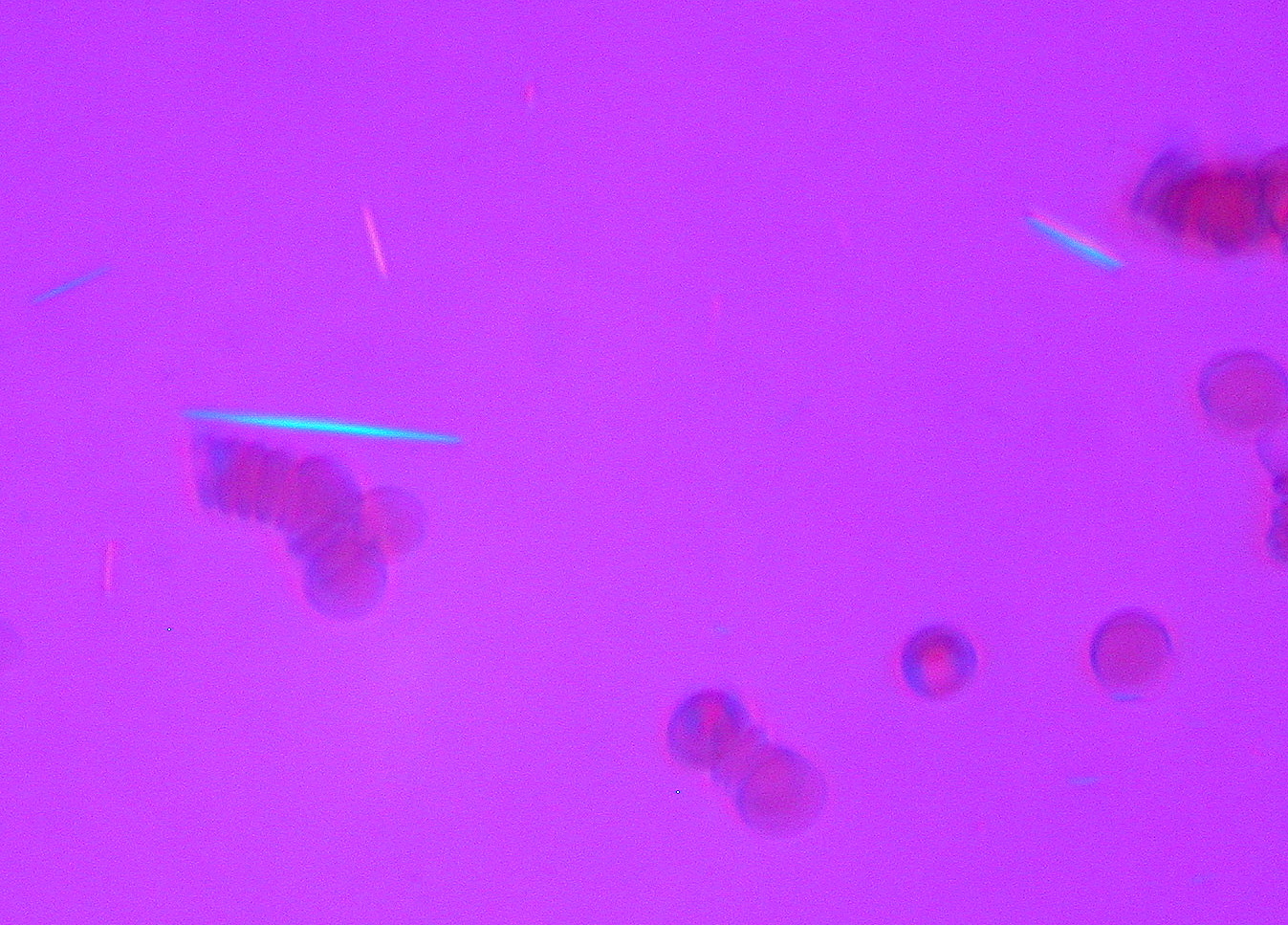
- Sign of birefringence. In a negatively birefringent crystal the fast axis lies along the long axis of the crystal. MSU crystals are needle shaped and strongly negatively birefringent. They appear yellow when aligned parallel to the slow axis of the compensator and blue when perpendicular to it (Zhang et al. 2016).
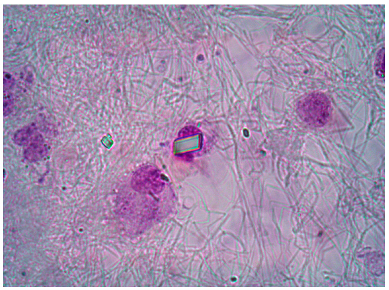
- CPP crystals are positively birefringent and appear blue when aligned with the slow vibration (Jokic et al. 2020). Their birefringence is weak, and they are best visualized by ordinary light microscopy (Jokic et al. 2020).
- Orientation dependence. Birefringence is strongest when the crystal is aligned with the slow axis. Misalignment weakens the signal, which matters most for small, weakly birefringent CPP crystals (FitzGerald et al. 2024).
Examination technique for synovial fluid crystals
- Wet preparation. One drop of well-mixed fluid is placed on a slide under a coverslip so that the fluid fills the coverslip area (Jokic et al. 2020).
- Screening and identification. The slide is screened at low power (100×) and crystals are identified at 400× or higher (Jokic et al. 2020).
- Sequence of light modes. In a study of 250 observations, five observers examined each sample under ordinary, simple polarized and compensated polarized light in that order. Observations under ordinary light matched the final diagnosis in 96.8% (242 of 250) (Bernal et al. 2023).
- Ordinary light for CPP. When CPP crystals were counted in a hemocytometer under ordinary light and the same area was then examined under polarized light, only 17–18% showed birefringence. For MSU crystals, the count under polarized light was 107–127% of the count under ordinary light, because polarized light revealed crystals missed under ordinary light (Ivorra et al. 1999). A search for CPP crystals conducted only under polarized light misses most of them (Ivorra et al. 1999).
- Concentration. Cytocentrifuged slides concentrate fluid components in a monolayer and increase crystal recovery. They also allow stained or unstained examination and retention of slides for training. A large number of cells makes crystal identification on cytocentrifuged slides more difficult (Jokic et al. 2020).
- Detection threshold. In fluids spiked with known crystals, the threshold for reliable identification of MSU and CPP crystals was 10–100 µg/mL. False-positive results were frequent (Gordon et al. 1989).
Optical and morphologic features of synovial fluid crystals
| Crystal | Shape and size | Polarized light | Main clinical setting |
|---|---|---|---|
| MSU | Needle shaped, 2–30 µm; frequently intracellular | Strong negative birefringence; yellow parallel to the slow axis | Acute and chronic gout |
| CPP | Rods (67%) and rhomboids (33%); median rod length 3.7 µm (range 1.0–9.8 µm) in Zell et al.; reported overall range 1–20 µm | Weak positive birefringence; blue parallel to the slow axis; most crystals nonbirefringent | CPPD disease, including acute CPP crystal arthritis and osteoarthritis with CPPD |
| BCP (including hydroxyapatite) | Individual crystals below light-microscopic resolution; clumps and globules 5–20 µm that appear as "shiny coins" | Nonbirefringent; clumps stain with alizarin red S | Osteoarthritis; Milwaukee shoulder-knee syndrome; periarthritis |
| Calcium oxalate | Tetragonal, bipyramidal, octahedral or envelope shaped, 15–20 µm; rod-like forms occur | Rod-like forms are positively birefringent | End-stage renal disease |
| Corticosteroid | Irregular, variable shapes depending on the injected agent | Birefringent; can resemble MSU or CPP | Recent intra-articular injection |
| Cholesterol | Large flat plates, 10–80 µm; smaller rod- and needle-shaped forms, 1–5 µm | Listed as not birefringent (Jokic et al. 2020); identified by plate shape | Chronic inflammatory effusions, especially long-standing rheumatoid arthritis; also osteoarthritis |
Sources: Yavorskyy et al. 2008; Zell et al. 2021; Ivorra et al. 1999; Zhang et al. 2016; Hoffman et al. 1982; FitzGerald et al. 2024; Jokic et al. 2020; Fam et al. 1981; Ettlinger and Hunder 1979.
Gout and monosodium urate crystals in synovial fluid
- Frequency. In 5,036 synovial fluid samples from a Swiss tertiary center, 15.3% contained MSU crystals. The male-to-female ratio among MSU-positive samples was 10.1:1 (Manigold and Leichtle 2025).
- Agreement between observers. Agreement for MSU identification among five observers was κ 0.88 (Bernal et al. 2023). After training, four observers identified MSU crystals with a sensitivity of 95.3% and a specificity of 97.2% (Lumbreras et al. 2005).
- Stability. MSU crystals remained detectable for 8 weeks of storage but became smaller, less birefringent and less numerous (Kerolus et al. 1989). A systematic review found MSU crystals stable regardless of storage time, temperature and preservative (Meyer et al. 2021).
- Consistency with the clinical diagnosis. In a Mexican series of 264 analyses, MSU crystals were present in 89.1% of fluids from patients referred with gout. CPP crystals were present in 7.27% of those fluids (Zamudio-Cuevas et al. 2021).
2015 ACR/EULAR gout classification criteria and the synovial fluid
- Entry criterion. At least one episode of swelling, pain or tenderness in a peripheral joint or bursa (Neogi et al. 2015).
- Sufficient criterion. MSU crystals in a symptomatic joint or bursa, or in a tophus, observed by a trained examiner. A patient who meets this criterion is classified as having gout without scoring (Neogi et al. 2015).
- No exclusion criteria. Gout coexists with other diseases, and synovial fluid microscopy can miss MSU crystals for technical, sampling or treatment reasons (Neogi et al. 2015).
- Scoring domains. Clinical features, laboratory results (serum urate and MSU-negative synovial fluid) and imaging (double-contour sign on ultrasound, urate on dual-energy CT and gout-related erosion on radiographs) (Neogi et al. 2015).
- Negative scores. A synovial fluid from a symptomatic joint or bursa that is MSU negative on polarizing microscopy by a trained examiner subtracts 2 points. A serum urate below 4 mg/dL (0.24 mmol/L) subtracts 4 points. A fluid that was not assessed scores 0 (Neogi et al. 2015).
- Threshold. The maximum score is 23, and a score of 8 or more classifies the patient as having gout. Sensitivity was 92% and specificity 89% (Neogi et al. 2015).
- Earlier criteria sets. Some earlier sets also accepted MSU crystals alone as fulfilling the criteria. The 2015 set added imaging and gave negative weight to an MSU-negative aspirate (Neogi et al. 2015).
Calcium pyrophosphate deposition disease and CPP crystals in synovial fluid
- EULAR 2011 terminology. CPPD is the umbrella term. It includes acute CPP crystal arthritis, osteoarthritis with CPPD and chronic CPP crystal inflammatory arthritis. Chondrocalcinosis is cartilage calcification detected by imaging or histology, most commonly due to CPPD (Zhang et al. 2011). Laboratory recommendations and older literature use the term pseudogout for CPP crystal arthritis (Jokic et al. 2020).
- Definitive diagnosis. Identification of CPP crystals in synovial fluid is the basis of a definitive diagnosis of CPPD (Zhang et al. 2011).
- Risk factors. Aging, osteoarthritis, primary hyperparathyroidism, hemochromatosis and hypomagnesemia. Familial forms are rare (Zhang et al. 2011).
- Frequency. CPP crystals were the most common crystal type in the Swiss series, present in 21.9% of 5,036 samples. They predominated in both inflammatory (58.9%) and noninflammatory (65.7%) crystal-positive samples (Manigold and Leichtle 2025).
- Crystal size and shape. Of 175 confirmed CPP crystals, 67% were rods and 33% rhomboids. Median rod length was 3.7 µm, and the median acute angle of rhomboids was 77.1°. Digital image enhancement revealed many smaller, less birefringent crystals that observers had not seen (Zell et al. 2021).
- Agreement between observers. Agreement for CPP identification among five observers was κ 0.69, lower than for MSU (Bernal et al. 2023). Trained observers identified CPP crystals with a sensitivity of 92.7% and a specificity of 92.1% (Lumbreras et al. 2005).
2023 ACR/EULAR CPPD disease classification criteria and the synovial fluid
The 2023 criteria are the first validated classification criteria for symptomatic CPPD disease (Abhishek et al. 2023). Earlier diagnostic criteria (Ryan and McCarty, 1960s) required both typical radiographic calcification and CPP crystals on polarized light microscopy for a definite diagnosis (Abhishek et al. 2023).
- Entry criterion. At least one episode of joint pain, swelling or tenderness (Abhishek et al. 2023).
- Exclusion criterion. All symptoms are more likely explained by another condition, such as rheumatoid arthritis, gout, psoriatic arthritis or osteoarthritis (Abhishek et al. 2023).
- Sufficient criteria. Crowned dens syndrome, or CPP crystals in synovial fluid. Either one classifies the patient as having CPPD disease (Abhishek et al. 2023).
- Weighted score. Without a sufficient criterion, a score above 56 points classifies the patient as having CPPD disease. The domains are clinical features, associated metabolic disorders, laboratory results and imaging (Abhishek et al. 2023).
- Performance. Applied as a whole, the criteria had a sensitivity of 97.8% and a specificity of 87.9% in the derivation cohort, and 99.2% and 92.5% in the validation cohort. The score threshold alone, in cases without a sufficient criterion, gave 92.2% and 87.9% in derivation (Abhishek et al. 2023).
| Synovial fluid result in a symptomatic joint (2023 ACR/EULAR CPPD criteria) | Effect on classification |
|---|---|
| CPP crystals present | Sufficient criterion; no scoring needed |
| CPP crystals absent on 2 or more occasions | −7 points |
| CPP crystals absent on 1 occasion | −1 point |
| Synovial fluid analysis not performed | 0 points |
Source: Abhishek et al. 2023 (criteria table). Analysis by an individual trained in compensated polarized light microscopy (Abhishek et al. 2023, Table 2 footnote).
- Metabolic domain. Hereditary hemochromatosis, primary hyperparathyroidism, hypomagnesemia, Gitelman syndrome, hypophosphatasia or a familial history of CPPD disease adds 6 points (Abhishek et al. 2023).
Basic calcium phosphate, calcium oxalate, cholesterol and corticosteroid crystals in synovial fluid
- Basic calcium phosphate. BCP crystals are present in the joint fluid of many patients with osteoarthritis (Yavorskyy et al. 2008). Individual crystals are below the resolution of the light microscope (about 1 µm). Only aggregated clumps are visible by light microscopy (Yavorskyy et al. 2008).
- Alizarin red S. Calcium forms a red chelate with alizarin red S, which stains hydroxyapatite clumps and CPP crystals regardless of their birefringence (Yavorskyy et al. 2008). Six observers showed many discrepancies on alizarin red S slides, and its value as a screening test for BCP was questioned (Gordon et al. 1989). Alizarin red-positive material was present in 9.5% of samples in the Swiss series (Manigold and Leichtle 2025).
- Milwaukee shoulder syndrome is a destructive arthropathy of the shoulder associated with BCP crystals (Yavorskyy et al. 2008).
- Calcium oxalate. Calcium oxalate crystals were identified in the synovial fluid of three patients with chronic renal failure. The crystals were pleomorphic and bipyramidal. Some were rod-like and positively birefringent and could be confused with CPP crystals under CPLM alone. Radiographic chondrocalcinosis in one patient was due to calcium oxalate (Hoffman et al. 1982).
- Cholesterol. Cholesterol crystals form flat rectangular plates in chronic inflammatory effusions, particularly rheumatoid arthritis (Jokic et al. 2020). In 16 fluids from 12 patients collected over 14 years, 10 patients had rheumatoid arthritis of a median duration of 12 years. The fluids were usually turbid, white or yellow and thick. Cholesterol crystals suggest severe persistent synovitis (Ettlinger and Hunder 1979). In 4 osteoarthritic knee effusions, large crystals (10–80 µm) were identified by their plate-like shape, and smaller rod- and needle-shaped crystals (1–5 µm) were also present. X-ray diffraction confirmed them as cholesterol (Fam et al. 1981). They have also been described in rheumatoid bursal fluid (Zhu and Chu 2022). They are listed as not birefringent (Jokic et al. 2020).
- Corticosteroid crystals. Birefringent crystals follow intra-articular corticosteroid injection and can cause a false-positive diagnosis of gout. Their irregular shape separates them from MSU needles (Zhang et al. 2016). Their appearance depends on the agent, and they can resemble MSU or CPP crystals (Jokic et al. 2020). In the Mexican series, corticosteroid crystals were present in 44.4% of fluids from rheumatoid arthritis and 42.9% from osteoarthritis (Zamudio-Cuevas et al. 2021).
- Artifacts that develop during storage. Alizarin red S-positive clumps, star-shaped arrays, plate-like structures, positively birefringent Maltese crosses and hematoidin crystals appeared in stored fluids that had not contained them initially (Kerolus et al. 1989).
Specimen collection and storage for synovial fluid crystal analysis
- Collection order. The Clinical and Laboratory Standards Institute order sends the first aliquot for biochemistry, the second for microscopy (cell count, differential and crystals) and the third for microbiology (Jokic et al. 2020).
- Containers. Tubes with oxalate, lithium heparin or lyophilized EDTA form crystal-like material that can be mistaken for MSU or calcium oxalate crystals (Jokic et al. 2020). EDTA was a suitable preservative in a systematic review, and fluid may also be stored at room temperature without preservative (Meyer et al. 2021).
- Prompt examination. In 50 fluids examined over 2 months, CPP crystals became much less abundant by the next day in 3 of 5 crystal-rich samples and dissolved completely by 3–8 weeks. MSU crystals persisted for 8 weeks. Clumps of apatite-like crystals persisted for months (Kerolus et al. 1989).
- Refrigerated and frozen storage. In 91 fluids stored at 4 °C, intracellular MSU crystals were found again in 97% at 24 hours and 96% at 72 hours. CPP crystals were found in 100% and 97%. After 2 months at −80 °C, MSU crystals were found in 81% and CPP crystals in 83%. No crystals appeared in samples that had been crystal free (Gálvez et al. 2002).
- Pooled evidence. CPP crystals are mostly stable for 24–48 hours and are more stable when refrigerated. Re-examination of an initially negative sample at 24 hours detects a small number of additional MSU and CPP cases. Centrifugation makes CPP identification easier (Meyer et al. 2021).
- Long-term storage. Dried cytospin preparations allowed delayed MSU analysis for at least 12 months. MSU crystal concentration fell over time at room temperature but not in refrigerated samples (Graf et al. 2013).
Quality assurance and reproducibility of synovial fluid crystal identification
| Study | Design | Main finding |
|---|---|---|
| Schumacher et al. 1986 | 30 fluids sent to 3 laboratories for crystal identification | Discrepant crystal reports in 7 of 11 patients with crystals |
| Hasselbacher 1987 | 26 hospital laboratories, 4 unknown fluids | CPP detected 6 times and MSU 39 times in 50 opportunities each |
| von Essen et al. 1998 | Annual cytocentrifuge slide surveys, 25–47 laboratories, 1989–1996 | About one error per laboratory per year; errors rose when crystals were few; no improvement with feedback |
| Gordon et al. 1989 | Spiked fluids, 6 observers | Mean sensitivity 69% for MSU and 82% for CPP; mean specificity 97% and 78% |
| Lumbreras et al. 2005 | 4 trained observers, 64 fluids | κ 0.93 for MSU and 0.79 for CPP against the expert |
| Berendsen et al. 2017 | Online image test, 110 participants | 81% identified all MSU images; 68% identified all CPP images; 23% classified all 22 non-MSU images as non-MSU |
| Bernal et al. 2023 | 5 observers, 50 fluids | Overall κ 0.74; no effect of delay beyond 24 hours or of expertise |
- Error rates exceed other laboratory fields. Errors in crystal identification surveys were much more frequent than in clinical chemistry or microbiology (von Essen et al. 1998).
- Items mistaken for crystals. The online test included cholesterol, depot methylprednisolone, calcium oxalate, rice bodies, liquid lipid, fibrin, Charcot-Leyden crystals and artifacts. Incorrect identification of non-MSU material was frequent (Berendsen et al. 2017).
- Newer methods. Single-shot computational polarized light microscopy increased detection of CPP crystals from 28% to 51% and of MSU crystals from 46% to 78% compared with CPLM images (FitzGerald et al. 2024). Lens-free polarized microscopy, Raman spectroscopy, Fourier transform infrared spectroscopy and scanning electron microscopy improve objectivity or resolution and remain mostly outside routine practice (Niessink et al. 2026).
Differential diagnosis of crystals and crystal-like material in synovial fluid
| Finding | Favors | Separating features |
|---|---|---|
| Needles, strong birefringence, yellow parallel to the slow axis | MSU | Negative sign; frequently intracellular |
| Small rods and rhomboids seen best in ordinary light, weak or absent birefringence, blue parallel | CPP | Positive sign; small size; most crystals nonbirefringent |
| Rod-like positively birefringent crystals with bipyramidal forms in a dialysis patient | Calcium oxalate | Envelope and bipyramidal shapes; renal failure |
| Irregular birefringent fragments after joint injection | Corticosteroid | Variable, irregular shapes; injection history |
| Nonbirefringent clumps, alizarin red positive | BCP | Clumps only; individual crystals not resolved |
| Maltese crosses, hematoidin or plates in an old specimen | Storage artifact | Absent at initial examination |
Sources: Zhang et al. 2016; Ivorra et al. 1999; Hoffman et al. 1982; Jokic et al. 2020; Yavorskyy et al. 2008; Kerolus et al. 1989.
Reporting of synovial fluid crystal analysis
- Elements. Crystal type, birefringence strength, shape, intracellular or extracellular location and number per high-power field (Jokic et al. 2020).
- Negative result. In the gout criteria, an MSU-negative fluid counts against gout only when a trained examiner performed polarizing microscopy (Neogi et al. 2015).
- Mixed crystals. MSU and CPP crystals can occur in the same fluid. In the Mexican series, MSU crystals were found in 9.09% of fluids from patients referred with CPPD (Zamudio-Cuevas et al. 2021).
- Crystals and infection. The finding of crystals does not exclude septic arthritis. Cell counts and culture results are interpreted together (Synovial fluid cell counts and cytology: septic, rheumatoid and traumatic effusions).
synovial fluid crystal identification lookup
Synovial Fluid Cell Counts and Cytology: Septic, Rheumatoid and Traumatic Effusions
Definition. Synovial fluid analysis comprises gross appearance, total leukocyte count, differential count, crystal search, Gram stain and culture of fluid aspirated from a joint or bursa. The leukocyte count and the neutrophil percentage classify a fluid as normal, noninflammatory, inflammatory, septic or hemorrhagic (Jokic et al. 2020). The synovial fluid leukocyte count and neutrophil percentage are the most informative laboratory results for septic arthritis available before Gram stain and culture results (Margaretten et al. 2007). Cytologic findings such as cytophagocytic mononuclear cells, eosinophils, lipid droplets and hemosiderin add diagnostic information in selected settings (Freemont and Denton 1985; Vázquez-Triñanes et al. 2013; Graham and Goldman 1978).
Related page: Synovial fluid crystal identification: compensated polarized light, gout, CPPD and other crystals (crystal identification, gout and CPPD) Related page: Benign Soft Tissue Tumors on FNA (fine-needle aspiration cytology of tenosynovial giant cell tumor)
Classification of synovial fluid by laboratory findings
| Group | Leukocytes per µL | Neutrophils | Appearance | Serum minus synovial glucose (mmol/L) | Typical disorders |
|---|---|---|---|---|---|
| Normal | <200 | <25% | Colorless to straw; clear | ≤0.6 | None |
| Noninflammatory | <2,000 | <25% | Light yellow; clear to slightly cloudy | <1.1 | Osteoarthritis, traumatic arthritis, osteonecrosis |
| Inflammatory | 2,000–50,000 | >50% | Yellow-white or gray; cloudy to milky | >1.1 (up to 4.4) | Rheumatoid arthritis, gout, CPPD, systemic lupus erythematosus |
| Septic | >50,000 | >75% (often >90%) | White, gray or yellow-green; purulent | >2.2 (up to 5.6) | Bacterial, fungal or mycobacterial infection |
| Hemorrhagic | Equal to blood | Equal to blood | Red-brown or xanthochromic | <1.1 | Trauma, hemophilia, anticoagulation, tumor, joint prosthesis |
Source: Jokic et al. 2020 (American Rheumatism Association cut-offs; 1 × 10⁶/L equals 1/µL).
- Normal volume. Up to about 3.5 mL of synovial fluid is present in a normal joint (Jokic et al. 2020).
- Overlap between groups. The total leukocyte count overlaps between disease groups. The combination of leukocyte count and neutrophil percentage separates noninflammatory, inflammatory and infectious disorders better than either value alone (Jokic et al. 2020).
- Neutrophil predominance. A neutrophil fraction above 80% is associated with bacterial arthritis and gout (Jokic et al. 2020).
- Lymphocyte predominance. Increased lymphocytes occur in early rheumatoid arthritis (Jokic et al. 2020).
- Paucicellular fluid. Fluids with fewer than 1,000 cells/µL occur in mechanical disorders (Dougados 1996).
Cell counting methods and preanalytical errors in synovial fluid
- Timing. Total and differential counts are performed within 1 hour of collection (Jokic et al. 2020).
- Loss of leukocytes with delay. The leukocyte count fell within a few hours of aspiration. In 4 mildly inflammatory fluids with initial counts of 3,150–6,200/µL, the count fell below 2,000/µL within 5–6 hours (Kerolus et al. 1989).
- Preservation. Accurate counts are obtained from fluid stored at 2–8 °C in EDTA tubes. Inflammatory fluids clot readily, and clotting affects the count (Jokic et al. 2020).
- Diluents. Erythrocytes are lysed with 0.3% hypotonic saline. Viscous fluid is diluted with hyaluronidase buffer for an even distribution in the counting chamber (Jokic et al. 2020). Dilution with acetic acid for cell counts was one of the technical errors found in a survey of 42 hospital laboratories (Hasselbacher 1987).
- Differential count. The differential count is performed on an air-dried cytocentrifuge preparation stained with May-Grünwald or Wright stain (Jokic et al. 2020).
- Automated analyzers. Body fluid modes on hematology analyzers shorten turnaround time. Fat globules, cartilage fragments and crystals cause pseudoleukocytosis or pseudoeosinophilia, and atypical or malignant cells are not classified. Unclear results, low counts and suspected interference are verified by microscopy (Jokic et al. 2020).
- Reproducibility. Leukocyte counts from 4 laboratories correlated only fairly with the reference laboratory (r 0.76–0.80). The differences reclassified 4 fluids between the inflammatory and noninflammatory groups (Schumacher et al. 1986). In the hospital laboratory survey, the reported count for a single fluid ranged from 2,467 to 12,000/µL (Hasselbacher 1987).
- Evidence base for cytology. A literature survey found that the evidence for synovial fluid cytology was mainly anecdotal, without data on sensitivity, specificity or reliability (Swan et al. 2002).
Septic arthritis: synovial fluid cell counts, Gram stain and culture
Likelihood ratios for synovial fluid results in native joint septic arthritis
| Synovial fluid result | Likelihood ratio (95% CI) |
|---|---|
| Leukocytes <25,000/µL | 0.32 (0.23–0.43) |
| Leukocytes ≥25,000/µL | 2.9 (2.5–3.4) |
| Leukocytes >50,000/µL | 7.7 (5.7–11.0) |
| Leukocytes >100,000/µL | 28.0 (12.0–66.0) |
| Neutrophils ≥90% | 3.4 (2.8–4.2) |
| Neutrophils <90% | 0.34 (0.25–0.47) |
Source: Margaretten et al. 2007 (14 studies, 6,242 patients, 653 with septic arthritis).
- Risk factors. Age, diabetes mellitus, rheumatoid arthritis, joint surgery, hip or knee prosthesis, skin infection and HIV-1 infection increase the probability of septic arthritis (Margaretten et al. 2007).
- Clinical findings. Joint pain (sensitivity 85%), a history of joint swelling (78%) and fever (57%) were the only findings present in more than half of patients (Margaretten et al. 2007).
- Gram stain. In a multicenter study from the UK and Ireland, Gram stain had a sensitivity of 0.26 and a specificity of 0.99 for culture- or PCR-proven septic arthritis. False-positive Gram stains occurred in 1%. A negative Gram stain does not exclude septic arthritis, whatever the crystal findings or leukocyte count (Smith et al. 2025).
- Culture containers. In 3,171 synovial fluid specimens, growth was obtained in 29.6% of samples inoculated into blood culture bottles and 16.1% of samples in sterile containers (Gürler et al. 2026).
- Organisms. Staphylococcus aureus (33.6%), coagulase-negative staphylococci (24.9%) and Pseudomonas aeruginosa (8.6%) were the most frequent isolates in native and prosthetic joint infections combined. Coagulase-negative staphylococci and Enterococcus faecalis were significantly more common in prosthetic joint infections (Gürler et al. 2026).
- Polymerase chain reaction (PCR). In 475 patients evaluated in 9 US emergency departments, PCR detected an organism in 18.5% and culture in 16.8%. For PCR-targeted organisms, sensitivity was 89.2% and specificity 95.3% against culture. PCR-positive, culture-negative results were more frequent after antibiotic pretreatment and with Neisseria gonorrhoeae (Steele et al. 2026).
Coexisting crystal arthritis and septic arthritis
- Frequency. Septic arthritis coexisted with crystal arthritis in 4% (45 of 1,116) of patients with crystal-induced arthritis in one hospital database. Monosodium urate crystals were present in 80% of these cases. The median synovial leukocyte count was 61,478/µL (IQR 33,600–131,030), and concomitant infection was very unlikely below 12,000/µL (Duangkum et al. 2025).
- Emergency department series. Of 140 patients with septic arthritis, 23.6% had concomitant crystal arthritis. Of 427 patients with crystal arthritis, 7.2% had concomitant septic arthritis. A synovial leukocyte cut-off of 32,000/µL gave 100% sensitivity and about 50% specificity for infection (Mukerji et al. 2024).
- Higher threshold proposal. In 358 aspirations, 3 joints had both crystals and infection, each with a count of at least 85,000/µL. A cut-off of 85,000/µL had 100% specificity and a positive predictive value of 12.0% in crystal-positive joints (Luo et al. 2020).
- Interpretation. Crystals in a fluid with a high leukocyte count do not exclude infection. Culture is required regardless of crystal findings (Smith et al. 2025; Duangkum et al. 2025).
Synovial fluid in prosthetic joint infection
| Definition | Synovial leukocyte criterion | Neutrophil criterion | Use of the result |
|---|---|---|---|
| 2018 International Consensus Meeting/MSIS definition (hip and knee) | >3,000 cells/µL scores 3 points | >80% scores 2 points | Preoperative score of 6 or more indicates infection; 2–5 requires intraoperative findings |
| EBJIS 2021 definition | >3,000 cells/µL confirms infection | >80% confirms infection | A positive confirmatory test alone defines infection in the hip and knee; published cut-offs range from 1,500 to 4,000 cells/µL and 65% to 80% neutrophils |
Sources: Parvizi et al. 2018; McNally et al. 2021.
- Earlier definition. The 2018 definition updated the 2011 Musculoskeletal Infection Society (MSIS) criteria. Its sensitivity was 97.7%, against 79.3% for the 2011 MSIS criteria, with a similar specificity of 99.5% (Parvizi et al. 2018).
- Other synovial markers in the 2018 score. Alpha-defensin (3 points), leukocyte esterase (3 points) and synovial C-reactive protein above 6.9 mg/L (1 point) (Parvizi et al. 2018). Two positive cultures or a sinus tract are major criteria and are diagnostic on their own (Parvizi et al. 2018).
- Limits. Prosthetic joint thresholds are affected by other conditions and may not apply to joints other than the hip and knee, particularly in the upper limb (McNally et al. 2021).
Rheumatoid arthritis and other inflammatory arthritides in synovial fluid
- Ragocytes. Ragocytes ("RA cells", "inclusion body cells") are synovial fluid phagocytes with cytoplasmic inclusions (Hollander et al. 1965). In rheumatoid synovial fluid, immunoelectron microscopy showed IgG, IgM and C3 in vacuolar inclusions of more than 75% of neutrophils and mononuclear phagocytes (Cherian and Schumacher 1983). Cytologic examination of fluid from rheumatoid knees showed ragocytes in just over half the cases. Effusions from osteoarthritis and mechanical derangement gave nonspecific findings, and the cytologist diagnosed only 13% of them correctly (Older et al. 1988).
- Prognostic subgroups. In serial rheumatoid effusions, the proportions of neutrophils and ragocytes correlated. Joints with a high ragocyte proportion had a significantly poorer outcome (Davis et al. 1988).
- Rheumatoid factor. Synovial fluid rheumatoid factor is found in about 60% of patients with rheumatoid arthritis. It is not a routine test (Jokic et al. 2020).
Cytophagocytic mononuclear cells (Reiter cells)
- Definition. Cytophagocytic mononuclear cells are macrophages containing whole apoptotic neutrophils. They were formerly called Reiter cells (Oliviero et al. 2023; Jones et al. 1993).
- Disease distribution. They are common in reactive arthritis, spondyloarthritis and crystal arthritides and rare in rheumatoid arthritis (Jones et al. 1993). Of 189 fluids with these cells, 69% came from patients with seronegative spondyloarthritis (Oliviero et al. 2023).
- Combination with mast cells. In 321 knee fluids, cytophagocytic mononuclear cells and mast cells occurred together only in reactive arthritis, psoriatic arthritis, ankylosing spondylitis and enteropathic arthritis (Freemont and Denton 1985).
- Mechanism. The cells clear apoptotic neutrophils in vivo. Pyknotic neutrophils increase rapidly in stored fluid, so fresh fluid is required for assessment (Jones et al. 1993).
Eosinophils and other atypical cells in synovial fluid
- Synovial fluid eosinophilia. Eosinophils were found in 10 of 982 arthrocenteses (1.02%). Minor eosinophilia (<10%) occurred in 3 hemorrhagic fluids. Major eosinophilia (>10%) occurred in 7 patients, only 2 of whom had peripheral eosinophilia. Most cases resolved promptly with nonsteroidal anti-inflammatory drugs (Vázquez-Triñanes et al. 2013).
- Referral for cytology. Plasma cells, eosinophils, lupus erythematosus cells and malignant cells are atypical findings that are referred for cytologic evaluation. Synovial lining cells have no clinical significance (Jokic et al. 2020).
Traumatic and hemorrhagic synovial effusions
- Hemarthrosis versus traumatic tap. A red-brown fluid with unevenly distributed color or a bloody streak suggests a traumatic tap (Jokic et al. 2020). Observation of the fluid as it enters the syringe separates hemarthrosis from a traumatic tap (Dougados 1996).
- Causes by age. Chondrocalcinosis and osteoarthritis are the most frequent causes of hemarthrosis in the elderly. Disorders of hemostasis and synovial tumors are mostly seen in children and young adults (Dougados 1996).
- Fat droplets. Fat droplets in synovial fluid signify joint trauma with damage to bone or adjacent supporting structures. Lipid globules inside and outside leukocytes, and a fatty top layer in the hematocrit spin of a hemorrhagic fluid, indicate a traumatic cause. Phagocytosis of lipid droplets produced synovial fluid leukocytosis in 2 patients with hemorrhagic effusions (Graham and Goldman 1978).
- Leukocyte count after hemorrhage. Fluids aspirated after traumatic hemorrhage usually have a low leukocyte count (Graham and Goldman 1978).
- Tenosynovial giant cell tumor. Synovial tumors, including tenosynovial giant cell tumor (TGCT), are a main cause of hemarthrosis in young adults (Dougados 1996). Aspiration of diffuse TGCT (pigmented villonodular synovitis) typically yields hemorrhagic or xanthochromic, serosanguineous ("brown, murky") fluid, and clear fluid does not exclude the diagnosis (Bouali et al. 2004). The 2013 WHO classification grouped these lesions as TGCT, localized and diffuse types; earlier names were giant cell tumor of tendon sheath and nodular tenosynovitis (localized) and pigmented villonodular synovitis and diffuse-type giant cell tumor (diffuse) (Healey et al. 2020). The 6th edition (2026) renames the chapter "fibrohistiocytic and synovial tumors", adds chondroid synoviocytic neoplasm and lists no change to the name or the localized and diffuse types of TGCT (Hornick et al. 2026). The cytology and genetics of the tumor are given in Benign Soft Tissue Tumors on FNA.
Differential diagnosis of synovial fluid by dominant cytologic finding
| Dominant finding | Leading diagnoses | Separating features |
|---|---|---|
| Neutrophils >90%, count >50,000/µL | Septic arthritis; crystal arthritis; both | Culture, Gram stain, crystal search; counts ≥85,000/µL with crystals raise concern for coexisting infection |
| Neutrophils with ragocytes | Rheumatoid arthritis | Clinical and serologic correlation |
| Cytophagocytic mononuclear cells with mast cells | Reactive arthritis and other spondyloarthritides | Fresh fluid; rare in rheumatoid arthritis |
| Eosinophils >10% | Eosinophilic synovitis of varied cause | Peripheral eosinophilia often absent |
| Lymphocytes | Early rheumatoid arthritis; lymphocytic arthritis | Low neutrophil fraction |
| Blood with fat droplets | Trauma with bone or supporting-structure injury | Fatty layer in the hematocrit spin |
| Uniform blood without fat; hemosiderin | Hemophilia, anticoagulation, tumor including TGCT, prosthesis | History; imaging of the joint |
| Uneven blood with a bloody streak | Traumatic tap | Clearing during aspiration |
Sources: Margaretten et al. 2007; Luo et al. 2020; Older et al. 1988; Freemont and Denton 1985; Jones et al. 1993; Vázquez-Triñanes et al. 2013; Jokic et al. 2020; Graham and Goldman 1978; Dougados 1996.
Reporting of synovial fluid cell counts and cytology
- Content. Fluid type, appearance, volume, leukocyte count, differential and the reference range or decision limit for each result (Jokic et al. 2020).
- Interpretive comments. Standardized comments cover preanalytical problems and the influence of the sample matrix, and the result is communicated directly to the clinician when it changes management (Jokic et al. 2020).
References
- Abhishek A, Tedeschi SK, Pascart T, et al. The 2023 ACR/EULAR classification criteria for calcium pyrophosphate deposition disease. Ann Rheum Dis. 2023;82(10):1248–1257. doi:10.1136/ard-2023-224575. PMID 37495237
- Berendsen D, Neogi T, Taylor WJ, et al. Crystal identification of synovial fluid aspiration by polarized light microscopy. An online test suggesting that our traditional rheumatologic competence needs renewed attention and training. Clin Rheumatol. 2017;36(3):641–647. doi:10.1007/s10067-016-3461-0. PMID 27837341
- Bernal JA, Andrés M, López-Salguero S, et al. Agreement among multiple observers on crystal identification by synovial fluid microscopy. Arthritis Care Res (Hoboken). 2023;75(3):682–688. doi:10.1002/acr.24874. PMID 36426584
- Bouali H, Deppert EJ, Leventhal LJ, Reeves B, Pope T. Pigmented villonodular synovitis: a disease in evolution. J Rheumatol. 2004;31(8):1659–1662. PMID 15290750
- Cherian PV, Schumacher HR Jr. Immunoelectronmicroscopic characterization of intracellular inclusions in synovial fluid cells of patients with rheumatoid arthritis. Ultrastruct Pathol. 1983;5(1):15–27. doi:10.3109/01913128309141815. PMID 6316601
- Davis MJ, Denton J, Freemont AJ, Holt PJ. Comparison of serial synovial fluid cytology in rheumatoid arthritis: delineation of subgroups with prognostic implications. Ann Rheum Dis. 1988;47(7):559–562. doi:10.1136/ard.47.7.559. PMID 3401052
- Dougados M. Synovial fluid cell analysis. Baillieres Clin Rheumatol. 1996;10(3):519–534. doi:10.1016/s0950-3579(96)80047-1. PMID 8876957
- Duangkum K, Saengmongkonpipat P, Tantiwong P, et al. Prevalence and characteristics of concomitant septic and crystal-induced arthritis: a hospital database and literature review. Rheumatol Immunol Res. 2025;6(3):179–187. doi:10.1515/rir-2025-0021. PMID 41050478
- Ettlinger RE, Hunder GG. Synovial effusions containing cholesterol crystals: report of 12 patients and review. Mayo Clin Proc. 1979;54(6):366–374. PMID 449407
- Fam AG, Pritzker KP, Cheng PT, Little AH. Cholesterol crystals in osteoarthritic joint effusions. J Rheumatol. 1981;8(2):273–280. PMID 7230158
- FitzGerald JD, Barrios C, Liu T, et al. A novel polarized light microscope for the examination of birefringent crystals in synovial fluid. Gout Urate Cryst Depos Dis. 2024;2(4):315–324. doi:10.3390/gucdd2040022. PMID 39840290
- Freemont AJ, Denton J. Disease distribution of synovial fluid mast cells and cytophagocytic mononuclear cells in inflammatory arthritis. Ann Rheum Dis. 1985;44(5):312–315. doi:10.1136/ard.44.5.312. PMID 4004360
- Gálvez J, Sáiz E, Linares LF, et al. Delayed examination of synovial fluid by ordinary and polarised light microscopy to detect and identify crystals. Ann Rheum Dis. 2002;61(5):444–447. doi:10.1136/ard.61.5.444. PMID 11959769
- Gordon C, Swan A, Dieppe P. Detection of crystals in synovial fluids by light microscopy: sensitivity and reliability. Ann Rheum Dis. 1989;48(9):737–742. doi:10.1136/ard.48.9.737. PMID 2478085
- Graf SW, Buchbinder R, Zochling J, Whittle SL. The accuracy of methods for urate crystal detection in synovial fluid and the effect of sample handling: a systematic review. Clin Rheumatol. 2013;32(2):225–232. doi:10.1007/s10067-012-2107-0. PMID 23138881
- Graham J, Goldman JA. Fat droplets and synovial fluid leukocytosis in traumatic arthritis. Arthritis Rheum. 1978;21(1):76–80. doi:10.1002/art.1780210113. PMID 623698
- Gürler M, Kırca F, Dinç B. Septic arthritis and prosthetic joint infections: microbial spectrum and evolving resistance patterns. J Bone Jt Infect. 2026;11(1):123–137. doi:10.5194/jbji-11-123-2026. PMID 41767587
- Hasselbacher P. Variation in synovial fluid analysis by hospital laboratories. Arthritis Rheum. 1987;30(6):637–642. doi:10.1002/art.1780300606. PMID 3606682
- Healey JH, Bernthal NM, van de Sande M. Management of tenosynovial giant cell tumor: a neoplastic and inflammatory disease. J Am Acad Orthop Surg Glob Res Rev. 2020;4(11):e20.00028. doi:10.5435/JAAOSGlobal-D-20-00028. PMID 33156160
- Hoffman GS, Schumacher HR, Paul H, et al. Calcium oxalate microcrystalline-associated arthritis in end-stage renal disease. Ann Intern Med. 1982;97(1):36–42. doi:10.7326/0003-4819-97-1-36. PMID 7092004
- Hollander JL, McCarty DJ Jr, Rawson AJ. The "R.A. cell", "ragocyte", or "inclusion body cell". Bull Rheum Dis. 1965;16(1):382–383. PMID 5828718
- Hornick JL, Bovée JVMG, Antonescu CR, et al. The 2026 World Health Organization classification of tumors of soft tissue and bone: 6th edition. Am J Surg Pathol. 2026 (online ahead of print). doi:10.1097/PAS.0000000000002624. PMID 42839882
- Ivorra J, Rosas J, Pascual E. Most calcium pyrophosphate crystals appear as non-birefringent. Ann Rheum Dis. 1999;58(9):582–584. doi:10.1136/ard.58.9.582. PMID 10460193
- Jokic A, Milevoj Kopcinovic L, Culej J, et al. Laboratory testing of extravascular body fluids: national recommendations on behalf of the Croatian Society of Medical Biochemistry and Laboratory Medicine. Part II – synovial fluid. Biochem Med (Zagreb). 2020;30(3):030501. doi:10.11613/BM.2020.030501. PMID 32774119
- Jones ST, Denton J, Holt PJ, Freemont AJ. Possible clearance of effete polymorphonuclear leucocytes from synovial fluid by cytophagocytic mononuclear cells: implications for pathogenesis and chronicity in inflammatory arthritis. Ann Rheum Dis. 1993;52(2):121–126. doi:10.1136/ard.52.2.121. PMID 8447691
- Kerolus G, Clayburne G, Schumacher HR Jr. Is it mandatory to examine synovial fluids promptly after arthrocentesis? Arthritis Rheum. 1989;32(3):271–278. doi:10.1002/anr.1780320308. PMID 2930602
- Lumbreras B, Pascual E, Frasquet J, et al. Analysis for crystals in synovial fluid: training of the analysts results in high consistency. Ann Rheum Dis. 2005;64(4):612–615. doi:10.1136/ard.2004.027268. PMID 15769916
- Luo TD, Jarvis DL, Yancey HB, et al. Synovial cell count poorly predicts septic arthritis in the presence of crystalline arthropathy. J Bone Jt Infect. 2020;5(3):118–124. doi:10.7150/jbji.44815. PMID 32566449
- Manigold T, Leichtle A. Predominance of calcium pyrophosphate crystals in synovial fluid samples of patients at a large tertiary center. Diagnostics (Basel). 2025;15(7):907. doi:10.3390/diagnostics15070907. PMID 40218257
- Margaretten ME, Kohlwes J, Moore D, Bent S. Does this adult patient have septic arthritis? JAMA. 2007;297(13):1478–1488. doi:10.1001/jama.297.13.1478. PMID 17405973
- Mastboom MJL, Palmerini E, Verspoor FGM, et al. Surgical outcomes of patients with diffuse-type tenosynovial giant-cell tumours: an international, retrospective, cohort study. Lancet Oncol. 2019;20(6):877–886. doi:10.1016/S1470-2045(19)30100-7. PMID 31029509
- McNally M, Sousa R, Wouthuyzen-Bakker M, et al. The EBJIS definition of periprosthetic joint infection. Bone Joint J. 2021;103-B(1):18–25. doi:10.1302/0301-620X.103B1.BJJ-2020-1381.R1. PMID 33380199
- Meyer MM, Marks LA, Aslam F. Clinical implications of synovial fluid specimen handling for crystal associated arthritides: a systematic review. Int J Rheum Dis. 2021;24(1):10–20. doi:10.1111/1756-185X.14019. PMID 33150706
- Mukerji S, Ryan P, Simmonds H, et al. Concomitant septic and crystal arthropathy: a single-centre 10-year retrospective observational study in New Zealand. N Z Med J. 2024;137(1597):67–78. doi:10.26635/6965.6510. PMID 38901050
- Neogi T, Jansen TL, Dalbeth N, et al. 2015 Gout classification criteria: an American College of Rheumatology/European League Against Rheumatism collaborative initiative. Ann Rheum Dis. 2015;74(10):1789–1798. doi:10.1136/annrheumdis-2015-208237. PMID 26359487
- Niessink T, Jansen TL, Janssen M, et al. Advances in synovial fluid analysis for the diagnosis of crystal arthropathies. Arthritis Care Res (Hoboken). 2026;78(6):788–797. doi:10.1002/acr.25698. PMID 41211614
- Older J, Rollinson P, Pike C. Cytological assessment of knee effusions. Arthroscopy. 1988;4(3):174–178. doi:10.1016/s0749-8063(88)80022-7. PMID 3166655
- Oliviero F, Galozzi P, Padoan A, et al. Seasonal variation and disease distribution of cytophagocytic mononuclear cells in synovial fluid: a medical record study. Exp Biol Med (Maywood). 2023;248(10):839–842. doi:10.1177/15353702231162078. PMID 37092758
- Parvizi J, Tan TL, Goswami K, et al. The 2018 definition of periprosthetic hip and knee infection: an evidence-based and validated criteria. J Arthroplasty. 2018;33(5):1309–1314.e2. doi:10.1016/j.arth.2018.02.078. PMID 29551303
- Schumacher HR Jr, Sieck MS, Rothfuss S, et al. Reproducibility of synovial fluid analyses. A study among four laboratories. Arthritis Rheum. 1986;29(6):770–774. doi:10.1002/art.1780290610. PMID 3718565
- Smith C, Maloney RJ, Wearmouth D, et al. Multi-centre evaluation of Gram stain in the diagnosis of septic arthritis. J Bone Jt Infect. 2025;10(2):61–71. doi:10.5194/jbji-10-61-2025. PMID 40330095
- Steele MT, Stubbs AM, Krishnadasan A, et al. Performance and projected impact of synovial fluid polymerase chain reaction testing for patients evaluated for septic arthritis. Clin Infect Dis. 2026 (online ahead of print). doi:10.1093/cid/ciag476. PMID 42671259
- Swan A, Amer H, Dieppe P. The value of synovial fluid assays in the diagnosis of joint disease: a literature survey. Ann Rheum Dis. 2002;61(6):493–498. doi:10.1136/ard.61.6.493. PMID 12006320
- Vázquez-Triñanes C, Sopeña B, González-González L, et al. Synovial fluid eosinophilia: a case series with a long follow-up and literature review. Rheumatology (Oxford). 2013;52(2):346–351. doi:10.1093/rheumatology/kes236. PMID 23041597
- von Essen R, Hölttä AM, Pikkarainen R. Quality control of synovial fluid crystal identification. Ann Rheum Dis. 1998;57(2):107–109. doi:10.1136/ard.57.2.107. PMID 9613340
- Yavorskyy A, Hernandez-Santana A, McCarthy G, McMahon G. Detection of calcium phosphate crystals in the joint fluid of patients with osteoarthritis – analytical approaches and challenges. Analyst. 2008;133(3):302–318. doi:10.1039/b716791a. PMID 18299743
- Zamudio-Cuevas Y, Martínez-Nava GA, Martínez-Flores K, et al. Synovial fluid analysis for the enhanced clinical diagnosis of crystal arthropathies in a tertiary care institution. Clin Rheumatol. 2021;40(8):3239–3246. doi:10.1007/s10067-021-05610-0. PMID 33598809
- Zell M, Aung T, Kaldas M, et al. Calcium pyrophosphate crystal size and characteristics. Osteoarthr Cartil Open. 2021;3(1):100133. doi:10.1016/j.ocarto.2020.100133. PMID 34386778
- Zhang W, Doherty M, Bardin T, et al. European League Against Rheumatism recommendations for calcium pyrophosphate deposition. Part I: terminology and diagnosis. Ann Rheum Dis. 2011;70(4):563–570. doi:10.1136/ard.2010.139105. PMID 21216817
- Zhang Y, Lee SY, Zhang Y, et al. Wide-field imaging of birefringent synovial fluid crystals using lens-free polarized microscopy for gout diagnosis. Sci Rep. 2016;6:28793. doi:10.1038/srep28793. PMID 27356625
- Zhu J, Chu CQ. Cholesterol crystals in rheumatoid bursal fluid. Rheumatology (Oxford). 2022;61(5):e132. doi:10.1093/rheumatology/keab561. PMID 34264345
Image credits
- Gout. Image: Ed Uthman, Wikimedia Commons, CC BY 2.0.
- Calcium pyrophosphate crystal in synovial fluid from a patient with osteoarthritis. Image: Oliviero Francesca et al., PMC open access (Int J Mol Sci. 2023 Dec 27;25(1):393. doi: 10.3390/ijms25010393) Figure 5, CC BY.
