Library › Laboratory hematology, anemias and hemoglobinopathies
Laboratory hematology, anemias and hemoglobinopathies
Last reviewed 2026-11-10 by a board-certified pathologist. 234 references.
Automated Blood Counts, Reticulocyte Count and Peripheral Smear Review
Definition. The complete blood count (CBC) is produced by automated hematology analyzers from K2- or K3-EDTA whole blood. Analyzers measure the red blood cell (RBC), white blood cell (WBC) and platelet counts, the hemoglobin (Hb) concentration and cell volume, and calculate the remaining red cell indices (Zandecki 2007b). Abnormal results and instrument flags are indications for review of a stained peripheral blood film or a manual differential (Palmer 2015).
Related page: Microcytic Anemias and Iron Studies (iron studies, thalassemia trait indices and sideroblastic anemia) Related page: Macrocytic Anemias and Vitamin B12 and Folate Testing (megaloblastic anemia and vitamin B12 and folate testing) Related page: Hemoglobin Variant Analysis and Laboratory Diagnosis of Thalassemia (hemoglobin variant analysis and thalassemia diagnosis) Related page: Hemolytic Anemias: Red Cell Membrane and Enzyme Defects and Paroxysmal Nocturnal Hemoglobinuria (hemolysis markers, membrane and enzyme disorders and PNH) Related page: Bone Marrow Failure, Pure Red Cell Aplasia and the Laboratory Approach to Cytopenias and Leukocyte Disorders (cytopenias, Duffy-null associated neutrophil count and leukocyte disorders) Related page: DIC, Thrombotic Microangiopathy and Viscoelastic Testing (ICSH schistocyte counting and thrombotic microangiopathy)
Measurement principles of hematology analyzers
- WBC differential. Analyzers use impedance and fluorescence flow cytometry to separate neutrophils, lymphocytes, monocytes, eosinophils and basophils. Many report a sixth population of immature granulocytes (promyelocytes, myelocytes and metamyelocytes) (Palmer 2015).
- Precision. The analyzer counts thousands of cells, whereas a manual differential counts 100–200. The automated differential is therefore more precise when no abnormal population is present (Palmer 2015).
- Hemoglobin. Hb is measured colorimetrically after lysis, by a modification of the cyanmethemoglobin method or with sodium lauryl sulfate or imidazole reagents (Zandecki 2007b). WHO 2024 recommends venous blood, automated analyzers and quality control for Hb measurement (WHO 2024).
- Platelet counting. Platelets are counted by impedance, by optical methods that combine volume with refractive index, by fluorescence staining of platelet RNA, or with a CD61 immunologic count on one analyzer (Zandecki 2007a).
- Measured and calculated red cell values. Laser-based analyzers measure Hb in individual red cells and report the cellular hemoglobin concentration mean (CHCM). Disagreement between CHCM and the calculated mean corpuscular hemoglobin concentration (MCHC) generates a flag (Zandecki 2007b). Analyzers that calculate the hematocrit (Hct) from the mean cell volume (MCV) carry any MCV error into the Hct and MCHC (Zandecki 2007b).
- Rule of three. Hb in g/dL is commonly taken as one-third of the Hct in %, and loss of agreement between the RBC count, Hb and Hct raises suspicion of cold agglutinins (Carneiro 2007; Balraam 2021).
Impedance and optical counting principles. Analyzers count and size cells by electrical impedance or by optical analysis. Coulter first applied impedance in 1949 to count cells and measure their volume. Hydrodynamic focusing, coincidence correction and lysing agents selective for leukocyte populations later refined the method. In optical counting, the amplitude of narrow-angle (forward) scatter correlates with cell volume, and scatter at wider angles reflects internal structural complexity. Optical red cell volume measurement is influenced by the biconcave disc shape and by the refractive index of each cell, which depends on its Hb concentration. Fluorescent dyes for nucleic acids are used for reticulocyte and nucleated red cell counts (Daves 2024).
Red cell indices
Red cell indices from measured Hb, RBC count and MCV.
| Index | Formula | Units | Source |
|---|---|---|---|
| Hematocrit (calculated) | MCV (fL) × RBC (× 10¹²/L) ÷ 1000 | L/L | Zandecki 2007b |
| Mean corpuscular hemoglobin (MCH) | Hb (g/L) ÷ RBC (× 10¹²/L) | pg | Zandecki 2007b |
| Mean corpuscular hemoglobin concentration (MCHC) | Hb (g/L) × 100 ÷ [MCV (fL) × RBC (× 10¹²/L)] | g/dL | Zandecki 2007b |
- MCHC as an alarm. An MCHC above 36 g/dL is uncommon on impedance analyzers. It indicates a spurious result unless dehydrated red cells are present, as in hereditary spherocytosis, Hb CC, Hb SC, Hb C/β-thalassemia, xerocytosis and warm autoimmune hemolytic anemia with spherocytes (Zandecki 2007b).
- Red cell distribution width (RDW). Anisocytosis is reflected in an increased RDW, and ICSH recommends the RDW as the reported measure of anisocytosis (Palmer 2015). MCV, MCHC and RDW differ between analyzers, and MCH is more stable (Fletcher 2022). RDW is reported as RDW-CV (%), the coefficient of variation of red cell volume, and as RDW-SD (fL), the width of the red cell volume histogram measured at the 20% height level (Katsaros 2020).
Analyzer evaluation and verification under ICSH 2014
The 2014 ICSH guideline replaced the 1994 guideline (Briggs 2014; ICSH 1994) and added digital image analysis, a flow cytometric reference differential and new cell indices. An independent evaluation precedes clinical use, and each laboratory performs a local validation (transference) before reporting results (Briggs 2014).
| Element | ICSH 2014 specification | Source |
|---|---|---|
| Specimen | Fresh K2- or K3-EDTA blood processed within 4–8 hours of venesection | Briggs 2014 |
| Carryover | High sample run 3 times followed by low sample run 3 times | Briggs 2014 |
| Method comparison | At least 250–300 samples for a full validation, one-third to one-half normal | Briggs 2014 |
| Reference differential | 2 × 200-cell manual differential by two examiners (CLSI H20-A2), with a third examiner when they disagree | Briggs 2014 |
Reporting units. ICSH recommends SI units and absolute counts for the WBC differential, reticulocytes and nucleated red cells when an analyzer provides them (Brereton 2016).
WBC differential reference method: ICSH 2026 flow cytometric method
- Current rule (ICSH 2026). The ICSH WBC reference method is a single-tube, eight-color, lyse-no-wash flow cytometric assay with the nucleic acid dye Syto16 and counting beads. It identifies lymphocytes, monocytes, neutrophils, eosinophils and basophils by positive markers, defines blasts by exclusion, and enumerates immature granulocytes and lymphocyte subsets (Hedley 2026).
- Performance. In 616 samples from four laboratories, the method performed equivalently to the manual reference differential and better for monocytes and basophils (Hedley 2026).
- Relationship to the previous rule. The previous reference method is the manual differential under CLSI H20-A2 (approved 1992, revised 2007, with a further revision in 2023). The flow method could replace it as the reference for evaluating automated differential counters, and it is intended for manufacturers and instrument validation (Hedley 2026).
- Monocyte lineage. ICSH 2026 classifies monocyte lineage cells into three categories: blasts and blast equivalents (monoblasts and promonocytes), immature monocytes and mature monocytes (Zini 2026).
Criteria for peripheral smear review
- Consensus rules. The International Consensus Group for Hematology Review tested 83 rules in 15 laboratories on 13,298 samples and consolidated them into 41 rules for first-time samples and for delta checks on repeat samples within 72 hours (Barnes 2005).
- Review rate. Morphologic or digital review is required in 10%–50% of samples because of analyzer flags (Hedley 2026).
- ICSH reporting policy. The automated differential is reported when no flags or abnormal populations are present. It may also be reported after film review confirms the automated values (Palmer 2015).
- Differential count size. A 100-cell differential is routine. The WHO classification cited by ICSH specifies a 200-cell differential in the workup of acute myeloid leukemia and myelodysplastic neoplasms (Palmer 2015).
- Digital morphology analyzers. These systems preclassify cells with artificial-intelligence algorithms and support remote review, archiving and competency assessment. Staining, magnification, display and file formats are not standardized. Reclassification and interpretation require a skilled morphologist (Kratz 2019).
ICSH 2015 grading of red cell, white cell and platelet morphology
ICSH grades most abnormalities in two tiers, moderate (2+) and many (3+). The grade "few" (1+) is reserved for schistocytes because small numbers are clinically significant (Palmer 2015). About 1,000 red cells are evaluated for a precise percentage of an abnormal shape (Palmer 2015). Analyzer MCV and MCH are preferred to film grading for red cell size and hemoglobinization (Palmer 2015).
| Feature | 2+ (moderate), % of cells | 3+ (many), % of cells | Source |
|---|---|---|---|
| Anisocytosis, macrocytes, microcytes, hypochromic cells | 11–20 | >20 | Palmer 2015 |
| Oval macrocytes | 2–5 | >5 | Palmer 2015 |
| Polychromasia, acanthocytes, echinocytes, elliptocytes, ovalocytes, spherocytes, stomatocytes, target cells, teardrop cells, basophilic stippling | 5–20 | >20 | Palmer 2015 |
| Bite cells, blister cells, irregularly contracted cells, sickle cells | 1–2 | >2 | Palmer 2015 |
| Schistocytes (1+ = <1%) | 1–2 | >2 | Palmer 2015 |
| Howell-Jolly bodies, Pappenheimer bodies | 2–3 | >3 | Palmer 2015 |
| Döhle bodies | 2–4 | >4 | Palmer 2015 |
| Neutrophil vacuolation, hypogranulation, hypergranulation | 4–8 | >8 | Palmer 2015 |
| Giant platelets | 11–20 | >20 | Palmer 2015 |
Red cell size, shape and inclusions
| Term | ICSH definition | Associated conditions | Source |
|---|---|---|---|
| Microcyte | Diameter <7 µm; MCV <80 fL | Iron deficiency, thalassemia | Palmer 2015 |
| Macrocyte | Diameter >8.5 µm; MCV >100 fL; round or oval | Vitamin B12 or folate deficiency, liver disease, myelodysplastic neoplasms | Palmer 2015 |
| Hypochromic cell | Central pallor >1/3 of the cell diameter | Iron deficiency, thalassemia | Palmer 2015 |
| Spherocyte | Diameter <6.5 µm, dense, without central pallor | Hereditary spherocytosis, ABO hemolytic disease, warm autoimmune hemolytic anemia, Clostridium perfringens sepsis, burns | Palmer 2015 |
| Elliptocyte; ovalocyte | Long axis more than twice the short axis; less than twice | Hereditary elliptocytosis, iron deficiency | Palmer 2015 |
| Target cell | Thin cell with central staining inside the pallor; moderate numbers lower the MCV | Liver disease, hemoglobinopathies, thalassemia | Palmer 2015 |
| Acanthocyte | Round, hyperchromic cell with 2–20 irregular projections | Liver disease, vitamin E deficiency, postsplenectomy, abetalipoproteinemia, McLeod phenotype | Palmer 2015 |
| Echinocyte | 10–30 short, regular projections | Liver and renal disease, pyruvate kinase deficiency, storage artifact | Palmer 2015 |
| Stomatocyte | Cup-shaped cell with slit-like pallor; two stomas in Southeast Asian ovalocytosis | Alcoholic liver disease, hereditary stomatocytosis | Palmer 2015 |
| Teardrop cell | Pear- or teardrop-shaped cell | Myelofibrosis | Palmer 2015 |
| Bite cell | Peripheral arcuate defect after splenic removal of Heinz bodies | G6PD-deficiency hemolysis; not specific to it | Palmer 2015; Bussing 2026; Shean 2026; Ward 1983 |
| Blister cell | Hemoglobin retracted into one half of the cell | G6PD-deficiency hemolysis; not specific to it | Palmer 2015; Bussing 2026; Qadah 2026; Shean 2026 |
| Sickle cell | Crescent shape from HbS polymerization | Sickle cell disease | Palmer 2015 |
| Basophilic stippling | Fine to coarse blue granules of aggregated ribosomes | Lead poisoning, hemoglobinopathies, thalassemia | Palmer 2015 |
| Howell-Jolly body | Single round inclusion of about 1 µm composed of DNA | Hyposplenism, hemolytic and megaloblastic anemia | Palmer 2015 |
| Pappenheimer bodies | Multiple basophilic ferritin aggregates, positive with Perls Prussian blue | Sideroblastic anemia, hemoglobinopathies, hyposplenism | Palmer 2015 |
| Hemoglobin crystals | Dense straight-edged crystals | Hb C and Hb SC disease | Palmer 2015 |
- Agglutination and rouleaux. Agglutination forms irregular grape-like clusters, usually from a cold-reacting antibody. Rouleaux are stacks of red cells that form when plasma protein concentration is high. Both are reported when present (Palmer 2015).
- Dimorphism. Two red cell populations, such as microcytic hypochromic and normochromic cells, appear as a dual population on the red cell histogram with an increased RDW (Palmer 2015).
- Nucleated red cells. Nucleated red cells are reported as an absolute count, with the WBC count corrected, or per 100 WBC (Palmer 2015).
Leukocyte changes and inherited anomalies
- Hypersegmentation. Normal neutrophils have 3–4 lobes. Hypersegmentation is any neutrophil with 6 or more lobes or more than 3% of neutrophils with 5 lobes when 100 neutrophils are counted (Palmer 2015). Its significance is covered in Macrocytic Anemias and Vitamin B12 and Folate Testing.
- Reactive changes. Toxic granulation (hypergranulation of primary granules), Döhle bodies and cytoplasmic vacuoles occur in infection and inflammation. Döhle bodies also occur with granulocyte colony-stimulating factor. Neutrophil vacuoles also form in alcohol toxicity and after prolonged EDTA storage (Palmer 2015).
- MYH9-related disease. Döhle-like inclusions accompanied by thrombocytopenia and giant platelets indicate MYH9-related disease, historically May-Hegglin anomaly (Palmer 2015). MYH9-related disease is autosomal dominant and is caused by variants in MYH9, which encodes nonmuscle myosin heavy chain IIA. The Epstein phenotype has macrothrombocytopenia without neutrophil inclusions, and the Fechtner phenotype adds hearing loss, cataract and nephropathy (Yan 2026).
- Pelger-Huët anomaly. Heterozygous LBR variants cause nuclear hyposegmentation, and homozygous pathogenic variants cause skeletal malformations (Hoffmann 2026). Hyposegmented neutrophils have condensed chromatin and a low nuclear-to-cytoplasmic ratio. They are counted as segmented neutrophils with an interpretive comment (Palmer 2015).
- Chédiak-Higashi syndrome. Giant cytoplasmic granules in granulocytes on blood and marrow films are pathognomonic. The syndrome is autosomal recessive, is caused by LYST variants and presents with partial oculocutaneous albinism and recurrent infections. An accelerated phase with hemophagocytic lymphohistiocytosis is a major complication (Naamane 2025; Amri 2025).
- Lymphocyte terms. ICSH uses "reactive lymphocyte" for benign causes and "abnormal lymphocyte" with a description for suspected clonal causes (Palmer 2015).
- Smudge cells. Smudge cells form from shearing of fragile cells during spreading. A film made with one part albumin to four parts blood can preserve them (Palmer 2015).
Spurious results on hematology analyzers
| Interference | Effect on the CBC | Recognition | Correction | Source |
|---|---|---|---|---|
| Cold agglutinins | Low RBC count, high MCV and MCHC >36 g/dL; Hb unaffected | The combination is almost pathognomonic; agglutinates on the film | Warming to 37 °C and prompt reanalysis | Zandecki 2007b; Topic 2018 |
| Warm autoantibody agglutination | Spurious MCV and RBC count | Agglutinates | Not reversed by warming | Zandecki 2007b |
| Lipemia (triglycerides ≥20 g/L, chylomicron-rich types I and V) | High Hb, MCH and MCHC | MCHC >36 g/dL; CHCM–MCHC disagreement | Isovolumetric replacement of plasma with isoosmotic diluent, or lipid extraction | Zandecki 2007b |
| Extreme leukocytosis (>50–100 × 10⁹/L) | High Hb from turbidity; RBC and MCV error above 100 × 10⁹/L | MCHC >36 g/dL | Depends on the analyzer | Zandecki 2007b |
| Hyperglycemia | High MCV and calculated Hct, low MCHC | Glucose above 20 mmol/L begins to raise the MCV | Depends on the analyzer | Zandecki 2007b; Planas 1985 |
| Hypernatremia; hyponatremia | Macrocytic hypochromic shift; microcytic hyperchromic shift | Electrolyte results | Depends on the analyzer | Zandecki 2007b |
| Cryoglobulins | High WBC and platelet counts, at times high Hb | Particles on scattergrams; precipitates on the film | Warming to 37 °C for at least 30 minutes; at times recollection kept at 37 °C | Zandecki 2007b |
| Nucleated red cells, platelet clumps, unlysed red cells | Falsely high WBC count | Flags; abnormal WBC scattergram | Analyzer correction or manual count | Zandecki 2007b |
| Microcytes (<36 fL), schistocytes, burn microspherocytes, bacteria, cytoplasmic fragments | Falsely high platelet count | Platelet curve fails to return to baseline at 20 fL; microcytes on the platelet histogram; high MPV or RDW | Film review, optical or fluorescence count, or CD61 count | Zandecki 2007a |
- Hyperglycemia mechanism. Glucose-loaded red cells swell when water enters them in the analyzer diluent, and their size normalizes within about 5 minutes (Zandecki 2007b). The effect depends on glucose concentration, temperature and instrument (Planas 1985).
- Tube underfilling. In 4-mL K2EDTA tubes, 1.0-mL and 2.0-mL draws agreed with a full draw on a Sysmex XE-2100. A 0.5-mL draw increased variation and produced a platelet bias of −5.5% (Xu 2010).
- Storage. Platelet volume increases during EDTA storage and changes with delayed analysis and excess anticoagulant (Palmer 2015; Zandecki 2007a).
Pseudothrombocytopenia, platelet satellitism and immature platelet fraction
- Mechanism. EDTA-dependent platelet agglutination is caused by autoantibodies to a cryptic epitope of glycoprotein IIb/IIIa that EDTA exposes. The antibodies do not react with platelets from patients with type I Glanzmann thrombasthenia (Zandecki 2007a).
- Frequency. The prevalence is 0.07%–0.20%, and 0.2% in plateletpheresis donors. Pseudothrombocytopenia also follows treatment with glycoprotein IIb/IIIa antagonists (Zandecki 2007a).
- Detection. A WBC-histogram clump flag had 90% sensitivity and 100% specificity in one study (Zandecki 2007a).
- Correction. Blood collected into trisodium citrate gives a count corrected for dilution, although clumping can recur in citrate. Agglutination that forms at room temperature is not reversed by warming. Aminoglycosides dissociate and prevent the aggregates. Heparin is unsuitable (Zandecki 2007a).
- Satellitism. Platelets adhere to neutrophils in EDTA in about 1 of 12,000 counts, and the platelet count falls by 50–100 × 10⁹/L (Zandecki 2007a).
- Immature platelet fraction (IPF). The IPF reference interval in the evaluating study was 1.1%–6.1%. Means were 22.3% in autoimmune thrombocytopenic purpura and 17.2% in acute thrombotic thrombocytopenic purpura, and the IPF fell as the count recovered (Briggs 2004).
Reticulocyte count and reticulocyte indices
- Definition. ICSH defines a reticulocyte as a non-nucleated red cell with at least two blue-staining particles, or one particle linked to a filament, after supravital staining (Gaur 2021). The adult reference fraction is 0.5%–2.5% and the infant fraction 2%–6% (Gaur 2021).
- Automated counting. Analyzers label reticulocyte RNA with fluorescent dyes such as thiazole orange or polymethine (Gaur 2021). Automated counting replaced manual microscopy and adds the immature reticulocyte fraction (IRF) and the reticulocyte production index (RPI) (Zhou 2026).
- Reference methods. ICSH proposed a reference method for reticulocyte counting based on the reticulocyte-to-red-cell ratio in 1998 (ICSH 1998). ICSH 2014 considers a flow cytometric reference method more precise and the manual method an accepted reference (Briggs 2014).
- Shift reticulocytes. In anemia, reticulocytes leave the marrow early and circulate longer before they lose their reticulum, so the reticulocyte count is adjusted for maturation time (Hillman 1969).
Corrected reticulocyte count and reticulocyte production index.
| Step | Formula or threshold | Source |
|---|---|---|
| Corrected reticulocyte count (CRC) | Reticulocytes (%) × patient Hct ÷ normal Hct (45 for adult men) | Chueh 2022 |
| Reticulocyte production index (RPI) | CRC ÷ maturation time in days; maturation time depends on Hct | Chueh 2022; Gaur 2021 |
| Maturation time used in the RPI | 1 day at Hct ≥40%; 1.5 days at 30%–39.9%; 2 days at 20%–29.9%; 2.5 days at <20% | Huang 2026 |
| Interpretation | RPI >3: appropriate marrow response; RPI <2: inadequate response | Chueh 2022 |
Reticulocyte hemoglobin content. Reticulocyte hemoglobin content is reported as CHr or Ret-He, and hypochromic red cells as %HYPO or %Hypo-He, depending on the analyzer (Fletcher 2022). Their use in iron deficiency is covered in Microcytic Anemias and Iron Studies.
Erythrocyte sedimentation rate and plasma viscosity
The Westergren method is the reference method for the erythrocyte sedimentation rate (ESR) (Kratz 2017; Cennamo 2024). In external quality assessment data from more than 6,000 laboratories, newer methods differed from Westergren by up to 142%. ICSH therefore recommends method verification and communication of method changes to clinical users (Kratz 2017).
Plasma viscosity and factors that lower the ESR. Plasma viscosity is unaffected by anemia or red cell size. It is expensive, not widely available and technically cumbersome. The ESR and plasma viscosity have been the most satisfactory monitors of the acute-phase response after the first 24 hours, and C-reactive protein may be the better indicator during the first 24 hours. Sickle cells, spherocytes, acanthocytes, anisocytosis, microcytosis, polycythemia, extreme leukocytosis, hypofibrinogenemia and a clotted sample lower the ESR (Brigden 1999).
Hemoglobin reference intervals and the definition of anemia
Current rule (WHO 2024). WHO set Hb cutoffs at the 5th percentile of healthy populations (WHO 2024).
| Population | WHO 2024 cutoff (g/L) | Previous WHO cutoff (WHO 2011) (g/L) | Source |
|---|---|---|---|
| Children 6–23 months | <105 | <110 (6–59 months as one group) | WHO 2024; WHO 2011 |
| Children 24–59 months | <110 | <110 | WHO 2024; WHO 2011 |
| Children 5–11 years | <115 | <115 | WHO 2024; WHO 2011 |
| Children 12–14 years | <120 | <120 | WHO 2024; WHO 2011 |
| Nonpregnant women 15–65 years | <120 | <120 (15 years and older) | WHO 2024; WHO 2011 |
| Men 15–65 years | <130 | <130 (15 years and older) | WHO 2024; WHO 2011 |
| Pregnancy, first and third trimesters | <110 | <110 (pregnancy, no trimester cutoffs) | WHO 2024; WHO 2011 |
| Pregnancy, second trimester | <105 | <110 | WHO 2024; WHO 2011 |
- Changes from WHO 2011. WHO 2024 lowered the cutoff for children 6–23 months and adopted a second-trimester cutoff of <105 g/L. WHO 2011 gave no trimester-specific cutoffs and noted a second-trimester fall of about 5 g/L (WHO 2024; WHO 2011). Data suggested a higher cutoff of 135 g/L for men, but WHO retained 130 g/L because of uncertain evidence (WHO 2024).
- Severity. Severity follows the 1989 WHO method. For nonpregnant women, mild anemia is 110–119 g/L, moderate 80–109 g/L and severe <80 g/L. For men, mild anemia is 110–129 g/L (WHO 2024).
- Adjustments. Hb is adjusted for elevation of residence (for example, by 14 g/L at 2000–2499 m) and for smoking. The adjustment is subtracted from the observed Hb or added to the cutoff (WHO 2024).
- No adjustment. WHO 2024 does not adjust cutoffs for inflammation or for genetic ancestry. A mean Hb 10 g/L lower has been reported in Black individuals than in White individuals (WHO 2024).
- Neonates. Between 22 and 40 weeks of gestation, Hct rose by 0.64% and Hb by 0.21 g/dL per week, and values declined linearly over the first 28 days after birth (Jopling 2009).
- Neonatal indices. Among 12,016 neonates on the first day of life, the MCV fell from 119 ± 7 fL at 25 weeks or less to 106 ± 4 fL at 40 weeks. The MCHC stayed near 34 g/dL (Christensen 2008). Red cells of healthy children are smaller than adult red cells (Palmer 2015).
Laboratory classification of anemia by MCV and reticulocyte response
| Pattern | MCV | Reticulocyte response | Main laboratory branches | Linked unit | Source |
|---|---|---|---|---|---|
| Microcytic | <80 fL | Interpreted with iron studies | Iron deficiency, thalassemia, sideroblastic anemia, anemia of inflammation | Microcytic Anemias and Iron Studies; Hemoglobin Variant Analysis and Laboratory Diagnosis of Thalassemia | Palmer 2015; Snook 2021 |
| Macrocytic, megaloblastic | >100 fL | Low | Vitamin B12 or folate deficiency | Macrocytic Anemias and Vitamin B12 and Folate Testing | Kaferle 2009 |
| Macrocytic, nonmegaloblastic | >100 fL | Low: drugs or alcohol; high: hemolysis or hemorrhage | Alcohol, drugs, liver disease, hypothyroidism, myelodysplastic neoplasms | Macrocytic Anemias and Vitamin B12 and Folate Testing | Kaferle 2009 |
| Any MCV with RPI >3 | Variable | Appropriate | Hemolysis or blood loss | Hemolytic Anemias: Red Cell Membrane and Enzyme Defects and Paroxysmal Nocturnal Hemoglobinuria | Chueh 2022 |
| Any MCV with RPI <2 | Variable | Inadequate | Production defect, marrow failure | Bone Marrow Failure, Pure Red Cell Aplasia and the Laboratory Approach to Cytopenias and Leukocyte Disorders | Chueh 2022 |
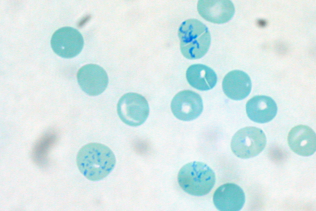
Microcytic Anemias and Iron Studies
Definition. Microcytic anemia is anemia with small red cells, defined by an MCV below 80 fL or a red cell diameter below 7 µm (Palmer 2015). Its main causes are iron deficiency, thalassemia, anemia of inflammation and sideroblastic anemia (Snook 2021; Palmer 2015). Iron deficiency is the most common cause of anemia worldwide (Fletcher 2022). The laboratory diagnosis of iron deficiency in adults and children follows the British Society for Haematology (BSH) good practice paper of 2021, published in 2022 (Fletcher 2022) and the World Health Organization (WHO) 2020 guideline on ferritin (WHO 2020).
Related page: Automated Blood Counts, Reticulocyte Count and Peripheral Smear Review (red cell indices, reticulocyte hemoglobin content and morphology grading) Related page: Hemoglobin Variant Analysis and Laboratory Diagnosis of Thalassemia (HbA2 measurement and alpha-thalassemia genotyping) Related page: Macrocytic Anemias and Vitamin B12 and Folate Testing (megaloblastic anemia and vitamin B12 and folate testing) Related page: Hemolytic Anemias: Red Cell Membrane and Enzyme Defects and Paroxysmal Nocturnal Hemoglobinuria (hemolysis markers)
Iron regulation by hepcidin
- Hepcidin and ferroportin. Hepcidin is a liver peptide secreted in response to iron loading and inflammation. It binds the iron exporter ferroportin on enterocytes, macrophages, hepatocytes and placental cells and causes its internalization and degradation. Cellular iron export then falls (Nemeth 2004).
- Direction of change. Low hepcidin causes tissue iron overload, and excess hepcidin causes hypoferremia and the anemia of inflammation (Nemeth 2004). Hepcidin-25 synthesis falls in iron deficiency and rises with inflammation (Fletcher 2022).
- Body iron distribution. Red cells contain about half of body iron at any one time (Fletcher 2022).
Laboratory tests of iron status
| Test | Principle | Interpretation and limits | Source |
|---|---|---|---|
| Serum ferritin | Immunoassay of the small fraction of storage ferritin that leaks into plasma | Directly related to reticuloendothelial iron stores in health; rises as an acute-phase protein and in kidney disease, liver disease and malignancy | Fletcher 2022 |
| Serum iron | Ferric iron bound to transferrin | High day-to-day variability; used only to calculate the transferrin saturation (TSAT) | Fletcher 2022 |
| Total iron-binding capacity (TIBC) and transferrin | TIBC by saturation with excess iron, or as unsaturated iron-binding capacity plus serum iron; transferrin by immunoassay | Rise in iron deficiency; transferrin is a negative acute-phase protein; specificity is poor | Fletcher 2022 |
| TSAT | Serum iron ÷ TIBC × 100 | Diurnal variation up to 70%; malnutrition and chronic disease lower transferrin and raise the TSAT | Fletcher 2022 |
| Soluble transferrin receptor (sTfR) | Receptor shed from erythroid precursors | Rises in iron deficiency; also rises with erythroid hyperplasia in hemolysis, megaloblastic anemia, thalassemia and hypoxia; assays lack standardization | Fletcher 2022 |
| Zinc protoporphyrin (ZPP) | Zinc incorporated into protoporphyrin when iron is unavailable | Falsely raised by hyperbilirubinemia, chronic renal failure and Hb <100 g/L; raised in lead poisoning and thalassemia | Fletcher 2022 |
| Reticulocyte hemoglobin content (CHr, Ret-He) | Hb content of reticulocytes, which circulate for 1–2 days | Early measure of iron-restricted erythropoiesis; limited when MCV >100 fL or thalassemia is present | Mast 2002; Fletcher 2022 |
| Percentage of hypochromic red cells (%HRC) | Red cells with Hb <280 g/L or <17 pg | Reflects iron status over the previous 3 months; requires analysis within 3–4 hours | Fletcher 2022 |
| Hepcidin | Mass spectrometry, radioimmunoassay or ELISA | Diurnal variation; methods are not yet standardized | Fletcher 2022; Diepeveen 2019 |
| Bone marrow iron stain | Stainable iron in the aspirate | Reference standard in diagnostic accuracy studies | Punnonen 1997; Mast 2002; Guyatt 1992 |
- Ferritin accuracy. In a systematic review of 55 studies with bone marrow examination, serum ferritin was the most powerful test for iron-deficiency anemia, with an area under the ROC curve of 0.95. Test properties differed in inflammatory, liver and neoplastic disease (Guyatt 1992). A ferritin below 15 µg/L had a positive likelihood ratio of 51.9 (WHO 2020).
- Ferritin and marrow iron. In healthy individuals, mean ferritin was 15.1 µg/L when marrow iron was absent and 70.4 µg/L when it was present (WHO 2020).
- Hepcidin harmonization. Without harmonization, the between-method coefficient of variation for hepcidin was 42.1% and 52.8% in two round robins. Calibration with a commutable secondary reference material reduced it to 11.0% and 19.1% (Diepeveen 2019).
- Reticulocyte hemoglobin content. In 78 patients with bone marrow examination, CHr had the highest sensitivity and specificity of the peripheral blood tests for absent marrow iron once patients with an MCV above 100 fL were excluded (Mast 2002).
Diagnostic thresholds for iron deficiency: BSH 2021 and WHO 2020
Current rules. BSH 2021 and WHO 2020 set the thresholds below. Ferritin reference ranges vary by method and population, so each laboratory establishes and reassesses its own (Fletcher 2022).
| Measure | BSH 2021 (Fletcher 2022) | WHO 2020 | Previous rule | Source |
|---|---|---|---|---|
| Ferritin, children <5 years | <12 µg/L indicates iron deficiency | <12 µg/L (0–59 months) | WHO 2011: <12 µg/L | Fletcher 2022; WHO 2020; WHO 2011 |
| Ferritin, age ≥5 years | <15 µg/L indicates iron deficiency; up to 30 µg/L can still be consistent with deficiency | <15 µg/L (5 years and older, including older persons) | WHO 2011: <15 µg/L | Fletcher 2022; WHO 2020; WHO 2011 |
| Ferritin with infection or inflammation | A normal ferritin does not exclude deficiency; <150 µg/L suggested as a threshold for further investigation | <30 µg/L under 5 years; <70 µg/L from 5 years (children, adolescents and adults) | WHO 2011: <30 µg/L in children under 5 years; no adult value | Fletcher 2022; WHO 2020; WHO 2011 |
| Ferritin in pregnancy | Outside the scope of the BSH paper | <15 µg/L in the first trimester | WHO 2011: ferritin of limited use in late pregnancy | Fletcher 2022; WHO 2020; WHO 2011 |
| TSAT | <16% supports iron deficiency when initial tests are inconclusive | Not addressed | BSG 2021: <20% indicates iron restriction | Fletcher 2022; Snook 2021 |
| Reticulocyte Hb content | <29 pg supports iron deficiency when initial tests are inconclusive | Not addressed | Published thresholds 25–30 pg | Fletcher 2022 |
| %HRC | >5% suggests iron deficiency | Not addressed | Not stated | Fletcher 2022 |
| Corrected ferritin formulas | Not recommended | Population correction factors for inflammation | Not stated | Fletcher 2022; WHO 2020 |
| sTfR, ZPP, hepcidin | Not recommended for investigating iron deficiency | Not addressed | Not stated | Fletcher 2022 |
- Red cell indices. Without thalassemia, an MCV, MCH or MCHC below the reference range can suggest iron deficiency. Normal indices do not exclude it, and the MCV is normal in up to 40% of unselected iron-deficient patients (Fletcher 2022).
- Previous British gastroenterology rule. The British Society of Gastroenterology (BSG) 2011 guideline used the laboratory lower limit of normal to define anemia and named ferritin the most powerful test for iron deficiency (Goddard 2011). The BSG 2021 guideline states that a ferritin below 15 µg/L indicates absent iron stores and below 30 µg/L low stores. It places the lower limit of most laboratories in the 15–30 µg/L range (Snook 2021).
- Iron overload. WHO 2020 retains ferritin >150 µg/L in females and >200 µg/L in males as indicating risk of iron overload in apparently healthy people aged 5 years and older (WHO 2011 gave the same values for adults). In adults who are not healthy, a ferritin above 500 µg/L may indicate risk of iron overload or other disease and requires further evaluation. Ferritin alone is not used to identify overload (WHO 2020; WHO 2011).
- Inflammation markers. WHO 2020 recommends measuring inflammation markers together with ferritin. A population analysis used C-reactive protein >5 mg/L or α1-acid glycoprotein >1 g/L to define inflammation (WHO 2020).
Iron-deficiency anemia: laboratory sequence and associated findings
- Nonanemic iron deficiency. Iron stores are depleted first, while the Hb remains in the reference range. In young women with menorrhagia, more than half had reduced iron stores and 25% were anemic (Snook 2021).
- Morphology. Anisocytosis precedes hypochromia and microcytosis. Elliptocytes, pencil cells and some target cells appear when Hb falls below 100–110 g/L (Fletcher 2022). A blood film is recommended when the diagnosis is uncertain (Fletcher 2022).
- Thrombocytosis. In children, iron deficiency can cause thrombocytosis that is inversely related to Hb and MCV and falls with treatment (Kodimyala 2026).
- Neutrophil hypersegmentation. Hypersegmentation was found in 30.4% of children with iron-deficiency anemia and normal vitamin B12 and folate, and in 9.5% of controls (Düzgün 2005). In young women, the association between hypersegmentation and iron deficiency was explained by low-normal folate (Manavifar 2014).
- Investigation of the cause. BSG 2021 requires iron studies to confirm deficiency before investigation. Initial investigation includes urinalysis, celiac serology and, in appropriate patients, bidirectional endoscopy. About a third of men and postmenopausal women with iron-deficiency anemia have an underlying lesion, most often in the gastrointestinal tract (Snook 2021).
Anemia of inflammation and combined deficiency
- Mechanism. Anemia of inflammation, also called anemia of chronic disease, occurs in infection, autoimmune disease, cancer, chronic kidney disease, heart failure, chronic lung disease and obesity. Cytokines and hepcidin block intestinal iron absorption and retain iron in macrophages, which restricts iron for erythropoiesis. Shortened red cell survival, a blunted erythropoietin response and inhibition of erythroid differentiation contribute (Weiss 2019).
- Laboratory pattern. The diagnosis is one of exclusion and is supported by hypoferremia with hyperferritinemia. Coexisting iron deficiency is harder to diagnose (Weiss 2019).
- sTfR in inflammation. In 129 anemic patients with marrow examination, sTfR was raised in most patients with iron-deficiency anemia and in those with depleted stores and inflammation. It stayed within reference limits in anemia of chronic disease (Punnonen 1997). The optimal ferritin decision limit was well above the conventional lower reference limit (Punnonen 1997).
- sTfR/log ferritin index. The index identified depleted marrow iron with an area under the ROC curve of 1.00 (Punnonen 1997). In a prospective multicenter study of 145 patients, the index outperformed sTfR alone (AUC 0.87 versus 0.74). Ferritin alone detected 41% of iron deficiency, and ferritin, sTfR and the index together detected 92% (Skikne 2011).
sTfR/log ferritin index (Access sTfR assay).
| Element | Value | Source |
|---|---|---|
| Formula | sTfR ÷ log₁₀ ferritin (µg/L) | Punnonen 1997; Skikne 2011 |
| sTfR cutoff | 21 nmol/L (1.55 mg/L) | Skikne 2011 |
| Index cutoff, sTfR in nmol/L | 14 | Skikne 2011 |
| Index cutoff, sTfR in mg/L | 1.03 | Skikne 2011 |
| Interpretation | Values above the cutoff indicate iron deficiency alone or with anemia of chronic disease | Skikne 2011 |
Iron studies in iron deficiency, anemia of inflammation and combined deficiency.
| Pattern | Ferritin | TSAT | sTfR | sTfR/log ferritin index | Source |
|---|---|---|---|---|---|
| Iron-deficiency anemia | Low | Low | High | High | Fletcher 2022; Skikne 2011 |
| Anemia of inflammation | Normal or high | Low | Within reference limits | Low | Weiss 2019; Punnonen 1997; Skikne 2011 |
| Combined iron deficiency and inflammation | Normal or mildly raised | Low | High | High | Punnonen 1997; Skikne 2011 |
- BSG view of functional iron deficiency. A TSAT below 20% indicates iron restriction. In chronic kidney disease, %HRC and reticulocyte Hb are better than TSAT for predicting response to intravenous iron (Snook 2021).
- Anemia of chronic kidney disease. KDIGO 2026 updates the 2012 guideline. It uses TSAT and ferritin thresholds to guide iron therapy. Correctable causes of anemia are addressed before an erythropoiesis-stimulating agent (ESA) or a hypoxia-inducible factor prolyl hydroxylase inhibitor is used. ESAs are first-line, and the upper Hb target during ESA maintenance is below 115 g/L (Babitt 2026; Hedayati 2026).
Thalassemia trait in the microcytosis differential
- Indices. In thalassemia, the MCV is reduced out of proportion to the anemia. BSG recommends hemoglobin analysis for microcytosis with normal iron studies to avoid unnecessary gastrointestinal investigation (Snook 2021).
- Effect of iron deficiency on HbA2. Iron deficiency affects the rate of HbA2 synthesis, so 16–20 weeks of iron therapy are given before HbA2 is measured again (Vehapoglu 2014).
- Comparative study. In 290 children with microcytic anemia (154 β-thalassemia trait, 136 iron deficiency), each index was applied with its published cutoff. HbA2 >3.5% after iron therapy defined thalassemia trait (Vehapoglu 2014).
Discriminant indices for β-thalassemia trait and iron deficiency (MCV in fL, RBC in × 10¹²/L, Hb in g/dL, RDW in %).
| Index | Formula | Favors β-thalassemia trait | Sensitivity for β-TT (%) | Specificity (%) | Youden index (%) | Source |
|---|---|---|---|---|---|---|
| Mentzer | MCV ÷ RBC | <13 | 98.7 | 82.3 | 81 | Vehapoglu 2014 |
| Ehsani | MCV − (10 × RBC) | <15 | 94.8 | 73.5 | 68.3 | Vehapoglu 2014 |
| RBC count | RBC | >5 | 94.8 | 70.5 | 65.3 | Vehapoglu 2014 |
| Sirdah | MCV − RBC − (3 × Hb) | <27 | 85.7 | 79.4 | 65 | Vehapoglu 2014 |
| RDW index | MCV × RDW ÷ RBC | <220 | 83.1 | 76.4 | 59.5 | Vehapoglu 2014 |
| Srivastava | MCH ÷ RBC | <3.8 | 85.7 | 72 | 57.7 | Vehapoglu 2014 |
| Green and King | MCV² × RDW ÷ (Hb × 100) | <65 | 83.1 | 73.5 | 56.6 | Vehapoglu 2014 |
| England and Fraser | MCV − (5 × Hb) − RBC − 3.4 | <0 | 66.2 | 85.3 | 51.4 | Vehapoglu 2014 |
| Shine and Lal | MCV² × MCH ÷ 100 | <1530 | 100 | 10.2 | 10.2 | Vehapoglu 2014 |
- Limits. No index reached 100% sensitivity and specificity. A high RBC count (>5.0 × 10¹²/L) occurred in 29.4% of children with iron deficiency. Studies in other populations ranked other indices first, which the authors attributed in part to differences in mutation spectrum (Vehapoglu 2014).
Sideroblastic anemias
- Ring sideroblast. A ring sideroblast is an erythroblast with at least 5 siderotic granules covering at least one-third of the nuclear circumference (Mufti 2008). At least 100 erythroid precursors of all maturation stages are counted to establish the percentage (Rodriguez-Sevilla 2022).
- Congenital forms. Congenital sideroblastic anemia arises from defects in heme biosynthesis, iron-sulfur cluster biogenesis or mitochondrial protein synthesis. Some patients respond to vitamin B6 (Zhao 2026). X-linked sideroblastic anemia from ALAS2 variants occurs in female carriers and is distinguished from myelodysplastic neoplasms with ring sideroblasts (Ducamp 2025).
- Alcohol and drugs. Up to one-third of patients with excessive chronic alcohol use may show ring sideroblasts, and alcohol and acetaldehyde exert a reversible toxic effect on δ-aminolevulinic acid synthase. Isoniazid and chloramphenicol are the drugs most often associated with ring sideroblasts, and linezolid, pyrazinamide, penicillamine, cycloserine, fusidic acid, melphalan, busulfan and triethylenetetramine have also been reported (Rodriguez-Sevilla 2022).
- Isoniazid. Isoniazid causes sideroblastic anemia by limiting pyridoxal 5′-phosphate available to erythroid 5-aminolevulinate synthase (ALAS2). It also inhibits ALAS2 directly (Fratz-Berilla 2017).
- Copper deficiency from zinc excess. In 37 published cases of zinc-induced hematologic toxicity, anemia was nearly universal, neutropenia was common and serum copper was low in every patient. Sources included supplements, denture adhesive creams and swallowed coins. Vacuolated precursors and ring sideroblasts led to frequent misdiagnosis as a myelodysplastic neoplasm. Stopping zinc and giving copper restored blood counts in most patients (Dutta 2026). Small vacuoles in hematopoietic precursors are a diagnostic feature (Chun 2025).
Lead poisoning
- Blood lead reference value. The Centers for Disease Control and Prevention (CDC) blood lead reference value for children is 3.5 µg/dL, set in 2021 at the 97.5th percentile of US children aged 1–5 years. The previous value, set in 2012, was 5 µg/dL (Ruckart 2021).
- Hematologic mechanism. Lead inhibits δ-aminolevulinic acid dehydratase (ALAD), ALA synthase and ferrochelatase, and ALAD is the most markedly affected. Ferrochelatase inhibition blocks insertion of iron into protoporphyrin IX and raises erythrocyte protoporphyrin and ZPP (Kim 2015; Fletcher 2022). Lead also lowers pyrimidine 5′-nucleotidase activity, and basophilic stippling serves as a biomarker of exposure (Kim 2015).
Iron-refractory iron deficiency anemia and hemochromatosis
- Iron-refractory iron deficiency anemia (IRIDA). IRIDA is a rare hereditary microcytic anemia caused by pathogenic TMPRSS6 variants. Plasma hepcidin is elevated, and the anemia resists oral iron partly or completely. Intravenous iron is often effective (Hoving 2025).
- Hemochromatosis. In HFE p.Cys282Tyr homozygotes, a TSAT >45% with ferritin >200 µg/L in women, or a TSAT >50% with ferritin >300 µg/L in men and postmenopausal women, is sufficient for the diagnosis (EASL 2022).
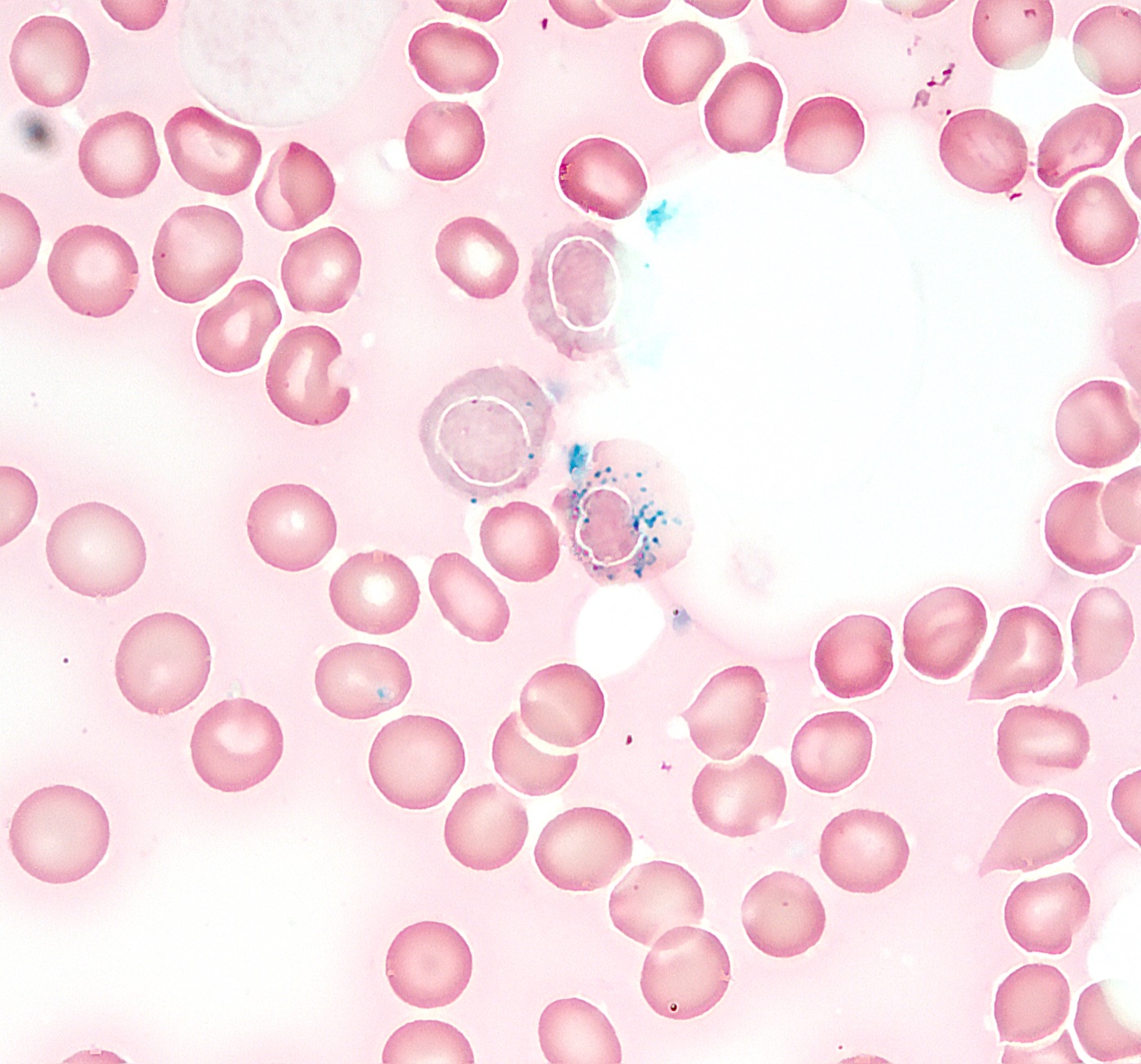
Macrocytic Anemias and Vitamin B12 and Folate Testing
Definition. Macrocytosis is an MCV above 100 fL. Its most common causes are alcohol use, vitamin B12 (cobalamin) and folate deficiency, and medications (Kaferle 2009). Megaloblastic anemia, with oval macrocytes and hypersegmented neutrophils, indicates vitamin B12 or folate deficiency as the most likely cause. Nonmegaloblastic macrocytosis is separated by the reticulocyte count into drug or alcohol effects and hemolysis or hemorrhage (Kaferle 2009). Vitamin B12 testing follows the British Society for Haematology (BSH) 2014 guideline (Devalia 2014) and the National Institute for Health and Care Excellence (NICE) guideline NG239 of 2024 for people over 16 years (NICE 2024).
Related page: Automated Blood Counts, Reticulocyte Count and Peripheral Smear Review (spurious macrocytosis, hypersegmentation definition and reticulocyte count) Related page: Microcytic Anemias and Iron Studies (iron studies, copper deficiency and sideroblastic anemia) Related page: Hemolytic Anemias: Red Cell Membrane and Enzyme Defects and Paroxysmal Nocturnal Hemoglobinuria (hemolysis markers and the hemolytic pattern) Related page: Bone Marrow Failure, Pure Red Cell Aplasia and the Laboratory Approach to Cytopenias and Leukocyte Disorders (pancytopenia and marrow failure)
Megaloblastic anemia: blood and marrow findings
- Persistence of hypersegmentation. In 14 patients with uncomplicated megaloblastic anemia, the mean neutrophil lobe count began to fall 11.4 days after treatment and normalized by 13.9 days. Hypersegmentation persisted after neutropenia and granulocyte folate had corrected (Nath 1979).
- Masked megaloblastic anemia. In 8 episodes of folate or vitamin B12 deficiency, the anemia was normocytic. Coexisting conditions included inflammatory or infectious illness, iron deficiency, thalassemia trait and renal failure. Hypersegmented neutrophils were seen in every episode, but a mean lobe count above 3.5 was present only once. Giant metamyelocytes were present in the marrow in every episode (Spivak 1982).
- Ineffective erythropoiesis. Vitamin B12 deficiency can present with pancytopenia, raised lactate dehydrogenase (LDH), indirect hyperbilirubinemia and low haptoglobin. An inappropriately low reticulocyte response, a negative direct antiglobulin test (DAT) and absent schistocytes separate ineffective erythropoiesis from peripheral hemolysis (Thannir 2026).
- Marrow. Signs of dyserythropoiesis in the marrow aspirate led to the diagnosis of pernicious anemia in a patient whose serum vitamin B12 was falsely normal (Ghammad 2023).
Biomarkers of vitamin B12 status
Four biomarkers assess vitamin B12 status: total B12, holotranscobalamin (holoTC, "active B12", the B12 bound to the transport protein transcobalamin), methylmalonic acid (MMA) and total homocysteine (Nexo 2024). No single gold standard defines deficiency, and the clinical picture is the most important factor in interpreting results (Devalia 2014).
| Test | What it measures | Confounders and limits | Source |
|---|---|---|---|
| Total serum B12 | All circulating cobalamin | No definitive cutoffs; method-dependent; local reference ranges; of limited value during pharmacologic B12 therapy | Devalia 2014; Nexo 2024 |
| HoloTC (active B12) | Cobalamin bound to transcobalamin, available for cellular uptake | Fewer methods; most assays traceable to the WHO reference standard; an indeterminate range remains | Marandola 2026; Devalia 2014 |
| MMA | Metabolite that accumulates when B12 is lacking | Rises as glomerular filtration falls, independent of B12 status; laboratory reference ranges | Åsberg 2025; NICE 2024 |
| Total homocysteine | Metabolite that accumulates in B12 or folate deficiency | Less specific than MMA; raised by folate deficiency | Devalia 2014; NICE 2024 |
| Serum folate | Circulating folate | BSH 2014: equivalent to red cell folate in diagnostic capability and the first-line test for folate status (Devalia 2014). An earlier review recommended red cell folate because serum folate fluctuates with dietary intake and red cell folate correlates better with folate stores (Kaferle 2009) | Devalia 2014; Kaferle 2009 |
- Factors that alter total or active B12. Over-the-counter B12 preparations raise total and active B12 without fully treating a deficiency. The combined oral contraceptive pill lowers total B12 without causing deficiency (NICE 2024). People of Black ethnicity may have a higher reference range for serum B12 than people of White or Asian ethnicity (NICE 2024).
- High B12. An unexpectedly high total B12 is usually of no clinical importance and is sometimes associated with underlying cancer (Nexo 2024).
- Renal adjustment. MMA adjusted to an estimated GFR of 100 mL/min/1.73 m² (MMA100) related less to renal function and had higher diagnostic accuracy than unadjusted MMA (Åsberg 2025).
- Combination testing. A total B12 combined with a metabolic marker, such as MMA, may be the most useful approach in daily practice (Nexo 2024).
Falsely normal B12 from anti-intrinsic factor antibodies
- Case evidence. In a patient with macrocytic anemia, raised homocysteine and MMA, and dyserythropoiesis, serum B12 was repeatedly normal by competitive-binding chemiluminescence. Anti-intrinsic factor antibodies were present at a very high level. An electrochemiluminescence method gave a very low B12, and removal of immunoglobulin with protein G restored a low result on the original method (Ghammad 2023).
- Arbiter method. Immunoassays can give falsely normal B12 results in the presence of anti-intrinsic factor or heterophile antibodies. A liquid chromatography–tandem mass spectrometry (LC-MS/MS) method identified falsely normal and falsely raised immunoassay results among 236 samples (Zhang 2023).
Vitamin B12 testing strategies of BSH 2014 and NICE 2024
Current rule (NICE NG239, 2024). Total B12 or active B12 is the initial test, with fixed thresholds. Laboratories may use their own validated thresholds where total B12 thresholds vary substantially (NICE 2024).
NICE NG239 interpretation of the initial vitamin B12 test.
| Total B12 | Active B12 (holoTC) | Interpretation | Source |
|---|---|---|---|
| <180 ng/L (133 pmol/L) | <25 pmol/L | Confirmed vitamin B12 deficiency | NICE 2024 |
| 180–350 ng/L (133–258 pmol/L) | 25–70 pmol/L | Indeterminate; possible deficiency | NICE 2024 |
| >350 ng/L (258 pmol/L) | >70 pmol/L | Deficiency unlikely | NICE 2024 |
| Element | BSH 2014 (Devalia 2014) | NICE NG239 (NICE 2024) |
|---|---|---|
| First-line test | Serum cobalamin; holoTC has potential as a first-line test | Total B12 or active B12 |
| Pregnancy | Not stated | Active B12 |
| Suspected nitrous oxide use | Not stated | Plasma homocysteine or serum MMA as the initial test |
| Cutoffs | Definitive cutoffs not possible; local reference ranges | Fixed thresholds in the table above, or locally validated thresholds |
| Second-line test | Plasma MMA; homocysteine less specific | Serum MMA when symptoms or signs are present and the initial result is indeterminate |
| Discordant or indeterminate results | Treatment not delayed when strong clinical features conflict with the result | Replacement considered for indeterminate results with rapidly progressive neurological or hematological features, autoimmune gastritis, gastric or ileal surgery, or pregnancy or breastfeeding, and started while MMA is awaited |
| Folate | Serum folate first-line; equivalent to red cell folate | Folate deficiency taken into account when homocysteine is interpreted |
- Sampling and treatment. Samples are taken before replacement begins. Replacement is not delayed in suspected megaloblastic anemia with neurological symptoms, especially subacute combined degeneration of the spinal cord (NICE 2024).
- Unlikely result with persisting symptoms. Other causes are investigated, and the initial test is repeated if symptoms persist after 3–6 months (NICE 2024).
Causes of vitamin B12 deficiency
- Risk factors. Low dietary intake, family history of vitamin B12 deficiency or an autoimmune condition, atrophic gastritis affecting the gastric body, celiac disease or another autoimmune condition, gastrectomy, terminal ileal resection, many bariatric operations, abdominal or pelvic radiotherapy, recreational nitrous oxide, and medicines including metformin, proton pump inhibitors, H2-receptor antagonists, colchicine, phenobarbital, pregabalin, primidone and topiramate (NICE 2024).
- Metformin. In a randomized trial of 390 insulin-treated patients with type 2 diabetes, metformin 850 mg three times daily for 4.3 years lowered B12 by 19% compared with placebo. Deficiency (<150 pmol/L) was 7.2 percentage points more common, with a number needed to harm of 13.8, and homocysteine rose (de Jager 2010).
- Acid suppression. In 25,956 patients with B12 deficiency and 184,199 controls, a supply of proton pump inhibitors for 2 years or more had an odds ratio of 1.65 and H2-receptor antagonists 1.25. Doses above 1.5 PPI pills daily had an odds ratio of 1.95 (Lam 2013).
- Nitrous oxide. In 81 patients (100 presentations) with recreational nitrous oxide toxicity, median homocysteine was 50 µmol/L and median MMA 0.68 µmol/L. For subacute combined degeneration, total B12 and holoTC had sensitivities of 22% and 5%, and homocysteine and MMA had sensitivities of 95% and 93% (Bhujel 2026). NICE recommends homocysteine or MMA as the initial test when nitrous oxide is the suspected cause (NICE 2024).
- Deficiency populations. Subclinical B12 deficiency affects 2.5%–26% of the general population, depending on the definition. Elderly individuals and populations with restricted intake of animal-source foods are at greatest risk (Green 2017).
Pernicious anemia and autoimmune gastritis
- Disease definition. Pernicious anemia is the late stage of autoimmune gastritis, with vitamin B12 deficiency and macrocytic anemia (Shah 2021). The main autoantigen of autoimmune atrophic gastritis is the β subunit of the gastric proton pump (H+/K+-ATPase) on parietal cells. The disease impairs iron and cobalamin absorption and raises the risk of type 1 gastric neuroendocrine neoplasms (Lahner 2026).
- Antibody performance. Parietal cell antibodies (PCA) are the most sensitive serum marker but give false positives in Helicobacter pylori infection and other autoimmune diseases (Shah 2021). By ELISA, PCA had 81.5% sensitivity and 90.3% specificity in one cohort (Myat 2026). Intrinsic factor antibodies (IFA) have specificity near 100% and low, variable sensitivity: below 30% in many studies, 37% by ELISA in one cohort and 50%–70% in other reports, rising to about 80% as pernicious anemia progresses. They appear later in the disease (Shah 2021; Myat 2026; Nagao 2017). A negative IFA result does not exclude autoimmune gastritis (NICE 2024).
- PCA method. Solid-phase assays with purified H+/K+-ATPase detected PCA with higher sensitivity than indirect immunofluorescence (IIF) on rodent or primate stomach (Tonegato 2024). In patients with B12 deficiency, PCA and no IFA, IIF positivity was associated with confirmed pernicious anemia with 92% specificity, and an IIF titer of 80 or more raised specificity to 96%. An immunodot assay was positive in nearly all patients with and without the disease (Lacombe 2026).
- NICE testing sequence. An IFA test is considered when autoimmune gastritis is suspected and no positive IFA result or relevant surgery is on record. When autoimmune gastritis is still suspected after a negative IFA result, NICE lists a PCA test, a gastrin level, an absorption test (CobaSorb) and gastroscopy with gastric body biopsy. Laboratories report what their IFA assay detects and how to interpret it (NICE 2024).
- Gastric function markers. Parietal cell loss causes hypergastrinemia, enterochromaffin-like cell hyperplasia and type 1 gastric neuroendocrine neoplasms at an estimated 0.4%–0.7% per year. A pepsinogen I below 70 µg/L with a pepsinogen I:II ratio below 3.0 had high sensitivity and specificity for severe corpus atrophy in high-incidence regions. Pepsinogen testing is not routinely available in the United States (Shah 2021).
- AGA 2021 workup. Histology compatible with autoimmune gastritis prompts PCA and IFA testing and evaluation for B12 and iron deficiency. A new diagnosis of pernicious anemia prompts endoscopy with topographic biopsies. Iron deficiency occurs in up to 50% of patients with corpus-predominant atrophic gastritis and often precedes B12 deficiency. Autoimmune thyroid disease is screened for (Shah 2021).
| Marker | Finding in autoimmune gastritis | Limitation | Source |
|---|---|---|---|
| IFA | Positive | Low and variable sensitivity (below 30% to 70%); specificity near 100% | Shah 2021; Myat 2026 |
| PCA | Positive | False positives with H pylori and other autoimmune diseases; IIF more specific than immunodot | Shah 2021; Lacombe 2026 |
| Gastrin | High | Reflects parietal cell loss | Shah 2021; NICE 2024 |
| Pepsinogen I and I:II ratio | <70 µg/L and <3.0 | Validated mainly in East Asian populations | Shah 2021 |
| Gastric body biopsy | Corpus-predominant atrophy with antral sparing | Requires endoscopy | Shah 2021 |
Nonmegaloblastic macrocytosis
| Cause | Laboratory features | Source |
|---|---|---|
| Alcohol use | Nonmegaloblastic macrocytosis with a low reticulocyte count; about 90% of people with alcoholism have macrocytosis (MCV 100–110 fL) before anemia develops, and the MCV returns to normal with abstinence | Kaferle 2009; Nagao 2017 |
| Liver disease, hypothyroidism | Nonmegaloblastic film | Kaferle 2009 |
| Reticulocytosis (hemolysis, hemorrhage) | Raised reticulocyte count; reticulocytes are larger than mature red cells | Kaferle 2009; Palmer 2015 |
| Zidovudine | Median MCV 114.6 fL at discontinuation; macrocytosis persisted in 37% at 2 years after stopping | Yu 2017 |
| Zidovudine, progression | Nonanemic macrocytosis progressed to anemia in 21.3% by 4 years | Nanthatanti 2022 |
| Hydroxyurea and antiretroviral therapy | An MCV above 130 fL narrows the differential to antiretroviral therapy, hydroxyurea, and vitamin B12 or folate deficiency | Nagao 2017 |
| Other medicines | Methotrexate, azathioprine, valproic acid, phenytoin, trimethoprim-sulfamethoxazole, metformin and other antineoplastic and antiviral drugs | Nagao 2017 |
| Myelodysplastic and myeloproliferative neoplasms | Primary marrow disorders | Kaferle 2009 |
| Spurious macrocytosis | Cold agglutinins and hyperglycemia, described in Automated Blood Counts, Reticulocyte Count and Peripheral Smear Review | Zandecki 2007 |
Inherited disorders of cobalamin transport and metabolism
- Remethylation disorders. Inherited defects of cobalamin or folate metabolism disrupt the methionine–homocysteine cycle and raise homocysteine, often with low methionine. Combined cobalamin defects also raise MMA. cblC (MMACHC) is the most common cobalamin-related remethylation disorder. Plasma total homocysteine, methionine, MMA, serum B12 and folate are measured in suspected cases, and newborn screening improves outcomes (Olivieri 2026).
- Transcobalamin deficiency. Transcobalamin (TCN2) deficiency is an autosomal recessive defect of B12 transport. It presents with megaloblastic anemia and neutropenia and can mimic a myelodysplastic neoplasm. Serum B12 and folate can be normal, and genetic testing confirms the diagnosis (Hu 2026).
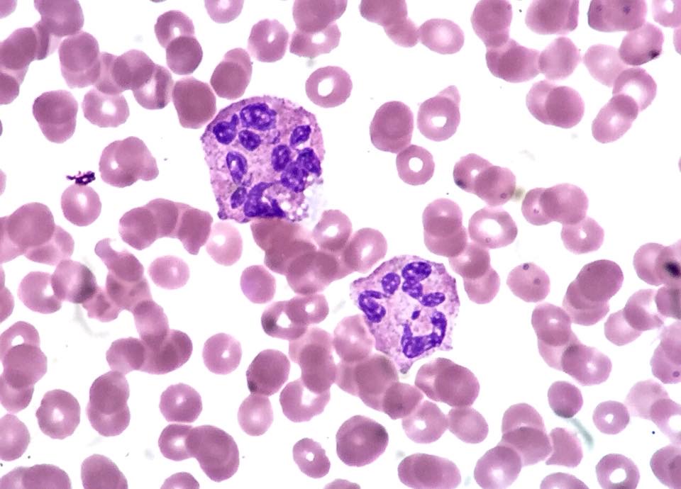
Hemoglobin Variant Analysis and Laboratory Diagnosis of Thalassemia
Definition. Hemoglobin disorders are inherited defects of globin. Structural variants (hemoglobinopathies) such as HbS, HbC and HbE alter the globin sequence. Thalassemias reduce or abolish the production of one globin chain (Therrell 2026). More than 1000 naturally occurring variants with single amino acid substitutions have been described (Thom 2013). Laboratory diagnosis combines the blood count and red cell indices, protein-based hemoglobin separation and quantitation, and DNA analysis. Protein methods identify most common variants and raise the suspicion of thalassemia, and DNA testing is required to confirm α-thalassemia in particular (Zhang 2025).
Related page: Automated Blood Counts, Reticulocyte Count and Peripheral Smear Review (red cell indices, target cells and supravital staining) Related page: Microcytic Anemias and Iron Studies (iron deficiency and the discriminant indices for microcytosis) Related page: Hemolytic Anemias: Red Cell Membrane and Enzyme Defects and Paroxysmal Nocturnal Hemoglobinuria (hemolysis markers, Heinz bodies and red cell enzyme defects) Related page: Hemolytic Disease of the Fetus and Newborn, Fetomaternal Hemorrhage, Rh Immune Globulin and Fetal and Neonatal Transfusion (Kleihauer-Betke and flow cytometric HbF testing for fetomaternal hemorrhage) Related page: Therapeutic Apheresis, ASFA Indications and Hematopoietic Progenitor Cell Products (red cell exchange and HbS targets in sickle cell disease) Related page: Immunohematology Methods in Red Cell Serology (red cell antigen phenotyping in patients with hemoglobin disorders)
Current guidelines for hemoglobinopathy screening and diagnosis
- British Society for Haematology (BSH) 2023. The BSH guideline on significant haemoglobinopathies (Bain 2023) is the current UK guideline. It succeeds the 2010 British Committee for Standards in Haematology guideline of the same title (Ryan 2010).
- International Council for Standardization in Haematology (ICSH). The 2012 ICSH recommendations describe the methods and pitfalls of HbA2 and HbF quantitation, with emphasis on automated high-performance liquid chromatography (HPLC) and capillary electrophoresis (CE). HbA2 measurement is essential for the routine identification of β-thalassemia carriers, and DNA analysis characterizes the mutation (Stephens 2012a). HbF measurement is needed for the diagnosis of δβ-thalassemia, hereditary persistence of fetal hemoglobin (HPFH) and sickle cell disease (SCD) (Stephens 2012b). A 2015 ICSH document sets out how HPLC and CE instruments are assessed for HbA2 quantitation. Carrier and noncarrier HbA2 levels differ only slightly, so the analyses must be precise and accurate (Stephens 2015).
- Analytical requirements at the action values. The UK national screening program sets action values of HbA2 3.5% and HbF 5% or 10% (depending on the MCH). An HPLC or CE system must therefore measure HbA2 and HbF accurately and precisely at these values and must detect variant peaks in the HbA2 window (Bain 2023). The Italian Society of Thalassemia and Haemoglobinopathies (SITE) best-practice document specifies cation-exchange HPLC with a coefficient of variation below 5% (Mandrile 2022).
Elements the 2023 guideline presents as new include mass spectrometry for variant identification and newborn screening, point-of-care devices for HbS, DNA testing for the sickle gene in neonates transfused in utero or early in the neonatal period in place of a repeat sample 4 months after transfusion, HbF monitoring during hydroxyurea (hydroxycarbamide) treatment or after gene therapy, and laboratory awareness of split peaks induced by voxelotor (Bain 2023).
Globin genes and the fetal-to-adult switch
- Gene clusters. The β-globin gene cluster on the short arm of chromosome 11 contains the β-globin gene (HBB), the δ-globin gene, the embryonic ε gene, the fetal Aγ and Gγ genes and a pseudogene (Cao 2010). The duplicated α-globin genes HBA1 and HBA2 lie on chromosome 16 (Zhang 2025).
- Adult hemoglobins. Adult blood contains predominantly HbA (α2β2) with minor fractions of HbA2 (α2δ2) and HbF (α2γ2) (Molina-Arrebola 2026).
- Switching. Newborns with sickle cell anemia are asymptomatic because of high HbF, and disease begins as HbF declines in the first year of life (Quinn 2025). BCL11A, MYB and KLF1 regulate HbF silencing (Sankaran 2013).
- Excess chains in α-thalassemia. Excess γ chains form γ4 tetramers (Hb Bart's) in fetal life, and excess β chains form β4 tetramers (HbH) in adult life (Harteveld 2010).
Protein methods for hemoglobin separation
At least two methods are used for the presumptive identification of an abnormal hemoglobin. Use of both HPLC and CE maximizes detection of variants and minimizes misinterpretation of artifacts (Traeger-Synodinos 2015). Each suspected variant is confirmed by a second analytical method (Molina-Arrebola 2026).
| Method | Principle | Strengths | Limitations | Source |
|---|---|---|---|---|
| Alkaline electrophoresis (cellulose acetate, pH 8.2–8.6) | Separation by charge | Low cost; separates A, F, S and C | HbA2, C, E and O comigrate; S comigrates with D; poor quantitation of A2 | Twumasi 2025; Zhang 2025 |
| Acid electrophoresis (citrate agar or agarose, pH 6.0–6.2) | Separation by charge at acid pH | Resolves some variants that comigrate at alkaline pH | Does not resolve E from A2 or D from G | Twumasi 2025; Zhang 2025 |
| Isoelectric focusing (IEF) | Migration to the isoelectric point in a pH gradient | Resolves C from E and O, and S from D and G; detects Hb Bart's and HbH; used in newborn screening | Does not separate E from C-Harlem or O-Arab, or G-Philadelphia from Lepore; imprecise for A2; manual | Zhang 2025 |
| Cation-exchange HPLC | Elution from a negatively charged resin by a buffer of increasing cation concentration; peaks assigned to retention-time windows | Quantitates A2 and F; separates A, A2, F, S, C, O-Arab, D-Punjab and G-Philadelphia | HbE, Korle-Bu and Lepore elute with A2; A2 falsely raised with HbS; windows vary by instrument and software | Zhang 2025 |
| Capillary zone electrophoresis (CZE) | Electro-osmotic flow in silica capillaries | Separates E from A2; not affected by glycated or acetylated hemoglobins; usable on dried blood spots and cord blood | Limited separation of S from D | Zhang 2025 |
| Mass spectrometry | Globin chain or peptide mass | Identifies variants missed by separation methods | Specialized workflow | Zhang 2025; Traeger-Synodinos 2015 |
- Agreement between HPLC and CE. In a meta-analysis of 1,881 paired measurements, the pooled HbA2 bias between HPLC and CE was 0.16% (0.37% in healthy individuals), enough to affect interpretation near the 3.5% threshold (Wei 2026).
- Variants missed by protein methods. Mass spectrometry or DNA analysis is used when the hematology suggests a variant that separation methods do not show. Hyperunstable variants may be found only by DNA methods (Traeger-Synodinos 2015).
Sickle solubility test
- Principle. Deoxygenated HbS is insoluble in a high-molarity phosphate buffer containing a reducing agent such as sodium dithionite, and the solution turns turbid. A sample containing only HbA stays clear (Twumasi 2025; Mendez-Marti 2024).
- No separation of trait from disease. The test detects HbS in heterozygous and homozygous states and does not distinguish them. Positive results are verified by electrophoresis, HPLC or molecular testing (Twumasi 2025). Positive, equivocal and negative solubility results are all confirmed by HPLC, CE or an alternative technique, and a solubility result reported before confirmation is labeled interim (Bain 2023).
- False negatives. False-negative results occur in neonates with high HbF, in severe anemia and after recent transfusion or red cell exchange (Twumasi 2025; Mendez-Marti 2024). The solubility kits mainly used in the UK detect HbS down to about 20%, and some detect it as low as 8% (Bain 2023). In one adult series of 220 samples, all 5 false negatives had HbS below 15%, and 4 followed red cell exchange (Mendez-Marti 2024). A solubility test is generally not indicated before 6 months of age (Bain 2023).
- False positives. False positives occur with high plasma protein levels and in anemic patients when double the volume of blood is used. A more concentrated sample or washed red cells avoid the latter (Bain 2023).
HbA2 quantitation and β-thalassemia trait
- Cutoff. In the BSH guideline, an HbA2 of 3.5% or more with an MCH below 27 pg indicates β-thalassemia heterozygosity. In the antenatal algorithm, HbA2 values of 3.1%–3.9% are indeterminate and lead to β-globin DNA analysis. An HbA2 above 4% with a normal MCH is investigated further as a possible milder carrier state, and some mild β+ mutations give HbA2 below 3.5% (Bain 2023). The European Molecular Genetics Quality Network (EMQN) guideline uses above 3.5% as the standard cutoff, with 3.1%–3.5% borderline depending on the method, reference range and coefficient of variation (Traeger-Synodinos 2015). Carriers may also show HbF of 1%–5% (Molina-Arrebola 2026).
- Red cell indices. An adult MCV below 79 fL and MCH below 27 pg indicate possible thalassemia heterozygosity. Each laboratory sets its own cutoffs (Traeger-Synodinos 2015). The BSH guideline recommends MCH for thalassemia screening because MCH is more stable than MCV. An MCH below 25 pg prompts assessment for α0-thalassemia heterozygosity, which takes family origin into account (Bain 2023).
- Elevated HbA2 from other causes. Variants that elute with HbA2, such as HbE, Hb Lepore and HbE/β-thalassemia, raise the apparent HbA2 (Anandani 2025; Zhang 2025). In a Thai cohort, variants with HbA2 of 3.6% or more were misinterpreted as β-thalassemia or HPFH in 50.6% of cases (Singha 2026).
- Variants that split or lower the HbA2 peak. α-chain or δ-chain variants split the HbA2 peak, and coexisting δ-thalassemia lowers it (Traeger-Synodinos 2015). Co-inheritance of δ-thalassemia can reduce the HbA2 of a β-thalassemia carrier to normal (Galanello 2010).
- HbA2 with HbS. With many HPLC systems, HbA2 is overestimated in the presence of HbS, and a raised HbA2 does not reliably separate HbSS from HbS/β0-thalassemia. HbAS is recognized by HbA greater than HbS, and HbS/β-thalassemia by HbS greater than HbA (Bain 2023).
- Co-inherited α-thalassemia. α-Thalassemia can normalize the red cell indices of a β-thalassemia carrier. HbA2 remains in the carrier range and is diagnostic, so HbA2 is always measured (Galanello 2010).
- Silent β-thalassemia. Very mild mutations with high residual β-chain output give normal red cell indices and normal or borderline HbA2 (Galanello 2010).
- Iron deficiency. The effect of iron deficiency on HbA2 is described in Microcytic Anemias and Iron Studies. The screen of an iron-deficient person is repeated once the person is iron replete, which may not be practical in an ongoing pregnancy (Traeger-Synodinos 2015).
| Hematologic finding | Possible interpretations | Source |
|---|---|---|
| MCV <79 fL and MCH <27 pg with normal HbA2 and HbF | Iron deficiency; heterozygous α-thalassemia; mild β-thalassemia variant; combined δ- and β-thalassemia; heterozygous εγδβ-thalassemia | Traeger-Synodinos 2015 |
| Normal or borderline reduced indices with raised HbA2 | Combined α- and β-thalassemia; β-thalassemia carrier with folate or vitamin B12 deficiency or hepatitis | Traeger-Synodinos 2015 |
| Normal or reduced indices with HbF >5% and normal or low HbA2 | Heterozygous δβ-thalassemia, Aγδβ-thalassemia or HPFH | Traeger-Synodinos 2015 |
| Normal indices with normal or borderline HbA2 | α-gene triplication, KLF1 variants or a mild β-thalassemia variant | Traeger-Synodinos 2015 |
| Severely reduced indices with raised HbA2 | More than four α-globin genes with heterozygous β-thalassemia | Traeger-Synodinos 2015 |
Clinical forms of β-thalassemia. Thalassemia major shows hemoglobin below 7 g/dL, MCV 50–70 fL and MCH 12–20 pg. Thalassemia intermedia shows hemoglobin of 7–10 g/dL, MCV 50–80 fL and MCH 16–24 pg. Rare dominant forms result from hyperunstable β-globin variants. They are suspected when thalassemia intermedia occurs with hematologically normal parents or with dominant transmission (Galanello 2010).
HbF quantitation and F-cell distribution
- HbF levels. An isolated HbF above 5% with microcytosis suggests δβ-thalassemia (Molina-Arrebola 2026). In δβ-thalassemia, large F cells can make microcytosis less evident (Traeger-Synodinos 2015).
- Cellular distribution. The distribution of HbF among red cells helps to diagnose HPFH and to assess fetomaternal hemorrhage (Stephens 2012b). HbF is normally heterocellular and enriched in F cells, which carry about 30% HbF each. In the common form of HbS-HPFH, HbF is pancellular and bulk HbF by HPLC is about 30% (Khandros 2026). F-cell percentages are measured by intracellular staining and flow cytometry (Khandros 2026).
α-Thalassemia: hematology and molecular diagnosis
- Genotype and severity. Silent carriers have one α-globin gene deleted, HbH disease three and hydrops fetalis four (Zhang 2025).
- Trait. Carriers have mild microcytic hypochromic anemia and a normal or slightly reduced HbA2, and molecular analysis is usually required for diagnosis. Carriers of -α3.7 can have normal hematology. Ferritin is measured because the trait resembles iron deficiency (Harteveld 2010).
- Hb Bart's in the newborn. Hb Bart's is detectable on routine analysis when α-globin synthesis falls below about 70% of normal in fetal life. It disappears rapidly after birth, and in HbH disease it is replaced by HbH (Harteveld 2010).
- HbH disease. Hemoglobin ranges from 2.6 to 13.3 g/dL, and HbH from 0.8% to 40%. HbH inclusions are shown by staining blood with 1% brilliant cresyl blue (Harteveld 2010). Nondeletional HbH disease is more severe than the common deletional forms (Harteveld 2010).
- Hb Bart's hydrops fetalis. Affected fetuses usually die in utero between 23 and 38 weeks or shortly after birth (Harteveld 2010).
- Hb Constant Spring and Hb Paksé. These termination-codon variants cause severe nondeletional HbH disease with α0-thalassemia deletions. They form only 0.1%–1.0% of total hemoglobin in heterozygotes, and conventional screening often misses them (Satthakarn 2026).
- Variant percentage and α-globin gene number. In carriers of HbS, HbD-Punjab, HbE and HbC, the variant percentage depends on the number of α-globin genes. Co-inherited α-thalassemia usually lowers it below about 35% for HbS, HbD-Punjab and HbC and below about 25% for HbE (Traeger-Synodinos 2015).
| Molecular method | Targets | Comment | Source |
|---|---|---|---|
| Gap-PCR | Common deletions: -α3.7, -α4.2, --SEA, --MED I, -(α)20.5, --FIL, --THAI | Fast; allele dropout can give false negatives in prenatal diagnosis | Traeger-Synodinos 2015 |
| MLPA or array CGH | Known and unknown deletions and duplications of the α- and β-globin clusters | Used when gap-PCR is negative and the hematology indicates a deletion | Traeger-Synodinos 2015 |
| Sequencing of HBA1 and HBA2 | Nondeletional mutations | α genes are about 1.2 kb and are sequenced in two overlapping fragments | Harteveld 2010 |
| Targeted mutation panels or HBB sequencing | β-Thalassemia mutations | Sequencing used when targeted analysis fails | Galanello 2010 |
When one partner carries a severe α-thalassemia defect and the other is a β-thalassemia carrier, the α-globin cluster of the β-thalassemia carrier is fully characterized (Traeger-Synodinos 2015).
Interpretation of common hemoglobin patterns
| Condition | Characteristic laboratory pattern | Source |
|---|---|---|
| β0-thalassemia homozygote | HbA absent; HbF 92%–95% | Galanello 2010 |
| β+ homozygote or β+/β0 | HbA 10%–30%; HbF 70%–90% | Galanello 2010 |
| Sickle cell trait (HbAS) | HbS below HbA; HbS below about 35% suggests co-inherited α-thalassemia | Traeger-Synodinos 2015; Ceacero-Marín 2026 |
| Sickle cell anemia (HbSS) | HbS predominant (mean 74.5%, with HbF 18.3%, in one HPLC series); HbA absent; HbF variable; HbA2 overestimated by HPLC in the presence of HbS | Rao 2010; Anandani 2025; Bain 2023 |
| HbS/β0-thalassemia | Resembles HbSS: HbA absent, HbS predominant, MCV reduced; HbA2 often raised, but HbA2 does not reliably separate HbS/β0 from HbSS, and family studies or HBB analysis are used | Anandani 2025; Bain 2023 |
| HbSC disease | About 50% HbS in red cells; HbF below 5%; HbC causes cation loss and red cell dehydration | Rees 2026 |
| HbSβ+-thalassemia | HbS predominates; HbA 5%–30%; HbA2 increased | Anandani 2025 |
| HbS-HPFH | HbF about 30% by HPLC; pancellular distribution | Khandros 2026 |
| HbS/HbD-Punjab and HbS/HbO-Arab | Sickling syndromes; D comigrates with S on alkaline gel and is separated by HPLC or IEF | Traeger-Synodinos 2015; Zhang 2025 |
| HbSD-Punjab | HbS and HbD-Punjab in about equal proportions (41.9% and 41% in one case); no HbA | Rao 2010; Bain 2023 |
| HbD-Punjab trait | HbD 28%–43% in the D window; HbA2 normal or low (mean 1.7% in one series) | Anandani 2025; Pant 2014; Rao 2010 |
| HbE heterozygote | HbE about 19%; lower with α0-thalassemia; an HbA2-window value above about 15% on HPLC suggests HbE trait, and 8% or more suggests Hb Lepore | Srisuwan 2026; Bain 2023 |
| HbE homozygote (HbEE) | HbE 77%–83% in the HbA2 window; HbF 2%–5% | Pant 2014 |
| HbE/β-thalassemia | Phenotype from thalassemia major to mild intermedia; HbE elutes in the A2 window on HPLC | Galanello 2010; Zhang 2025 |
| Hb Lepore | δβ fusion with low expression; elutes in the A2 window on HPLC | Zhang 2025 |
| Hb Bart's hydrops fetalis | Hb Bart's present; HbF absent | Harteveld 2010 |
- HbE. HbE results from a G→A substitution in codon 26 of HBB. It is a structural variant and also activates a cryptic splice site with abnormal mRNA processing (Olivieri 2011). HbE causes microcytosis through reduced β-chain synthesis (Zhang 2025).
- Transfusion. Hemoglobin analysis after a recent transfusion is potentially misleading and is repeated 4 months after transfusion. When the result is needed urgently, DNA testing is used (Bain 2023). Donor HbA alters the patient's hemoglobin pattern. Sample identification for hemoglobinopathy testing records transfusion in the previous 4 months (Traeger-Synodinos 2015). In HbSC disease, posttransfusion targets apply to HbS plus HbC: an HbS target below 30% corresponds to HbS + HbC below 30% (Rees 2026).
Unstable and high oxygen affinity hemoglobins
- Unstable hemoglobins. Unstable variants denature and precipitate as Heinz bodies, which cause hemolysis. Most are not detected by conventional testing, and next-generation sequencing that includes the globin genes establishes the diagnosis (Rizzuto 2021). The isopropanol stability test detects unstable hemoglobins by their reduced solubility in an organic solvent. It can identify variants missed by HPLC and electrophoresis (Twumasi 2025). Heinz bodies may be shown by supravital staining (Medri 2022).
- High oxygen affinity hemoglobins. These variants cause erythrocytosis through tissue hypoxia and increased erythropoietin production (Bento 2014). A left-shifted oxygen dissociation curve with a low P50 supported the diagnosis historically, and DNA sequencing of the globin genes is the confirmatory test (Repp 2026).
Laboratory approach to erythrocytosis
| Mechanism | Laboratory findings | Genes | Source |
|---|---|---|---|
| Primary familial erythrocytosis | Low erythropoietin | EPOR | Bento 2014 |
| High oxygen affinity hemoglobin | Low P50; normal or raised erythropoietin | HBB, HBA1, HBA2 | Bento 2014; Repp 2026; Giannella 2026 |
| Low 2,3-bisphosphoglycerate production | Erythrocytosis from tissue hypoxia | BPGM | Bento 2014 |
| Hypoxia-sensing pathway defects | Increased erythropoietin production | VHL, EPAS1, EGLN1 | Bento 2014; Giannella 2026 |
The molecular cause remains unknown in about 70% of patients with congenital erythrocytosis (Bento 2014). Primary erythrocytosis most often reflects JAK2-mutated polycythemia vera (Giannella 2026).
Methemoglobin, sulfhemoglobin and hemoglobin M
- Definition and cyanosis. Normal blood contains less than 1% methemoglobin (MetHb). Cyanosis appears when MetHb exceeds about 1.5 g/dL (Iolascon 2021).
- Oximetry. PaO2 on arterial blood gas is normal, and the calculated arterial saturation appears normal or high. Most pulse oximeters give a fixed reading of about 85% when MetHb is significant (Iolascon 2021; Twine 2026). Co-oximetry with 4 or more wavelengths measures the hemoglobin species directly and is the diagnostic test (Twine 2026).
- Cyanide reaction. MetHb absorbs at 630–635 nm, and conversion to cyanmethemoglobin by cyanide abolishes this band in the Evelyn-Malloy method (Iolascon 2021; Twine 2026). Sulfhemoglobin absorbs at 614 nm and does not react with potassium cyanide. Older co-oximeters can misread it as MetHb (Twine 2026).
- Cytochrome b5 reductase deficiency. This autosomal recessive disorder results from biallelic CYB5R3 variants. Type I is confined to red cells. Type II includes severe neurologic disease. MetHb is typically 20%–30% in both types (Iolascon 2021). The enzyme assay (ferricyanide acceptor, NADH oxidation at 340 nm) is the gold standard for separating hereditary from acquired methemoglobinemia, and residual activity is usually below 20% of normal (Iolascon 2021).
- Hemoglobin M. HbM variants are autosomal dominant, usually with tyrosine replacing the proximal or distal histidine. Patients are cyanotic and usually otherwise asymptomatic, with MetHb of 12.5%–25% (Iolascon 2021). Electrophoresis at pH 7.1 can identify HbM variants (Iolascon 2021). Some are electrophoretically silent, and some α-chain variants mimic sulfhemoglobin on co-oximetry, so diagnosis usually rests on globin gene DNA analysis (Twine 2026).
- Other causes. Unstable hemoglobins and G6PD deficiency can raise MetHb. Dapsone, nitrites, benzocaine and prilocaine cause acquired methemoglobinemia (Iolascon 2021).
Antenatal and partner carrier screening for hemoglobin disorders
- The World Health Organization (WHO) advises screening for thalassemia and other hemoglobin disorders in all individuals of reproductive age (Zhang 2025).
- The American College of Obstetricians and Gynecologists (ACOG) recommends offering hemoglobinopathy testing to persons planning pregnancy or at the initial prenatal visit when no prior results are available (Zhang 2025).
- Prenatal diagnosis is offered for thalassemia major, the sickle cell syndromes (including S/C, S/β-thalassemia, S/D-Punjab, S/O-Arab and S/E) and Hb Bart's hydrops fetalis (Traeger-Synodinos 2015).
Newborn screening for hemoglobin disorders
- United States. The original US Recommended Uniform Screening Panel (RUSP), approved in 2008, included sickle cell anemia (SCD-SS), HbSC disease and the sickle β0- and β+-thalassemias. Universal hemoglobin newborn screening was mandated in all states by 2006 (Therrell 2026). HbH disease was nominated as a core condition in 2010, and the nomination was not approved. Most US programs detect HbH disease through Hb Bart's (Therrell 2026).
- Methods. Most programs use IEF or HPLC for the first tier, sometimes with a second-tier molecular test (Therrell 2026).
- Reporting patterns. Hemoglobins are reported in order of abundance. FA is normal, FAS indicates HbA above HbS, and FSA indicates HbS above HbA (Shook 2020). FS, FSC and FSA are screen-positive SCD patterns (Awuonda 2026).
- FSA in practice. Among 31 newborns with an initial FSA pattern, all 8 with a persistent FSA pattern on venous retesting proved to have HbAS (Shook 2020).
- Preanalytic pitfalls. Transfusion before the screen alters the hemoglobin pattern. Iowa repeats the screen about 56 days after transfusion. Infants born before 37 weeks are rescreened, and all false positives in the Iowa series occurred at 32 weeks or less (Jilek 2024). Infants with only HbF on screening are referred for molecular testing in most states (Jilek 2024).
- Confirmation. Newborns with a variant are retested on a venous sample, for example at 2–4 weeks by CZE followed by acid electrophoresis or IEF (Shook 2020).
Hemolytic Anemias: Red Cell Membrane and Enzyme Defects and Paroxysmal Nocturnal Hemoglobinuria
Definition. Hemolytic anemia is anemia caused by shortened red cell survival. The laboratory markers of hemolysis are increased reticulocytes, increased lactate dehydrogenase (LDH), reduced haptoglobin and unconjugated hyperbilirubinemia. The direct antiglobulin test (DAT) separates autoimmune forms from nonimmune forms, and smear examination is central to the diagnosis of congenital membrane defects and thrombotic microangiopathies (Barcellini 2015). The nonimmune hemolytic anemias diagnosed with specialized laboratory tests are the hereditary red cell membrane disorders, the red cell enzyme deficiencies and paroxysmal nocturnal hemoglobinuria (PNH).
Related page: Automated Blood Counts, Reticulocyte Count and Peripheral Smear Review (reticulocyte count, red cell morphology and inclusions) Related page: Blood Component Selection and Serologic Workup of Autoantibodies (DAT workup, warm and cold autoantibodies, drug-induced immune hemolysis) Related page: Transfusion Reactions (acute and delayed hemolytic transfusion reactions) Related page: DIC, Thrombotic Microangiopathy and Viscoelastic Testing (microangiopathic hemolytic anemia, thrombotic microangiopathy and schistocyte counting) Related page: Hemoglobin Variant Analysis and Laboratory Diagnosis of Thalassemia (unstable hemoglobins and Heinz body testing) Related page: Bone Marrow Failure, Pure Red Cell Aplasia and the Laboratory Approach to Cytopenias and Leukocyte Disorders (PNH clones in aplastic anemia) Related page: Thrombophilia Testing and Antiphospholipid Antibody Testing (PNH testing in unusual-site thrombosis)
Laboratory markers of hemolysis and their limits
| Marker | Finding in hemolysis | Limits and confounders | Source |
|---|---|---|---|
| Reticulocyte count | Increased as the marrow compensates | Inadequate or absent with marrow involvement, iron or vitamin deficiency, infection or an autoimmune reaction against erythroid precursors; reticulocytopenia occurs in about 20% of adults and 39% of children with autoimmune hemolytic anemia (AIHA) | Barcellini 2015 |
| LDH | Red cells contain mainly LDH-1 and LDH-2; slightly increased in extravascular hemolysis and 4–5 times the upper reference limit in intravascular hemolysis | Increased in tissue necrosis, myocardial infarction, hepatitis, tumors and vitamin B12 or folate deficiency | Barcellini 2015 |
| Haptoglobin | Reduced in intravascular and extravascular hemolysis | Reduced in liver impairment, malnutrition and congenital hypohaptoglobinemia; in AIHA it is the most sensitive marker and the last to normalize | Barcellini 2015 |
| Haptoglobin ≤25 mg/dL | Sensitivity 83% and specificity 96% for hemolytic disease in 100 admitted patients | Retrospective nephelometric study | Marchand 1980 |
| Unconjugated bilirubin | Increased, usually to no more than 4 mg/dL | Values above 4 mg/dL indicate acute massive hemolysis, coexisting Gilbert syndrome (about 5% of the population) or reduced hepatic function | Barcellini 2015 |
| Hemosiderinuria | Present in intravascular hemolysis, for example PNH | Typical of intravascular hemolysis with a marked LDH increase | Barcellini 2015 |
| Ferritin | Increased in chronic hemolysis | Acute-phase reactant; may be low in PNH because of urinary iron loss | Barcellini 2015 |
Bilirubin in anemia. In anemic patients the bilirubin upper reference limit (ULN) is corrected as ULN × patient hematocrit / 45, and a small increase above the corrected limit is abnormal (Barcellini 2015).
Intravascular and extravascular hemolysis patterns
| Feature | Intravascular pattern | Extravascular pattern | Source |
|---|---|---|---|
| Bilirubin | Smaller increase; part of the heme binds hemopexin and is catabolized in the liver | Main marker of extravascular hemolysis | Barcellini 2015 |
| Urine | Hemoglobinuria, dark tea to black or cherry red; hemosiderinuria | No hemoglobinuria | Hill 2017; Barcellini 2015 |
| Examples | PNH, prosthetic valve hemolysis, complement-activating AIHA | Warm AIHA, congenital membrane and enzyme defects | Barcellini 2015 |
DAT in the classification of hemolysis. The DAT patterns of warm AIHA, cold agglutinin disease and mixed AIHA, antibody identification and drug-induced immune hemolysis are described in Blood Component Selection and Serologic Workup of Autoantibodies. A DAT-negative hemolytic anemia is investigated with membrane, enzyme and PNH tests.
Hereditary spherocytosis: screening and confirmatory tests
The International Council for Standardization in Haematology (ICSH) guideline groups hereditary spherocytosis (HS) and hereditary elliptocytosis (HE) as defects of the cytoskeleton and hereditary stomatocytosis as a defect of cation permeability. Sodium dodecyl sulfate–polyacrylamide gel electrophoresis (SDS-PAGE) of membrane proteins, monovalent cation flux measurement and molecular analysis are specialist tests (King 2015). The British Society for Haematology (BSH) 2011 update states that the eosin-5′-maleimide (EMA) binding test has been validated in several studies and that biochemical or genetic analysis is rarely needed for atypical cases (Bolton-Maggs 2012).
EMA binding test. EMA binds covalently to Lys-430 on the first extracellular loop of band 3, and the flow cytometer measures the fluorescence of intact labeled red cells. Fluorescence is reduced in HS, congenital dyserythropoietic anemia type II, Southeast Asian ovalocytosis and cryohydrocytosis. The original study reported a sensitivity of 92.7% and a specificity of 99.1% for HS (King 2000).
EMA binding ratio.
| Step | Specification | Source |
|---|---|---|
| Ratio | Patient mean channel fluorescence (MCF) / mean MCF of 6 normal control samples | Hunt 2015 |
| Cutoff, FC500 cytometer | 0.918 (specificity 98.7%, sensitivity 95.6%); gray area 0.868–0.918 | Hunt 2015 |
| Cutoff, FACSCanto II cytometer | 0.925 (specificity 97.1%, sensitivity 100%); gray area 0.859–0.925 | Hunt 2015 |
| Gray-area result | Interpreted with the clinical presentation and family history, or with a family study | Hunt 2015 |
Expressed as a percentage fluorescence reduction, an optimal cutoff of 16.5% gave a sensitivity of 96.0% and a specificity of 96.9% in 181 referred samples. False-positive EMA results were mainly associated with severe microcytosis or AIHA (Biaggioni 2026).
| Test (principle) | Sensitivity and specificity for HS | Source |
|---|---|---|
| EMA binding (band 3 fluorescence) | Sensitivity 93%, specificity 98%; sensitivity independent of the defective protein and clinical phenotype | Bianchi 2012 |
| Acidified glycerol lysis test (AGLT) | Sensitivity 95% | Bianchi 2012 |
| Pink test | Sensitivity 91% | Bianchi 2012 |
| NaCl osmotic fragility, fresh blood | Sensitivity 68%; 53% in compensated cases | Bianchi 2012 |
| NaCl osmotic fragility, incubated blood | Sensitivity 81%; 64% in compensated cases | Bianchi 2012 |
| EMA plus AGLT | Identified all 150 patients with HS | Bianchi 2012 |
Osmotic gradient ektacytometry. Osmotic gradient ektacytometry measures red cell deformability (elongation index) across an osmolality gradient. Omin is the osmolality at minimal elongation and reflects the surface-to-volume ratio. EImax is maximal deformability. Ohyper reflects cell hydration. In HS the curve shows decreased EImax and increased Omin, with a variable Ohyper (Vives-Corrons 2026).
Hereditary spherocytosis in neonates. An MCHC above 36.5–37 g/dL and an MCHC:MCV ratio (HS index) above 0.36 suggest HS in a newborn. The absence of spherocytes on the newborn smear does not exclude HS, and EMA binding can be used to make the diagnosis (Achenjang 2025).
Genetics. Dominant variants in ANK1, SPTB and SLC4A1 typically cause mild to moderate HS. Recessive SPTA1 or EPB42 variants often cause more severe disease (Vives-Corrons 2026).
Hereditary elliptocytosis and pyropoikilocytosis
HE results from defects that impair spectrin self-association or disrupt the horizontal cytoskeletal network. In HE, hydration is usually normal and EImax is markedly reduced. The ektacytometry curve is flattened and trapezoidal (Vives-Corrons 2026). Hereditary pyropoikilocytosis (HPP) shows microspherocytosis, poikilocytosis and unusual thermal sensitivity of red cells. Spectrin extracted from HPP red cells contains excess dimers, and the dimers fail to reassociate into tetramers (Liu 1981). SDS-PAGE detects spectrin variants in HE (King 2000).
Stomatocytosis, xerocytosis and Southeast Asian ovalocytosis
- Dehydrated hereditary stomatocytosis (hereditary xerocytosis). It is autosomal dominant and usually presents as compensated hemolytic anemia with macrocytosis. The pleiotropic syndrome includes pseudohyperkalemia and perinatal edema. Heterozygous missense PIEZO1 variants increase cation transport in erythroid cells (Andolfo 2013). PIEZO1 encodes a mechanotransduction channel (Zarychanski 2012). KCNN4, which encodes the Gardos channel, is a second gene (Andolfo 2016).
- Ektacytometry and MCHC. In xerocytosis the MCHC can exceed typical HS values, and the ektacytometry curve is left-shifted with a low Ohyper and preserved EImax (Vives-Corrons 2026).
- Splenectomy. Splenectomy is contraindicated in xerocytosis because of thrombotic risk (Vives-Corrons 2026).
- Southeast Asian ovalocytosis (SAO). SAO results from deletion of codons 400–408 of the band 3 gene. The ovalocytes are rigid and resist invasion by malaria parasites (Jarolim 1991). EMA binding is reduced in SAO (King 2000).
Glucose-6-phosphate dehydrogenase deficiency
Glucose-6-phosphate dehydrogenase (G6PD) deficiency affects an estimated 500 million people. It presents as neonatal jaundice, as acute hemolytic anemia triggered by fava beans, infection or drugs, or rarely as chronic nonspherocytic hemolytic anemia. The G6PD gene is on the X chromosome, and over 230 variants have been identified at the molecular level (Luzzatto 2024).
WHO classification of G6PD variants: 2024 classes and the 1985 classes
The 1967 World Health Organization (WHO) study group classification, endorsed with slight changes in 1985, used residual activity and clinical phenotype. The 2022 panel merged former classes II and III because activity in hemizygous males with common variants varied across the 10% threshold, and both classes carry the same risks (Luzzatto 2024).
| Previous class (1985) | Definition | Source |
|---|---|---|
| I | Severe enzyme deficiency with chronic nonspherocytic hemolytic anemia | Luzzatto 2024 |
| II | Severe enzyme deficiency (<10% of normal) | Luzzatto 2024 |
| III | Moderate enzyme deficiency (10%–60% of normal) | Luzzatto 2024 |
| IV | Normal activity (60%–150% of normal) | Luzzatto 2024 |
| V | Increased activity (more than twice normal) | Luzzatto 2024 |
| Current class (WHO 2024) | Median G6PD activity (% of normal) | Clinical manifestations | Source |
|---|---|---|---|
| A | <20% | Chronic hemolytic anemia | Luzzatto 2024 |
| B | <45% | Neonatal jaundice; acute hemolytic anemia triggered by medicines, fava beans or infection | Luzzatto 2024 |
| C | >60% | No hemolysis | Luzzatto 2024 |
| U | Any | Uncertain clinical significance (temporary assignment) | Luzzatto 2024 |
- A variant with less than 20% activity is in class A only when it causes chronic hemolytic anemia. It is in class B when it causes acute hemolysis and in class U when its clinical effect is unknown (Luzzatto 2024).
- Class V was dropped because it rested on a single case report (Luzzatto 2024).
- The classification applies to variants. Median activity is measured in hemizygous males or homozygous females. A variant with median activity between 45% and 60% is placed in class U until its risk is known (Luzzatto 2024).
- The new classification does not change WHO recommendations on primaquine and tafenoquine, which depend on the G6PD activity of each person (Luzzatto 2024).
- Class B includes G6PD A−, a former class III variant. The merger aligns the classification with the neonatal hyperbilirubinemia risk of former class III variants and of heterozygous female neonates (Watchko 2025).
G6PD assays and their limits
| Test | Principle and performance | Limits | Source |
|---|---|---|---|
| Quantitative spectrophotometric assay | Gold-standard measurement of residual activity | Activity varies with biological, technical and methodological factors, including white cell interference | Luzzatto 2024 |
| Fluorescent spot test (FST) | Qualitative fluorescence | With results reported as absent or present, the test cannot identify heterozygous females; grading intermediate fluorescence improves detection | Nadarajan 2011 |
| Qualitative rapid test | Binary result | Classifies hemizygous males; may misclassify females with intermediate activity | Ley 2015 |
| Quantitative point-of-care biosensor | Activity normalized to hemoglobin | Pooled meta-analysis of 12,678 paired measurements; the STANDARD G6PD test performed better than other devices | Sadhewa 2025; da Silva 2026 |
| Cytochemical assay | Stains individual red cells | Only method that separates G6PD-normal from G6PD-deficient cells in a mosaic heterozygote | Ley 2015 |
- Heterozygous females. Random X inactivation gives heterozygous females a mosaic of normal and deficient red cells. The deficient fraction ranges from nearly 0% to nearly 100%, so a heterozygote with 80% deficient cells may have more severe hemolysis than a heterozygote with a variant of lower mean activity (Luzzatto 2024).
- Reticulocytes. Reticulocytes have about 3–8 times the G6PD activity of mature red cells. A deficient patient with reticulocytosis may be misclassified as normal (Taylor 2021).
- Retesting after hemolysis. During an acute hemolytic episode, the most deficient red cells are destroyed first, and the reticulocytes that follow have higher G6PD activity, so enzyme assays may give a falsely normal result. Testing is repeated several months after the episode, when a new red cell population is in the circulation (Bussing 2026; Taylor 2021).
- Smear. During acute oxidant hemolysis, Heinz bodies (supravital stain), bite cells and blister cells appear (Bussing 2026; Qadah 2026). They also occur with unstable hemoglobins (Shean 2026), and in one comparison eccentrocytes were more frequent than bite cells in G6PD-deficient patients (Ward 1983).
Drugs whose labels require G6PD testing
| Drug | Label requirement | Source |
|---|---|---|
| Rasburicase | Contraindicated in G6PD deficiency because hydrogen peroxide is a by-product of uric acid conversion to allantoin; patients at higher risk are screened before therapy; can cause methemoglobinemia | FDA rasburicase label 2026 |
| Tafenoquine | All patients are tested before prescribing; contraindicated with G6PD deficiency or unknown status; trial patients had activity of at least 70% of the site median | FDA tafenoquine label 2026 |
| Primaquine | G6PD testing is performed before use; contraindicated in severe deficiency; in severe anemia the test is postponed until recovery | FDA primaquine label 2026 |
Pyruvate kinase deficiency and other glycolytic and nucleotide defects
Disease. Pyruvate kinase (PK) deficiency is the most common cause of chronic congenital nonspherocytic hemolytic anemia, with an estimated prevalence of 1 in 100,000 to 1 in 300,000 (Al-Samkari 2024). It is autosomal recessive. More than 300 PKLR variants are known, most of them missense (Bianchi 2019).
Guidelines. The 2024 international guideline used GRADE methodology and includes recommendations on diagnosis and genetics (Al-Samkari 2024). Before 2024, diagnostic recommendations came from a 2019 consensus endorsed by EuroBloodNet, which specifies the assay methods (Bianchi 2019). The 2024 guideline recommends PK deficiency testing in all patients with non-immune hemolytic anemia after hemoglobin and membrane disorders are excluded (A1). Initial testing uses either PKLR molecular analysis or PK enzyme activity, since both perform similarly (A2). The sensitivity of the enzyme assay, about 80%–90%, rises above 95% when PK is compared with another age-dependent red cell enzyme as the PK:hexokinase ratio (A2). A diagnosis made by enzyme assay is confirmed by PKLR genotyping, because activity alone cannot separate carriers from affected patients or primary from secondary deficiency (A3). Novel variants or variants of uncertain significance are confirmed by reduced enzyme activity (A4). PK activity is not used to predict severity (A5) (Al-Samkari 2024).
Laboratory features. Reticulocytosis is not proportional to the severity of hemolysis, and reticulocytes rise sharply after splenectomy. Unconjugated bilirubin is usually below 5 mg/dL. Osmotic fragility is normal or altered and is not informative. Ferritin and transferrin saturation may be increased out of proportion to transfusion history (Bianchi 2019). Increased 2,3-bisphosphoglycerate shifts the oxygen dissociation curve to the right (Bianchi 2019).
Assay methods. The ICSH spectrophotometric assay is the reference method, performed at 37 °C. Molecular diagnosis requires 1 homozygous or 2 compound heterozygous PKLR variants. Genotyping is recommended before splenectomy (Bianchi 2019).
| Cause of a falsely normal PK result | Laboratory response | Source |
|---|---|---|
| Marked reticulocytosis (PK activity depends on red cell age) | Ratio of PK to hexokinase (HK) or G6PD activity, or comparison with a control of similar reticulocyte count | Bianchi 2019 |
| Recently transfused donor red cells | Assay delayed as long as possible; time since transfusion recorded | Bianchi 2019 |
| Incomplete leukocyte and platelet removal (leukocyte PK is more active) | Red cells purified by α-cellulose and microcrystalline cellulose filtration or by buffy coat removal | Bianchi 2019 |
| Kinetically abnormal mutant enzyme | Additional kinetic studies or PKLR genotyping | Bianchi 2019 |
In one PK-deficient patient with 55% reticulocytes, PK activity was within the reference interval while the PK/HK ratio was 3.9, compared with 18.5 in controls (Bianchi 2019). PK activity fell by 20% after 7 days of sample storage (Bianchi 2019).
Transfusion and the PK assay: current and previous rules. Current rule (2024): PK enzyme assays are unreliable within 90 days of a red cell transfusion (Al-Samkari 2024). Enzyme activity can be falsely normal after recent transfusion, and the diagnosis is confirmed by PKLR genotyping (Fattizzo 2021). Previous rule (2019 consensus): a minimum of 50 days after transfusion was considered a safe period, with an estimated donor red cell contamination of about 7%–14%, and 120 days was the minimum for complete clearance (Bianchi 2019).
Pyrimidine 5′-nucleotidase deficiency. This autosomal recessive nonspherocytic hemolytic anemia shows persistent basophilic stippling. Red cells contain large amounts of pyrimidine nucleotides, and reduced glutathione is increased. Heterozygous carriers are hematologically normal and difficult to detect (Vives i Corrons 2000).
Paroxysmal nocturnal hemoglobinuria
Pathogenesis. PNH is a clonal hematopoietic stem cell disease caused by somatic PIGA mutations. PIGA is on Xp22.2, so one mutation is sufficient in both sexes. Loss of glycosylphosphatidylinositol (GPI) anchors removes CD55 and CD59 from blood cells and makes PNH red cells susceptible to complement-mediated intravascular hemolysis. About 0.003% of granulocytes in most healthy people are GPI-deficient (Hill 2017).
Classification. The International PNH Interest Group recognizes classical PNH with hemolysis or thrombosis, PNH in the setting of another bone marrow disorder such as aplastic anemia or myelodysplastic neoplasm, and subclinical PNH with a small clone and no evidence of hemolysis or thrombosis (Hill 2017). PNH clones in aplastic anemia are covered in Bone Marrow Failure, Pure Red Cell Aplasia and the Laboratory Approach to Cytopenias and Leukocyte Disorders.
Indications for testing. Flow cytometry is requested for DAT-negative hemolytic anemia, aplastic anemia or unexplained pancytopenia, and unexplained thrombosis with other evidence of PNH (Hill 2017). The 2026 Italian consensus recommends testing with 2 or more suggestive signs or symptoms, or with DAT-negative hemolysis, idiopathic venous thrombosis at atypical sites, or primary bone marrow dysfunction (Fattizzo 2026). The diagnosis of PNH combines the flow cytometry result with laboratory and clinical features (Dezern 2018).
PNH flow cytometry guidelines: ICCS 2010 and ICCS/ESCCA 2018
Flow cytometric PNH testing follows the 2018 International Clinical Cytometry Society (ICCS) and European Society for Clinical Cell Analysis (ESCCA) consensus guidelines, which updated the 2010 ICCS guideline (Sutherland 2018; Borowitz 2010).
| Element | ICCS 2010 | ICCS/ESCCA 2018 | Source |
|---|---|---|---|
| Analytic sensitivity | Routine analysis detects a population of 1% or more; high-sensitivity analysis detects as few as 0.01% | Focuses on optimized and validated high-sensitivity assays for red cells, neutrophils and monocytes | Borowitz 2010; Sutherland 2018 |
| Structure | Single document with indications, panels, gating, interpretation and validation | Four parts: clinical utility; reagents and assay optimization; data analysis and reporting; validation and quality assurance | Borowitz 2010; Dezern 2018; Sutherland 2018; Illingworth 2018; Oldaker 2018 |
The 2026 Italian consensus recommends high-sensitivity testing of neutrophils and red cells with a lower limit of quantification of 0.01% (Fattizzo 2026). With standardized sensitive protocols, one technique serves both routine diagnosis and high-resolution analysis, and the 2010 separation between routine and high-sensitivity analysis has lost its significance (Brando 2019).
| Population size | Report wording | Source |
|---|---|---|
| >1% | PNH clone, with the cell types stated | Brando 2019 |
| 0.1%–1% | Minor PNH population, with the cell type stated | Brando 2019 |
| <0.1% | Rare GPI-deficient cells, with the cell type stated | Brando 2019 |
| Every report | Achieved sensitivity for each cell subset; presence of type II red cells | Brando 2019 |
High-sensitivity PNH panels
| Lineage | Reagents | Reported sensitivity | Source |
|---|---|---|---|
| Red cells | CD235a with CD59 | Type III PNH red cells at 0.01% | Sutherland 2015 |
| Reticulocytes | Gating on immature CD71-positive red cells | Similar to the red cell assay | Sutherland 2015 |
| Neutrophils | Fluorescent aerolysin (FLAER) with CD24, gated on CD15 | 0.01% | Sutherland 2015; Chan 2018 |
| Monocytes | FLAER with CD14, gated on CD64 | 0.04%–0.05% | Sutherland 2015; Chan 2018 |
- FLAER. FLAER is an inactive fluorescent variant of aerolysin that binds the GPI anchor itself. In 8 patients with PNH, FLAER detected equal or higher proportions of PNH granulocytes and monocytes than anti-CD59 (Brodsky 2000). FLAER is used on nucleated cells. Red cells are not stained because glycophorin binds aerolysin and interferes with the assay (Hill 2017).
- Red cell types. Type III red cells are CD59-negative and are the most abnormal PNH red cells. Type II red cells show intermediate complement sensitivity. CD55 alone cannot separate type II from type III cells (Chan 2018; Gupta 2007).
- Red cell clone size. Hemolysis of PNH red cells and red cell transfusion can lead to underestimation of the red cell clone (Chan 2018). PNH red cells that lack CD55 and CD59 can circulate for up to 60 days in some patients (Hill 2017).
Complement inhibitors and C3 deposition on PNH red cells
Untreated PNH red cells carry no C3. In all 41 patients treated with the anti-C5 antibody eculizumab, a fraction of CD59-negative red cells carried C3. The C3-positive fraction correlated with the reticulocyte count and the hematologic response, and chromium-51 studies showed splenic and hepatic red cell uptake (Risitano 2009). C3 fragments on PNH red cells are detected by flow cytometry and by the DAT (Hill 2017). Reticulocytes often remain increased during eculizumab treatment because extravascular hemolysis persists (Barcellini 2015). Under-dosing of eculizumab is shown by transfusion need or increased LDH (Hill 2017). The 2026 consensus defines disease activity as LDH above 1.5 times the upper reference limit with PNH signs or symptoms, and it recommends annual clone retesting (Fattizzo 2026).
Historical lytic tests for PNH
Ham observed in 1937 that PNH red cells hemolyzed in acidified serum, which became the first diagnostic test (Hill 2017). The Ham and sucrose lysis tests became positive only at 5% or more type III red cells and detected fewer clones than flow cytometry (Gupta 2007).
Nonimmune acquired hemolysis
| Cause | Laboratory features | Source |
|---|---|---|
| Clostridium perfringens bacteremia | Massive intravascular hemolysis in 7%–15% of cases; mortality 70%–100% with intravascular hemolysis | Suzaki 2023 |
| Wilson disease | Acute liver failure with DAT-negative hemolytic anemia in 8 of 51 children (15.7%), all adolescent girls | Pop 2021 |
| Mechanical heart valves | Mild intravascular hemolysis in 30% of patients with 2 mechanical prostheses; LDH above normal in all groups, with normal reticulocytes and haptoglobin | Ismeno 1999 |
| Spur cell anemia in cirrhosis | Acanthocytes and spur cells from altered membrane cholesterol-to-phospholipid ratios; splenic trapping; poor prognosis; liver transplantation is the only treatment | Privitera 2016 |
Microangiopathic hemolytic anemia and schistocyte counting are covered in DIC, Thrombotic Microangiopathy and Viscoelastic Testing.
G6PD variant class lookup (WHO 2024)
Bone Marrow Failure, Pure Red Cell Aplasia and the Laboratory Approach to Cytopenias and Leukocyte Disorders
Definition. Bone marrow failure is a reduction in blood cell production by the marrow. Aplastic anemia (AA) is its prototype: pancytopenia with a hypocellular bone marrow, diagnosed after alternative causes such as hypoplastic myelodysplastic syndrome (MDS) are excluded (Kulasekararaj 2024). Pure red cell aplasia (PRCA) is an isolated normocytic anemia with severe reticulocytopenia and absent or nearly absent erythroid precursors in the marrow (Means 2023). Inherited bone marrow failure syndromes (IBMFS) arise from germline defects in DNA repair, telomere maintenance or ribosome function (Singh 2026).
Related page: Automated Blood Counts, Reticulocyte Count and Peripheral Smear Review (automated counts, pseudothrombocytopenia, reticulocyte count and smear review) Related page: Hemolytic Anemias: Red Cell Membrane and Enzyme Defects and Paroxysmal Nocturnal Hemoglobinuria (PNH flow cytometry, reagents, gating and clone-size reporting) Related page: Macrocytic Anemias and Vitamin B12 and Folate Testing (vitamin B12 and folate deficiency as causes of pancytopenia) Related page: Kell, Duffy, Kidd, MNS, Lewis, I, P1PK and Other Blood Group Systems, Nomenclature and Genotyping (Duffy antigens and the Fy(a−b−) phenotype) Related page: DIC, Thrombotic Microangiopathy and Viscoelastic Testing (consumptive thrombocytopenia and thrombotic microangiopathy) Related page: Anticoagulant Monitoring, DOAC Measurement and Heparin-Induced Thrombocytopenia Testing (heparin-induced thrombocytopenia)
Aplastic anemia: diagnostic criteria and workup
Current guideline. The British Society for Haematology (BSH) published its adult AA guideline in 2024 (Kulasekararaj 2024). The previous BSH adult guideline dates from 2016 (Killick 2016), with a paediatric amendment in 2018 (Samarasinghe 2018). The 2024 guideline uses the Camitta criteria for severity and states that molecular cytogenomic testing helps separate immune-mediated AA from IBMFS (Kulasekararaj 2024).
Blood counts. Diagnosis requires at least 2 of the following: hemoglobin <100 g/L, platelet count <50 × 10⁹/L and absolute neutrophil count (ANC) <1.5 × 10⁹/L (Kulasekararaj 2024). Monocytopenia prompts investigation for hairy cell leukemia or GATA2 deficiency (Kulasekararaj 2024).
Marrow. A good-quality trephine biopsy of at least 2 cm is required to assess cellularity and exclude an abnormal infiltrate, and tangential biopsies are avoided because subcortical marrow is normally hypocellular. Where hypocellularity is patchy, an overall average cellularity below 30% is required, after lymphocytes and plasma cells are excluded (Kulasekararaj 2024). Mild erythroid dysplasia is common in chronic AA. Dysplastic megakaryocytes are not a feature of AA (Jang 2026), and micromegakaryocytes or dysplastic megakaryocytes favor MDS (Singh 2026). Immunohistochemistry for p53 (TP53), CD34 and CD117 can help separate hypoplastic MDS from AA (Jang 2026).
Cytogenetics. Abnormal cytogenetic clones, such as del(13q) and trisomy 8, which may be transient, occur in up to 12% of patients with otherwise typical AA at diagnosis. In children monosomy 7 may indicate MDS. In adults monosomy 7 can also occur in AA, and a new abnormality during follow-up indicates clonal evolution (Kulasekararaj 2024). Monosomy 7 or del(7q) predicts a poor response to immunosuppressive therapy and progression to MDS. Trisomy 8 and del(13q) are associated with a better response (Jang 2026).
Molecular studies. Myeloid NGS panels help separate AA from hypocellular MDS and detect clonal evolution. Somatic mutations occur in 20%–30% of patients with idiopathic AA and do not by themselves indicate MDS. Peripheral blood leukocyte telomere length screens for telomere gene disorders, and short telomeres also occur in acquired AA with reduced stem cell reserve (Kulasekararaj 2024).
| Investigation | Purpose | Source |
|---|---|---|
| Blood count, blood film and reticulocyte count | Defines the cytopenias; low reticulocytes indicate a marrow cause | Jang 2026 |
| Aspirate and trephine biopsy (≥2 cm) | Cellularity, dysplasia, infiltration and fibrosis | Kulasekararaj 2024 |
| Karyotype, with FISH for chromosomes 5, 7, 8 and 13 if it fails | Abnormal clones in up to 12% | Kulasekararaj 2024 |
| High-sensitivity PNH flow cytometry | Detects GPI-deficient clones | Singh 2026; Fattizzo 2026 |
| Viral studies (parvovirus B19, EBV, CMV, HIV, hepatitis viruses and others) | Separates postviral aplasia | Singh 2026 |
| DEB chromosome breakage study | All patients under 50 years; older patients when FA is suspected; transplant candidates and siblings of patients with FA | Kulasekararaj 2024 |
| Telomere length and germline gene panels | Identify telomere biology disorders and other IBMFS | Jang 2026; DeCleene 2026 |
| Antinuclear and anti-dsDNA antibodies; liver tests | Exclude lupus-related pancytopenia and hepatitis | Jang 2026 |
Severity grading of aplastic anemia (modified Camitta criteria)
TOOL CANDIDATE: Camitta severity of aplastic anemia (BSH 2024)
| Grade | Marrow | Peripheral blood | Source |
|---|---|---|---|
| Severe AA (SAA) | Cellularity <25%, or 25%–50% with <30% residual hematopoietic cells | At least 2 of 3: ANC <0.5 × 10⁹/L; platelets <20 × 10⁹/L; absolute reticulocytes <60 × 10⁹/L by automated count | Kulasekararaj 2024 |
| Very severe AA (VSAA) | As for SAA | As for SAA, with ANC <0.2 × 10⁹/L | Kulasekararaj 2024 |
| Nonsevere AA (NSAA) | AA | Criteria for SAA or VSAA not met | Kulasekararaj 2024 |
Previous rule. The Camitta reticulocyte criterion was set on manual counts, and the absolute reticulocyte threshold was <20 × 10⁹/L (Singh 2026). BSH 2024 states that automated counting overestimates the count against the manual Camitta levels and sets the threshold at <60 × 10⁹/L by automated count (Kulasekararaj 2024).
VSAA is associated with lower overall survival than SAA (Singh 2026).
PNH clones. Small PNH clones are detected by flow cytometry in up to 50% of patients with AA, usually without hemolysis. A PNH clone, especially a large one present in all lineages, helps exclude inherited AA. PNH screening is performed at diagnosis. A negative screen is repeated at 6 months and then annually. A positive screen is repeated every 3 months for the first year (Kulasekararaj 2024). PNH flow cytometry, its interpretation and the recommended lower limit of quantification are described in Hemolytic Anemias: Red Cell Membrane and Enzyme Defects and Paroxysmal Nocturnal Hemoglobinuria.
Inherited bone marrow failure syndromes
Monocytopenia increases the likelihood of an IBMFS. Next-generation sequencing (NGS) panels identify the known IBMFS genes (Jang 2026). Short stature, cleft palate, cardiac, renal or genitourinary malformations and abnormalities of the bones, nails, teeth, skin and eyes prompt evaluation of children with AA for an IBMFS (Singh 2026). Immunosuppressive therapy is ineffective in AA caused by a telomere biology disorder (TBD) (DeCleene 2026).
Fanconi anemia and the diepoxybutane chromosome breakage test
About 75% of patients with Fanconi anemia (FA) have morphologic findings. These include short stature, upper limb malformations (typically of the thumb), café-au-lait macules, microcephaly, and genitourinary and ophthalmologic anomalies. Marrow failure is progressive and usually begins in childhood. Some patients have no marrow failure (Da Silva 2026).
- Principle. Lymphocytes are cultured with a bifunctional alkylating agent that induces DNA interstrand crosslinks. FA cells show increased chromosome breakage and radial figures (Da Silva 2026).
- Thresholds in one cohort. At 0.5 µg/mL 1,2,3,4-diepoxybutane (DEB), controls showed at most 0.28 breaks per cell and patients with FA showed 0.96–15.37 breaks per cell. A positive result required ≥0.96 breaks per cell together with radial figures (Da Silva 2026).
- Mosaicism. Mosaicism can cause false-negative results. Two patients with intermediate values of 0.68 and 0.86 breaks per cell were classified as mosaic FA.
Telomere biology disorders and flow-FISH telomere length
Telomere shortening in peripheral leukocytes occurs in dyskeratosis congenita and also in acquired AA with low marrow reserve (Jang 2026).
| Element | Finding | Source |
|---|---|---|
| Basic flow-FISH test | Telomere length in total lymphocytes and granulocytes | DeCleene 2026 |
| Detailed flow-FISH test | Adds naïve T, memory T, B and NK lymphocyte subsets | DeCleene 2026 |
| Very low telomere length | <1st percentile for age | DeCleene 2026 |
| Very low length in lymphocytes, earlier reference cohort | Sensitivity 97%, specificity 91% for a TBD | DeCleene 2026 |
| Very low length in ≥3 lymphocyte subsets, earlier reference cohort | Sensitivity 98%, specificity 94% | DeCleene 2026 |
Shwachman-Diamond syndrome and GATA2 deficiency
- Shwachman-Diamond syndrome (SDS). SDS is an autosomal recessive disorder with exocrine pancreatic insufficiency, hematologic dysfunction and skeletal abnormalities. Most variants in SBDS at 7q11 arise by gene conversion from the adjacent pseudogene SBDSP. Gene-conversion variants were found in 89% of unrelated patients (Boocock 2003). Neutropenia is the most frequent cytopenia (Han 2023; Welte 2026).
- GATA2 deficiency. Haploinsufficiency of GATA2 causes monocytopenia with dendritic cell, B-cell and NK-cell deficiency, mycobacterial infections and familial MDS or acute myeloid leukemia (AML). MDS occurred in 84% of 57 patients (Spinner 2014).
Pure red cell aplasia: congenital and acquired
Diamond-Blackfan anemia
Diamond-Blackfan anemia (DBA) is a ribosomopathy caused by a heterozygous variant in 1 of 20 ribosomal protein genes. Defective ribosomal RNA maturation stabilizes p53 and causes erythroid apoptosis (Da Costa 2020).
| Criteria | Elements | Source |
|---|---|---|
| Diagnostic | Age <1 year; macrocytic anemia without other significant cytopenias; reticulocytopenia; normal marrow cellularity with few erythroid precursors | Vlachos 2008 |
| Major supporting | Gene variant described in classical DBA; positive family history | Vlachos 2008 |
| Minor supporting | Elevated erythrocyte adenosine deaminase (eADA) activity; congenital anomalies of classical DBA; elevated hemoglobin F; no evidence of another IBMFS | Vlachos 2008 |
About 80% of patients with DBA have elevated eADA activity, and genetic testing of ribosomal protein genes identifies variants in about 60% (Utsugisawa 2021). Family members are evaluated with a blood count, red cell indices, eADA activity and hemoglobin F (Vlachos 2008).
Acquired pure red cell aplasia
Acquired PRCA has an estimated annual incidence of 1.06 per million (Lobbes 2023). It may be primary, or secondary to immune dysregulation, infection, neoplasms or drugs (Means 2023). Thymoma, large granular lymphocytic leukemia and autoimmune disease are the main secondary associations studied in treatment series (Lobbes 2023).
| Cause | Laboratory finding | Source |
|---|---|---|
| Parvovirus B19 | Giant pronormoblasts with viral cytopathic change; confirmed by serology or PCR | Yadav 2026 |
| Thymoma | Secondary PRCA; thymectomy performed for the tumor | Lobbes 2023 |
| Recombinant erythropoietin therapy | Neutralizing anti-erythropoietin antibodies that inhibit erythroid colony formation; 13 patients with chronic renal failure | Casadevall 2002 |
| Major or bidirectional ABO-incompatible HSCT | Persistent recipient anti-A or anti-B from residual host plasma cells | Marco-Ayala 2021 |
Major ABO incompatibility means that the recipient has isohemagglutinins against donor red cells (Matteocci 2024). Post-transplant PRCA is often self-limited and can resolve over weeks to years (Marco-Ayala 2021).
Congenital dyserythropoietic anemias
The congenital dyserythropoietic anemias (CDAs) are inherited anemias with ineffective erythropoiesis. All types show anisocytosis and poikilocytosis. CDA type I is macrocytic, and types II and III are usually normocytic (Roy 2019).
| Type | Gene and inheritance | Marrow and laboratory findings | Source |
|---|---|---|---|
| CDA I | Biallelic CDAN1 (about 80%) or C15orf41; about 10% unexplained | Erythroid hyperplasia; 2.4%–10% binucleate late erythroblasts; internuclear bridges in 1%–8% of intermediate erythroblasts; abnormalities in ≥20% of erythroblasts required; "spongy" heterochromatin on electron microscopy | Roy 2019 |
| CDA II | Biallelic SEC23B (autosomal recessive) | Binucleate and multinucleate erythroblasts with nuclei of equal size; hypoglycosylation of band 3 and other red cell proteins and lipids; endoplasmic reticulum double-membrane remnants | Schwarz 2009; Bianchi 2016 |
| CDA III | Dominant KIF23 P916R | Usually normocytic anemia | Roy 2019 |
Marrow examination separates CDA from DBA, since both present with reticulocytopenic anemia (Roy 2019). Patients with CDA II develop splenomegaly, gallstones and iron overload (Schwarz 2009). Eosin-5-maleimide (EMA) binding is reduced in CDA II as well as in hereditary spherocytosis, so a low EMA result alone does not separate the two (King 2000). Dyserythropoiesis is often misdiagnosed in hemolytic anemias, and genetic analysis confirms the diagnosis when it is uncertain (Roy 2022).
Neutropenia: definitions, Duffy-null neutrophil counts and workup
Absolute neutrophil count and severity grades
The ANC is calculated as the total white blood cell count multiplied by the fraction of neutrophils (Reich 2009). Two adult cutoffs are in use: ANC <1.5 × 10⁹/L in expert guidance published in 2026 (Welte 2026) and ANC <1.8 × 10⁹/L in a review of isolated chronic neutropenia in adults (Njue 2025). Severity grades are given in the table. Chronic neutropenia persists for more than 3 months (Njue 2025).
TOOL CANDIDATE: absolute neutrophil count with severity grade and Duffy-null interval
| Step | Specification | Source |
|---|---|---|
| ANC | ANC (× 10⁹/L) = WBC (× 10⁹/L) × neutrophils (%) / 100 | Reich 2009 |
| Neutropenia cutoff | ANC <1.5 × 10⁹/L (Welte 2026) or ANC <1.8 × 10⁹/L (Njue 2025) | Welte 2026; Njue 2025 |
| Mild neutropenia | ANC 1.0 × 10⁹/L to the cutoff | Welte 2026; Njue 2025 |
| Moderate neutropenia | ANC 0.5–1.0 × 10⁹/L | Welte 2026; Njue 2025 |
| Severe neutropenia | ANC <0.5 × 10⁹/L | Welte 2026; Njue 2025 |
| Agranulocytosis | ANC <0.2 × 10⁹/L | Njue 2025 |
| Duffy-null adult reference interval, US multicenter | 1.03–7.30 × 10⁹/L (18 years and older) | Merz 2026 |
| Duffy-null pediatric reference intervals, US multicenter | 0.53–4.50 (6 months–2 years); 0.78–4.67 (3–6 years); 0.87–5.34 (7–12 years); 0.90–4.89 (13–17 years), all × 10⁹/L | Merz 2026 |
| Duffy-null adult reference intervals, multinational | Namibia 0.82–6.37; Saudi Arabia 1.14–5.29; United Kingdom 1.185–5.462; United States 1.21–5.39, all × 10⁹/L | Hibbs 2026 |
Duffy-null associated neutrophil count
Current term and intervals. Duffy-null associated neutrophil count (DANC) is a 30%–40% lower neutrophil count without increased infection risk in individuals with the Duffy-null phenotype (Tsiamita 2025). The ACKR1 promoter variant and the Fy(a−b−) phenotype are described in Kell, Duffy, Kidd, MNS, Lewis, I, P1PK and Other Blood Group Systems, Nomenclature and Genotyping. In 6,005 African American participants, homozygosity for the null allele of rs2814778 accounted for the lower neutrophil count (Reich 2009). In a multinational study of 8,018 healthy Duffy-null adults, institutional intervals misclassified 21.7%–50.9% of these individuals as neutropenic, and ANC did not differ between Black and non-Black Duffy-null participants (Hibbs 2026). A US multicenter study of 1,412 healthy Duffy-null participants verified the adult interval and established the first pediatric intervals. (Merz 2026).
Previous term. "Benign ethnic neutropenia" described an ANC <1.5 × 10⁹/L without increased infection risk in people of African, Arab or Yemenite Jewish ancestry (Merz 2023). The term "Duffy-null associated neutrophil count" has been proposed to replace it (Sierra 2026).
Laboratory implementation. Among 269 patients with ANC <1.5 × 10⁹/L referred for Duffy red cell phenotyping after exclusion of secondary causes, 86.6% were Fy(a−b−) (Sierra 2026).
Laboratory workup of chronic neutropenia
| Tier | Tests | Source |
|---|---|---|
| First line | Blood count with differential and film; liver and kidney tests; immunoglobulins; vitamin B12 and folate; C-reactive protein; lymphocyte subsets by flow cytometry; hepatitis B and C, CMV, EBV, parvovirus and HIV; thyroid tests; anti-neutrophil, antinuclear and antiphospholipid antibodies; flow cytometry and T-cell receptor clonality for large granular lymphocytes; ferritin; rheumatoid factor | Welte 2026 |
| Second line | Copper; serum protein electrophoresis; complement; bone marrow examination with NGS panels for myeloid neoplasms | Njue 2025 |
- Drug-induced agranulocytosis. Idiosyncratic agranulocytosis presents as severe neutropenia with fever or sepsis and its incidence approaches 1% of patients treated with clozapine (Rattay 2021). Neutrophils reappear in the blood about 4–7 days after the drug is stopped when marrow promyelocytes and myelocytes are preserved (Welte 2026).
- Autoimmune neutropenia. Anti-neutrophil antibodies are detected by the granulocyte immunofluorescence test (GIFT). A positive GIFT supports the diagnosis and is insufficient alone (Niznik 2026). Adults with acquired neutropenia can have autoimmune disease without detectable anti-neutrophil antibodies (Welte 2026).
- Large granular lymphocytes. The large granular lymphocyte syndrome resembles chronic idiopathic neutropenia and is identified by flow cytometry (Welte 2026).
- Clonal cytopenia. Clonal cytopenia of undetermined significance is defined by unexplained cytopenia with myeloid neoplasm-associated somatic mutations at a variant allele fraction ≥2% (Njue 2025).
Congenital and cyclic neutropenia
- Severe congenital neutropenia (SCN). SCN is defined by ANC <0.5 × 10⁹/L on 3 occasions over 6 months, early severe bacterial infections and maturation arrest of granulopoiesis at the promyelocyte or myelocyte stage. Its incidence is about 1 in 200,000 births. Heterozygous ELANE variants cause about 50% of cases in patients of Central European and North American ancestry (Vandenberghe 2011). Homozygous HAX1 variants cause autosomal recessive SCN, including the original Kostmann pedigree (Klein 2007). GFI1 variants cause SCN with striking monocytosis (Vandenberghe 2011).
- Leukemia risk in SCN. The cumulative incidence of MDS or AML was 21% after 10 years of G-CSF treatment. Acquired truncating CSF3R mutations identify a subgroup at high risk of leukemic conversion (Vandenberghe 2011).
- Cyclic neutropenia. Neutrophil counts oscillate with a 21-day period between near-normal values and zero, and monocytes, platelets, lymphocytes and reticulocytes cycle with the same frequency. The disease is autosomal dominant and caused by variants in ELA2 (now ELANE) (Horwitz 1999). Diagnosis uses blood counts 3 days a week for 6 weeks, with nadirs below 0.2 × 10⁹/L (Guarino 2023).
Leukemoid reaction and chronic myeloid leukemia
A leukemoid reaction is the main differential diagnosis of chronic myeloid leukemia (CML) when the leukocyte count is about 50 × 10⁹/L with mature neutrophilia and a marked left shift. Toxic granulation, Döhle bodies, absence of basophilia, a normal or increased leukocyte alkaline phosphatase (LAP) score and the absence of splenomegaly favor a leukemoid reaction (Suttorp 2021). In chronic-phase CML, basophils are usually 5%–10%, the LAP score is decreased, and the platelet count is normal or increased (Suttorp 2021). Detection of the Philadelphia chromosome or the BCR::ABL1 fusion establishes CML (Suttorp 2021). Serum granulocyte colony-stimulating factor (G-CSF) is usually raised in paraneoplastic leukemoid reactions and low in chronic neutrophilic leukemia, whose LAP score is raised (Elbaz Younes 2025).
Neutrophil function disorders
| Disorder | Defect | Laboratory test | Source |
|---|---|---|---|
| Chronic granulomatous disease (CGD) | NADPH oxidase; no superoxide production | Dihydrorhodamine 123 (DHR) flow cytometry after phorbol myristate acetate stimulation | Vowells 1995; Leiding 2026 |
| Leukocyte adhesion deficiency type I (LAD-I) | ITGB2 variants; absent or deficient CD18 (β2 integrin) | Flow cytometry for neutrophil CD18 and CD11a/CD11b; ITGB2 sequencing | Heimall 2026 |
| Myeloperoxidase (MPO) deficiency | Absent or partial neutrophil MPO | Abnormal cluster position on the peroxidase channel of peroxidase-based analyzers; MPO index | Kutter 1998 |
Dihydrorhodamine flow cytometry for chronic granulomatous disease
- Method. The whole blood DHR assay with phorbol myristate acetate detects the mixed normal and abnormal phagocyte populations of the X-linked carrier state (Vowells 1995).
- Nitroblue tetrazolium test. The NBT test is largely outdated because it is qualitative (Leiding 2026).
- False abnormal DHR results. Eosinophilia can produce a falsely abnormal DHR result, and acetaminophen and sepsis can transiently suppress oxidative activity. MPO deficiency, severe glucose-6-phosphate dehydrogenase deficiency and Rac2 deficiency also give abnormal DHR results and are separated by genetic testing (Leiding 2026).
- X-linked carriers. Lyonization produces variable proportions of oxidase-active and oxidase-inactive neutrophils. Carriers with <20% superoxide-producing neutrophils are susceptible to infection with Aspergillus, Salmonella and Nocardia (Leiding 2026).
Leukocyte adhesion deficiency type I
LAD-I is autosomal recessive. Flow cytometry is prompted by marked leukocytosis and neutrophilia, delayed umbilical cord separation, omphalitis or a family history (Heimall 2026).
Current severity definitions (2026 consensus). Severe LAD-I is defined as <2% of expected neutrophil CD18 expression, or ≥2% CD18 with <2% CD11a and/or CD11b expression. Moderate LAD-I is defined as 2%–30% of expected CD18 expression. In symptomatic patients, clinical phenotype, ITGB2 genetics and family history are considered with these values (Heimall 2026).
Previous definitions. Severity was classified by the percentage of CD18-positive neutrophils alone: <2% severe and ≥2% to <30% moderate (Heimall 2026).
Myeloperoxidase deficiency on automated analyzers
Peroxidase-based analyzers separate leukocytes by peroxidase activity and volume. MPO deficiency shifts the neutrophil cluster and is quantified by the MPO index. About half of individuals with total deficiency had no typical symptoms (Kutter 1998).
Monocytosis and basophilia: definitions and laboratory approach
- Monocytosis. The adult absolute monocyte count ranges from 0.2 to 0.8 × 10⁹/L. Causes are reactive or clonal. Transient reactive monocytosis follows marrow recovery after chemotherapy, splenectomy, myocardial infarction, exercise and acute infection, and the count is repeated after the acute illness has resolved (Mangaonkar 2021).
- CMML threshold: current and previous rules. The WHO 5th edition requires persistent absolute (≥0.5 × 10⁹/L) and relative (≥10%) monocytosis for chronic myelomonocytic leukemia (CMML). The previous absolute threshold was 1.0 × 10⁹/L. With 0.5 to <1.0 × 10⁹/L monocytes, a clonal cytogenetic or molecular abnormality and dysplasia in at least one lineage are required (Khoury 2022).
- Monocyte subsets. Flow cytometry separates classical (MO1, CD14++ CD16−), intermediate (MO2, CD14+ CD16+) and nonclassical (MO3, CD14dim CD16+) monocytes. CMML shows a classical fraction above 94%. Rheumatologic and other reactive conditions expand MO2 and reduce the accuracy of these cutoffs (Mangaonkar 2021). Abnormal partitioning of monocyte subsets is a supporting criterion for CMML in the WHO 5th edition (Khoury 2022).
- Basophilia. Absolute basophilia is usually defined as more than 100 basophils/µL, and relative basophilia as more than 1% (2% in some laboratories) of leukocytes on microscopy. At least 500 leukocytes are counted on a good-quality smear to determine the basophil percentage. Hyperbasophilia is a persistent count above 1,000/µL documented over at least 8 weeks. It is almost always associated with an underlying myeloid neoplasm, and its stepwise workup starts with BCR::ABL1 and JAK2 V617F testing of blood leukocytes and serum tryptase (Valent 2017).
Lymphocytosis and lymphocytopenia
- Infectious mononucleosis. The reactive (atypical) lymphocytes of Epstein-Barr virus (EBV) infectious mononucleosis are cytotoxic CD8-positive T cells. Heterophile antibody tests have relatively high specificity and low sensitivity, especially in children younger than 5 years (Naughton 2025).
- Monoclonal B-cell lymphocytosis (MBL). Chronic lymphocytic leukemia (CLL) requires more than 5,000 CLL-phenotype cells/µL. Asymptomatic persons with fewer CLL-phenotype cells have MBL. MBL was found by flow cytometry in 5.1% of persons aged 62–80 years with a normal blood count and in 13.9% of persons with lymphocytosis (Rawstron 2008).
Eosinophilia: definitions and laboratory workup
| Term | Definition | Source |
|---|---|---|
| Eosinophilia | Absolute eosinophil count ≥0.5 × 10⁹/L | Wigerinck 2026 |
| Hypereosinophilia (HE) | Absolute eosinophil count >1.5 × 10⁹/L on 2 occasions at least 4 weeks apart | Szymczyk 2024 |
| Tissue HE | Eosinophils >20% of nucleated marrow cells, or extensive tissue infiltration | Szymczyk 2024 |
| HE of undetermined significance | HE with no underlying disease and no hypereosinophilic syndrome | Valent 2012 |
The International Cooperative Working Group on Eosinophil Disorders (ICOG-EO) criteria of 2011 are the current classification (Szymczyk 2024; Valent 2012).
- Reactive causes. Strongyloides infection is suspected with eosinophilia lasting more than 2 weeks and gastrointestinal or skin symptoms (Wigerinck 2026). Drug reaction with eosinophilia and systemic symptoms (DRESS) begins 2–8 weeks after a new drug and has a mortality of 5%–10% (Wigerinck 2026). Primary or secondary adrenal insufficiency causes eosinophilia through cortisol deficiency (Szymczyk 2024).
- Lymphocyte-variant HE. Aberrant T cells (CD3+CD4−CD8− or CD3−CD4+) produce interleukin-5 (Szymczyk 2024).
- Clonal screening. Serum vitamin B12 and tryptase are noninvasive markers of an underlying myeloid neoplasm (Wigerinck 2026). The FIP1L1::PDGFRA fusion is cytogenetically cryptic and is detected by RT-PCR and FISH (Wigerinck 2026).
Hemophagocytic lymphohistiocytosis
HLH-2004 added low or absent NK-cell activity, hyperferritinemia and high soluble interleukin-2 receptor to the 5 HLH-94 criteria. Five of the 8 criteria are required unless family history or molecular diagnosis is consistent with HLH (Henter 2007).
TOOL CANDIDATE: HLH-2004 diagnostic criteria count
| Criterion | Threshold | Source |
|---|---|---|
| Molecular diagnosis | A molecular diagnosis consistent with HLH fulfills the criteria alone | Henter 2007 |
| 1. Fever | Present | Henter 2007; Ali 2026 |
| 2. Splenomegaly | Present | Henter 2007; Ali 2026 |
| 3. Cytopenias in ≥2 of 3 lineages | Hemoglobin <90 g/L (infants <4 weeks: <100 g/L); platelets <100 × 10⁹/L; neutrophils <1.0 × 10⁹/L | Henter 2007; Ali 2026 |
| 4. Hypertriglyceridemia and/or hypofibrinogenemia | Fasting triglycerides ≥3.0 mmol/L (≥265 mg/dL); fibrinogen ≤1.5 g/L | Henter 2007; Ali 2026 |
| 5. Hemophagocytosis | In bone marrow, spleen or lymph nodes | Henter 2007; Ali 2026 |
| 6. NK-cell activity | Low or absent by local reference values | Henter 2007; Ali 2026 |
| 7. Ferritin | ≥500 µg/L | Henter 2007; Ali 2026 |
| 8. Soluble CD25 (soluble interleukin-2 receptor) | ≥2,400 U/mL | Henter 2007; Ali 2026; Pençe 2026 |
| Result | ≥5 of 8 criteria fulfilled | Henter 2007; Ali 2026 |
- Hemophagocytosis. Hemophagocytosis is neither pathognomonic nor required for the diagnosis (Nedel 2026).
- Ferritin. In 6,340 intensive care patients, 41.4% had ferritin above 500 µg/L and fewer than 1% had HLH (Nedel 2026).
- Soluble interleukin-2 receptor. Assays are not internationally standardized, and research assays reporting in ng/mL cannot use the U/mL threshold (Pençe 2026).
- HScore. The HScore combines 9 clinical, laboratory and cytologic variables for reactive hemophagocytic syndrome. A score of ≥250 corresponded to a 99% probability (Fardet 2014). At a cutoff of 169, sensitivity was 82% and specificity 87% in secondary HLH cohorts (Nedel 2026).
References
- Achenjang NS, Jadczak E, Ryan RM, Nock ML. Hereditary spherocytosis: review of presentation at birth. Children (Basel). 2025;12(9):1207. doi:10.3390/children12091207. PMID 41007072
- Al-Samkari H, Shehata N, Lang-Robertson K, et al. Diagnosis and management of pyruvate kinase deficiency: international expert guidelines. Lancet Haematol. 2024;11(3):e228–e239. doi:10.1016/S2352-3026(23)00377-0. PMID 38330977
- Ali SB, King J, Lucas M. A narrative review of drug-induced haemophagocytic lymphohistiocytosis: a rare but relevant differential diagnosis. Int Arch Allergy Immunol. 2026:1–13. doi:10.1159/000550922. PMID 41770659
- Amri Y, Chouchene S, Foddha H, et al. Comprehensive analysis of a novel LYST mutation in a Tunisian patient with Chediak-Higashi syndrome. BMC Med Genomics. 2025;18(1):95. doi:10.1186/s12920-025-02145-0. PMID 40426172
- Anandani G, Motiani A, Goswami P, Sonagra A. Challenges associated with the identification of abnormal hemoglobin variants utilizing the high-performance liquid chromatograph technique: a prospective study in a hospital setting in Gujarat. Int J Appl Basic Med Res. 2025;15(3):197–205. doi:10.4103/ijabmr.ijabmr_70_25. PMID 40937029
- Andolfo I, Alper SL, De Franceschi L, et al. Multiple clinical forms of dehydrated hereditary stomatocytosis arise from mutations in PIEZO1. Blood. 2013;121(19):3925–3935. doi:10.1182/blood-2013-02-482489. PMID 23479567 (Andolfo 2013)
- Andolfo I, Russo R, Gambale A, Iolascon A. New insights on hereditary erythrocyte membrane defects. Haematologica. 2016;101(11):1284–1294. doi:10.3324/haematol.2016.142463. PMID 27756835 (Andolfo 2016)
- Åsberg A, Lian IA, Mikkelsen G, et al. Testing MMA100 — the eGFR-adjusted concentration of methylmalonic acid in plasma. Scand J Clin Lab Invest. 2025;85(8):619–623. doi:10.1080/00365513.2025.2585469. PMID 41220341
- Awuonda BO, Kiyaga C, Chirande L, et al. Newborn screening results for sickle cell disease from the ASH Consortium on Newborn Screening in Africa (CONSA). Blood Adv. 2026;10(9):3200–3206. doi:10.1182/bloodadvances.2026019796. PMID 41811970
- Babitt JL, Berns JS, Bozkurt B, et al. Executive summary of the KDIGO 2026 clinical practice guideline for the management of anemia in chronic kidney disease (CKD). Kidney Int. 2026;109(1):44–56. doi:10.1016/j.kint.2025.06.005. PMID 41485807
- Bain BJ, Daniel Y, Henthorn J, et al. Significant haemoglobinopathies: a guideline for screening and diagnosis: a British Society for Haematology guideline. Br J Haematol. 2023;201(6):1047–1065. doi:10.1111/bjh.18794. PMID 37271570
- Balraam KVV, Masood A, Garg N, Somasundaram V. Coomb's negative cold agglutinin disease: a rare report of an incidentally detected case. Asian J Transfus Sci. 2021;15(2):233–236. doi:10.4103/ajts.AJTS_78_20. PMID 34908761
- Barcellini W, Fattizzo B. Clinical applications of hemolytic markers in the differential diagnosis and management of hemolytic anemia. Dis Markers. 2015;2015:635670. doi:10.1155/2015/635670. PMID 26819490
- Barnes PW, McFadden SL, Machin SJ, Simson E. The international consensus group for hematology review: suggested criteria for action following automated CBC and WBC differential analysis. Lab Hematol. 2005;11(2):83–90. PMID 16024331
- Bento C, Percy MJ, Gardie B, et al. Genetic basis of congenital erythrocytosis: mutation update and online databases. Hum Mutat. 2014;35(1):15–26. doi:10.1002/humu.22448. PMID 24115288
- Bhujel L, Chiew AL, Jagarlamudi B, et al. Relationships among nitrous oxide exposure, neurological injury and biomarkers. Br J Clin Pharmacol. 2026;92(11):3959–3971. doi:10.1002/bcp.70699. PMID 42476595
- Biaggioni PA, López Romero AC. Evaluation of the eosin-5′-maleimide flow cytometry test for the diagnosis of hereditary spherocytosis. Eur J Haematol. 2026;117(2):459–466. doi:10.1111/ejh.70204. PMID 42046260
- Bianchi P, Fermo E, Glader B, et al. Addressing the diagnostic gaps in pyruvate kinase deficiency: consensus recommendations on the diagnosis of pyruvate kinase deficiency. Am J Hematol. 2019;94(1):149–161. doi:10.1002/ajh.25325. PMID 30358897 (Bianchi 2019)
- Bianchi P, Fermo E, Vercellati C, et al. Diagnostic power of laboratory tests for hereditary spherocytosis: a comparison study in 150 patients grouped according to molecular and clinical characteristics. Haematologica. 2012;97(4):516–523. doi:10.3324/haematol.2011.052845. PMID 22058213 (Bianchi 2012)
- Bianchi P, Schwarz K, Högel J, et al. Analysis of a cohort of 101 CDAII patients: description of 24 new molecular variants and genotype-phenotype correlations. Br J Haematol. 2016;175(4):696–704. doi:10.1111/bjh.14271. PMID 27471141
- Bolton-Maggs PH, Langer JC, Iolascon A, et al. Guidelines for the diagnosis and management of hereditary spherocytosis—2011 update. Br J Haematol. 2012;156(1):37–49. doi:10.1111/j.1365-2141.2011.08921.x. PMID 22055020
- Boocock GR, Morrison JA, Popovic M, et al. Mutations in SBDS are associated with Shwachman-Diamond syndrome. Nat Genet. 2003;33(1):97–101. doi:10.1038/ng1062. PMID 12496757
- Borowitz MJ, Craig FE, Digiuseppe JA, et al. Guidelines for the diagnosis and monitoring of paroxysmal nocturnal hemoglobinuria and related disorders by flow cytometry. Cytometry B Clin Cytom. 2010;78(4):211–230. doi:10.1002/cyto.b.20525. PMID 20533382
- Brando B, Gatti A, Preijers F. Flow cytometric diagnosis of paroxysmal nocturnal hemoglobinuria: pearls and pitfalls – a critical review article. EJIFCC. 2019;30(4):355–370. PMID 31814811
- Brereton M, McCafferty R, Marsden K, et al. Recommendation for standardization of haematology reporting units used in the extended blood count. Int J Lab Hematol. 2016;38(5):472–482. doi:10.1111/ijlh.12563. PMID 27565952
- Brigden ML. Clinical utility of the erythrocyte sedimentation rate. Am Fam Physician. 1999;60(5):1443–1450. PMID 10524488
- Briggs C, Kunka S, Hart D, Oguni S, Machin SJ. Assessment of an immature platelet fraction (IPF) in peripheral thrombocytopenia. Br J Haematol. 2004;126(1):93–99. doi:10.1111/j.1365-2141.2004.04987.x. PMID 15198738 (Briggs 2004)
- Brodsky RA, Mukhina GL, Li S, et al. Improved detection and characterization of paroxysmal nocturnal hemoglobinuria using fluorescent aerolysin. Am J Clin Pathol. 2000;114(3):459–466. doi:10.1093/ajcp/114.3.459. PMID 10989647
- Bussing K, Brehl N, Inman A, Davidson D. Educational case: glucose-6-phosphate dehydrogenase (G6PD) deficiency anemia. Acad Pathol. 2026;13(3):100282. doi:10.1016/j.acpath.2026.100282. PMID 42542589
- Cao A, Galanello R. Beta-thalassemia. Genet Med. 2010;12(2):61–76. doi:10.1097/GIM.0b013e3181cd68ed. PMID 20098328
- Carneiro IA, Drakeley CJ, Owusu-Agyei S, Mmbando B, Chandramohan D. Haemoglobin and haematocrit: is the threefold conversion valid for assessing anaemia in malaria-endemic settings? Malar J. 2007;6:67. doi:10.1186/1475-2875-6-67. PMID 17519019
- Casadevall N, Nataf J, Viron B, et al. Pure red-cell aplasia and antierythropoietin antibodies in patients treated with recombinant erythropoietin. N Engl J Med. 2002;346(7):469–475. doi:10.1056/NEJMoa011931. PMID 11844847
- Ceacero-Marín D, Cortés Bosch de Basea A, Puig-Pey Comas I, Vidal Pla M, Sánchez Navarro L. A machine-learning capillary electrophoresis-based pattern to identify suspected sickle cell trait and α-thalassemia co-inheritance. Adv Lab Med. 2026;7(3):202–210. doi:10.1515/almed-2026-0045. PMID 42713326
- Cennamo M, Giuliano L, Arrigoni G, et al. Method comparison of erythrocyte sedimentation rate automated systems, the VES-MATIC 5 (DIESSE) and Test 1 (ALIFAX), with the reference method in routine practice. J Clin Med. 2024;13(3):847. doi:10.3390/jcm13030847. PMID 38337540
- Chan RC, Leung RH, Posadas A, et al. High sensitivity 8-color flow cytometry assay for paroxysmal nocturnal hemoglobinuria granulocyte and monocyte detections. Biomed Rep. 2018;8(3):224–234. doi:10.3892/br.2018.1047. PMID 29599976
- Christensen RD, Jopling J, Henry E, Wiedmeier SE. The erythrocyte indices of neonates, defined using data from over 12,000 patients in a multihospital health care system. J Perinatol. 2008;28(1):24–28. doi:10.1038/sj.jp.7211852. PMID 17972890
- Chueh HW, Hwang SM, Shim YJ, et al. Korean clinical practice guidelines for the diagnosis of hereditary hemolytic anemia. Blood Res. 2022;57(2):86–94. doi:10.5045/br.2022.2021224. PMID 35593002
- Chun N, Aman S, Xu D, et al. Anemia due to unexpected zinc-induced copper deficiency. Hematol Rep. 2025;17(4):35. doi:10.3390/hematolrep17040035. PMID 40700102
- Da Costa L, Leblanc T, Mohandas N. Diamond-Blackfan anemia. Blood. 2020;136(11):1262–1273. doi:10.1182/blood.2019000947. PMID 32702755
- da Silva FRM, Vizzoni AG, Mendes-de-Almeida D, et al. Evaluation of the diagnostic accuracy of the quantitative point-of-care SD Biosensor Standard G6PD test for assessment of G6PD deficiency in infectious diseases. Int J Lab Hematol. 2026;48(2):440–446. doi:10.1111/ijlh.70030. PMID 41276984
- Da Silva JD, Oliveira C, Neto N, et al. Clinical, cytogenetic, and molecular insights from 32 years of the Portuguese Fanconi anemia cohort. Genet Med Open. 2026;4:104491. doi:10.1016/j.gimo.2026.104491. PMID 42824411
- Daves M, Roccaforte V, Lombardi F, et al. Modern hematology analyzers: beyond the simple blood cells count (with focus on the red blood cells). J Lab Precis Med. 2024;9:4. doi:10.21037/jlpm-23-32
- de Jager J, Kooy A, Lehert P, et al. Long term treatment with metformin in patients with type 2 diabetes and risk of vitamin B-12 deficiency: randomised placebo controlled trial. BMJ. 2010;340:c2181. doi:10.1136/bmj.c2181. PMID 20488910
- DeCleene NF, Nguyen DT, Kirk SE, et al. The diagnostic performance of the basic versus the detailed telomere flow FISH test in young patients with aplastic anaemia. Br J Haematol. 2026;208(3):1093–1104. doi:10.1111/bjh.70155. PMID 40988089
- Devalia V, Hamilton MS, Molloy AM; British Committee for Standards in Haematology. Guidelines for the diagnosis and treatment of cobalamin and folate disorders. Br J Haematol. 2014;166(4):496–513. doi:10.1111/bjh.12959. PMID 24942828
- Dezern AE, Borowitz MJ. ICCS/ESCCA consensus guidelines to detect GPI-deficient cells in paroxysmal nocturnal hemoglobinuria (PNH) and related disorders part 1 – clinical utility. Cytometry B Clin Cytom. 2018;94(1):16–22. doi:10.1002/cyto.b.21608. PMID 29236352
- Diepeveen LE, Laarakkers CMM, Martos G, et al. Provisional standardization of hepcidin assays: creating a traceability chain with a primary reference material, candidate reference method and a commutable secondary reference material. Clin Chem Lab Med. 2019;57(6):864–872. doi:10.1515/cclm-2018-0783. PMID 30485171
- Ducamp S, Campagna DR, Sendamarai AK, et al. X-linked sideroblastic anemia in females. Blood. 2025;145(14):1583–1587. doi:10.1182/blood.2024024475. PMID 39912603
- Dutta A, Chaudhary V, Kumari S, et al. Zinc-induced hematologic toxicities: a systematic review of descriptive studies. Biol Trace Elem Res. 2026;204(9):6628–6642. doi:10.1007/s12011-026-05136-z. PMID 42087025
- Düzgün S, Yildirmak Y, Cetinkaya F. Neutrophil hypersegmentation and thrombocytosis in children with iron deficiency anemia. Turk J Pediatr. 2005;47(3):251–254. PMID 16250310
- Elbaz Younes I, Mroz P, Tashakori M, Hamed A, Sen S. Chronic neutrophilic leukemia: advances in diagnosis, genetic insights, and management strategies. Cancers (Basel). 2025;17(2):227. doi:10.3390/cancers17020227. PMID 39858009
- European Association for the Study of the Liver. EASL Clinical Practice Guidelines on haemochromatosis. J Hepatol. 2022;77(2):479–502. doi:10.1016/j.jhep.2022.03.033. PMID 35662478 (EASL 2022)
- Expert Panel on Cytometry of the International Council for Standardization in Haematology. Proposed reference method for reticulocyte counting based on the determination of the reticulocyte to red cell ratio. Clin Lab Haematol. 1998;20(2):77–79. doi:10.1046/j.1365-2257.1998.00104.x. PMID 9681216 (ICSH 1998)
- Fardet L, Galicier L, Lambotte O, et al. Development and validation of the HScore, a score for the diagnosis of reactive hemophagocytic syndrome. Arthritis Rheumatol. 2014;66(9):2613–2620. doi:10.1002/art.38690. PMID 24782338
- Fattizzo B, Giannotta JA, Cecchi N, Barcellini W. Confounding factors in the diagnosis and clinical course of rare congenital hemolytic anemias. Orphanet J Rare Dis. 2021;16(1):415. doi:10.1186/s13023-021-02036-4. PMID 34627331 (Fattizzo 2021)
- Fattizzo B, Marchetti M, Cannizzo E, et al. Expert consensus on the diagnosis and monitoring of paroxysmal nocturnal hemoglobinuria (PNH): an algorithmic approach in an era of new treatments. Eur J Haematol. 2026. doi:10.1111/ejh.70294. PMID 42675626
- Fletcher A, Forbes A, Svenson N, Thomas DW; British Society for Haematology. Guideline for the laboratory diagnosis of iron deficiency in adults (excluding pregnancy) and children. Br J Haematol. 2022;196(3):523–529. doi:10.1111/bjh.17900. PMID 34693519
- Fratz-Berilla EJ, Breydo L, Gouya L, et al. Isoniazid inhibits human erythroid 5-aminolevulinate synthase: molecular mechanism and tolerance study with four X-linked protoporphyria patients. Biochim Biophys Acta Mol Basis Dis. 2017;1863(2):428–439. doi:10.1016/j.bbadis.2016.11.011. PMID 27838491
- Galanello R, Origa R. Beta-thalassemia. Orphanet J Rare Dis. 2010;5:11. doi:10.1186/1750-1172-5-11. PMID 20492708
- Gaur M, Sehgal T. Reticulocyte count: a simple test but tricky interpretation! Pan Afr Med J. 2021;40:3. doi:10.11604/pamj.2021.40.3.31316. PMID 34650653
- Ghammad W, Sarthou A, Dutkiewicz M, et al. Anémie de Biermer avec dosage de vitamine B12 faussement normal par interférence des anticorps anti-facteur intrinsèque : à propos d'un cas. Ann Biol Clin (Paris). 2023. doi:10.1684/abc.2023.1834. PMID 37987415
- Giannella A, Vianello F, Zoletto S, et al. From variant detection to interpretation in idiopathic erythrocytosis: a structured approach applied to a clinical cohort. Br J Haematol. 2026;209(2):837–841. doi:10.1111/bjh.70597. PMID 42285891
- Goddard AF, James MW, McIntyre AS, Scott BB; British Society of Gastroenterology. Guidelines for the management of iron deficiency anaemia. Gut. 2011;60(10):1309–1316. doi:10.1136/gut.2010.228874. PMID 21561874
- Green R, Allen LH, Bjørke-Monsen AL, et al. Vitamin B12 deficiency. Nat Rev Dis Primers. 2017;3:17040. doi:10.1038/nrdp.2017.40. PMID 28660890
- Guarino AD, Luglio G, Imperatore N, et al. Cyclic neutropenia mimicking Crohn's disease: two case reports and a narrative review. J Clin Med. 2023;12(19):6323. doi:10.3390/jcm12196323. PMID 37834967
- Gupta R, Pandey P, Choudhry R, et al. A prospective comparison of four techniques for diagnosis of paroxysmal nocturnal hemoglobinuria. Int J Lab Hematol. 2007;29(2):119–126. doi:10.1111/j.1751-553X.2006.00838.x. PMID 17474884
- Guyatt GH, Oxman AD, Ali M, et al. Laboratory diagnosis of iron-deficiency anemia: an overview. J Gen Intern Med. 1992;7(2):145–153. doi:10.1007/BF02598003. PMID 1487761
- Han X, Lu S, Gu C, et al. Clinical features, epidemiology, and treatment of Shwachman-Diamond syndrome: a systematic review. BMC Pediatr. 2023;23(1):503. doi:10.1186/s12887-023-04324-3. PMID 37803383
- Harteveld CL, Higgs DR. Alpha-thalassaemia. Orphanet J Rare Dis. 2010;5:13. doi:10.1186/1750-1172-5-13. PMID 20507641
- Hedayati SS, Babitt JL, Berns JS, et al. Evaluation and management of anemia in chronic kidney disease: synopsis of the Kidney Disease: Improving Global Outcomes 2026 clinical practice guideline. Ann Intern Med. 2026. doi:10.7326/ANNALS-26-02326. PMID 42735412
- Hedley BD, Keeney M, Gambell P, et al. White blood cell enumeration and differential by flow cytometry: the ICSH WBC reference method. Int J Lab Hematol. 2026;48(1):93–101. doi:10.1111/ijlh.14553. PMID 40936290
- Heimall J, González-Granado LI, Booth C, et al. International clinical consensus on leukocyte adhesion deficiency-I: modified Delphi analysis. J Allergy Clin Immunol Glob. 2026;5(3):100682. doi:10.1016/j.jacig.2026.100682. PMID 42011429
- Henter JI, Horne A, Aricó M, et al. HLH-2004: diagnostic and therapeutic guidelines for hemophagocytic lymphohistiocytosis. Pediatr Blood Cancer. 2007;48(2):124–131. doi:10.1002/pbc.21039. PMID 16937360
- Hibbs SP, Chipare I, Halawani AJ, et al. Multinational assessment of absolute neutrophil counts and white blood cell counts among healthy Duffy-null adults. Blood. 2026;147(3):290–298. doi:10.1182/blood.2025029359. PMID 41191521
- Hill A, DeZern AE, Kinoshita T, Brodsky RA. Paroxysmal nocturnal haemoglobinuria. Nat Rev Dis Primers. 2017;3:17028. doi:10.1038/nrdp.2017.28. PMID 28516949
- Hillman RS. Characteristics of marrow production and reticulocyte maturation in normal man in response to anemia. J Clin Invest. 1969;48(3):443–453. doi:10.1172/JCI106001. PMID 5773082
- Hoffmann K, Vayá A, Ricart Vayá JM, et al. A homozygous lamin B receptor variant resulting in Pelger-Huët anomaly without skeletal dysplasia. Nucleus. 2026;17(1):2720062. doi:10.1080/19491034.2026.2720062. PMID 42622427
- Horwitz M, Benson KF, Person RE, et al. Mutations in ELA2, encoding neutrophil elastase, define a 21-day biological clock in cyclic haematopoiesis. Nat Genet. 1999;23(4):433–436. doi:10.1038/70544. PMID 10581030
- Hoving V, Donker AE, Schols SEM, et al. How I treat iron-refractory iron deficiency anaemia: an expert opinion-based treatment guidance for children and adults. Br J Haematol. 2025;206(4):1067–1076. doi:10.1111/bjh.20030. PMID 39985323
- Hu X, Xu H, Xie L. Transcobalamin II deficiency mimicking myelodysplastic syndrome in a child: a case report. Front Pediatr. 2026;14:1757303. doi:10.3389/fped.2026.1757303. PMID 42038245
- Huang L, Lin B, Zhu G, et al. Age- and sex-specific normative values for reticulocyte indices and their relation to early-stage CKM syndrome. BMC Cardiovasc Disord. 2026;26(1):802. doi:10.1186/s12872-026-06185-x. PMID 42374229
- Hunt L, Greenwood D, Heimpel H, et al. Toward the harmonization of result presentation for the eosin-5′-maleimide binding test in the diagnosis of hereditary spherocytosis. Cytometry B Clin Cytom. 2015;88(1):50–57. doi:10.1002/cyto.b.21187. PMID 25227211
- ICSH Expert Panel on Cytometry. Guidelines for the evaluation of blood cell analysers including those used for differential leucocyte and reticulocyte counting and cell marker applications. Clin Lab Haematol. 1994;16(2):157–174. PMID 7955924 (ICSH 1994)
- Illingworth A, Marinov I, Sutherland DR, et al. ICCS/ESCCA consensus guidelines to detect GPI-deficient cells in paroxysmal nocturnal hemoglobinuria (PNH) and related disorders part 3 – data analysis, reporting and case studies. Cytometry B Clin Cytom. 2018;94(1):49–66. doi:10.1002/cyto.b.21609. PMID 29236350 (Illingworth 2018)
- International Council for Standardization in Haematology, Writing Group; Briggs C, Culp N, et al. ICSH guidelines for the evaluation of blood cell analysers including those used for differential leucocyte and reticulocyte counting. Int J Lab Hematol. 2014;36(6):613–627. doi:10.1111/ijlh.12201. PMID 24666725 (Briggs 2014)
- Iolascon A, Bianchi P, Andolfo I, et al. Recommendations for diagnosis and treatment of methemoglobinemia. Am J Hematol. 2021;96(12):1666–1678. doi:10.1002/ajh.26340. PMID 34467556
- Ismeno G, Renzulli A, Carozza A, et al. Intravascular hemolysis after mitral and aortic valve replacement with different types of mechanical prostheses. Int J Cardiol. 1999;69(2):179–183. doi:10.1016/s0167-5273(99)00024-8. PMID 10549841
- Jang JH, Kwong YL, Issaragrisil S, et al. Evidence- and opinion-based consensus from Delphi round 1 of the Asian aplastic anemia recommendation development. Int J Hematol. 2026;124(4):521–537. doi:10.1007/s12185-026-04240-z. PMID 42484952
- Jarolim P, Palek J, Amato D, et al. Deletion in erythrocyte band 3 gene in malaria-resistant Southeast Asian ovalocytosis. Proc Natl Acad Sci U S A. 1991;88(24):11022–11026. doi:10.1073/pnas.88.24.11022. PMID 1722314
- Jilek R, Marcy J, Johnson C, et al. Iowa Newborn Screening Program experience with hemoglobinopathy screening over the last two decades and its increasing global relevance. Int J Neonatal Screen. 2024;10(1):21. doi:10.3390/ijns10010021. PMID 38535125
- Jopling J, Henry E, Wiedmeier SE, Christensen RD. Reference ranges for hematocrit and blood hemoglobin concentration during the neonatal period: data from a multihospital health care system. Pediatrics. 2009;123(2):e333–e337. doi:10.1542/peds.2008-2654. PMID 19171584
- Kaferle J, Strzoda CE. Evaluation of macrocytosis. Am Fam Physician. 2009;79(3):203–208. PMID 19202968
- Katsaros M, Paschos P, Giouleme O. Red cell distribution width as a marker of activity in inflammatory bowel disease: a narrative review. Ann Gastroenterol. 2020;33(4):348–354. doi:10.20524/aog.2020.0486. PMID 32624654
- Khandros E, Steinberg MH. One cell at a time: HbF distribution in sickle cell disease. Blood Adv. 2026;10(4):1278–1280. doi:10.1182/bloodadvances.2025018293. PMID 41686455
- Khoury JD, Solary E, Abla O, et al. The 5th edition of the World Health Organization Classification of Haematolymphoid Tumours: myeloid and histiocytic/dendritic neoplasms. Leukemia. 2022;36(7):1703–1719. doi:10.1038/s41375-022-01613-1. PMID 35732831
- Killick SB, Bown N, Cavenagh J, et al. Guidelines for the diagnosis and management of adult aplastic anaemia. Br J Haematol. 2016;172(2):187–207. doi:10.1111/bjh.13853. PMID 26568159
- Kim HC, Jang TW, Chae HJ, et al. Evaluation and management of lead exposure. Ann Occup Environ Med. 2015;27:30. doi:10.1186/s40557-015-0085-9. PMID 26677413
- King MJ, Behrens J, Rogers C, et al. Rapid flow cytometric test for the diagnosis of membrane cytoskeleton-associated haemolytic anaemia. Br J Haematol. 2000;111(3):924–933. PMID 11122157
- King MJ, Garçon L, Hoyer JD, et al. ICSH guidelines for the laboratory diagnosis of nonimmune hereditary red cell membrane disorders. Int J Lab Hematol. 2015;37(3):304–325. doi:10.1111/ijlh.12335. PMID 25790109
- Klein C, Grudzien M, Appaswamy G, et al. HAX1 deficiency causes autosomal recessive severe congenital neutropenia (Kostmann disease). Nat Genet. 2007;39(1):86–92. doi:10.1038/ng1940. PMID 17187068
- Kodimyala R, Kahwash B, Nicol K, et al. Iron deficiency associated thrombocytosis may reach very high levels in children and usually shows inverse correlation with hemoglobin and MCV: report of a pediatric case and a brief literature review. Pediatr Dev Pathol. 2026;29(2):162–165. doi:10.1177/10935266251404130. PMID 41410297
- Kratz A, Lee SH, Zini G, et al. Digital morphology analyzers in hematology: ICSH review and recommendations. Int J Lab Hematol. 2019;41(4):437–447. doi:10.1111/ijlh.13042. PMID 31046197
- Kratz A, Plebani M, Peng M, et al. ICSH recommendations for modified and alternate methods measuring the erythrocyte sedimentation rate. Int J Lab Hematol. 2017;39(5):448–457. doi:10.1111/ijlh.12693. PMID 28497537
- Kulasekararaj A, Cavenagh J, Dokal I, et al. Guidelines for the diagnosis and management of adult aplastic anaemia: a British Society for Haematology guideline. Br J Haematol. 2024;204(3):784–804. doi:10.1111/bjh.19236. PMID 38247114
- Kutter D. Prevalence of myeloperoxidase deficiency: population studies using Bayer-Technicon automated hematology. J Mol Med (Berl). 1998;76(10):669–675. doi:10.1007/s001090050266. PMID 9766844
- Lacombe V, Urbanski G. Not all anti-parietal cell antibody tests are equal for diagnosing pernicious anemia. Clin Chem Lab Med. 2026;64(1):158–165. doi:10.1515/cclm-2025-0671. PMID 40955495
- Lahner E, Lenti MV, Massironi S, et al. Autoimmune gastritis: diagnosis, clinical management and natural history. A position paper by the Autoimmune gastRitis Italian netwOrk Study grOup (ARIOSO). Dig Liver Dis. 2026;58(1):38–50. doi:10.1016/j.dld.2025.10.015. PMID 41198445
- Lam JR, Schneider JL, Zhao W, Corley DA. Proton pump inhibitor and histamine 2 receptor antagonist use and vitamin B12 deficiency. JAMA. 2013;310(22):2435–2442. doi:10.1001/jama.2013.280490. PMID 24327038
- Leiding JW, Pettiford LH, Chang CC, et al. How I treat: chronic granulomatous disease. J Hum Immun. 2026;2(3):e20250162. doi:10.70962/jhi.20250162. PMID 42170004
- Ley B, Luter N, Espino FE, et al. The challenges of introducing routine G6PD testing into radical cure: a workshop report. Malar J. 2015;14:377. doi:10.1186/s12936-015-0896-8. PMID 26416229
- Liu SC, Palek J, Prchal J, Castleberry RP. Altered spectrin dimer-dimer association and instability of erythrocyte membrane skeletons in hereditary pyropoikilocytosis. J Clin Invest. 1981;68(3):597–605. doi:10.1172/jci110293. PMID 7276161
- Lobbes H, Lega JC, Le Guenno G, et al. Treatment strategy for acquired pure red cell aplasia: a systematic review and meta-analysis. Blood Adv. 2023;7(21):6451–6465. doi:10.1182/bloodadvances.2023010587. PMID 37624775
- Luzzatto L, Bancone G, Dugué PA, et al. New WHO classification of genetic variants causing G6PD deficiency. Bull World Health Organ. 2024;102(8):615–617. doi:10.2471/BLT.23.291224. PMID 39070600
- Manavifar L, Keramati MR, Jamali J, et al. The association between neutrophilic hypersegmentation and iron deficiency with regard to folate status in 16-30 year-old women. Clin Lab. 2014;60(3):517–522. doi:10.7754/clin.lab.2013.130146. PMID 24697133
- Mandrile G, Barella S, Giambona A, et al. First and second level haemoglobinopathies diagnosis: best practices of the Italian Society of Thalassemia and Haemoglobinopathies (SITE). J Clin Med. 2022;11(18):5426. doi:10.3390/jcm11185426. PMID 36143073
- Mangaonkar AA, Tande AJ, Bekele DI. Differential diagnosis and workup of monocytosis: a systematic approach to a common hematologic finding. Curr Hematol Malig Rep. 2021;16(3):267–275. doi:10.1007/s11899-021-00618-4. PMID 33880680
- Marandola M, Napoli G, Leggeri S, et al. Holotranscobalamin vs total vitamin B12: current evidence and laboratory challenges. J Appl Lab Med. 2026;11(6):1572–1588. doi:10.1093/jalm/jfag131. PMID 42752574
- Marchand A, Galen RS, Van Lente F. The predictive value of serum haptoglobin in hemolytic disease. JAMA. 1980;243(19):1909–1911. PMID 7365971
- Marco-Ayala J, Gómez-Seguí I, Sanz G, et al. Pure red cell aplasia after major or bidirectional ABO incompatible hematopoietic stem cell transplantation: to treat or not to treat, that is the question. Bone Marrow Transplant. 2021;56(4):769–778. doi:10.1038/s41409-020-01124-6. PMID 33188257
- Mast AE, Blinder MA, Lu Q, Flax S, Dietzen DJ. Clinical utility of the reticulocyte hemoglobin content in the diagnosis of iron deficiency. Blood. 2002;99(4):1489–1491. doi:10.1182/blood.v99.4.1489. PMID 11830506
- Matteocci A, Pierelli L. Immuno-hematologic complexity of ABO-incompatible allogeneic HSC transplantation. Cells. 2024;13(10):814. doi:10.3390/cells13100814. PMID 38786038
- Means RT Jr. Pure red cell aplasia: the second hundred years. Am J Med Sci. 2023;366(3):160–166. doi:10.1016/j.amjms.2023.06.009. PMID 37327996
- Medri C, Méndez A, Hammerer-Lercher A, Rovó A, Angelillo-Scherrer A. Unstable hemoglobin Montreal II uncovered in an adult with unexplained hemolysis exacerbated by a presumed viral infection: a case report. J Med Case Rep. 2022;16(1):145. doi:10.1186/s13256-022-03374-y. PMID 35397565
- Mendez-Marti SR, Zik C, Alan S, Wang H, Ershler WB. Sickle cell screening in adults: a current review of point-of-care testing. J Hematol. 2024;13(3):53–60. doi:10.14740/jh1272. PMID 38993742
- Merz LE, Fink A, Han H, et al. Establishing absolute neutrophil count and white blood cell count reference intervals for the Duffy null population. Blood Adv. 2026. doi:10.1182/bloodadvances.2026020900. PMID 42986295 (Merz 2026)
- Merz LE, Hibbs SP. Invisible infrastructure in haematology: neutrophil reference ranges and the Duffy-null phenotype. Hemasphere. 2023;7(11):e972. doi:10.1097/HS9.0000000000000972. PMID 37908860 (Merz 2023)
- Molina-Arrebola MA, Sánchez-Crespo A, Alarcón-Gallo AM, et al. Evolution of hemoglobinopathy diagnosis over 30 years in southern Spain: impact of migration and laboratory screening strategies. Lab Med. 2026;57(5):lmag045. doi:10.1093/labmed/lmag045. PMID 42551921
- Mufti GJ, Bennett JM, Goasguen J, et al. Diagnosis and classification of myelodysplastic syndrome: International Working Group on Morphology of myelodysplastic syndrome (IWGM-MDS) consensus proposals for the definition and enumeration of myeloblasts and ring sideroblasts. Haematologica. 2008;93(11):1712–1717. doi:10.3324/haematol.13405. PMID 18838480
- Myat YM, Thein KZ, Oo TH. Pernicious anemia: basic pathophysiology and diagnostic challenges in neuropsychiatric patients. Hematol Rep. 2026;18(4):47. doi:10.3390/hematolrep18040047. PMID 42496446
- Naamane MA, Harrach A, Boussif S, et al. Contribution of cytology to the diagnosis of Chediak-Higashi syndrome. Cureus. 2025;17(6):e86597. doi:10.7759/cureus.86597. PMID 40704228
- Nadarajan V, Shanmugam H, Sthaneshwar P, et al. Modification to reporting of qualitative fluorescent spot test results improves detection of glucose-6-phosphate dehydrogenase (G6PD)-deficient heterozygote female newborns. Int J Lab Hematol. 2011;33(5):463–470. doi:10.1111/j.1751-553X.2011.01309.x. PMID 21501392
- Nagao T, Hirokawa M. Diagnosis and treatment of macrocytic anemias in adults. J Gen Fam Med. 2017;18(5):200–204. doi:10.1002/jgf2.31. PMID 29264027
- Nanthatanti N, Charoenphak S, Sungkanuparph S. Progression of non-anemic macrocytosis to anemia in HIV-positive persons receiving zidovudine-containing regimens in a resource-limited setting. Int J STD AIDS. 2022;33(14):1193–1198. doi:10.1177/09564624221132399. PMID 36254724
- Nath BJ, Lindenbaum J. Persistence of neutrophil hypersegmentation during recovery from megaloblastic granulopoiesis. Ann Intern Med. 1979;90(5):757–760. doi:10.7326/0003-4819-90-5-757. PMID 434675
- National Institute for Health and Care Excellence. Vitamin B12 deficiency in over 16s: diagnosis and management. NICE guideline NG239. London: NICE; 2024. https://www.nice.org.uk/guidance/ng239 (NICE 2024)
- Naughton P, Harte JV, Lucey B, et al. Thrombocytopenia and neutropenia in Epstein-Barr virus infectious mononucleosis: a retrospective cohort study. Biomol Biomed. 2025;26(6):951–958. doi:10.17305/bb.2025.12998. PMID 41353761
- Nedel W. Hemophagocytic lymphohistiocytosis in critically ill patients: a guide for clinicians. World J Crit Care Med. 2026;15(3):120840. doi:10.5492/wjccm.120840. PMID 42707831
- Nemeth E, Tuttle MS, Powelson J, et al. Hepcidin regulates cellular iron efflux by binding to ferroportin and inducing its internalization. Science. 2004;306(5704):2090–2093. doi:10.1126/science.1104742. PMID 15514116
- Nexo E, Parkner T. Vitamin B12-related biomarkers. Food Nutr Bull. 2024;45(1 Suppl):S28–S33. doi:10.1177/03795721241227114. PMID 38987873
- Niznik S, Frizinsky S, Hag Yehia S, et al. Longitudinal follow-up of autoimmune neutropenia in adults: a single-cohort experience. Front Immunol. 2026;17:1956807. doi:10.3389/fimmu.2026.1956807. PMID 42923408
- Njue L, Porret N, Andres M, et al. Isolated chronic neutropenia in adults: causes, diagnostic work-up, and management. A narrative review. J Clin Med. 2025;14(21):7495. doi:10.3390/jcm14217495. PMID 41226891
- Oldaker T, Whitby L, Saber M, et al. ICCS/ESCCA consensus guidelines to detect GPI-deficient cells in paroxysmal nocturnal hemoglobinuria (PNH) and related disorders part 4 – assay validation and quality assurance. Cytometry B Clin Cytom. 2018;94(1):67–81. doi:10.1002/cyto.b.21615. PMID 29251828
- Olivieri G, Bordugo A, Burnyte B, et al. First revision of the guidelines for the diagnosis and management of remethylation disorders. J Inherit Metab Dis. 2026;49(4):e70177. doi:10.1002/jimd.70177. PMID 42231716
- Olivieri NF, Pakbaz Z, Vichinsky E. Hb E/beta-thalassaemia: a common & clinically diverse disorder. Indian J Med Res. 2011;134:522–531. PMID 22089616
- Palmer L, Briggs C, McFadden S, et al. ICSH recommendations for the standardization of nomenclature and grading of peripheral blood cell morphological features. Int J Lab Hematol. 2015;37(3):287–303. doi:10.1111/ijlh.12327. PMID 25728865
- Pant L, Kalita D, Singh S, et al. Detection of abnormal hemoglobin variants by HPLC method: common problems with suggested solutions. Int Sch Res Notices. 2014;2014:257805. doi:10.1155/2014/257805. PMID 27351019
- Pençe İ, Tuğrul HC, Pençe ME, et al. Diagnostic and prognostic value of ferritin and soluble interleukin-2 receptor α in critically ill children with hyperferritinemia. Diagnostics (Basel). 2026;16(16):2600. doi:10.3390/diagnostics16162600. PMID 42651003
- Planas AT, Van Voolen GA, Kelly LA. Hyperglycemic macrocytosis in electronically determined mean corpuscular volume. Use of three different automatic cell counters. Ann Clin Lab Sci. 1985;15(4):286–291. PMID 4037700
- Pop TL, Grama A, Stefanescu AC, Willheim C, Ferenci P. Acute liver failure with hemolytic anemia in children with Wilson's disease: genotype-phenotype correlations? World J Hepatol. 2021;13(10):1428–1438. doi:10.4254/wjh.v13.i10.1428. PMID 34786177
- Privitera G, Meli G. An unusual cause of anemia in cirrhosis: spur cell anemia, a case report with review of literature. Gastroenterol Hepatol Bed Bench. 2016;9(4):335–339. PMID 27895861
- Punnonen K, Irjala K, Rajamäki A. Serum transferrin receptor and its ratio to serum ferritin in the diagnosis of iron deficiency. Blood. 1997;89(3):1052–1057. PMID 9028338
- Qadah T. Inherited anemia: a contemporary clinical and lab review. Healthcare (Basel). 2026;14(20):3404. doi:10.3390/healthcare14203404. PMID 42940449
- Quinn CT, Ware RE. The modern use of hydroxyurea for children with sickle cell anemia. Haematologica. 2025;110(5):1061–1073. doi:10.3324/haematol.2023.284633. PMID 39781621
- Rao S, Kar R, Gupta SK, Chopra A, Saxena R. Spectrum of haemoglobinopathies diagnosed by cation exchange-HPLC & modulating effects of nutritional deficiency anaemias from north India. Indian J Med Res. 2010;132:513–519. doi:10.4103/0971-5916.73390. PMID 21150000
- Rattay B, Benndorf RA. Drug-induced idiosyncratic agranulocytosis: infrequent but dangerous. Front Pharmacol. 2021;12:727717. doi:10.3389/fphar.2021.727717. PMID 34483939
- Rawstron AC, Bennett FL, O'Connor SJ, et al. Monoclonal B-cell lymphocytosis and chronic lymphocytic leukemia. N Engl J Med. 2008;359(6):575–583. doi:10.1056/NEJMoa075290. PMID 18687638
- Rees DC, Brousse V, de Montalembert M, et al. EHA guidelines on the management of children and adults with hemoglobin SC disease. Hemasphere. 2026;10(10):e70506. doi:10.1002/hem3.70506. PMID 42840806
- Reich D, Nalls MA, Kao WH, et al. Reduced neutrophil count in people of African descent is due to a regulatory variant in the Duffy antigen receptor for chemokines gene. PLoS Genet. 2009;5(1):e1000360. doi:10.1371/journal.pgen.1000360. PMID 19180233
- Repp ML, McMahon B. Hemoglobin San Diego: a case report and review of the literature. Am J Case Rep. 2026;27:e951364. doi:10.12659/AJCR.951364. PMID 41978276
- Risitano AM, Notaro R, Marando L, et al. Complement fraction 3 binding on erythrocytes as additional mechanism of disease in paroxysmal nocturnal hemoglobinuria patients treated by eculizumab. Blood. 2009;113(17):4094–4100. doi:10.1182/blood-2008-11-189944. PMID 19179465
- Rizzuto V, Koopmann TT, Blanco-Álvarez A, et al. Usefulness of NGS for diagnosis of dominant beta-thalassemia and unstable hemoglobinopathies in five clinical cases. Front Physiol. 2021;12:628236. doi:10.3389/fphys.2021.628236. PMID 33613322
- Rodriguez-Sevilla JJ, Calvo X, Arenillas L. Causes and pathophysiology of acquired sideroblastic anemia. Genes (Basel). 2022;13(9):1562. doi:10.3390/genes13091562. PMID 36140729
- Roy NBA, Babbs C. The pathogenesis, diagnosis and management of congenital dyserythropoietic anaemia type I. Br J Haematol. 2019;185(3):436–449. doi:10.1111/bjh.15817. PMID 30836435
- Roy NBA, Da Costa L, Russo R, et al. The use of next-generation sequencing in the diagnosis of rare inherited anaemias: a joint BSH/EHA good practice paper. Hemasphere. 2022;6(6):e739. doi:10.1097/HS9.0000000000000739. PMID 35686139 (Roy 2022)
- Ruckart PZ, Jones RL, Courtney JG, et al. Update of the blood lead reference value — United States, 2021. MMWR Morb Mortal Wkly Rep. 2021;70(43):1509–1512. doi:10.15585/mmwr.mm7043a4. PMID 34710078
- Ryan K, Bain BJ, Worthington D, et al. Significant haemoglobinopathies: guidelines for screening and diagnosis. Br J Haematol. 2010;149(1):35–49. doi:10.1111/j.1365-2141.2009.08054.x. PMID 20067565
- Sadhewa A, Satyagraha AW, Alam MS, et al. Performance of quantitative point-of-care tests to measure G6PD activity: an individual participant data meta-analysis. PLoS Negl Trop Dis. 2025;19(3):e0012864. doi:10.1371/journal.pntd.0012864. PMID 40132008
- Samarasinghe S, Veys P, Vora A, et al. Paediatric amendment to adult BSH guidelines for aplastic anaemia. Br J Haematol. 2018;180(2):201–205. doi:10.1111/bjh.15066. PMID 29285764
- Sankaran VG, Orkin SH. The switch from fetal to adult hemoglobin. Cold Spring Harb Perspect Med. 2013;3(1):a011643. doi:10.1101/cshperspect.a011643. PMID 23209159
- Satthakarn S, Srisuwan W, Panyasai S. Performance evaluation of a Premier Resolution HPLC system for detecting hemoglobin Constant Spring, hemoglobin Paksé, and coexisting α- and β-thalassemia mutations. J Clin Lab Anal. 2026;40(13):e70267. doi:10.1002/jcla.70267. PMID 42179205
- Schwarz K, Iolascon A, Verissimo F, et al. Mutations affecting the secretory COPII coat component SEC23B cause congenital dyserythropoietic anemia type II. Nat Genet. 2009;41(8):936–940. doi:10.1038/ng.405. PMID 19561605
- Shah SC, Piazuelo MB, Kuipers EJ, Li D. AGA clinical practice update on the diagnosis and management of atrophic gastritis: expert review. Gastroenterology. 2021;161(4):1325–1332.e7. doi:10.1053/j.gastro.2021.06.078. PMID 34454714
- Shean RC, Agarwal A, Rets AV. Unstable hemoglobin, a rare but significant cause of hemolytic anemia: recognition of peripheral smear findings is crucial for diagnosis. Int J Lab Hematol. 2026;48(2):237–239. doi:10.1111/ijlh.70043. PMID 41399032
- Shook LM, Haygood D, Quinn CT. Clinical utility of confirmatory genetic testing to differentiate sickle cell trait from sickle-β+-thalassemia by newborn screening. Int J Neonatal Screen. 2020;6(1):7. doi:10.3390/ijns6010007. PMID 32064363
- Sierra LS, Núñez NF, Querol MS, et al. Systematic Duffy phenotyping reveals high prevalence of Duffy-null genotype among neutropenic patients. Adv Lab Med. 2026;7(3):238–240. doi:10.1515/almed-2026-0059. PMID 42713531
- Singh A, Srivastava P, Singh A, et al. Aplastic anemia in focus: insights into diagnosis and emerging therapies. Ann Afr Med. 2026;25(5):1025–1032. doi:10.4103/aam.aam_196_25. PMID 41190508
- Singha K, Srivorakun H, Yamsri S, et al. Common hemoglobin variants affecting the diagnosis of β-thalassemia: a large cohort data at a single center. PLoS One. 2026;21(4):e0346729. doi:10.1371/journal.pone.0346729. PMID 41996320
- Skikne BS, Punnonen K, Caldron PH, et al. Improved differential diagnosis of anemia of chronic disease and iron deficiency anemia: a prospective multicenter evaluation of soluble transferrin receptor and the sTfR/log ferritin index. Am J Hematol. 2011;86(11):923–927. doi:10.1002/ajh.22108. PMID 21812017
- Snook J, Bhala N, Beales ILP, et al. British Society of Gastroenterology guidelines for the management of iron deficiency anaemia in adults. Gut. 2021;70(11):2030–2051. doi:10.1136/gutjnl-2021-325210. PMID 34497146
- Spinner MA, Sanchez LA, Hsu AP, et al. GATA2 deficiency: a protean disorder of hematopoiesis, lymphatics, and immunity. Blood. 2014;123(6):809–821. doi:10.1182/blood-2013-07-515528. PMID 24227816
- Spivak JL. Masked megaloblastic anemia. Arch Intern Med. 1982;142(12):2111–2114. PMID 7138159
- Srisuwan W, Satthakarn S, Panyasai S. Assessment of a high-resolution HPLC system to quantify HbE for α0-thalassemia diagnosis in HbE heterozygotes. Ann Lab Med. 2026;46(5):528–538. doi:10.3343/alm.2025.0552. PMID 41672632
- Stephens AD, Angastiniotis M, Baysal E, et al. ICSH recommendations for the measurement of haemoglobin A2. Int J Lab Hematol. 2012;34(1):1–13. doi:10.1111/j.1751-553X.2011.01368.x. PMID 21974826 (Stephens 2012a)
- Stephens AD, Angastiniotis M, Baysal E, et al. ICSH recommendations for the measurement of haemoglobin F. Int J Lab Hematol. 2012;34(1):14–20. doi:10.1111/j.1751-553X.2011.01367.x. PMID 21914140 (Stephens 2012b)
- Stephens AD, Colah R, Fucharoen S, et al. ICSH recommendations for assessing automated high-performance liquid chromatography and capillary electrophoresis equipment for the quantitation of HbA2. Int J Lab Hematol. 2015;37(5):577–582. doi:10.1111/ijlh.12413. PMID 26372049
- Sutherland DR, Illingworth A, Keeney M, Richards SJ. High-sensitivity detection of PNH red blood cells, red cell precursors, and white blood cells. Curr Protoc Cytom. 2015;72:6.37.1–6.37.29. doi:10.1002/0471142956.cy0637s72. PMID 25827482 (Sutherland 2015)
- Sutherland DR, Illingworth A, Marinov I, et al. ICCS/ESCCA consensus guidelines to detect GPI-deficient cells in paroxysmal nocturnal hemoglobinuria (PNH) and related disorders part 2 – reagent selection and assay optimization for high-sensitivity testing. Cytometry B Clin Cytom. 2018;94(1):23–48. doi:10.1002/cyto.b.21610. PMID 29236353 (Sutherland 2018)
- Suttorp M, Millot F, Sembill S, Deutsch H, Metzler M. Definition, epidemiology, pathophysiology, and essential criteria for diagnosis of pediatric chronic myeloid leukemia. Cancers (Basel). 2021;13(4):798. doi:10.3390/cancers13040798. PMID 33672937
- Suzaki A, Hayakawa S. Clinical and microbiological features of fulminant haemolysis caused by Clostridium perfringens bacteraemia: unknown pathogenesis. Microorganisms. 2023;11(4):824. doi:10.3390/microorganisms11040824. PMID 37110247
- Szymczyk A, Jaworski J, Podhorecka M. The challenge of diagnosing and classifying eosinophilia and eosinophil disorders: a review. Cent Eur J Immunol. 2024;49(1):60–69. doi:10.5114/ceji.2024.136512. PMID 38812609
- Taylor WRJ, Kim S, Kheng S, et al. Dynamics of G6PD activity in patients receiving weekly primaquine for therapy of Plasmodium vivax malaria. PLoS Negl Trop Dis. 2021;15(9):e0009690. doi:10.1371/journal.pntd.0009690. PMID 34495956
- Thannir S, Kutcher J, Jayakumar J, et al. Not all hemolysis is hemolytic: a case of pernicious anemia. J Investig Med High Impact Case Rep. 2026;14:23247096261446486. doi:10.1177/23247096261446486. PMID 42027074
- Therrell BL. Newborn screening for hemoglobinopathies and thalassemias: brief history, recent activities, and global status—2026. Int J Neonatal Screen. 2026;12(1):8. doi:10.3390/ijns12010008. PMID 41718422
- Thom CS, Dickson CF, Gell DA, Weiss MJ. Hemoglobin variants: biochemical properties and clinical correlates. Cold Spring Harb Perspect Med. 2013;3(3):a011858. doi:10.1101/cshperspect.a011858. PMID 23388674
- Tonegato M, Panozzo MP, Antico A, Bizzaro N. Improving the diagnosis of autoimmune gastritis: from parietal cell antibodies to H+/K+ ATPase antibodies. Diagnostics (Basel). 2024;14(16):1721. doi:10.3390/diagnostics14161721. PMID 39202208
- Topic A, Milevoj Kopcinovic L, Bronic A, Pavic M. Effect of cold agglutinins on red blood cell parameters in a trauma patient: a case report. Biochem Med (Zagreb). 2018;28(3):031001. doi:10.11613/BM.2018.031001. PMID 30429683
- Traeger-Synodinos J, Harteveld CL, Old JM, et al. EMQN best practice guidelines for molecular and haematology methods for carrier identification and prenatal diagnosis of the haemoglobinopathies. Eur J Hum Genet. 2015;23(4):426–437. doi:10.1038/ejhg.2014.131. PMID 25052315
- Tsiamita O, Aiken L, Badat M, et al. Medical haematology trial eligibility and the Duffy null-associated neutrophil count: a cross-sectional study. Hemasphere. 2025;9(10):e70236. doi:10.1002/hem3.70236. PMID 41058754
- Twine AJ, Rees DC. Methaemoglobinaemia: from pathophysiology to contemporary clinical management. Br J Haematol. 2026;209(3):900–910. doi:10.1111/bjh.70647. PMID 42381511
- Twumasi S, Opoku A, Ayirebi AA, Serwaa E, Antonio DNM. A comparative analysis of haemoglobinopathy diagnostic techniques: the Ghanaian picture and the way forward: a narrative review. Health Sci Rep. 2025;8(9):e71223. doi:10.1002/hsr2.71223. PMID 40937010
- US Food and Drug Administration. Primaquine phosphate tablets prescribing information (Sanofi-Aventis). DailyMed, published 2026-02-23. https://dailymed.nlm.nih.gov/dailymed/drugInfo.cfm?setid=1bfbf4ae-81b8-4160-a00d-6322aadd4b59 (FDA primaquine label 2026)
- US Food and Drug Administration. Rasburicase (Elitek) prescribing information. DailyMed, published 2026-08-04. https://dailymed.nlm.nih.gov/dailymed/drugInfo.cfm?setid=0ae10bc4-6b65-402f-9db5-2d7753054922 (FDA rasburicase label 2026)
- US Food and Drug Administration. Tafenoquine (Krintafel) prescribing information. DailyMed, published 2026-07-03. https://dailymed.nlm.nih.gov/dailymed/drugInfo.cfm?setid=5cf989d5-36f5-4561-a30b-9fcb9deb6b6a (FDA tafenoquine label 2026)
- Utsugisawa T, Uchiyama T, Toki T, et al. Enzymatic changes in red blood cells of Diamond-Blackfan anemia. Tohoku J Exp Med. 2021;255(1):49–55. doi:10.1620/tjem.255.49. PMID 34526430
- Valent P, Klion AD, Horny HP, et al. Contemporary consensus proposal on criteria and classification of eosinophilic disorders and related syndromes. J Allergy Clin Immunol. 2012;130(3):607–612. doi:10.1016/j.jaci.2012.02.019. PMID 22460074
- Valent P, Sotlar K, Blatt K, et al. Proposed diagnostic criteria and classification of basophilic leukemias and related disorders. Leukemia. 2017;31(4):788–797. doi:10.1038/leu.2017.15. PMID 28090091
- Vandenberghe P, Beel K. Severe congenital neutropenia, a genetically heterogeneous disease group with an increased risk of AML/MDS. Pediatr Rep. 2011;3 Suppl 2:e9. doi:10.4081/pr.2011.s2.e9. PMID 22053285
- Vehapoglu A, Ozgurhan G, Demir AD, et al. Hematological indices for differential diagnosis of beta thalassemia trait and iron deficiency anemia. Anemia. 2014;2014:576738. doi:10.1155/2014/576738. PMID 24818016
- Vives i Corrons JL. Chronic non-spherocytic haemolytic anaemia due to congenital pyrimidine 5′ nucleotidase deficiency: 25 years later. Baillieres Best Pract Res Clin Haematol. 2000;13(1):103–118. doi:10.1053/beha.1999.0060. PMID 10916681
- Vives-Corrons JL, Krishnevskaya E. Hereditary spherocytosis: linking ion transport defects to osmotic gradient ektacytometry profiles—a review. Int J Mol Sci. 2026;27(2):721. doi:10.3390/ijms27020721. PMID 41596371
- Vlachos A, Ball S, Dahl N, et al. Diagnosing and treating Diamond Blackfan anaemia: results of an international clinical consensus conference. Br J Haematol. 2008;142(6):859–876. doi:10.1111/j.1365-2141.2008.07269.x. PMID 18671700
- Vowells SJ, Sekhsaria S, Malech HL, et al. Flow cytometric analysis of the granulocyte respiratory burst: a comparison study of fluorescent probes. J Immunol Methods. 1995;178(1):89–97. doi:10.1016/0022-1759(94)00247-t. PMID 7829869
- Ward PC, Schwartz BS, White JG. Heinz-body anemia: "bite cell" variant—a light and electron microscopic study. Am J Hematol. 1983;15(2):135–146. doi:10.1002/ajh.2830150205. PMID 6613984
- Watchko JF, Bhutani VK. Revised World Health Organization (WHO) classification of G6PD gene variants: relevance to neonatal hyperbilirubinemia. Semin Fetal Neonatal Med. 2025;30(1):101619. doi:10.1016/j.siny.2025.101619. PMID 40023662
- Wei Y, Gu M, Su Y, Xi J, Ren Y, Hu Z. Evaluation of analytical agreement between high-performance liquid chromatography and capillary electrophoresis for haemoglobin A2 quantification: a meta-analysis. Malays J Med Sci. 2026;33(2):80–91. doi:10.21315/mjms-11-2025-818. PMID 42953850
- Weiss G, Ganz T, Goodnough LT. Anemia of inflammation. Blood. 2019;133(1):40–50. doi:10.1182/blood-2018-06-856500. PMID 30401705
- Welte K, Zeidler C, Skokowa J, et al. Diagnosis and management of neutropenia in adults: expert guidance. Br J Haematol. 2026;209(2):432–440. doi:10.1111/bjh.70640. PMID 42366919
- Wigerinck S, Vandenberghe P. Eosinophilia and hypereosinophilia: a practical approach to navigating a broad differential diagnosis. Eur J Haematol. 2026;117(6):1485–1499. doi:10.1111/ejh.70299. PMID 42677931
- World Health Organization. Guideline on haemoglobin cutoffs to define anaemia in individuals and populations. Geneva: WHO; 2024. ISBN 9789240088542. https://iris.who.int/handle/10665/376196. PMID 38530913 (WHO 2024)
- World Health Organization. Haemoglobin concentrations for the diagnosis of anaemia and assessment of severity. Vitamin and Mineral Nutrition Information System. Geneva: WHO; 2011. WHO/NMH/NHD/MNM/11.1. https://iris.who.int/handle/10665/85839 (WHO 2011)
- World Health Organization. Serum ferritin concentrations for the assessment of iron status and iron deficiency in populations. Vitamin and Mineral Nutrition Information System. Geneva: WHO; 2011. WHO/NMH/NHD/MNM/11.2. https://iris.who.int/handle/10665/85843 (WHO 2011)
- World Health Organization. WHO guideline on use of ferritin concentrations to assess iron status in individuals and populations. Geneva: WHO; 2020. ISBN 9789240000124. https://iris.who.int/handle/10665/331505. PMID 33909381 (WHO 2020)
- Xu M, Robbe VA, Jack RM, Rutledge JC. Under-filled blood collection tubes containing K2EDTA as anticoagulant are acceptable for automated complete blood counts, white blood cell differential, and reticulocyte count. Int J Lab Hematol. 2010;32(5):491–497. doi:10.1111/j.1751-553X.2009.01211.x. PMID 20041968
- Yadav N, Kulkarni JD, J G, et al. Parvovirus B19-associated hematological complications: two case scenarios of pure red cell aplasia and hemophagocytic lymphohistiocytosis. Cureus. 2026;18(6):e111355. doi:10.7759/cureus.111355. PMID 42495469
- Yan X, Li Z, Huang S, et al. A case report of a family with MYH9 gene mutation-related disease in an ethnic minority group and literature review. BMC Med Genomics. 2026;19(1):73. doi:10.1186/s12920-026-02347-0. PMID 41904450
- Yu I, Greenberg RN, Crawford TN, Thornton AC, Myint T. Persistence of macrocytosis after discontinuation of zidovudine in HIV-infected patients. J Int Assoc Provid AIDS Care. 2017;16(5):512–515. doi:10.1177/2325957417702486. PMID 28393661
- Zandecki M, Genevieve F, Gerard J, Godon A. Spurious counts and spurious results on haematology analysers: a review. Part I: platelets. Int J Lab Hematol. 2007;29(1):4–20. doi:10.1111/j.1365-2257.2006.00870.x. PMID 17224004 (Zandecki 2007a)
- Zandecki M, Genevieve F, Gerard J, Godon A. Spurious counts and spurious results on haematology analysers: a review. Part II: white blood cells, red blood cells, haemoglobin, red cell indices and reticulocytes. Int J Lab Hematol. 2007;29(1):21–41. doi:10.1111/j.1365-2257.2006.00871.x. PMID 17224005 (Zandecki 2007b)
- Zarychanski R, Schulz VP, Houston BL, et al. Mutations in the mechanotransduction protein PIEZO1 are associated with hereditary xerocytosis. Blood. 2012;120(9):1908–1915. doi:10.1182/blood-2012-04-422253. PMID 22529292
- Zhang C, Chen VC, Osa-Andrews B, Cao J. Emerging technologies and advanced strategies in hemoglobin defect screening. J Clin Med. 2025;14(16):5690. doi:10.3390/jcm14165690. PMID 40869515
- Zhang R, Ma X, Zou Y, et al. Total serum vitamin B12 (cobalamin) LC-MS/MS assay as an arbiter of clinically discordant immunoassay results. Clin Chem Lab Med. 2023;61(1):86–92. doi:10.1515/cclm-2022-0523. PMID 36103653
- Zhao D, Yang J, Meng D, et al. Congenital sideroblastic anemia: unravelling molecular pathogenesis and advancing precision therapeutics. Gene. 2026;995:150120. doi:10.1016/j.gene.2026.150120. PMID 41903915
- Zhou H, Huang Y, Zhuang R, et al. Automated reticulocyte counting: advances, standardization challenges, and clinical accessibility. Clin Chem Lab Med. 2026;64(2):269–279. doi:10.1515/cclm-2025-1032. PMID 41103190
- Zini G, Chang YH, d'Onofrio G, et al. ICSH recommendations for monocyte cell lineage morphologic identification, nomenclature harmonization, and utilization as a biomarker. Int J Lab Hematol. 2026;48(1):12–25. doi:10.1111/ijlh.70029. PMID 41297912
Image credits
- Reticulocytes. Image: Ed Uthman, MD, pathologist, Houston, Texas, USA, Wikimedia Commons, CC BY 3.0.
- Ring sideroblast. Image: Makysm, Wikimedia Commons, CC0.
- Megaloblastic anemia. Image: Ed Uthman, Wikimedia Commons, CC BY 2.0.


