Library › Cervical cytology and the Bethesda System
Cervical cytology and the Bethesda System
Last reviewed 2026-11-04 by a board-certified pathologist. 92 references.
The Bethesda System for Reporting Cervical Cytology (TBS)
Definition. The Bethesda System is a standard terminology and report format for cervical and vaginal cytology. It uses descriptive interpretations that correspond to the two-tier histologic terms low-grade and high-grade squamous intraepithelial lesion (LSIL and HSIL). The current terminology is TBS 2014 (3rd edition). The 3rd edition atlas was published in 2015.
Clinical use of the Bethesda System for Reporting Cervical Cytology
- Use: TBS terminology is used for cervical and vaginal cytology from screening and follow-up, in conventional and liquid-based preparations. The same squamous terminology is also used for anal cytology.
- Purpose: each interpretation links to the ASCCP 2019 risk-based management guidelines (see Management linkage).
History
- 1988: first Bethesda workshop. It replaced the Papanicolaou class system and the dysplasia/CIN terms in cytology reports.
- 1991 and 2001: revisions. The 2001 revision removed the "satisfactory but limited by" adequacy category and the ASCUS qualifiers "favor reactive" and "favor neoplastic". It created ASC-US and ASC-H.
- 2014: third revision, TBS 2014 (3rd edition). The atlas appeared in 2015.
Report format (TBS 2014)
| Component | Content |
|---|---|
| Specimen type | Conventional smear, liquid-based preparation, or other |
| Specimen adequacy | Satisfactory for evaluation (presence or absence of the endocervical/transformation-zone component and any quality indicator, such as partially obscuring blood or inflammation), or unsatisfactory for evaluation (rejected and not processed, or processed and examined; the reason is stated) |
| General categorization (optional) | Negative for intraepithelial lesion or malignancy (NILM); other (see interpretation/result); epithelial cell abnormality (squamous or glandular) |
| Interpretation/result | NILM with optional non-neoplastic findings and organisms; other; epithelial cell abnormalities; other malignant neoplasms |
| Adjunctive testing | Method and result of any HPV or other test, written so the clinician can understand it |
| Computer-assisted interpretation | Device and result, when an automated device was used |
| Educational notes and comments (optional) | Short suggestions consistent with published management guidelines, with references when useful |
Interpretation/result categories
Negative for intraepithelial lesion or malignancy
- Non-neoplastic findings (optional to report):
- non-neoplastic cellular variations: squamous metaplasia, keratotic changes, tubal metaplasia, atrophy, pregnancy-associated changes
- reactive cellular changes associated with inflammation (includes typical repair), lymphocytic (follicular) cervicitis, radiation, or an intrauterine contraceptive device (IUD)
- glandular cells status post hysterectomy
- Organisms:
- Trichomonas vaginalis
- fungal organisms morphologically consistent with Candida species
- shift in flora suggestive of bacterial vaginosis
- bacteria morphologically consistent with Actinomyces species
- cellular changes consistent with herpes simplex virus
- cellular changes consistent with cytomegalovirus (TBS 2014 Online Atlas, Figs 2.64 and 2.65)
Other
- Endometrial cells in a female 45 years or older. The report states whether the squamous findings are negative. TBS 2014 raised the age from 40 to 45.
Epithelial cell abnormalities: squamous
| Category | Encompasses |
|---|---|
| Atypical squamous cells of undetermined significance (ASC-US) | Changes suggestive of SIL that are insufficient for a definitive interpretation |
| Atypical squamous cells, cannot exclude HSIL (ASC-H) | Changes suggestive of HSIL that are insufficient for a definitive interpretation |
| Low-grade squamous intraepithelial lesion (LSIL) | HPV cytopathic effect, mild dysplasia, CIN 1 |
| High-grade squamous intraepithelial lesion (HSIL) | Moderate and severe dysplasia, carcinoma in situ, CIN 2 and CIN 3; "with features suspicious for invasion" is added when invasion is suspected |
| Squamous cell carcinoma | Keratinizing and nonkeratinizing types |
Epithelial cell abnormalities: glandular
- Atypical: endocervical cells, endometrial cells, or glandular cells, each not otherwise specified (NOS) or specified in comments
- Atypical, favor neoplastic: endocervical cells or glandular cells. There is no "favor neoplastic" option for endometrial cells.
- Endocervical adenocarcinoma in situ (AIS)
- Adenocarcinoma: endocervical, endometrial, extrauterine, or NOS
Other malignant neoplasms
- The report specifies the type, for example small cell neuroendocrine carcinoma, melanoma, lymphoma or carcinosarcoma.
Reporting principles
- Every relevant finding is reported. An organism and an epithelial abnormality are both listed. The general category is the most significant abnormality.
- The SIL terms are shared with histology. The LAST project adopted the cytologic terms LSIL and HSIL for biopsies. Histology can add the CIN subcategory, for example HSIL (CIN 2).
- LSIL-H is not a TBS category. TBS 2014 reviewed "LSIL, cannot exclude HSIL" and did not adopt it.
- The term "atypical" is restricted to ASC and AGC. Reactive changes are reported under NILM.
- ASC is subdivided only into ASC-US and ASC-H. Qualifiers such as "favor reactive" were removed in 2001.
Main changes in TBS 2014
- Endometrial cells are reported from age 45 (previously 40).
- LSIL-H was considered and was not adopted.
- Adequacy thresholds were kept from 2001, with added guidance (TBS 2014 Online Atlas, Figs 1.3–1.7):
- an optional quality indicator, "borderline/low squamous cellularity", for 5,000–20,000 cells in a liquid-based preparation
- flexibility below 5,000 cells after radiation, chemotherapy or hysterectomy, and in atrophy
- fewer than 2,000 cells is unsatisfactory in most circumstances
- New material: updated anal cytology and ancillary testing chapters, expanded explanatory notes on non-neoplastic findings, and an expanded atlas with liquid-based images. Chapter 12 is "Risk Assessment Approach to Management" (Wentzensen et al., in TBS 2014; doi:10.1007/978-3-319-11074-5_12).
Common misclassifications in the Bethesda System for Reporting Cervical Cytology
| Pair | Distinguishing point |
|---|---|
| Reactive changes vs ASC-US | Reactive nuclei are about 1.5–2 times the area of a normal intermediate nucleus with smooth contours; ASC-US nuclei are about 2.5–3 times that area with slight hyperchromasia |
| ASC-US vs LSIL | LSIL needs nuclear enlargement of more than 3 times the area of an intermediate nucleus, hyperchromasia and contour irregularity; koilocytes support LSIL |
| ASC-H vs HSIL | ASC-H applies when HSIL-type cells are few or equivocal; HSIL is definitive |
| Atypical endometrial cells vs endometrial cells (other) | The "other" category is for benign-appearing endometrial cells; atypical endometrial cells are reported as AGC |
| Glandular cells after hysterectomy vs AGC | Benign glandular cells after hysterectomy are reported under NILM |
Ancillary testing within the Bethesda System for Reporting Cervical Cytology
HPV testing. Results are reported in the adjunctive-testing section.
- FDA-approved DNA assays include Hybrid Capture 2, Cervista, cobas, BD Onclarity and Alinity m. Aptima detects HPV mRNA.
- Genotyping differs by assay. cobas reports HPV 16 and HPV 18 separately. Aptima genotyping reports HPV 16 and pooled HPV 18/45. Onclarity reports extended genotypes.
- The Enduring Guidelines on extended genotyping (Massad et al. 2025; PMID 39791481) base management on CIN 3+ risks calculated from Onclarity data and the ASCCP 2019 action thresholds:
- HPV 16 or 18 positive: colposcopy
- HPV 45, 33/58, 31, 52, 35/39/68 or 51 positive (16 and 18 negative): triage with cytology or dual stain
- HPV 56/59/66 only, primary HPV screening: repeat HPV testing in 1 year
- during follow-up, genotype-based management is acceptable for patients without prior high-grade cytology or CIN 2+/AIS histology. ASCCP 2019 management is recommended for patients with these results and after CIN 2+ treatment.
p16/Ki-67 dual-stain cytology (CINtec PLUS) is performed on the residual liquid-based specimen. The FDA approved it on 10 March 2020 (PMA P190024) for use with the cobas 4800 HPV Test on ThinPrep PreservCyt specimens:
- primary HPV screening, ages 25–65, positive for the 12 "other" high-risk types
- cotesting with NILM cytology, ages 30–65
It is the only FDA-approved dual stain. Later PMA supplements were not reviewed.
Automated screening devices are recorded in the computer-assisted interpretation section.
Management of the Bethesda System for Reporting Cervical Cytology
- ASCCP 2019 risk-based guidelines use the cytology result, the current HPV result and prior history to estimate the risk of CIN 3+. The cytology category alone does not set management.
- Action thresholds (immediate CIN 3+ risk unless stated):
- colposcopy at ≥4%
- expedited treatment preferred at ≥60% and acceptable at 25% to <60% (nonpregnant patients 25 years or older)
- 1-year return at a 5-year risk ≥0.55%; 3-year return at 0.15% to <0.55%; 5-year return below 0.15%
- Dual stain (Clarke 2024): dual stain is acceptable for triage of HPV-positive results from primary screening, or from cotesting with NILM, ASC-US or LSIL cytology.
- dual stain positive: colposcopy
- dual stain negative: 1-year HPV-based follow-up
- HPV 16 or 18 positive, or high-grade cytology: colposcopy regardless of the dual-stain result
- Benign endometrial cells (ASCCP 2019, section G.4): no further evaluation is recommended for asymptomatic premenopausal patients. Endometrial assessment is recommended for postmenopausal patients.
- Unsatisfactory cytology (ASCCP 2023 update): repeat age-based screening as soon as convenient and no later than 4 months. A negative HPV result obtained with unsatisfactory cytology is not considered valid (Perkins 2024).
- Immediate CIN 3+ risk with no prior history (Egemen 2020, Table 1A):
| Cytology | HPV positive | HPV negative | Usual management (HPV positive) |
|---|---|---|---|
| NILM | 2.1% | — | Repeat HPV-based testing in 1 year (colposcopy for HPV 16 or 18) |
| ASC-US | 4.4% | — | Colposcopy |
| LSIL | 4.3% | — | Colposcopy |
| ASC-H | 26% | 3.4% | Colposcopy |
| AGC | 26% | 1.1% | Colposcopy and endocervical sampling; endometrial sampling when indicated |
| HSIL | 49% | 25% | Colposcopy or expedited treatment |
- HPV-negative ASC-H, AGC and HSIL still go to colposcopy. Their HPV-negative risks explain this.
- HPV16-positive HSIL has an immediate CIN 3+ risk of 60%, so expedited treatment is preferred (Perkins 2020, section E.3).
- Prior negative HPV screen: HPV-positive ASC-US after a documented negative HPV-based screen has an immediate CIN 3+ risk of 2.0% and is managed with 1-year follow-up (Egemen 2020, Table 1B).
- Patients younger than 25 have separate recommendations (Perkins 2020, section K.1).
- Pregnant patients use the same clinical action thresholds as nonpregnant patients. Endocervical curettage, endometrial biopsy and treatment without biopsy are unacceptable in pregnancy (Perkins 2020, section K.2).
Bethesda System for Reporting Cervical Cytology, epithelial cell abnormality lookup
Bethesda result and HPV result to immediate CIN 3+ risk (ASCCP 2019)
Cervical Specimen Adequacy
Definition. The Bethesda System, TBS 2014 (3rd edition), reports every cervical cytology specimen as satisfactory or unsatisfactory for evaluation. Adequacy depends on the number of well-preserved, well-visualized squamous cells and on the proportion of cells obscured. The transformation-zone (TZ) component is a quality indicator. Its absence does not make a specimen unsatisfactory. The numeric thresholds are unchanged from TBS 2001. TBS 2014 added explanatory guidance on borderline cellularity and on special settings.
Clinical features of cervical specimen adequacy
- Setting: every cervical and vaginal cytology specimen from screening or follow-up receives an adequacy statement.
- Purpose: the statement indicates whether a negative result is reliable and whether the test must be repeated.
Adequacy statement
| Statement | Use |
|---|---|
| Satisfactory for evaluation | Adequate squamous cellularity. The report states whether a TZ component is present or absent and notes any quality indicator (for example, partially obscured by blood or inflammation, or borderline/low squamous cellularity) |
| Unsatisfactory: specimen rejected and not processed | Specimen not processed. The reason is stated, such as an unlabeled vial or slide, a mismatch with the requisition, or a broken slide that cannot be repaired |
| Unsatisfactory: specimen processed and examined | Specimen processed but unsatisfactory for evaluation of epithelial abnormality. The reason is stated, such as scant squamous cellularity or obscuring factors |
Squamous cellularity
| Preparation | Minimum estimated number of well-preserved, well-visualized squamous cells |
|---|---|
| Conventional smear | 8,000–12,000 |
| Liquid-based preparation (ThinPrep, SurePath) | 5,000 |
- Squamous and squamous metaplastic cells count. Endocervical cells, inflammatory cells and completely obscured cells do not (TBS 2014 Online Atlas, Figs 1.1 and 1.11).
- Borderline/low squamous cellularity is an optional quality indicator for liquid-based preparations with an estimated 5,000–20,000 cells (Atlas Figs 1.3 and 1.4).
- Conventional smears are estimated by comparison with the TBS reference images of known cellularity. A 4× field with about 150 cells meets the minimum only if every field looks like it; about 75 cells per 4× field is unsatisfactory (Atlas Figs 1.12–1.16).
- Liquid-based preparations are estimated from the average cell number in at least 10 fields along a diameter of the preparation at 40×, with the TBS conversion tables (Atlas Figs 1.2–1.4).
- ThinPrep: about 4 cells per 40× field corresponds to slightly over 5,000 cells.
- SurePath: about 11 cells per 40× field corresponds to 5,000–10,000 cells. Fewer than 8 per field is unsatisfactory.
- The threshold is an estimate. Laboratories do not count every cell.
- Lower cellularity in special settings (Atlas Figs 1.5 and 1.7–1.10):
- Atrophic specimens with fewer than 5,000 cells "may still be considered adequate at the discretion of the laboratory".
- After radiation, chemotherapy, hysterectomy or trachelectomy, the 5,000-cell threshold is not applied rigidly.
- Fewer than 2,000 cells is unsatisfactory in most circumstances, including these settings.
Abnormal cells and adequacy
- A specimen with abnormal cells (ASC-US, AGC or worse) is never reported as unsatisfactory, whatever its cellularity or degree of obscuring.
Transformation-zone component
- Definition: at least 10 well-preserved endocervical or squamous metaplastic cells, singly or in clusters.
- Reporting: "endocervical/transformation-zone component present" or "absent".
- Absence is a quality indicator. The specimen remains satisfactory.
- Atrophy: parabasal-type cells can be difficult to distinguish from squamous metaplastic cells, and the laboratory may elect to add a comment (Atlas Fig 1.22).
- Post-hysterectomy vaginal specimens have no TZ, and the statement is omitted.
Obscuring factors
- Causes: blood, inflammatory cells, thick areas of cell overlap, poor fixation (air-drying in conventional smears), excess lubricant, and cytolysis. Excess lubricant can also cause unsatisfactory ThinPrep slides (Atlas Fig 1.25).
- Thresholds:
- more than 75% of squamous cells obscured: unsatisfactory, provided no abnormal cells are present. The estimate refers to squamous cells, and slide area is not the measure (Atlas Fig 1.23).
- 50–75% obscured: satisfactory, with a statement that the specimen is partially obscured
- Liquid-based preparation reduces obscuring by blood and mucus. Reprocessing bloody ThinPrep specimens with a diluted glacial acetic acid wash can yield a satisfactory sample and can reduce the unsatisfactory rate by more than half (Atlas Fig 1.6). Some studies report an effect of reprocessing on HPV testing.
Differential of adequacy problems
| Finding | Adequacy statement |
|---|---|
| Liquid-based preparation with about 3,000 squamous cells and no abnormal cells | Unsatisfactory, processed and examined (scant squamous cellularity) |
| Scant specimen containing HSIL cells | Satisfactory; HSIL is reported |
| 60% of squamous cells obscured by blood, adequate visible cellularity | Satisfactory, partially obscured by blood |
| 85% of squamous cells obscured by inflammation, no abnormal cells | Unsatisfactory, processed and examined (obscuring inflammation) |
| Adequate squamous cells, fewer than 10 endocervical or metaplastic cells | Satisfactory, TZ component absent |
| Unlabeled vial | Unsatisfactory, rejected and not processed |
Ancillary testing in cervical specimen adequacy
- HPV testing on an unsatisfactory specimen. ASCCP states that interpretation of an HPV result when cellularity is insufficient has not been validated (Perkins 2020, section G.2). The 2023 update states that a negative HPV result obtained with unsatisfactory cytology should not be considered a valid result (Perkins 2024).
- Internal controls differ by assay. cobas and Onclarity include a human β-globin cellularity control. Aptima uses a non-cellular internal control. Hybrid Capture 2 has no cellular control.
- A positive HPV result on an unsatisfactory specimen guides management.
Management of cervical specimen adequacy (ASCCP 2019 and the 2023 update)
- Unsatisfactory cytology with no HPV result, an unknown result, or a negative HPV result: repeat age-based screening as soon as convenient and no later than 4 months (BIII; Perkins 2024). After cotesting, both cytology and HPV are repeated. HPV triage is not recommended.
- Treatment before the repeat test: treatment of atrophy or of a specific infection is acceptable (CIII; Perkins 2020, section G.2).
- Unsatisfactory cytology with a positive HPV result, 25 years or older: for cotesting with an HPV-positive result without genotyping, or positive for types other than HPV 16 or 18, repeat cytology in 2–4 months or colposcopy is acceptable (BII). Direct colposcopy is recommended for HPV 16 or 18 positivity (BII). The 2023 update did not change this pathway.
- Two consecutive unsatisfactory results: colposcopy.
- Absent or insufficient TZ component (Perkins 2020, section G.3):
- ages 21–29 with NILM: routine screening. HPV triage after cytology-only screening is unacceptable (DIII).
- age 30 or older, NILM and HPV negative: routine screening
- age 30 or older, NILM, HPV result unknown: HPV testing is preferred (BIII). Repeat cytology in 3 years is acceptable if HPV testing is not performed (BIII).
- HPV positive: management according to the HPV and cytology result
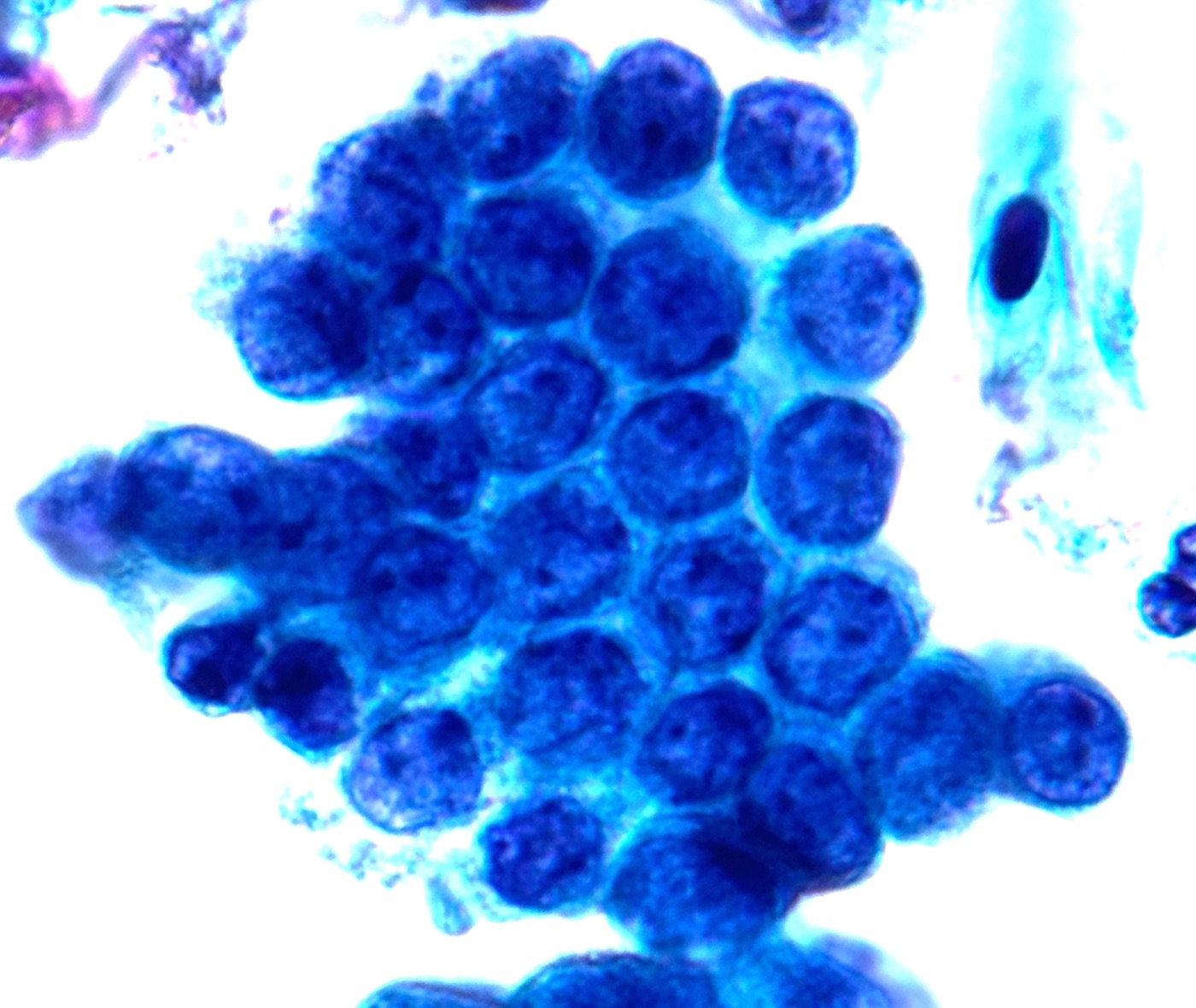
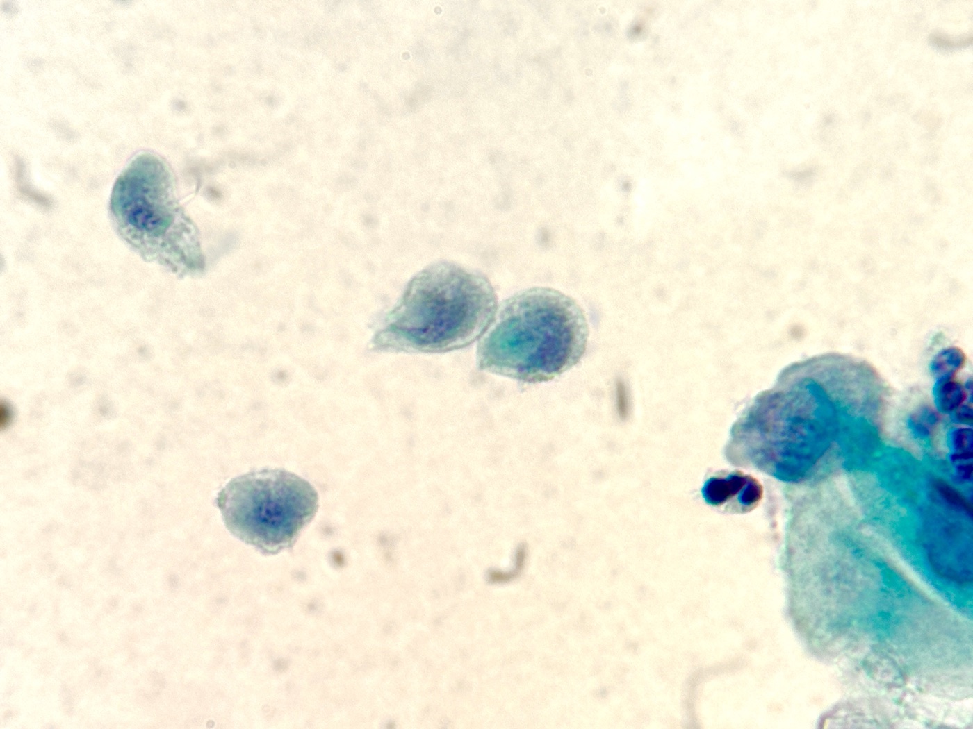
Reactive Cellular Changes in Cervical Cytology
Definition. Reactive cellular changes are benign cellular changes caused by inflammation, repair, radiation or an intrauterine contraceptive device (IUD). The Bethesda System, TBS 2014 (3rd edition), reports them under negative for intraepithelial lesion or malignancy (NILM), as non-neoplastic findings that are optional to report. The term "atypical" is reserved for ASC and AGC, so it is never used for reactive changes.
Clinical features of reactive cellular changes in cervical cytology
- Settings: infection, cervicitis, recent biopsy or excision, pelvic radiation and IUD use.
- Significance: reactive changes are benign. The diagnostic task is to separate them from ASC, SIL, AGC and carcinoma.
Bethesda subtypes (under NILM)
- reactive cellular changes associated with inflammation (includes typical repair)
- lymphocytic (follicular) cervicitis
- reactive cellular changes associated with radiation
- reactive cellular changes associated with an IUD
Cytomorphology of reactive cellular changes in cervical cytology
Inflammation (squamous and endocervical cells)
- Nuclear enlargement: about 1.5–2 times the area of a normal intermediate squamous cell nucleus.
- Nuclei: round to oval, smooth contours, evenly distributed fine chromatin; slight hyperchromasia is acceptable.
- Nucleoli: single or multiple, sometimes prominent.
- Occasional binucleation or multinucleation.
- Cytoplasm: polychromasia, vacuoles, and small perinuclear halos without the sharp peripheral thickening of koilocytes (Atlas Fig 2.36).
- Background: neutrophils, sometimes with an organism such as Trichomonas vaginalis.
- Reactive endocervical cells can show more nuclear size variation and conspicuous nucleoli. Flat sheets, smooth nuclear contours and admixed neutrophils support a benign process (Atlas Figs 2.32 and 2.33).
Typical repair
- Architecture: flat, cohesive monolayer sheets with nuclear polarity maintained, in a streaming pattern.
- Few single cells. Intact cell borders.
- Nuclei: enlarged, smooth, with fine chromatin and prominent nucleoli; mitoses can be present.
- Settings: after biopsy or excision, cervicitis, IUD use, and other epithelial injury.
- Atypical repair: reparative changes with significant overlap, dyscohesion, pleomorphism or loss of polarity are reported as ASC-US, or as ASC-H if invasive carcinoma is a morphologic consideration (Atlas Fig 4.18). Atypical repair in glandular cells is reported as AGC, according to the cell type (Atlas Fig PT.9).
Radiation
- Cytomegaly with proportional nuclear enlargement, so the nuclear-to-cytoplasmic (N/C) ratio is normal to low because of the increased cytoplasm (Atlas Fig 2.43). In liquid-based preparations the cytoplasm rounds up, and the apparent N/C ratio is slightly higher (Atlas Fig 2.44).
- Bi- and multinucleation, bizarre cell shapes, and prominent nucleoli.
- Chromatin: smudgy, or granular and evenly distributed (Atlas Fig 2.43). Slight hyperchromasia can be present.
- Cytoplasm: vacuoles and two-tone (polychromatic) staining.
- Other agents: some chemotherapeutic agents cause similar cytomegaly.
IUD-associated changes
- Small clusters of glandular cells (about 5–15 cells) with abundant vacuolated cytoplasm. A large vacuole can displace the nucleus and produce a signet-ring appearance.
- Single epithelial cells with a high N/C ratio, which resemble HSIL cells. Nucleoli can be prominent (Atlas Fig 2.47).
- Background: usually clean. Calcified debris and Actinomyces-like organisms can be present.
- Source: endometrial or endocervical cells altered by chronic irritation from the device.
Lymphocytic (follicular) cervicitis
- Polymorphous lymphoid cells with tingible-body macrophages. The finding resembles lymphoma and high-grade lesions in liquid-based preparations.
Differential diagnosis of reactive cellular changes in cervical cytology
| Entity | Features favoring the entity | Features favoring the reactive process |
|---|---|---|
| Nonkeratinizing squamous cell carcinoma | Many single cells, syncytia with loss of polarity, irregular coarse chromatin, macronucleoli, tumor diathesis | Cohesive flat sheets, polarity, smooth membranes, fine chromatin, no single atypical cells |
| ASC-US | Nuclei about 2.5–3 times an intermediate nucleus, slight hyperchromasia, irregular chromatin | Nuclei 1.5–2 times, smooth, fine chromatin |
| LSIL | Nuclear enlargement more than 3 times, hyperchromasia, raisinoid nuclei, koilocytes with sharp cavities | Small perinuclear halos without peripheral cytoplasmic thickening |
| HSIL (versus IUD cells) | Many single cells with coarse chromatin and irregular membranes, no nucleoli | Few single cells with nucleoli, vacuolated glandular clusters, IUD history |
| Adenocarcinoma (versus IUD cells) | Many cells, crowded three-dimensional groups, macronucleoli, tumor diathesis | Few small clusters, vacuoles |
| Recurrent carcinoma (versus radiation) | High N/C ratio, coarse chromatin, tumor diathesis | Cytomegaly with a normal to low N/C ratio, smudged chromatin, vacuoles |
| Herpes simplex virus | Multinucleation with molding, margination and ground-glass chromatin, Cowdry A inclusions | Multinucleated cells without molding or ground-glass nuclei |
| Multinucleated histiocytes (versus HSV) | — | Foamy or finely vacuolated cytoplasm, bean-shaped nuclei without molding or margination (Atlas Fig 2.27) |
| Cytomegalovirus | Large single basophilic intranuclear inclusion in glandular or stromal cells (Atlas Fig 2.64) | No viral inclusions |
| Atrophy with inflammation ("postmenopausal atypia") | — | Mild nuclear enlargement without hyperchromasia in parabasal-type cells is NILM (Atlas Fig 4.19) |
Ancillary testing in reactive cellular changes in cervical cytology
- Under ASCCP 2019, management of a reactive NILM result depends on the HPV result. Reflex HPV testing is not indicated for NILM.
- p16/Ki-67 dual stain is validated only for triage of HPV-positive screening results in asymptomatic patients (Clarke 2024; FDA PMA P190024). It is not validated for radiation-altered or other reactive cells.
- No immunostain defines a reactive change. Diagnosis is morphologic.
Management of reactive cellular changes in cervical cytology
- A reactive change does not alter management. Follow-up depends on the HPV result and history, as for any NILM result.
- NILM with a negative HPV test: return to routine screening.
- NILM with a positive HPV test: repeat HPV-based testing in 1 year, or colposcopy for HPV 16 or 18 positivity. Dual stain is an acceptable triage option for other high-risk types and for HPV-positive results without genotyping (Clarke 2024).
- dual stain positive: colposcopy
- dual stain negative: 1-year HPV-based follow-up
- Atypical repair reported as ASC or AGC follows the guideline for that category.
- Specific organisms are treated clinically. TBS states that Actinomyces-like organisms are usually seen with an IUD and that their clinical significance remains unclear. In an asymptomatic patient, management is expectant and the IUD can remain in place.
Atypical Squamous Cells of Undetermined Significance (ASC-US)
Definition. ASC-US comprises cytologic changes suggestive of a squamous intraepithelial lesion (SIL) that are quantitatively or qualitatively insufficient for a definitive interpretation. ASC-US is one of the two subcategories of atypical squamous cells (ASC) in the Bethesda System, TBS 2014 (3rd edition). It is an interpretation of uncertainty and has no single histologic counterpart. Most ASC-US results reflect reactive change, HPV infection or LSIL, and a minority reflect HSIL.
Clinical features of atypical squamous cells of undetermined significance (ASC-US)
- Frequency: ASC-US is the most common epithelial cell abnormality in cervical cytology.
- Significance: management depends on the HPV result, the patient's age and prior history (see Management linkage).
Cytomorphology of atypical squamous cells of undetermined significance (ASC-US) (TBS 2014 criteria)
- Cell type: mature squamous cells (superficial or intermediate). Atypical squamous metaplastic cells are included when the change suggests LSIL (TBS 2014 Online Atlas, Fig 4.14). Atypia in immature metaplastic cells is ASC-H.
- Nuclear size: about 2.5–3 times the area of a normal intermediate squamous cell nucleus (about 35 µm²), or about twice the area of a squamous metaplastic cell nucleus (about 50 µm²).
- N/C ratio: slightly increased.
- Nuclei: minimal hyperchromasia and slight irregularity in chromatin distribution or nuclear shape. Contour irregularity is permitted and is not required.
- Atypical parakeratosis: nuclear abnormalities in cells with dense orangeophilic cytoplasm are included (Atlas Figs 4.15 and 4.16).
- Liquid-based preparations: the cytoplasm rounds up, so the apparent N/C ratio is slightly higher than in conventional smears (Atlas Fig 2.44).
Common patterns
- intermediate cells with nuclear enlargement and slight hyperchromasia, short of LSIL
- incomplete koilocytes (perinuclear halos with limited nuclear atypia)
- atypical parakeratosis (small orangeophilic cells with enlarged, hyperchromatic nuclei)
- atypia in atrophic specimens: nuclear enlargement with hyperchromasia and irregular contours in a postmenopausal patient. Mild enlargement without hyperchromasia is NILM (Atlas Fig 4.19).
- atypical repair: reparative changes with significant overlap, dyscohesion, pleomorphism or loss of polarity (Atlas Fig 4.18)
Laboratory benchmarks
- ASC rate: the CAP 2006 survey median ASC-US rate was 4.3%; the total ASC rate is about 4–5% (Eversole 2010).
- ASC:SIL ratio: the CAP 2006 median ratio of ASC to SIL plus carcinoma was 1.5, up from 1.4 in 2003 (Eversole 2010). A ratio below 3:1 is a commonly used upper limit.
- HPV-positive rate of ASC-US: about 35–55%. The CAP 2006 laboratory median was 36.6% (Moriarty 2008), with similar rates in 2012 (Zhao 2015). In the KPNC cotest cohort, aged 25–65, it was 55% (Egemen 2020, Table 1A).
- Monitoring of each cytologist's ASC rate and HPV-positive rate of ASC-US is a quality-assurance tool.
Differential diagnosis of atypical squamous cells of undetermined significance (ASC-US)
| Entity | Key distinguishing features |
|---|---|
| Reactive changes (NILM) | Nuclei 1.5–2 times an intermediate nucleus, smooth contours, fine chromatin, nucleoli |
| LSIL | Nuclei more than 3 times an intermediate nucleus with definite hyperchromasia and irregular contours; koilocytes with sharp cavities and peripheral cytoplasmic thickening; binucleation |
| ASC-H | Atypia in immature (metaplastic-size) cells with high N/C ratios |
| Atrophy (NILM) | Uniform parabasal-type cells with fine chromatin; blue blobs; nuclear enlargement without hyperchromasia |
| Trichomonas halos (NILM) | Small perinuclear halos with mild nuclear enlargement, organisms present |
| Parakeratosis (NILM) | Small orangeophilic cells with small dense pyknotic nuclei, without enlargement |
| Air-drying artifact (conventional smears) | Pale, enlarged, poorly defined nuclei and cytoplasmic staining artifacts |
Ancillary testing in atypical squamous cells of undetermined significance (ASC-US)
- Reflex HPV testing on the residual liquid-based specimen is the preferred triage of ASC-US. The ASCUS-LSIL Triage Study (ALTS) established HPV triage:
- a single enrollment HPV test (Hybrid Capture 2) identified 92.4% of women with CIN 3
- the HPV triage arm referred 55.6% of women to colposcopy
- immediate colposcopy had a sensitivity of 53.6% for cumulative CIN 3 over 2 years
- HPV assays: DNA assays include Hybrid Capture 2, Cervista, cobas, BD Onclarity and Alinity m; Aptima detects mRNA. Genotyping differs by assay: cobas reports HPV 16 and 18, Aptima reports HPV 16 and pooled 18/45, and Onclarity reports extended genotypes.
- p16/Ki-67 dual stain (Clarke 2024): in cotesting, dual stain is acceptable for HPV-positive NILM, ASC-US or LSIL.
- dual stain positive: colposcopy
- dual stain negative: 1-year return, for HPV results without genotyping or positive for the 12 other high-risk types
- HPV 16 or 18 positive: colposcopy regardless of the dual-stain result
- The original FDA label for cotesting covers only HPV-positive NILM at ages 30–65 (PMA P190024). Use with ASC-US is supported by the guideline.
Management of atypical squamous cells of undetermined significance (ASC-US) (ASCCP 2019 and updates)
| Setting | Recommended management |
|---|---|
| ASC-US, HPV positive, 25 years or older (immediate CIN 3+ risk 4.4%; 5-year risk 7.3%) | Colposcopy |
| ASC-US, HPV negative, 25 years or older (5-year CIN 3+ risk 0.40%) | Repeat HPV-based testing in 3 years |
| ASC-US, HPV result unavailable, 25 years or older | Reflex HPV testing preferred. Patients still screened with cytology alone follow the 2012 guidelines, under which repeat cytology in 1 year is acceptable (Perkins 2024; Massad 2013) |
| ASC-US, HPV positive, after a documented negative HPV-based screen within about 5 years (immediate CIN 3+ risk 2.0%; 5-year risk 3.8%) | Repeat HPV-based testing in 1 year (Egemen 2020, Table 1B; Perkins 2020, section E.1) |
| ASC-US, ages 21–24 | HPV-positive ASC-US, or ASC-US without HPV testing: repeat cytology alone at 1 and 2 years. Reflex HPV negative: repeat cytology in 3 years. Colposcopy for high-grade cytology at any point, or for low-grade cytology that persists at 2 years (Perkins 2020, section K.1) |
| ASC-US, pregnant patient | Colposcopy at the same clinical action thresholds as for nonpregnant patients; endocervical curettage, endometrial biopsy and treatment without biopsy are unacceptable (Perkins 2020, section K.2) |
| ASC-US, immunocompromised patient | Colposcopy for all HPV-positive ASC-US. If HPV testing is not performed, repeat cytology in 6–12 months (Perkins 2020, section K.3) |
- Colposcopy is recommended at an immediate CIN 3+ risk of ≥4%. The risk values are from Egemen 2020, Table 1A (HPV positive, unknown history).
- A second consecutive HPV-positive result after HPV-negative ASC-US leads to colposcopy.
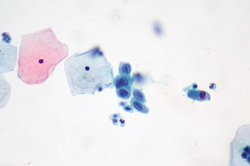
Atypical Squamous Cells, Cannot Exclude HSIL (ASC-H)
Definition. ASC-H comprises cytologic changes suggestive of a high-grade squamous intraepithelial lesion (HSIL) that are insufficient for a definitive interpretation. ASC-H is the second subcategory of atypical squamous cells (ASC) in the Bethesda System, TBS 2014 (3rd edition). The cells are few, immature, or both. ASC-H has a higher risk of CIN 2+ than ASC-US and is managed with colposcopy regardless of the HPV result.
Cytomorphology of atypical squamous cells, cannot exclude HSIL (ASC-H) (TBS 2014 criteria)
- Cell size: small cells, about the size of squamous metaplastic cells.
- Arrangement: single cells or small fragments of fewer than 10 cells. In liquid-based preparations the cells are mainly single and can be scarce.
- Nuclei: about 1.5–2.5 times the area of a normal metaplastic cell nucleus (TBS 2014 Online Atlas, Fig 4.20).
- N/C ratio: high, approaching that of HSIL.
- Chromatin and contour: hyperchromasia, irregular chromatin and focal irregularity of nuclear shape, which fall short of definitive HSIL.
Patterns
- Atypical immature metaplasia: small metaplastic-type cells with dense cytoplasm, high N/C ratios and slight hyperchromasia.
- Crowded sheet pattern: crowded groups with loss of polarity, where the individual cells are difficult to assess.
- Atypical repair: reported as ASC-H when invasive carcinoma is a morphologic consideration; other atypical repair is ASC-US (Atlas Fig 4.18).
- Post-radiation atypia: nuclear abnormality beyond the expected radiation effect.
- Atypia in atrophy: parabasal-type cells with hyperchromasia and irregular contours. Repeat sampling after topical estrogen, or adjunctive HPV testing, can clarify ASC-H versus atrophy (Atlas Figs 4.28 and PT.32).
- Atypical parakeratosis: small orangeophilic cells with markedly atypical nuclei.
Clinical and laboratory data
- Significance: ASC-H has a higher risk of CIN 2+ than ASC-US, and every ASC-H result leads to colposcopy.
- Proportion of ASC: ASC-H makes up about 5–10% of all ASC interpretations. In the KPNC cotest cohort, 4,557 of 60,394 ASC results (about 7.5%) were ASC-H (Egemen 2020, Table 1A).
- Outcome (KPNC): the immediate CIN 3+ risk is about 20% for all ASC-H (889 of 4,557) and 26% for HPV-positive ASC-H. HPV-positive ASC-H has an immediate CIN 2+ risk of 50% (Egemen 2020, Table 1A; Perkins 2020, section E.3).
- HPV positivity: about 50–85%. The CAP 2006 laboratory median was 50.0% (Moriarty 2008), and the KPNC rate was 83% (3,766 of 4,557; Egemen 2020, Table 1A).
Differential diagnosis of atypical squamous cells, cannot exclude HSIL (ASC-H)
| Entity | Key distinguishing features |
|---|---|
| HSIL | Definite and more numerous high-N/C cells with coarse chromatin and marked contour irregularity |
| ASC-US | Atypia in mature (intermediate or superficial) cells with lower N/C ratios |
| Squamous metaplasia (NILM) | Uniform cells with dense cytoplasm, smooth nuclei and fine chromatin; cytoplasmic processes in some cells |
| Atrophy (NILM) | Flat sheets of uniform parabasal-type cells, fine chromatin, smooth contours, blue blobs, granular background |
| Transitional metaplasia (NILM) | Streaming sheets of cells with longitudinal grooves, perinuclear halos and fine chromatin |
| Histiocytes | Bean-shaped eccentric nuclei with foamy cytoplasm, single and loosely aggregated (Atlas Fig PT.35) |
| Endometrial cells | Small three-dimensional clusters with scant cytoplasm and degenerated nuclei, which are much smaller than HSIL nuclei (Atlas Fig 4.32) |
| Endocervical cells seen end-on | High apparent N/C ratio; apical mucin appears on focusing up and down (Atlas Fig 4.34) |
| Tubal metaplasia (NILM) | Cilia and terminal bars in crowded groups |
| Lymphocytic (follicular) cervicitis | Polymorphous lymphoid cells with tingible-body macrophages |
| IUD-associated changes | Single high-N/C cells with nucleoli and vacuolated glandular clusters in a patient with an IUD |
Ancillary testing in atypical squamous cells, cannot exclude HSIL (ASC-H)
- HPV testing refines the risk estimate, and colposcopy is recommended with either result.
- p16/Ki-67 dual stain does not change management. Colposcopy is recommended regardless of the result.
- p16 immunohistochemistry on the colposcopic biopsy follows LAST criteria. Block-type p16 staining supports HSIL in biopsies with an H&E differential of HSIL versus its mimics.
Management of atypical squamous cells, cannot exclude HSIL (ASC-H) (ASCCP 2019 and updates)
- ASC-H at any age, with any HPV result: colposcopy (AII; Perkins 2020, section H.2).
- immediate CIN 3+ risk: 26% with a positive HPV test and 3.4% with a negative HPV test; 5-year risks are 33% and 3.8% (Egemen 2020, Table 1A)
- rationale: HPV-negative ASC-H has an immediate CIN 3+ risk of 3.4% but an immediate cancer risk of 0.69%, against 0.92% for HPV-positive ASC-H. Because the immediate cancer risk is disproportionately high compared with the CIN 3+ risk, colposcopy is recommended for all ASC-H, as in the 2012 guidelines (Perkins 2020, section H.2).
- HPV-negative ASC-H after a prior negative HPV screen has an immediate CIN 3+ risk of 2.8% and still leads to colposcopy (Egemen 2020, Table 1B).
- Expedited treatment: HPV-positive ASC-H (immediate CIN 3+ risk 26%) falls in the band of 25% to <60%. For nonpregnant patients 25 years or older in this band, colposcopy with biopsy and expedited treatment are both acceptable after shared decision-making. Expedited treatment is preferred only at an immediate CIN 3+ risk of ≥60%, for example HPV16-positive HSIL (Perkins 2020, section E.3; Egemen 2020, Table 1A).
- Patients aged 21–24: colposcopy. After colposcopy showing less than CIN 2, cytology is repeated at 1 year, with colposcopy for ASC-H or higher (Perkins 2020, section K.1).
- Pregnant patients: colposcopy at the same clinical action thresholds as for nonpregnant patients. Endocervical curettage is unacceptable (EIII), and expedited treatment is not performed in pregnancy (Perkins 2020, section K.2).
- CIN 1 or less after ASC-H (Perkins 2020, section I.4):
- Review of the cytologic, histologic and colposcopic findings is acceptable (CIII).
- Observation with HPV-based testing at 1 year is recommended if colposcopy fully visualized the squamocolumnar junction and the upper limit of any lesion and any endocervical sampling was negative. A diagnostic excisional procedure is not recommended (BII).
- Negative tests at 1 year lead to repeat testing at 2 years. Persistent ASC-H at 2 years leads to a diagnostic excisional procedure.
Low-Grade Squamous Intraepithelial Lesion (LSIL)
Definition. LSIL is the Bethesda System category for squamous cells that show the cytopathic effect of human papillomavirus (HPV) or mild dysplasia. It includes the older terms koilocytic atypia, condyloma, mild dysplasia and cervical intraepithelial neoplasia grade 1 (CIN 1). Its histologic counterpart is LSIL in the two-tier Lower Anogenital Squamous Terminology (LAST). The criteria below follow TBS 2014 (3rd edition).
Clinical features and biology
- LSIL represents a productive HPV infection. Viral assembly takes place in maturing squamous cells, so the abnormal cells have mature (intermediate- or superficial-type) cytoplasm.
- High-risk and low-risk HPV types both cause LSIL. Low-risk types 6 and 11 cause condyloma acuminatum.
- Most LSIL and CIN 1 lesions regress, especially in young patients. In patients aged 13–22 years, LSIL regressed in 61% at 12 months and in 91% at 36 months (Moscicki et al. 2004).
- Cytology cannot predict which HPV type is present. HPV testing and genotyping supply that information.
Cytomorphology of low-grade squamous intraepithelial lesion (LSIL) in cervical cytology (TBS 2014 criteria)
- Pattern: cells occur singly or in sheets. The abnormal cells are mature squamous cells with abundant, well-defined cytoplasm.
- Nuclear criteria (required):
- nuclear enlargement more than 3 times the area of a normal intermediate-cell nucleus
- mild to moderate variation in nuclear size and shape
- binucleation and multinucleation, which are common
- hyperchromasia of variable degree, with evenly distributed chromatin; the chromatin is coarsely granular, smudged or opaque in some cells
- nucleoli absent or inconspicuous
- nuclear membranes slightly irregular or smooth
- Koilocytosis: a sharply outlined, clear perinuclear cavity with a dense, peripheral rim of cytoplasm. Koilocytes are characteristic of LSIL. Their presence is optional for the diagnosis.
- Perinuclear clearing alone does not qualify. A cavity without the nuclear changes above is reported as NILM.
- Keratinized LSIL cells: cells with dense orangeophilic cytoplasm qualify when they show the same nuclear abnormalities.
- Liquid-based preparations: the same criteria apply. Abnormal cells are dispersed among normal cells. Nuclear-size comparison with adjacent normal intermediate cells on the same slide is the most practical reference.
Differential diagnosis of low-grade squamous intraepithelial lesion (LSIL) in cervical cytology
| Entity | Distinguishing features | Bethesda report |
|---|---|---|
| Glycogenated intermediate cells | Yellow, glycogen-filled cytoplasm; ragged halo edge; normal-sized nuclei | NILM |
| Inflammatory halos (for example, Trichomonas vaginalis) | Small, narrow halos with indistinct edges; slight nuclear enlargement with smooth membranes | NILM, organism |
| ASC-US | Nuclear enlargement about 2.5–3 times a normal intermediate nucleus; slight hyperchromasia; features quantitatively short of LSIL | ASC-US |
| HSIL | Smaller, less mature cells; high N/C ratio; marked hyperchromasia; deep membrane grooves and indentations | HSIL |
| Coexisting LSIL and a few immature atypical cells | Definite LSIL plus rare small cells with a high N/C ratio | LSIL and ASC-H, both reported. "LSIL, cannot exclude HSIL" is not a TBS 2014 category |
| Herpes simplex virus effect | Multinucleation with nuclear molding, ground-glass chromatin with margination, intranuclear inclusions | NILM, organism |
| Radiation effect | Cytomegaly with a preserved N/C ratio; cytoplasmic vacuoles and polychromasia; multinucleation | NILM, reactive changes |
Ancillary testing in low-grade squamous intraepithelial lesion (LSIL) in cervical cytology
- HPV testing: reflex high-risk HPV testing on the residual liquid-based specimen is used for management in patients 25 years and older. HPV testing is not recommended for patients 21–24 years with LSIL, because the result does not change management.
- HPV genotyping: HPV 16 and 18 have the highest risk. The 2019 guidelines incorporate partial genotyping into risk estimates.
- p16 immunocytochemistry: outside the TBS 2014 criteria. LSIL is a morphologic diagnosis.
- p16/Ki-67 dual stain: a cytology-based biomarker test for triage of HPV-positive results. A positive result is at least one cell that co-expresses cytoplasmic p16 and nuclear Ki-67, whatever its morphology (assay labeling; Wentzensen et al. 2015). The Enduring Consensus Cervical Cancer Screening and Management Guidelines Committee (ASCCP-led) states that dual stain is acceptable for triage of HPV-positive results. The recommendation applies only to the FDA-approved CINtec PLUS Cytology assay (Clarke et al. 2024).
- Dual-stain triage of HPV-positive LSIL (Clarke et al. 2024):
- HPV 16 or 18 positive: colposcopy, whatever the dual-stain result
- other high-risk types, or HPV positive without genotyping: dual stain positive leads to colposcopy; dual stain negative leads to a 1-year return
- dual stain is not recommended for ASC-H, AGC or HSIL cytology
- Self-collected vaginal HPV samples (Wentzensen et al. 2025): clinician-collected cervical specimens are preferred, and self-collection is acceptable for primary screening. Cytology and dual stain cannot be performed on a self-collected sample.
- HPV 16 or 18 positive: colposcopy, with cytology and biopsies taken at that visit
- HPV 45, 33/58, 31, 52, 35/39/68, 51 or pooled other types (HPV 16/18 negative): clinician-collected cytology or dual stain for triage
- HPV 56/59/66 alone: repeat HPV testing in 1 year
- p16 in follow-up biopsies (LAST): LAST recommends against routine p16 for a biopsy with a morphologic interpretation of negative, CIN 1 or CIN 3. Any p16-positive area must meet H&E criteria for a high-grade lesion before it is reinterpreted. A morphologic LSIL with block-positive p16 therefore remains LSIL (Darragh et al. 2012).
Management of low-grade squamous intraepithelial lesion (LSIL) in cervical cytology (ASCCP 2019 risk-based guidelines and updates)
- Principle: management follows the patient's immediate risk of CIN 3+, estimated from the current result and prior history. Colposcopy is recommended at an immediate CIN 3+ risk of ≥4%.
- HPV-positive LSIL, unknown screening history (age ≥25): colposcopy. The immediate CIN 3+ risk is 4.3%, and the 5-year risk is 6.9% (Egemen et al. 2020, Table 1A).
- HPV-positive LSIL after a documented negative HPV test in the prior screening round: 1-year follow-up. The immediate CIN 3+ risk is 2.1% (Egemen et al. 2020, Table 1B).
- HPV-positive LSIL with a negative dual stain (non-16/18 types or no genotyping): 1-year return (Clarke et al. 2024).
- HPV-negative LSIL: repeat HPV-based testing in 1 year, whatever the history. The immediate CIN 3+ risk is 1.1% with unknown history and 0.44% after a prior negative HPV test (Egemen et al. 2020, Tables 1A and 1B).
- Age 21–24 years: repeat cytology alone at 1 and 2 years. Colposcopy follows for HSIL, ASC-H, AGC or AIS at any point, and for ASC-US or worse at 2 years.
- Pregnancy: management uses the same clinical action thresholds as for nonpregnant patients, so HPV-positive LSIL leads to colposcopy. Endocervical curettage, endometrial biopsy and treatment without biopsy are unacceptable. Postpartum colposcopy takes place no earlier than 4 weeks after delivery (Perkins et al. 2020, Section K.2).
- After colposcopy with CIN 1 or less: HPV-based testing in 1 year.
- Histologic CIN 1 after a low-grade referral: observation is preferred. Treatment is acceptable only for CIN 1 that persists for at least 2 years (Perkins et al. 2020).
High-Grade Squamous Intraepithelial Lesion (HSIL)
Definition. HSIL is the Bethesda System category for squamous precursor lesions with a high risk of progression to invasive carcinoma. It includes moderate dysplasia, severe dysplasia and carcinoma in situ, which correspond to CIN 2 and CIN 3. Its histologic counterpart is HSIL in the two-tier LAST terminology. The criteria below follow TBS 2014 (3rd edition).
Clinical features and biology
- HSIL is a transforming HPV infection. High-risk HPV E6 and E7 oncoproteins inactivate p53 and retinoblastoma protein (pRb), which causes cell-cycle dysregulation and p16 overexpression.
- HPV 16 is the most common type in HSIL and in squamous cell carcinoma.
- Cytology cannot separate CIN 2 from CIN 3 reliably. Bethesda therefore reports a single HSIL category.
Cytomorphology of high-grade squamous intraepithelial lesion (HSIL) in cervical cytology (TBS 2014 criteria)
- Cell size and maturity: HSIL cells are smaller and less mature than LSIL cells. Nuclear size overlaps with LSIL. The cytoplasmic area is smaller, so the N/C ratio is high.
- Arrangement: single cells, sheets, or syncytial aggregates (hyperchromatic crowded groups).
- Nuclear criteria:
- marked hyperchromasia; some nuclei are normochromatic or hypochromatic
- fine or coarsely granular chromatin, evenly distributed
- irregular nuclear contours, with prominent indentations or grooves
- nucleoli usually absent; they appear when HSIL extends into endocervical glands or in an inflamed background
- Cytoplasm: lacy and delicate (immature), or dense and metaplastic. It is occasionally mature and densely keratinized (keratinizing HSIL).
Patterns
| Pattern | Features | Main mimics |
|---|---|---|
| Dysplastic-cell (metaplastic) pattern | Moderate amounts of dense cytoplasm; clear-cut nuclear enlargement and membrane irregularity | Immature squamous metaplasia, ASC-H |
| Small-cell pattern | Basal-type cells near the size of metaplastic or parabasal cells; scant cytoplasm; very high N/C ratio; single or in groups | Histiocytes, endometrial cells, lymphocytes, intrauterine device (IUD) cells |
| Hyperchromatic crowded groups | Syncytial groups with loss of polarity and disordered, overlapping nuclei; peripheral cells flattened | Endometrial cells, adenocarcinoma in situ (AIS), atrophy, squamous cell carcinoma |
| Keratinizing pattern | Pleomorphic keratinized cells with dense orangeophilic cytoplasm and dark nuclei; atypical parakeratosis | Keratinizing squamous cell carcinoma |
- Liquid-based preparations: abnormal cells are fewer and more often single (Alrajjal et al. 2021). Small-cell HSIL can be missed at low magnification.
- HSIL in endocervical glands: groups with peripheral flattening of nuclei, dense cytoplasm and no columnar features. This pattern mimics AIS (see endocervical-ais).
HSIL with features suspicious for invasion
- TBS 2014 allows the qualifier "HSIL with features suspicious for invasion" when the cells suggest invasion but the features fall short of a definite squamous cell carcinoma diagnosis.
- Suggestive features include:
- focal tumor diathesis (granular necrotic debris, lysed blood, fibrin)
- coarse, irregularly distributed chromatin
- nucleoli or macronucleoli
- bizarre or markedly keratinized cell shapes, such as tadpole or fiber cells
- The qualifier indicates that colposcopy should include evaluation for invasive carcinoma.
Differential diagnosis of high-grade squamous intraepithelial lesion (HSIL) in cervical cytology
| Entity | Distinguishing features | Bethesda report |
|---|---|---|
| ASC-H | Few atypical immature cells; features equivocal for HSIL | ASC-H |
| Immature squamous metaplasia | Cobblestone sheets; uniform round nuclei; smooth membranes; fine pale chromatin; cytoplasmic processes | NILM |
| Atrophy | Flat sheets of parabasal cells with uniform nuclei; preserved polarity; no membrane irregularity | NILM |
| Histiocytes | Eccentric, bean-shaped nuclei; fine pale chromatin; foamy cytoplasm; CD68 positive | NILM |
| Endometrial cells | Tight three-dimensional balls; small uniform nuclei; apoptosis; scant vacuolated cytoplasm | NILM (report if age ≥45) |
| IUD-associated cells | Single cells with high N/C ratios and large cytoplasmic vacuoles; history of IUD | NILM, reactive changes |
| Follicular cervicitis | Polymorphous lymphoid cells; tingible-body macrophages | NILM |
| Endocervical AIS | Columnar cells; feathering, rosettes and strips; elongated stratified nuclei | AIS |
| Squamous cell carcinoma | Macronucleoli; irregularly distributed, clumped chromatin; tumor diathesis; bizarre shapes | Squamous cell carcinoma |
Ancillary testing in high-grade squamous intraepithelial lesion (HSIL) in cervical cytology
- HPV testing: an HSIL interpretation requires colposcopy or treatment whatever the HPV result. The HPV result changes the risk estimate, and evaluation is required with either result.
- p16/Ki-67 dual stain: HSIL cells co-express p16 and Ki-67. Dual stain is a triage test for HPV-positive screening results. Its use with ASC-H, AGC or HSIL cytology is not recommended, and a result obtained in that setting does not guide management (Clarke et al. 2024).
- p16 in biopsies (LAST):
- block-positive p16 is strong, continuous staining of the basal layer extending upward over at least the lower third of the epithelium and laterally over a significant distance
- p16 is recommended to separate HSIL from mimics (immature metaplasia, atrophy, repair, tangential sectioning)
- p16 is recommended to classify a lesion with CIN 2 morphology: block-positive is HSIL, negative or patchy is LSIL
- p16 is recommended when a biopsy read as LSIL or less follows a cytology result with a high risk of missed high-grade disease: HSIL, ASC-H, ASC-US/HPV 16-positive or AGC (NOS)
- any p16-positive area must meet H&E criteria for a high-grade lesion before it is reinterpreted as HSIL (Darragh et al. 2012)
- Cytology-histology correlation: an HSIL cytology result with a negative or low-grade biopsy requires review of the cytology, histology and colposcopy findings.
Management of high-grade squamous intraepithelial lesion (HSIL) in cervical cytology (ASCCP 2019 risk-based guidelines and updates)
- Risk thresholds (nonpregnant, age ≥25): expedited treatment is preferred at an immediate CIN 3+ risk of ≥60% and acceptable at 25% to <60%. Expedited treatment means an excisional procedure without a confirmatory biopsy.
- HPV-positive HSIL, any genotype, unknown history: the immediate CIN 3+ risk is 49% (CIN 2+ 77%). At this risk, expedited treatment and colposcopy with biopsy are both acceptable (Egemen et al. 2020, Table 1A; Perkins et al. 2020, Section E.3).
- Expedited treatment is preferred in two situations:
- HPV 16-positive HSIL: immediate CIN 3+ risk 60%, cancer risk 8.1%
- HPV-positive HSIL in a patient who is rarely or never screened (usually no screening in more than 5 years): immediate CIN 3+ risk 64%
- HPV-negative HSIL: with unknown history, the immediate CIN 3+ risk is 25%, and colposcopy or expedited treatment is acceptable. After a documented prior negative HPV screen, HPV-negative HSIL has a risk of 14% and leads to colposcopy, and HPV-positive HSIL has a risk of 32% (colposcopy or expedited treatment) (Egemen et al. 2020, Table 1B).
- Age 21–24: colposcopy. Treatment without histologic confirmation is not recommended (Perkins et al. 2020, Section K.1).
- HSIL cytology with CIN 1 or less on biopsy: observation with colposcopy and cytology at 1 and 2 years. Diagnostic excision is recommended if HSIL persists for 2 years or the squamocolumnar junction is not fully seen.
- Histologic HSIL: observation is preferred for CIN 2, and treatment is recommended for CIN 3.
- Pregnancy: colposcopy. Endocervical curettage, endometrial biopsy and treatment without biopsy are unacceptable. Treatment is deferred unless invasive cancer is suspected. Histologic HSIL diagnosed in pregnancy is followed by colposcopy every 12–24 weeks (preferred), or colposcopy is deferred to the postpartum period (acceptable). Postpartum re-evaluation takes place no earlier than 4 weeks after delivery (Perkins et al. 2020, Section K.2).
- HSIL cytology with CIN 1 or less on biopsy (age ≥25): an immediate diagnostic excisional procedure and observation with HPV-based testing and colposcopy at 1 year are both acceptable. Observation requires that colposcopy fully showed the squamocolumnar junction and the upper limit of any lesion, and that any endocervical sample was less than CIN 2. Review of the cytology, histology and colposcopy findings is also acceptable. A diagnostic excision is recommended for HSIL cytology at the 1-year or 2-year visit (Perkins et al. 2020, Section I.4).
Squamous Cell Carcinoma in Cervical Cytology
Definition. Squamous cell carcinoma (SCC) is an invasive carcinoma with squamous differentiation. TBS 2014 (3rd edition) reports it as a single category and describes keratinizing and nonkeratinizing patterns. WHO Female Genital Tumours, 5th edition (2020), divides cervical SCC into HPV-associated and HPV-independent types, and accepts SCC, NOS when p16 or HPV testing is not performed.
Clinical features and classification
- Presentation: abnormal or postcoital bleeding, discharge or a visible cervical mass. Early tumors can be asymptomatic and found by screening.
- HPV-associated SCC accounts for most cases. HPV 16 is the most common type, followed by HPV 18.
- HPV-independent SCC is very rare, has no known precancerous lesion and is usually p16 negative. A subset shows TP53 mutation or aberrant p53 expression; other reported mutations (PIK3CA, STK11, GNAS) are inconsistent (Höhn et al. 2021; Regauer et al. 2022). No therapeutic or prognostic difference from HPV-associated SCC has been established for the cervix (Höhn et al. 2021).
- Cytologic clue to HPV status: none is reliable. p16 IHC or HPV testing is the only means.
- Grading: WHO 5th states that there is no established grading system for cervical SCC (Höhn et al. 2021).
- Staging: FIGO 2018 allows imaging and pathology findings to assign stage. Lateral extent no longer defines stage IA. Lymph node involvement is stage IIIC (Bhatla et al. 2019, with erratum).
Cytomorphology of squamous cell carcinoma in cervical cytology (TBS 2014 criteria)
Keratinizing pattern
- Relatively few cells, mostly single; aggregates are less common.
- Marked variation in cell size and shape: caudate (tadpole) cells, spindle cells and elongated fiber cells.
- Dense orangeophilic cytoplasm on the Pap stain; keratin pearls.
- Nuclei of variable size, often dark and opaque (pyknotic); coarsely granular, irregularly distributed chromatin where the chromatin is visible.
- Macronucleoli are less frequent than in the nonkeratinizing pattern.
- Hyperkeratosis or atypical parakeratosis can accompany the tumor.
- Tumor diathesis can be present. It is less prominent than in nonkeratinizing SCC.
Nonkeratinizing pattern
- Single cells or syncytial aggregates with poorly defined cell borders.
- Cells can be smaller than HSIL cells and share most HSIL features.
- Coarsely clumped, irregularly distributed chromatin with parachromatin clearing.
- Prominent nucleoli or macronucleoli.
- Tumor diathesis is common.
Tumor diathesis
- Composition: granular necrotic debris, lysed and old blood, fibrin and inflammatory cells.
- Conventional smears: diathesis spreads across the background and can obscure tumor cells.
- Liquid-based preparations: diathesis is reduced. It appears as clinging diathesis, which adheres to the edges of cell groups.
Differential diagnosis of squamous cell carcinoma in cervical cytology
| Entity | Distinguishing features | Bethesda report |
|---|---|---|
| Atrophy with granular background | Flat sheets of parabasal cells with uniform nuclei; granular basophilic background ("pseudodiathesis"); blue blobs; small degenerated orangeophilic cells with pyknotic nuclei | NILM |
| Keratinizing HSIL or atypical parakeratosis | Small keratinized cells with dense nuclei; no tumor diathesis; limited pleomorphism | HSIL or ASC-H, by degree (ASC-US when the change is mild) |
| HSIL with features suspicious for invasion | HSIL with focal diathesis or irregular chromatin; features short of carcinoma | HSIL with features suspicious for invasion |
| Repair | Flat, cohesive monolayer sheets with streaming; prominent nucleoli; smooth membranes; few single cells; no necrosis | NILM, reactive changes |
| Radiation effect | Cytomegaly with a preserved N/C ratio; vacuoles; polychromasia; history of radiation | NILM, reactive changes |
| Adenocarcinoma | Columnar or cuboidal cells; vacuolated or mucinous cytoplasm; eccentric nuclei; acinar or papillary groups | Adenocarcinoma |
| Small cell neuroendocrine carcinoma | Cells 2–3 times a lymphocyte; scant cytoplasm; nuclear molding; finely granular chromatin; crush artifact; necrosis | Other malignant neoplasm |
| Follicular cervicitis | Polymorphous lymphoid cells; tingible-body macrophages | NILM |
Ancillary testing in squamous cell carcinoma in cervical cytology
- HPV testing: a negative HPV test does not exclude SCC. Causes of a false-negative result include HPV-independent tumors, low viral load in necrotic tumors and sampling error.
- p16 immunohistochemistry: WHO 5th recommends p16 IHC as the surrogate for HPV status. Diffuse block-type staining supports HPV-associated SCC. HPV molecular testing is not required for diagnosis. HPV in situ hybridization (DNA or RNA) confirms HPV association when needed (Höhn et al. 2021).
- Squamous markers: p40 and p63 (nuclear), CK5/6.
- Separation from adenocarcinoma: mucin stains, CEA and an absence of p40 favor adenocarcinoma.
- Separation from small cell neuroendocrine carcinoma: synaptophysin, chromogranin and INSM1; a high Ki-67 index.
- Cell block: a residual liquid-based specimen can be used for a cell block when immunostaining is needed.
Management of squamous cell carcinoma in cervical cytology
- Cytology showing SCC: prompt tissue diagnosis, with biopsy of any visible lesion. The 2019 ASCCP risk tables include SCC within "HSIL+" cytology and give no separate SCC pathway. HSIL+ cytology leads to colposcopy or expedited treatment by risk band (Egemen et al. 2020; Perkins et al. 2020).
- Visible or palpable cervical lesion: biopsy is required whatever the cytology result.
- Confirmed invasive carcinoma: referral to gynecologic oncology. Staging follows FIGO 2018.
- Laboratory quality: a carcinoma diagnosis on cytology is correlated with the histology result. Discordant cases are reviewed.
Atypical Glandular Cells (AGC)
Definition. AGC is the Bethesda System category for endocervical or endometrial cells with nuclear atypia that exceeds reactive or reparative change and lacks the features of adenocarcinoma in situ (AIS) or invasive adenocarcinoma. The terms below follow TBS 2014 (3rd edition).
Bethesda glandular categories
- Atypical
- endocervical cells (NOS, or specify in comments)
- endometrial cells (NOS, or specify in comments)
- glandular cells (NOS, or specify in comments)
- Atypical, favor neoplastic
- endocervical cells
- glandular cells
- Endocervical adenocarcinoma in situ
- Adenocarcinoma: endocervical, endometrial, extrauterine, or not otherwise specified
Atypical endometrial cells have no "favor neoplastic" subcategory.
Clinical significance
- In pooled data, 29% of AGC results were followed by a significant lesion on histology: HSIL 11.1%, LSIL 8.5%, AIS 2.9%, endometrial hyperplasia 1.4% and malignancy 5.2% (Schnatz et al. 2006, which used the older term AGUS). The most common significant lesion is a squamous intraepithelial lesion. The 2019 ASCCP summary gives CIN 2+ 9%, AIS 3–4% and invasive cancer 2–3% (Perkins et al. 2020, Section G.1).
- Among malignancies after AGC, endometrial adenocarcinoma was the most common (57.6%), followed by cervical adenocarcinoma (23.6%) and ovarian or tubal carcinoma (6.4%) (Schnatz et al. 2006).
- Cancers of the endometrium, fallopian tube, ovary and other sites are found especially in older patients with a negative HPV test (Perkins et al. 2020, Section G.1). Premenopausal patients more often have CIN or AIS.
- HPV-negative AGC can still represent endometrial carcinoma, HPV-independent endocervical adenocarcinoma, or extrauterine carcinoma.
- Immediate CIN 3+ risk (KPNC data): HPV-positive AGC (all categories) 26%; HPV-negative AGC 1.1%; HPV-positive AGC favor neoplasia or adenocarcinoma 55% (Egemen et al. 2020, Table 1A). A positive HPV test, especially for HPV 18, indicates a higher risk of CIN 2+.
Cytomorphology of atypical glandular cells (AGC) in cervical cytology (TBS 2014 criteria)
Atypical endocervical cells, NOS
- Sheets and strips with some crowding, nuclear overlap or pseudostratification.
- Nuclear enlargement up to 3–5 times the area of normal endocervical nuclei.
- Mild variation in nuclear size and shape; mild hyperchromasia; mild chromatin irregularity.
- Nucleoli are present in some cells. Mitoses are rare.
- Cytoplasm is fairly abundant. Cell borders are often discernible.
Atypical endocervical cells, favor neoplastic
- Morphology that falls short of AIS in quantity or quality.
- Crowded sheets and strips with nuclear overlap and pseudostratification.
- Rare rosettes or feathering.
- Enlarged, hyperchromatic nuclei; occasional mitoses; increased N/C ratio; ill-defined cell borders.
Atypical endometrial cells
- Small groups, usually 5–10 cells.
- Nuclei slightly larger than those of normal endometrial cells; mild hyperchromasia; small nucleoli.
- Scant, occasionally vacuolated cytoplasm with ill-defined borders.
Atypical glandular cells, NOS and favor neoplastic
- Used when the cells cannot be assigned to an endocervical or endometrial origin.
Differential diagnosis of atypical glandular cells (AGC) in cervical cytology
| Entity | Distinguishing features | Bethesda report |
|---|---|---|
| Reactive endocervical cells (inflammation, IUD, polyp) | Flat honeycomb sheets; abundant cytoplasm; enlarged nuclei with nucleoli; fine, even chromatin; minimal overlap | NILM, reactive changes |
| Tubal metaplasia | Cilia and terminal bars; mixed ciliated, secretory and peg cells; fine chromatin | NILM |
| Lower uterine segment or directly sampled endometrium | Large biphasic fragments of glands and stroma; tubular glands; mitoses possible in proliferative phase | NILM |
| Shed benign endometrial cells | Small three-dimensional balls of small cells; apoptosis; "exodus" pattern in days 6–10 | NILM; report if age ≥45 |
| Cervical endometriosis | Endometrial-type glands with stromal cells; hemosiderin | NILM or AGC, by degree |
| Arias-Stella reaction | Pregnancy; large cells with abundant clear or vacuolated cytoplasm; enlarged, smudged nuclei | NILM (correlate) |
| Microglandular hyperplasia | Hormonal setting; small vacuolated cells; bland nuclei | NILM |
| HSIL in endocervical glands | Peripheral flattening of nuclei; dense cytoplasm; no columnar shape or feathering | HSIL |
| Endocervical AIS | Feathering, rosettes and pseudostratified strips; mitoses; apoptotic bodies; clean background | AIS |
| Adenocarcinoma (endocervical, endometrial, extrauterine) | Macronucleoli; irregular chromatin; three-dimensional clusters; tumor diathesis (endocervical and endometrial); clean background (extrauterine) | Adenocarcinoma |
Ancillary testing in atypical glandular cells (AGC) in cervical cytology
- HPV testing: HPV testing does not triage AGC. A positive result supports an HPV-associated lesion (CIN or AIS). A negative result does not exclude endometrial, HPV-independent endocervical or extrauterine carcinoma.
- p16 and Ki-67 on cell block or biopsy: diffuse p16 and high Ki-67 support AIS. Tubal metaplasia shows negative or focal p16 and low Ki-67.
- Endometrial origin: ER, PR and vimentin positivity, with a low Ki-67 index, favor endometrial cells. CD10 marks endometrial stroma. Endometrial epithelium and endometriosis can show extensive p16 (more than 50% of cells in 3 of 7 endometriosis cases), so p16 alone does not separate endometrial cells from AIS (Cameron et al. 2002).
- p16/Ki-67 dual stain: the 2024 dual-stain recommendations cover triage of HPV-positive screening results. Dual stain with AGC cytology is not recommended, and a result obtained in that setting does not guide management (Clarke et al. 2024).
Management of atypical glandular cells (AGC) in cervical cytology (ASCCP 2019 guidelines)
The 2019 guidelines treat AGC as a rare cytology result and keep the 2012 AGC algorithm. Colposcopy is recommended whatever the HPV result.
Initial evaluation
- All AGC subcategories except atypical endometrial cells:
- colposcopy with endocervical sampling, whatever the HPV result
- endometrial sampling at age ≥35 years, or under 35 with risk factors for endometrial neoplasia (abnormal bleeding, obesity, chronic anovulation)
- triage by reflex HPV testing is not recommended, and triage by repeat cytology is unacceptable (Perkins et al. 2020, Section G.1)
- Atypical endometrial cells:
- endometrial and endocervical sampling first (preferred)
- colposcopy at the same visit is acceptable; if the endometrial sample is negative, colposcopy follows
- Age under 25: colposcopy is recommended for AGC. Endometrial sampling depends on age ≥35 or risk factors (Perkins et al. 2020, Section K.1).
- Pregnancy: colposcopy. Endocervical curettage and endometrial sampling are unacceptable.
Subsequent management
- AGC NOS or atypical endocervical cells NOS, with no CIN 2+, AIS or cancer on initial evaluation: cotesting at 1 and 2 years. If both are negative, cotesting at 3 years. Any abnormal result leads to colposcopy.
- Atypical glandular, endocervical or endometrial cells NOS, with CIN 2+ and no glandular neoplasia: management follows the guideline for histologic HSIL.
- AGC favor neoplastic, or AIS cytology, with no invasive disease on initial evaluation: diagnostic excisional procedure. The specimen should be intact with interpretable margins. Endocervical sampling above the excisional bed is preferred.
Endocervical Adenocarcinoma In Situ (AIS)
Definition. Endocervical AIS is a noninvasive glandular lesion of the cervix. Malignant-appearing columnar cells replace the normal endocervical epithelium of surface and glands without stromal invasion. HPV-associated AIS is the precursor of HPV-associated endocervical adenocarcinoma. TBS 2014 (3rd edition) reports AIS as a separate glandular category.
Clinical features and classification
- WHO 5th (2020) separates AIS, HPV-associated (several variants) from AIS, HPV-independent, gastric type (gastric-type AIS) (Höhn et al. 2021).
- Stratified mucin-producing intraepithelial lesion (SMILE) is no longer an independent entity. It falls within HPV-associated AIS.
- Gastric-type AIS is p16 negative or patchy and HPV negative. It is the precursor of gastric-type adenocarcinoma, which has an unfavorable prognosis. HPV-based screening does not detect it. Gastric-type adenocarcinoma can still show p16 positivity (Höhn et al. 2021). In the IECC cohort of 409 endocervical adenocarcinomas, 10% were gastric type (Stolnicu et al. 2018).
- HPV 18 and HPV 16 are the main types in HPV-associated AIS. HPV 18 is relatively more frequent in glandular lesions than in squamous lesions.
- Coexisting squamous lesion: HSIL accompanies AIS in about half of cases. Up to 60% of AIS is found incidentally in excisions performed for CIN 2 or CIN 3 (cited in Munro et al. 2017).
- Location: AIS begins at the transformation zone, often arises within the endocervical canal and can extend high into it. Skip lesions occur (Teoh et al. 2020).
- Detection: cytology detects glandular lesions less reliably than squamous lesions.
Cytomorphology of endocervical adenocarcinoma in situ (AIS) in cervical cytology (TBS 2014 criteria)
- Architecture:
- sheets, clusters, pseudostratified strips and rosettes, with crowding and nuclear overlap
- loss of the honeycomb pattern
- feathering: nuclei protrude from the edges of groups
- rosettes: cells arranged radially around a central lumen
- strips: palisaded, pseudostratified nuclei ("picket fence")
- Nuclei:
- enlarged, oval or elongated, variable in size, stratified
- hyperchromatic, with evenly dispersed, coarsely granular chromatin
- nucleoli small or inconspicuous
- mitoses and apoptotic bodies are common
- Cytoplasm: reduced mucin; increased N/C ratio.
- Background: clean, without tumor diathesis. A coexisting squamous lesion can be present.
- Liquid-based preparations: in a comparison of AIS and invasive endocervical adenocarcinoma specimens, only architectural features differed between preparations. Large groups and single cells were more frequent in SurePath preparations and conventional smears. Feathering was more frequent in conventional smears. Individual cytologic features were identical, and mitoses and apoptotic debris were seen with equal frequency in all preparations (Belsley et al. 2008).
- Variants: intestinal-type AIS with goblet cells. Mucin-depleted (endometrioid-like) cells are a feature of usual-type HPV-associated AIS; WHO 5th does not keep a separate endometrioid-type AIS.
Differential diagnosis of endocervical adenocarcinoma in situ (AIS) in cervical cytology
| Entity | Distinguishing features | Bethesda report |
|---|---|---|
| Tubal metaplasia | Cilia and terminal bars; ciliated, secretory and peg cells; crowded but finely granular chromatin; mitoses rare | NILM |
| Endometrial cells, lower uterine segment | Large biphasic fragments of tubular glands and stroma; small round nuclei; mitoses possible in proliferative phase | NILM |
| Shed benign endometrial cells | Tight three-dimensional balls; small uniform nuclei; apoptosis; stromal core in exodus | NILM; report if age ≥45 |
| Cervical endometriosis | Endometrial-type glands with stromal cells; hemosiderin-laden macrophages | NILM or AGC, by degree |
| Reactive endocervical cells | Flat honeycomb sheets; abundant cytoplasm; nucleoli; fine, even chromatin | NILM, reactive changes |
| HSIL in endocervical glands | Peripheral flattening of nuclei; dense cytoplasm; no columnar shape, feathering or rosettes | HSIL |
| Invasive endocervical adenocarcinoma | Macronucleoli; irregularly distributed chromatin with clearing; three-dimensional clusters; single cells; tumor diathesis | Adenocarcinoma, endocervical |
| Endometrial adenocarcinoma | Small three-dimensional groups; vacuolated cytoplasm with neutrophils; nucleoli; watery diathesis | Adenocarcinoma, endometrial |
The Bethesda manual notes that AIS and invasive adenocarcinoma cannot always be separated on cytology. Diathesis and macronucleoli favor invasion. On histology, Silva pattern A adenocarcinoma is also difficult to separate from AIS (κ = 0.23) (Höhn et al. 2021).
Ancillary testing in endocervical adenocarcinoma in situ (AIS) in cervical cytology
- p16 immunohistochemistry: diffuse block-type staining in HPV-associated AIS. Tubal metaplasia shows negative or focal p16. Endometriosis shows variable and sometimes extensive p16 (more than 50% of cells in 3 of 7 cases), so strong p16 does not exclude endometriosis (Cameron et al. 2002).
- Ki-67: high proliferation index in AIS; low in tubal metaplasia. MIB1 labeled more than 10% of cells in 86% of glandular intraepithelial neoplasia and fewer than 10% in tubal metaplasia (Cameron et al. 2002).
- Bcl-2: diffusely positive in tubo-endometrial metaplasia and endometriosis; negative or focal in AIS (Cameron et al. 2002).
- Combined panel: p16 with Bcl-2 and Ki-67 separates AIS from endometriosis more reliably than p16 alone.
- ER and PR: often reduced or lost in AIS; retained in tubal metaplasia and endometrial cells.
- Endometrial markers: vimentin, ER, PR and CD10 (stroma) support endometrial origin.
- HPV testing and HPV in situ hybridization: positive in HPV-associated AIS; negative in HPV-independent (gastric-type) lesions.
Management of endocervical adenocarcinoma in situ (AIS) in cervical cytology (ASCCP 2019 guidelines)
AIS on cytology
- Colposcopy with endocervical sampling.
- Endometrial sampling at age ≥35, or younger with risk factors for endometrial neoplasia.
- If the initial workup shows no invasive disease: a diagnostic excisional procedure.
AIS on histology
- Diagnostic excision is required to exclude invasion, even when hysterectomy is planned.
- The excision should give an intact specimen with interpretable margins. Endocervical sampling above the excisional bed is preferred.
- Hysterectomy is the preferred management for all patients with histologic AIS. Fertility-sparing management is acceptable only with negative margins (Perkins et al. 2020, Section J).
- Positive margins: re-excision to achieve negative margins is preferred, even when hysterectomy is planned.
- Fertility preservation, negative margins: surveillance with cotesting and endocervical sampling every 6 months for at least 3 years, then annually for at least 2 years, or until hysterectomy. After 5 years of consistently negative results, surveillance every 3 years from the sixth year is acceptable. Positive results during surveillance make hysterectomy at completion of childbearing the preferred option (Perkins et al. 2020, Section J).
- AIS in pregnancy: referral to a gynecologic oncologist is preferred. Management by a gynecologist skilled in the colposcopic diagnosis and treatment of AIS is acceptable (Perkins et al. 2020, Section K.2).
- The Society of Gynecologic Oncology review (Teoh et al. 2020) makes the same recommendations on excision, margins and hysterectomy. ASCCP endorses it.
Endocervical Adenocarcinoma in Cervical Cytology
Definition. Endocervical adenocarcinoma is an invasive carcinoma of the cervix with glandular differentiation. The WHO Female Genital Tumours, 5th edition (2020), divides it into HPV-associated (HPVA) and HPV-independent (HPVI) adenocarcinomas, following the International Endocervical Adenocarcinoma Criteria and Classification (IECC; Stolnicu et al. 2018). The Bethesda System for Reporting Cervical Cytology, TBS 2014 (3rd edition), reports it as adenocarcinoma, endocervical.
Classification (WHO 5th, 2020)
- HPV-associated adenocarcinoma:
- usual type, the most common endocervical adenocarcinoma (73% of the IECC cohort)
- mucinous types: mucinous NOS, intestinal, signet-ring cell, and invasive stratified mucin-producing carcinoma (iSMILE)
- villoglandular pattern
- HPV-independent adenocarcinoma:
- gastric type, the most common HPVI type (10% of the IECC cohort); it includes the well-differentiated form formerly called minimal deviation adenocarcinoma (adenoma malignum)
- clear cell type
- mesonephric type
- HPVI adenocarcinoma, NOS
- Endometrioid adenocarcinoma of the cervix is extraordinarily rare (Stolnicu et al. 2018). In almost all cases, a tumor with this appearance is a mucin-depleted variant of HPVA adenocarcinoma (Höhn et al. 2021).
- IECC principle: apical mitoses and apoptotic bodies, visible at low magnification, identify HPVA tumors. Tumors without these features are HPVI and are further typed by cytoplasmic features (Stolnicu et al. 2018).
- Precursors: WHO 2020 separates HPV-associated adenocarcinoma in situ (AIS) from HPV-independent gastric-type AIS (Höhn et al. 2021). HPV-associated AIS precedes HPVA adenocarcinoma. Lobular endocervical glandular hyperplasia (LEGH) is a putative precursor of gastric-type adenocarcinoma (Kondo et al. 2005; Talia and McCluggage 2018).
Clinical features of endocervical adenocarcinoma in cervical cytology
- HPVA adenocarcinoma: associated with high-risk HPV, mainly HPV 18 and HPV 16, followed by HPV 45. HPV 18 is relatively more frequent in glandular than in squamous neoplasia.
- Gastric-type adenocarcinoma:
- occurs in significantly older patients than HPVA adenocarcinoma (Stolnicu et al. 2018); the median age in one series was 52 years (Turashvili et al. 2019)
- presents with watery vaginal discharge or a barrel-shaped cervix in some patients
- associated with Peutz-Jeghers syndrome (germline STK11) in a subset
- more aggressive and less responsive to chemotherapy than HPVA adenocarcinoma (Turashvili et al. 2019)
- spreads to the ovary, omentum and peritoneum, where it can mimic metastatic pancreaticobiliary carcinoma (Carleton et al. 2016)
- HPV-negative, so HPV-based screening does not detect it
- Screening limitations: cytology detects glandular lesions less reliably than squamous lesions. Lesions high in the endocervical canal are sampled less often.
- Staging: FIGO 2018 staging applies to both HPVA and HPVI adenocarcinoma (Bhatla et al. 2019).
- Silva pattern of invasion (A, B, C) stratifies the risk of nodal metastasis in HPVA adenocarcinoma. It applies only to HPVA tumors and is not recommended for HPVI adenocarcinoma (Alvarado-Cabrero et al. 2021).
Cytomorphology of endocervical adenocarcinoma in cervical cytology (TBS 2014 criteria)
HPV-associated (usual type) adenocarcinoma
- Abundant abnormal cells, columnar in shape, in sheets, three-dimensional clusters, syncytial aggregates and strips.
- Feathering and rosettes can be present, as in AIS.
- Nuclei: enlarged and pleomorphic, with irregularly distributed chromatin, parachromatin clearing and irregular nuclear membranes.
- Macronucleoli.
- Tumor diathesis: necrotic, granular debris and old blood. In liquid-based preparations the diathesis clings to the edges of cell groups ("clinging diathesis").
- Coexisting squamous lesion: abnormal squamous cells from a coexisting HSIL or squamous cell carcinoma can be present.
Features that separate invasive adenocarcinoma from AIS
| Feature | AIS | Invasive adenocarcinoma |
|---|---|---|
| Nucleoli | Small or inconspicuous | Macronucleoli |
| Chromatin | Coarse, evenly distributed | Irregularly distributed, with clearing |
| Background | Clean | Tumor diathesis |
| Architecture | Strips, rosettes, feathering | The same, plus three-dimensional clusters and single cells |
The Bethesda manual states that AIS and invasive adenocarcinoma cannot always be separated on cytology.
Gastric-type adenocarcinoma
- Sheets and three-dimensional clusters of cells with abundant pale, foamy or vacuolated cytoplasm and distinct cell borders (Turashvili et al. 2019).
- Round to oval nuclei with moderate pleomorphism and one or more prominent nucleoli.
- Intracytoplasmic golden-yellow mucin on Papanicolaou stain reflects a gastric (pyloric) mucin phenotype. Japanese series described it in adenoma malignum and in LEGH-associated adenocarcinoma (Hata et al. 2002; Kondo et al. 2005).
- Golden-yellow mucin also occurs in benign LEGH and pyloric gland metaplasia. It is a warning sign that requires histologic assessment, and it does not establish malignancy by itself (Hata et al. 2002).
- Well-differentiated tumors have bland nuclei. The cells resemble benign endocervical cells, and the Pap test can be reported as negative or atypical glandular cells (Greenland et al. 2021).
Other HPVI types
- Clear cell type: cells with clear or granular cytoplasm, hobnail cells and hyaline globules.
- Mesonephric type: small cuboidal cells in tubules, with luminal eosinophilic secretions; no mucin.
Differential diagnosis of endocervical adenocarcinoma in cervical cytology
| Entity | Distinguishing features | Bethesda report |
|---|---|---|
| Endocervical AIS | Feathering, rosettes, strips; small nucleoli; clean background | Endocervical adenocarcinoma in situ |
| Endometrial adenocarcinoma | Small three-dimensional groups; scant vacuolated cytoplasm; intracytoplasmic neutrophils; watery diathesis | Adenocarcinoma, endometrial |
| Extrauterine adenocarcinoma | Papillary clusters, psammoma bodies; clean background | Adenocarcinoma, extrauterine |
| HSIL involving endocervical glands | Peripheral flattening; dense cytoplasm; no columnar shape | HSIL |
| Reactive or repair changes | Flat sheets, streaming cells, prominent nucleoli, smooth membranes, no diathesis | NILM |
| Microglandular hyperplasia | Bland cells with vacuoles and neutrophils; hormone use | NILM |
| Benign endocervical cells (versus gastric type) | Honeycomb sheets; small, uniform basal nuclei | NILM |
Ancillary testing in endocervical adenocarcinoma in cervical cytology
| Marker | HPVA (usual type) | Gastric type | Endometrial endometrioid carcinoma |
|---|---|---|---|
| p16 | Diffuse, block-type | Negative or patchy in most; block-type in a minority | Patchy |
| High-risk HPV (ISH or PCR) | Positive | Negative | Negative |
| ER / PR | Negative or weak | Negative | Positive |
| Vimentin | Negative | Negative | Positive |
| CEA | Positive | Positive | Negative or focal |
| MUC6, HIK1083 | Negative | Positive | Negative |
| p53 | Wild-type | Aberrant in a subset | Wild-type in most |
- Block-type p16 without HPV: in the IECC cohort, 37% of HPV-independent adenocarcinomas overexpressed p16, and 3% contained HPV (Stolnicu et al. 2018). Two HPV-negative gastric-type adenocarcinomas showed diffuse block-type p16 (Carleton et al. 2016). Diffuse p16 alone therefore does not establish an HPVA tumor.
- High-risk HPV RNA in situ hybridization identified HPVA adenocarcinoma better than p16 immunohistochemistry: sensitivity 0.955 versus 0.872, specificity 0.968 versus 0.632 (Stolnicu et al. 2018).
- Gastric type (Carleton et al. 2016):
- CK7 positive in 47 of 47
- PAX8 positive in 32 of 47 (about two-thirds; 20 diffuse, 12 focal). PAX8 helps separate gastric-type adenocarcinoma from pancreaticobiliary carcinoma, which is usually PAX8 negative.
- CDX2 positive in 24 of 47, mostly focal
- mutation-type p53 in 19 of 46
- HER2 negative in 25 of 26. A second series, scored by strong complete or lateral/basolateral membranous staining in ≥10% of tumor cells, found HER2 overexpression in 17.2% and amplification in 15.5% (Wang et al. 2023).
- HPV testing on the cytology vial separates HPVA from HPVI tumors. A negative HPV result does not exclude adenocarcinoma.
Management of endocervical adenocarcinoma in cervical cytology (ASCCP 2019 guidelines)
- Bethesda report: adenocarcinoma, endocervical. The report states when AIS and invasive adenocarcinoma cannot be separated.
- Risk-based management: "CIN 3+" includes CIN 3, AIS and cancer. Atypical glandular cells, AIS and adenocarcinoma on cytology lead to colposcopy with endocervical sampling, and endometrial sampling at age ≥35 or with risk factors for endometrial neoplasia (Perkins et al. 2020).
- HPV-negative glandular cytology: colposcopy is recommended for atypical glandular cells whatever the HPV result (Perkins et al. 2020, Section G.1). Gastric-type and other HPVI adenocarcinomas are HPV negative.
- Confirmed invasive carcinoma: referral to gynecologic oncology. Staging follows FIGO 2018 (Bhatla et al. 2019).
Endometrial Adenocarcinoma in Cervical Cytology
Definition. Endometrial adenocarcinoma in cervical cytology is the presence of malignant glandular cells shed from an endometrial carcinoma in a Pap test. The Bethesda System for Reporting Cervical Cytology, TBS 2014 (3rd edition), reports it as adenocarcinoma, endometrial. The main histologic types seen are endometrioid carcinoma and serous carcinoma, classified by the WHO Female Genital Tumours, 5th edition (2020).
Clinical features of endometrial adenocarcinoma in cervical cytology
- Patients: most are postmenopausal and present with abnormal uterine bleeding. Risk factors for endometrioid carcinoma include obesity, unopposed estrogen and Lynch syndrome. Serous carcinoma affects older patients and arises in an atrophic endometrium.
- The Pap test is not a screening test for endometrial carcinoma. Conventional smear screening has had little impact on its detection (Selvaggi 2005).
- Background by type and grade: in 15 endometrial carcinomas on ThinPrep, a classic tumor diathesis was present in 8 (53%), mostly high-grade tumors. The diathesis was absent in 6 (40%), mostly low-grade tumors, and 1 case showed precipitated protein in a watery background (Selvaggi 2005).
- Benign-appearing endometrial cells are reported in a female 45 years or older (TBS 2014). In a postmenopausal patient they lead to endometrial assessment (Perkins et al. 2020, Section G.4).
- WHO 2020 molecular classification of endometrial carcinoma: POLE-ultramutated, mismatch repair-deficient, p53-abnormal and no specific molecular profile. Serous carcinoma belongs to the p53-abnormal group in almost all cases.
- Staging: FIGO 2023 incorporates histologic type and molecular class. Positive peritoneal washing cytology is not considered for staging purposes (Berek et al. 2023).
Cytomorphology of endometrial adenocarcinoma in cervical cytology (TBS 2014 criteria)
Endometrioid adenocarcinoma
- Few abnormal cells in low-grade tumors; more in high-grade tumors.
- Single cells and small, tight three-dimensional clusters.
- Nuclei: enlarged, with variation in size and loss of polarity. Nuclear size increases with tumor grade.
- Nucleoli: small to prominent, larger in high-grade tumors.
- Cytoplasm: scant, cyanophilic and vacuolated.
- Intracytoplasmic neutrophils (neutrophil emperipolesis) within vacuoles.
- Watery diathesis: a finely granular, thin tumor diathesis. It is variable in conventional smears and less evident in liquid-based preparations, where it clings to the edges of cell groups.
- Comparison with endocervical adenocarcinoma: endometrial cells are smaller, form smaller groups and have less cytoplasm. Columnar shape, feathering and strips favor endocervical origin.
Serous carcinoma
- Papillary clusters and single cells with large, pleomorphic, hyperchromatic nuclei and macronucleoli.
- More abnormal cells than low-grade endometrioid carcinoma.
- Psammoma bodies in some cases.
- Tumor diathesis in some cases. A clean background with papillary clusters and psammoma bodies favors an extrauterine (tubo-ovarian) source.
- The cytologic features resemble those of tubo-ovarian high-grade serous carcinoma. Imaging and endometrial sampling locate the primary.
Other types
- Clear cell carcinoma: cells with clear or granular cytoplasm and prominent nucleoli.
- Carcinosarcoma: high-grade carcinoma cells with malignant spindle cells.
Differential diagnosis of endometrial adenocarcinoma in cervical cytology
| Entity | Distinguishing features | Bethesda report |
|---|---|---|
| Shed benign endometrial cells | Tight balls of small, uniform cells; small nuclei; apoptosis; exodus pattern in the first half of the cycle | NILM; reported at age ≥45 |
| Atypical endometrial cells | Small groups with slightly enlarged nuclei and small nucleoli; too few or too subtle for carcinoma | Atypical endometrial cells |
| Histiocytes | Kidney-shaped nuclei, foamy cytoplasm, loose groups; CD68 positive | NILM |
| Intrauterine device effect | Small clusters of vacuolated glandular cells, occasional single cells with high N/C ratio; device history | NILM |
| Endocervical adenocarcinoma | Columnar cells; feathering, strips and rosettes; abundant cytoplasm; HPV positive (usual type) | Adenocarcinoma, endocervical |
| Extrauterine adenocarcinoma | Papillary clusters, psammoma bodies; clean background | Adenocarcinoma, extrauterine |
| Microglandular hyperplasia | Bland cells with vacuoles and neutrophils; hormone use | NILM |
Ancillary testing in endometrial adenocarcinoma in cervical cytology
| Marker | Endometrial endometrioid carcinoma | Endometrial serous carcinoma | HPVA endocervical adenocarcinoma |
|---|---|---|---|
| ER / PR | Positive | Variable | Negative or weak |
| Vimentin | Positive | Variable | Negative |
| CEA | Negative or focal | Variable | Positive |
| p16 | Patchy | Diffuse, block-type | Diffuse, block-type |
| p53 | Wild-type in most | Aberrant | Wild-type |
| High-risk HPV (ISH or PCR) | Negative | Negative | Positive |
| WT1 | Negative | Usually negative | Negative |
- Diffuse p16 is not specific for HPV. Endometrial serous carcinoma also shows block-type p16, and so do a minority of gastric-type endocervical adenocarcinomas (Carleton et al. 2016). High-risk HPV RNA in situ hybridization identified HPVA endocervical adenocarcinoma with a sensitivity of 0.955 and a specificity of 0.968 (Stolnicu et al. 2018).
- Molecular classification: FIGO 2023 encourages complete molecular classification (POLE mutation, mismatch repair deficiency, p53 abnormality, no specific molecular profile) in all endometrial carcinomas, for prognostic risk-group stratification (Berek et al. 2023). Mismatch repair status is assessed by immunohistochemistry for MLH1, PMS2, MSH2 and MSH6.
- Ancillary tests are performed on the endometrial biopsy or curettage in most cases. Immunostains on the Pap test residual material are used less often.
Management of endometrial adenocarcinoma in cervical cytology (ASCCP 2019 guidelines)
- Bethesda report: adenocarcinoma, endometrial.
- Atypical endometrial cells: endometrial and endocervical sampling; colposcopy at the same visit or after a negative endometrial result (Perkins et al. 2020, Section G.1).
- Other atypical glandular cell categories: colposcopy with endocervical sampling, and endometrial sampling at age ≥35 or with risk factors for endometrial neoplasia (Perkins et al. 2020, Section G.1).
- Benign endometrial cells in a postmenopausal patient: endometrial assessment (Perkins et al. 2020, Section G.4).
Normal Cells and Non-Neoplastic Cellular Variations in Cervical Cytology
Definition. A cervical sample contains squamous cells from the ectocervix and vagina, metaplastic and endocervical cells from the transformation zone and endocervical canal, and sometimes endometrial cells and histiocytes. The Bethesda System, TBS 2014 (3rd edition), groups benign adaptive and physiologic changes as non-neoplastic cellular variations under negative for intraepithelial lesion or malignancy (NILM). The listed variations are squamous metaplasia, keratotic changes, tubal metaplasia, atrophy and pregnancy-associated changes (Nayar 2015).
Squamous cell types in cervical cytology
Squamous maturation depends on hormonal status. Estrogen promotes maturation to superficial cells. Progesterone arrests maturation at the intermediate layer. In low-estrogen states, parabasal cells predominate at the surface (Nayar 2015).
| Cell type | Cytoplasm | Nucleus | Typical hormonal setting |
|---|---|---|---|
| Superficial | Large, polygonal, thin; eosinophilic or orangeophilic; sometimes keratohyaline granules | Small, dense, pyknotic | Estrogen effect; proliferative phase |
| Intermediate | Large, polygonal, thin; cyanophilic; edges often folded | Round to oval, vesicular, finely granular chromatin; about 35 µm² in area | Luteal phase, pregnancy, progestin use |
| Navicular (glycogenated intermediate) | Boat-shaped, thickened edges; abundant yellow glycogen | Intermediate-type nucleus, often eccentric | Pregnancy and other progestational states |
| Parabasal | Smaller, round to oval, dense cyanophilic | Round, relatively larger; higher N/C ratio than intermediate cells | Postmenopausal, postpartum and prepubertal atrophy |
- The intermediate cell nucleus is the reference standard. TBS 2014 grades nuclear enlargement against its area of about 35 µm². Reactive nuclei measure about 1.5–2 times, ASC-US nuclei 2.5–3 times and LSIL nuclei more than 3 times this area (Nayar 2015).
- Cytolysis follows Lactobacillus overgrowth in the luteal phase and pregnancy. Intermediate cells lyse and leave bare nuclei and cytoplasmic debris (Nayar 2015).
Squamous metaplastic cells in cervical cytology
- Origin: the transformation zone, where reserve cells replace columnar epithelium with squamous epithelium.
- Immature metaplastic cells: round to oval cells, single or in cobblestone sheets. The cytoplasm is dense and cyanophilic. Cytoplasmic processes appear in conventional smears where cells were pulled apart ("spider cells").
- Nuclei: round, slightly larger than intermediate nuclei (about 50 µm²), with fine, even chromatin and smooth membranes. Small chromocenters or nucleoli can be present.
- Mature metaplastic cells acquire more cytoplasm and resemble intermediate cells.
- Significance: metaplastic cells, with endocervical cells, form the transformation-zone component. Adequacy criteria for the transformation-zone component are given under specimen adequacy in cervical cytology.
- Main mimics: ASC-H and small-cell HSIL. Metaplastic cells keep smooth membranes and fine chromatin. ASC-H and HSIL show hyperchromasia and membrane irregularity.
Glandular and other cell types in cervical cytology
Endocervical cells in cervical samples
- Arrangement: flat honeycomb sheets seen en face, and palisaded strips ("picket fence") in profile.
- Cytoplasm: columnar, finely vacuolated mucinous cytoplasm.
- Nuclei: basal, round to oval, uniform, with fine chromatin and small nucleoli. Bare nuclei are common, and multinucleation occurs.
- Reactive endocervical cells show nuclear enlargement and prominent nucleoli in flat sheets (described under reactive cellular changes in cervical cytology).
Endometrial cells in cervical samples
- Exfoliated endometrial cells: small, tight three-dimensional balls of cells with scant cytoplasm. The nuclei are about the size of an intermediate cell nucleus or smaller. Apoptotic debris and cytoplasmic vacuoles are common.
- Exodus: in the first half of the cycle, large balls with a central core of stromal cells and a rim of glandular cells appear.
- Directly sampled endometrium: brushing the lower uterine segment, or sampling after a cone excision, yields large sheets of glandular cells with attached spindle stromal cells and capillaries. These fragments can mimic AIS or HSIL.
Histiocytes are single cells with eccentric bean-shaped nuclei, fine chromatin and foamy or finely vacuolated cytoplasm. They accompany menstrual endometrium, atrophy and inflammation. Multinucleated histiocytes are frequent after menopause.
| Feature | Endocervical cells | Exfoliated endometrial cells | Directly sampled endometrium | Histiocytes |
|---|---|---|---|---|
| Arrangement | Honeycomb sheets, palisaded strips | Tight three-dimensional balls | Large sheets with stroma and vessels | Single or loose groups |
| Nuclear size | Similar to or larger than intermediate nucleus | Smaller than or equal to intermediate nucleus | Small; crowded within tubular glands | Similar to intermediate nucleus |
| Cytoplasm | Columnar, mucinous | Scant, vacuolated | Scant | Abundant, foamy |
| Distinctive finding | Basal nuclei, mucin | Apoptosis; exodus balls | Spindle stromal cells | Bean-shaped nuclei |
| Main mimic | AIS | HSIL, endometrial adenocarcinoma | AIS, HSIL | Small-cell HSIL |
Keratotic changes in cervical cytology
| Finding | Cytomorphology | Report |
|---|---|---|
| Hyperkeratosis | Anucleate mature polygonal squames, sometimes with empty nuclear outlines ("ghost" nuclei) | NILM |
| Typical parakeratosis | Miniature superficial cells, single or in plaques; dense orangeophilic or eosinophilic cytoplasm; small pyknotic nuclei | NILM |
| Atypical parakeratosis | Miniature keratinized cells with enlarged, hyperchromatic or irregular nuclei | ASC-US, ASC-H or SIL by nuclear features |
Source: Nayar 2015.
Tubal metaplasia in cervical cytology
- Cytomorphology: crowded, pseudostratified columnar cells with cilia and terminal bars. Ciliated, secretory and intercalated (peg) cells are mixed. The nuclei are oval and can be enlarged and crowded, but the chromatin is fine and mitoses are rare.
- Site: the upper endocervical canal. Wider use of endocervical brushes increased the number of glandular interpretations, and many of them proved to be tubal metaplasia (Novotny 1992).
- Significance: tubal metaplasia is a frequent benign mimic of endocervical glandular neoplasia. In a review of 50 smears initially interpreted as endocervical glandular dysplasia, tubal metaplasia was the histologic explanation in 19 of 29 (66%) cases with conization or hysterectomy and in 19 of 21 (90%) cases with biopsy or endocervical curettage (Novotny 1992).
- The p16 and Ki-67 profile that separates tubal metaplasia from AIS is given under endocervical adenocarcinoma in situ.
Atrophy in cervical cytology
- Settings: menopause, the postpartum period, lactation, long-acting progestin (DMPA) use and prepubertal age.
- Architecture: flat sheets of parabasal cells with preserved polarity and evenly spaced nuclei. Sheets can be large and syncytium-like in conventional smears.
- Nuclei: uniform, round to oval, with fine chromatin. Mild nuclear enlargement without hyperchromasia is compatible with NILM.
- Degenerative changes:
- degenerated parabasal cells with orangeophilic cytoplasm and pyknotic nuclei (pseudoparakeratosis)
- bare nuclei
- "blue blobs", which are dense, amorphous, basophilic bodies formed by degenerated parabasal cells or condensed mucus
- Background: granular basophilic debris, inflammatory cells and multinucleated histiocytes. In atrophic vaginitis this background resembles a tumor diathesis.
- Liquid-based preparations: the granular background is less prominent and adheres to cell groups.
| Feature | Atrophy | HSIL | Squamous cell carcinoma |
|---|---|---|---|
| Architecture | Flat sheets; preserved polarity | Hyperchromatic crowded groups; disordered nuclei | Single cells and syncytia; bizarre shapes |
| Chromatin | Fine, even; pyknotic when degenerated | Hyperchromatic, granular | Coarse, irregularly distributed |
| Nuclear membranes | Smooth | Irregular, grooved | Irregular |
| Nucleoli | Absent or small | Usually absent | Present, often macronucleoli |
| Background | Granular debris, histiocytes, blue blobs | Usually clean | True tumor diathesis with fresh and old blood |
Pregnancy-associated changes in cervical cytology
Ectropion increases squamous metaplasia. Endocervical gland hyperplasia and the Arias-Stella reaction produce hypervacuolated cells with atypical nuclei. Decidual and trophoblastic cells can shed (Michael 1997).
| Cell | Cytomorphology | Main mimic |
|---|---|---|
| Navicular cell | Glycogenated intermediate cell; boat-shaped; yellow glycogen | None of significance |
| Decidual cell | Large single cell; abundant granular or finely vacuolated cytoplasm; large nucleus with a prominent nucleolus; degenerated forms have smudged, dark chromatin | HSIL, squamous cell carcinoma |
| Arias-Stella reaction cell | Large glandular cell; abundant clear or hypervacuolated cytoplasm; enlarged, hyperchromatic nucleus, sometimes with intranuclear pseudoinclusions | AGC, clear cell carcinoma |
| Cytotrophoblast | Small to intermediate single cell; high N/C ratio; dense chromatin | HSIL |
| Syncytiotrophoblast | Large multinucleated cell with dense cytoplasm and many dark nuclei | Herpes simplex virus effect, multinucleated histiocytes |
| Hyperplastic endocervical cells | Large sheets; vacuolated cytoplasm; enlarged nuclei with nucleoli | AGC |
- Pregnancy-related changes in abnormal smears: of 278 abnormal cervical smears from pregnant, preabortal, postpartum and postabortal patients, 98 (35%) had surgical correlation. On review of the other 180, most atypical interpretations were explained by pregnancy-related changes (Michael 1997).
- True lesions still occur in pregnancy. In the same series, biopsy confirmed dysplasia in 11 HSIL cases, 7 of them CIN 2 or CIN 3 (Michael 1997).
- Management in pregnancy follows the ASCCP 2019 guidelines for each Bethesda category. Endocervical curettage and endometrial biopsy are unacceptable in pregnancy (Perkins 2020).
Reporting of non-neoplastic cellular variations in cervical cytology
Non-neoplastic cellular variations are reported under NILM. Reporting them is optional. Benign-appearing endometrial cells are reported under "Other" from age 45 (Nayar 2015). Lymphocytic (follicular) cervicitis, IUD-associated changes and glandular cells after hysterectomy are other non-neoplastic findings, described under reactive cellular changes in cervical cytology.
Maturation index in cervical and vaginal cytology
The maturation index expresses the percentages of parabasal, intermediate and superficial cells in a vaginal sample as a measure of estrogen effect. A predominance of parabasal cells without superficial cells indicates low circulating estrogen. A predominance of superficial cells indicates higher estrogen levels (Lindau 2017). The maturation value is a weighted single score derived from the same cell counts (Meisels 1967). Population norms for these measures date from 1960s clinical cohorts (Lindau 2017). The vaginal maturation index remains an optional objective measure in the evaluation of postmenopausal vulvovaginal atrophy (Pérez-López 2021).
Artifacts and uncommon findings in cervical cytology
| Finding | Description | Significance |
|---|---|---|
| Cornflake artifact | Artifact that forms when the mounting medium begins to evaporate before coverslipping. Poor dehydration during staining and drying after xylene immersion cause it, and high humidity promotes it | Technical artifact; no diagnostic meaning (Okayama 2022; Okayama 2026) |
| Hematoidin crystals | Crystals of a hemoglobin breakdown pigment | Benign; more common than once thought and often associated with pregnancy or pregnancy-like states (Capaldo 1983) |
| Carpet beetle larval parts | Larval hairs with characteristic spear-headed tips (hastisetae) | Contaminant introduced at or near the time of sampling; found in asymptomatic patients (Bryant 1994) |
| Environmental contaminants | Pollen grains and arthropod parts; the fungus Alternaria has also been reported in a Pap test | Reported as contaminants (Martínez-Girón 2017a; Martínez-Girón 2017b; Erşahin 2006) |
Differential diagnosis of benign cells that mimic epithelial cell abnormalities in cervical cytology
| Benign finding | Abnormality it mimics | Separating features |
|---|---|---|
| Immature squamous metaplasia | ASC-H, HSIL | Cobblestone sheets; smooth membranes; fine pale chromatin |
| Atrophy | HSIL, squamous cell carcinoma | Preserved polarity; uniform nuclei; no true diathesis |
| Pseudoparakeratosis in atrophy | Atypical parakeratosis, keratinizing carcinoma | Pyknotic small nuclei; no nuclear enlargement |
| Tubal metaplasia | AIS, AGC | Cilia and terminal bars; fine chromatin; rare mitoses |
| Directly sampled lower uterine segment | AIS, HSIL | Attached stroma and capillaries; tubular glands; bland chromatin |
| Exfoliated endometrial cells | HSIL, endometrial adenocarcinoma | Small nuclei; tight balls; apoptosis; menstrual timing |
| Decidual cells | HSIL, squamous cell carcinoma | Abundant granular cytoplasm; single nucleolus; pregnancy |
| Arias-Stella reaction | AGC, clear cell carcinoma | Pregnancy; few cells; degenerated chromatin |
| Cytotrophoblast | HSIL | Pregnancy; association with syncytiotrophoblast |
Organisms in Cervical Cytology
Definition. The Bethesda System, TBS 2014 (3rd edition), reports organisms under negative for intraepithelial lesion or malignancy (NILM). The six named categories are Trichomonas vaginalis; fungal organisms morphologically consistent with Candida species; shift in flora suggestive of bacterial vaginosis; bacteria morphologically consistent with Actinomyces species; cellular changes consistent with herpes simplex virus (HSV); and cellular changes consistent with cytomegalovirus (CMV). The wording "morphologically consistent with" indicates identification by morphology alone (Nayar 2015).
Clinical role of organisms in cervical cytology
- Screening purpose: the Pap test is a cancer screening test. Organisms are incidental findings.
- Trichomoniasis: the CDC states that neither conventional nor liquid-based Pap tests are diagnostic tests for trichomoniasis. A patient with T. vaginalis on a Pap test is retested with a sensitive test, such as a nucleic acid amplification test (NAAT), and treated if infection is confirmed (Workowski 2021).
- Bacterial vaginosis: the CDC states that cervical Pap tests have no clinical utility for diagnosing bacterial vaginosis because of low sensitivity and specificity (Workowski 2021). Clinical diagnosis uses the Amsel criteria: at least 3 of thin homogeneous discharge, clue cells on wet mount, vaginal fluid pH >4.5, and a fishy odor with 10% KOH (Amsel 1983).
- Actinomyces: organisms are usually seen in patients with an intrauterine contraceptive device (IUD). About 7% of IUD users have Actinomyces-like organisms on a Pap test. The finding has low sensitivity, specificity and positive predictive value for pelvic actinomycosis. Asymptomatic patients need neither antimicrobial treatment nor IUD removal (Westhoff 2007).
Cytomorphology of organisms in cervical cytology
Trichomonas vaginalis
- Organism: pear-shaped, oval or round cyanophilic organism, about 15–30 µm.
- Nucleus: pale, vesicular, eccentric and elongated. Its presence is required for the interpretation.
- Cytoplasm: eosinophilic granules are often visible.
- Flagella: usually not visible in conventional smears. They are seen more often in liquid-based preparations.
- Liquid-based preparations: organisms are smaller and rounder, and the nucleus and granules are easier to see.
- Associated findings:
- Leptothrix, a long, thin, filamentous bacterium, is often present with Trichomonas
- small perinuclear halos in squamous cells ("trich halos") and neutrophil clusters on squamous cells
- Accuracy: in 203 patients tested with both a liquid-based Pap test and culture, the Pap test had a sensitivity of 61.4% and a specificity of 99.4%. The positive predictive value was 96.4% and the negative predictive value 90.8% (Lara-Torre 2003). The authors concluded that a positive result warranted treatment without further testing. The CDC 2021 guideline supersedes that conclusion and recommends confirmation with a sensitive test (Workowski 2021).
Candida species
- Organisms: budding yeasts, about 3–7 µm, and pseudohyphae, which are eosinophilic to gray-brown on Papanicolaou stain.
- "Shish kebab" pattern: pseudohyphae pierce strings of squamous cells that are pulled along the organism.
- Candida glabrata forms small budding yeasts without pseudohyphae.
- Background: neutrophils and fragmented squamous cells. The background can be clean.
Shift in flora suggestive of bacterial vaginosis
- Background: a filmy layer of small coccobacilli covers the slide.
- Clue cells: squamous cells covered by a layer of coccobacilli that obscures the cell membrane.
- Lactobacilli: absent or markedly decreased.
- Inflammation: neutrophils are usually scant.
- Liquid-based preparations: the coccobacilli remain on the squamous cells, and the background is clean.
Actinomyces species
- Low power: tangled clumps of filamentous organisms ("cotton balls"), often with neutrophils at the edge.
- High power: thin filaments with acute-angle branching. The filaments radiate from the center and can show swollen ends.
- Clinical association: IUD use. In a review of reported pelvic actinomycotic abscesses with an IUD, 92 cases from 63 reports were found. The patients averaged 37 years of age and 8 years of IUD use (Fiorino 1996).
Herpes simplex virus
- Nuclear changes ("three Ms"):
- multinucleation
- molding of nuclei
- margination of chromatin with a ground-glass nuclear appearance
- Cowdry type A inclusions: dense eosinophilic intranuclear inclusions surrounded by a halo. They indicate a later stage of infection.
- Cell type: squamous and metaplastic cells. Single cells with ground-glass nuclei also occur.
Cytomegalovirus
- Cell type: usually endocervical or other glandular cells, and stromal or endothelial cells.
- Cytomegaly: markedly enlarged cells with a single large basophilic intranuclear inclusion and a halo ("owl eye").
- Cytoplasmic inclusions: small granular basophilic inclusions.
- Occurrence: rare in cervical cytology (Nayar 2015).
Summary of organisms reported in cervical cytology
| Organism | Size, shape or cells affected | Key feature | Bethesda wording |
|---|---|---|---|
| Trichomonas vaginalis | 15–30 µm; pear-shaped to round | Eccentric vesicular nucleus; eosinophilic granules | Trichomonas vaginalis |
| Candida species | Yeasts 3–7 µm; pseudohyphae | Pseudohyphae with aligned squamous cells ("shish kebab") | Fungal organisms morphologically consistent with Candida spp. |
| Bacterial vaginosis flora | Small coccobacilli | Clue cells; loss of lactobacilli | Shift in flora suggestive of bacterial vaginosis |
| Actinomyces species | Filaments in clumps | Acute-angle branching; cotton-ball clumps | Bacteria morphologically consistent with Actinomyces spp. |
| Herpes simplex virus | Viral effect in squamous cells | Multinucleation, molding, margination; Cowdry A inclusions | Cellular changes consistent with herpes simplex virus |
| Cytomegalovirus | Viral effect in glandular or stromal cells | Single large intranuclear inclusion; small cytoplasmic inclusions | Cellular changes consistent with cytomegalovirus |
Organisms in conventional and liquid-based preparations
| Organism | Conventional smear | Liquid-based preparation |
|---|---|---|
| Trichomonas vaginalis | Pear-shaped organisms; nucleus and granules often faint; flagella rarely seen | Smaller, rounder organisms; nucleus and granules clearer; flagella seen more often |
| Candida species | Pseudohyphae spread across the smear; "shish kebab" strings of squamous cells | Yeasts and pseudohyphae with attached squamous cells |
| Shift in flora | Filmy coccobacillary background over the whole slide | Clean background; coccobacilli remain on squamous cells as clue cells |
| Actinomyces species | Large cotton-ball clumps at low power | Clumps of branching filaments, often with neutrophils at the edge |
| HSV and CMV | Viral changes in single cells and small groups | Same nuclear criteria |
Source: Nayar 2015.
Reporting of organisms in cervical cytology
- Wording: the report uses the TBS phrases, such as "Fungal organisms morphologically consistent with Candida spp."
- General category: a specimen with an organism and no epithelial abnormality is categorized as NILM.
- Combined findings: an organism with ASC-US or another abnormality is reported with both findings, for example "LSIL; Trichomonas vaginalis". The epithelial abnormality determines the general category.
- Reactive changes: reporting of inflammation-associated reactive changes that accompany an organism is optional.
- Obscuring inflammation from an organism is recorded as a quality indicator when it partly obscures the specimen (Nayar 2015).
- Organisms do not change cervical cancer screening management. Follow-up is determined by the cytology category and the HPV result (Perkins 2020).
- Inflammation and organisms do not make a specimen unsatisfactory unless they obscure more than 75% of the squamous cells (Nayar 2015).
Differential diagnosis of organisms in cervical cytology
| Organism or finding | Mimic | Separating features |
|---|---|---|
| Trichomonas vaginalis | Degenerated cytoplasmic fragments, mucus blobs, small parabasal cells, leukocytes | Only Trichomonas has an eccentric vesicular nucleus with eosinophilic granules |
| Candida pseudohyphae | Fibrin strands, mucus strands, cytoplasmic strands | Pseudohyphae have regular segments and constrictions and stain eosinophilic to gray-brown |
| Candida yeasts | Red blood cells, contaminant spores | Yeasts bud and have a refractile wall |
| Clue cells | Squamous cells with overlying Lactobacillus or cytolysis debris | Clue cells are covered by coccobacilli; Lactobacillus forms long rods, and cytolysis leaves bare nuclei |
| Actinomyces | Leptothrix | Leptothrix forms long, thin, nonbranching filaments and occurs with Trichomonas |
| Actinomyces | Pseudoactinomycotic radiate granules | Radiate granules have no central branching filaments and contain glycoproteins, lipid and calcium (Bhagavan 1982) |
| HSV | Multinucleated histiocytes | Histiocytes have foamy cytoplasm and bean-shaped nuclei without molding or margination |
| HSV | Multinucleated reactive endocervical or squamous cells | Reactive cells lack molding, margination and ground-glass chromatin |
| HSV | Syncytiotrophoblast in pregnancy | Syncytiotrophoblast has dense cytoplasm and dark nuclei without ground-glass chromatin |
| HSV | HSIL and squamous cell carcinoma | Viral nuclei are ground-glass and smooth; carcinoma nuclei show coarse chromatin and nucleoli |
| CMV | Reactive endocervical cells with prominent nucleoli | CMV inclusions are large, single and surrounded by a halo |
Ancillary testing for organisms in cervical cytology
- Trichomonas: NAATs detect more infections than wet mount. Wet mount has a sensitivity of 44%–68% compared with culture (Workowski 2021).
- Bacterial vaginosis: the diagnosis uses the Amsel criteria or the Gram stain (Workowski 2021).
- HSV: nucleic acid amplification testing of a genital lesion is the preferred confirmatory test (Workowski 2021).
Other Malignant Neoplasms and Metastatic Carcinoma in Cervical Cytology
Definition. The Bethesda System, TBS 2014 (3rd edition), reports malignancies other than squamous cell carcinoma and primary adenocarcinoma under other malignant neoplasms, with the type specified when possible. Examples are small cell neuroendocrine carcinoma, sarcoma, carcinosarcoma, lymphoma and melanoma. Metastatic and other extrauterine adenocarcinomas are reported in the glandular category as adenocarcinoma, extrauterine (Nayar 2015). These tumors are uncommon in cervical cytology. Their recognition depends on cells that differ from the squamous and glandular lesions of the cervix and endometrium.
Reporting of other malignant neoplasms in cervical cytology
| Tumor | WHO 2020 classification | Bethesda report |
|---|---|---|
| Small cell neuroendocrine carcinoma | Neuroendocrine carcinoma, small cell type | Other malignant neoplasms (specified) |
| Large cell neuroendocrine carcinoma | Neuroendocrine carcinoma, large cell type | Other malignant neoplasms (specified) |
| Carcinosarcoma of the uterine corpus | Classified among endometrial carcinomas | Other malignant neoplasms (specified) |
| Embryonal rhabdomyosarcoma and other sarcomas | Mesenchymal tumors | Other malignant neoplasms (specified) |
| Lymphoma and leukemia | Hematolymphoid tumors | Other malignant neoplasms (specified) |
| Melanoma, primary or metastatic | Melanocytic tumors | Other malignant neoplasms (specified) |
| Metastatic adenocarcinoma (ovary, tube, peritoneum, colon, breast, stomach) | Classified by primary site | Adenocarcinoma, extrauterine |
| Metastatic nonglandular carcinoma (for example, urothelial) | Classified by primary site | Other malignant neoplasms (specified) |
Sources: WHO 2020; Nayar 2015.
An adenocarcinoma whose site of origin cannot be suggested is reported as adenocarcinoma, NOS (Nayar 2015).
Metastatic and extrauterine carcinoma in cervical cytology
Extrauterine tumors reach a cervical sample by direct extension (endometrium, bladder, rectum and vagina), by transtubal and peritoneal spread (ovarian, tubal and peritoneal carcinomas), or by lymphovascular and hematogenous spread (breast, gastrointestinal tract and kidney) (Nayar 2015).
Published series of extrauterine malignancy in cervical cytology
- Gupta 1999 (33 cases): ovary, gastrointestinal tract and breast were the primary sites in 28 of 33 cases (85%). Adenocarcinoma made up 29 cases (88%). At first review, 21 cases were reported as adenocarcinoma consistent with metastasis and 7 as atypical glandular cells. The diagnostic yield was highest when the primary tumor was already known. Tumor diathesis was not significantly associated with primary or metastatic origin.
- Tandon 2024 (104 cases): extrauterine malignancy accounted for 104 of 11,674 Pap smears. An extrauterine origin was suggested in 47.1%, malignancy without a suggested origin was reported in 30.9%, and 22.0% were reported as atypical glandular cells. The Pap smear was the first investigation leading to the diagnosis in 56 cases. Adenocarcinomas of ovarian, colorectal and vaginal origin were the most common. Urothelial and breast carcinoma, melanoma, acute leukemia and anaplastic large cell lymphoma also occurred.
Cytomorphologic clues to an extrauterine origin
- normal squamous and glandular cells in the background, without SIL or AIS
- few malignant cells, often in clusters with marked atypia
- usually no necrosis in the background (Tandon 2024)
- cell morphology foreign to the cervix and endometrium, such as papillae with psammoma bodies, tall columnar cells or single signet-ring cells
- a known primary tumor elsewhere (Gupta 1999)
Cytologic patterns of metastatic carcinoma by primary site
| Primary site | Cytomorphology | Supporting features |
|---|---|---|
| Ovary, fallopian tube, peritoneum (high-grade serous carcinoma) | Three-dimensional papillary clusters; large pleomorphic nuclei; macronucleoli; vacuolated cytoplasm; psammoma bodies | Adnexal mass; elevated CA-125; clean background |
| Colorectum | Tall columnar cells with elongated, hyperchromatic, palisaded nuclei; "dirty" necrosis | History of colorectal carcinoma; direct invasion of the vagina or cervix |
| Breast (lobular carcinoma) | Small single cells and short chains; intracytoplasmic lumina; signet-ring forms | Known breast carcinoma (Mallow 1997; Pambuccian 2000) |
| Stomach (signet-ring cell carcinoma) | Single signet-ring cells with mucin vacuoles | Known gastric carcinoma |
| Endometrium (direct extension) | Small three-dimensional clusters, vacuolated cytoplasm, nucleoli; watery diathesis | Described under endometrial adenocarcinoma in cervical cytology |
Sources: Nayar 2015; Mallow 1997; Pambuccian 2000.
- Psammoma bodies alone are rare and usually benign. In 201,231 screening smears, psammoma bodies were found in 6 (0.003%). Four had benign causes (IUD, ovarian inclusion cysts, ovarian cystadenofibroma). Two accompanied malignant cells from ovarian serous carcinoma and psammocarcinoma (Pusiol 2008).
- Immunocytochemistry on a cell block from residual liquid-based material helps assign the primary site, with clinical and radiologic correlation (Tandon 2024).
Small cell neuroendocrine carcinoma in cervical cytology
Small cell neuroendocrine carcinoma is a rare, aggressive cervical carcinoma (WHO 2020). In 20 cervical small cell carcinomas, 17 (85%) expressed HPV 16 or HPV 18 mRNA. Of 18 tumors with neuroendocrine differentiation, 14 expressed HPV 18 mRNA (Stoler 1991).
Cytomorphology of cervical small cell neuroendocrine carcinoma
- Arrangement: single cells, loosely cohesive sheets and gland-like aggregates.
- Cells: small to large, with scant cytoplasm.
- Nuclei: hyperchromatic, pleomorphic and angulated, with nuclear molding and smearing (crush artifact).
- Proliferation: frequent mitoses and karyorrhectic debris.
- Detection data: in a series of 13 cases, 6 had a pretreatment smear with numerous malignant cells. None of the 6 was diagnosed as small cell carcinoma: 3 were reported as malignant cells NOS and 3 as adenocarcinoma. In the other 7 cases the smears had been reported as negative, and review identified 2 of them as false negatives (Zhou 1998).
Immunohistochemistry of cervical small cell neuroendocrine carcinoma
- Neuroendocrine markers (synaptophysin, chromogranin A, CD56) confirm the diagnosis on a cell block or biopsy.
- Cervical compared with pulmonary small cell carcinoma (23 cervical, 56 pulmonary tumors): diffuse strong TTF-1 was more frequent in pulmonary tumors (P = .003). Synaptophysin expression was higher in cervical tumors (P = .007). p53 overexpression occurred in 1 of 23 (4.3%) cervical tumors and 42.9% of pulmonary tumors. A Ki-67 index ≥80% occurred in 9 of 23 (39.1%) cervical tumors and 94.6% of pulmonary tumors (Liu 2018).
Differential diagnosis of cervical small cell neuroendocrine carcinoma
| Entity | Cytomorphology | Separating features |
|---|---|---|
| Small cell neuroendocrine carcinoma | Molded hyperchromatic nuclei; scant cytoplasm; crush artifact; mitoses; karyorrhexis | Neuroendocrine markers positive |
| Small-cell HSIL | Single immature cells and hyperchromatic crowded groups; high N/C ratio | Irregular membranes without molding, karyorrhexis or necrosis |
| Lymphoma | Discohesive monotonous lymphoid cells; lymphoglandular bodies | No molding; lymphoid markers positive |
| Follicular cervicitis | Polymorphous lymphoid cells; tingible-body macrophages | Mixed small and large lymphocytes; no molding |
| Exfoliated endometrial cells | Tight balls of small cells; apoptosis | Uniform small nuclei; no mitoses or necrosis |
| Nonkeratinizing squamous cell carcinoma | Syncytia; coarse chromatin; macronucleoli; diathesis | Abundant dense cytoplasm; tumor diathesis |
Sarcomas and carcinosarcoma in cervical cytology
- Carcinosarcoma: malignant epithelial cells together with malignant spindle cells. Heterologous elements, such as rhabdomyoblasts or cartilage, are occasionally present.
- Embryonal rhabdomyosarcoma: a polypoid cervical tumor of children and young adults.
- Cytology shows small round to spindle cells with hyperchromatic nuclei. Rhabdomyoblasts have eccentric dense eosinophilic cytoplasm.
- Desmin, myogenin and MyoD1 confirm skeletal muscle differentiation.
- DICER1 alterations were present in 18 of 19 (95%) uterine embryonal rhabdomyosarcomas. Six of 12 patients tested had a germline alteration, so genetic referral is recommended. The median age was 30 years (range 2.5–69) (de Kock 2020).
- Other sarcomas, such as leiomyosarcoma, endometrial stromal sarcoma and adenosarcoma, are rarely represented in cervical cytology. Pleomorphic spindle cells in a cervical sample raise the possibility of sarcoma, carcinosarcoma, melanoma or sarcomatoid carcinoma.
Lymphoma and leukemia in cervical cytology
- Primary cervical lymphoma: fewer than 60 cases had been reported by 2005. In a series of 6 patients, the median age was 52 years and 3 had an abnormal Pap test within 6 months of diagnosis. Across 64 reported patients, 43 had stage IE disease (Chan 2005).
- Secondary involvement: systemic lymphoma and leukemia, including anaplastic large cell lymphoma and acute leukemia, also appear in cervical samples (Tandon 2024).
- Cytomorphology:
- discohesive, monotonous atypical lymphoid cells
- nuclear membrane irregularity, coarse chromatin and nucleoli
- lymphoglandular bodies in the background of conventional smears
- Main mimic: lymphocytic (follicular) cervicitis, which shows a polymorphous population with a spectrum of lymphoid maturation and tingible-body macrophages (described under reactive cellular changes in cervical cytology). Monotony of the lymphoid population is the separating feature. Tingible-body macrophages alone do not exclude lymphoma, because high-grade lymphomas also contain them.
- Lineage is confirmed by immunocytochemistry on a cell block or by tissue biopsy (Tandon 2024).
Melanoma in cervical cytology
- Origin: primary mucosal melanoma of the cervix or vagina, or metastasis from a cutaneous primary. In one case, metastatic melanoma was found on a routine Pap smear 3 years after a cutaneous melanoma and led to the discovery of widespread disease (Shachner 2018).
- Cytomorphology:
- discohesive single cells, epithelioid or spindle
- eccentric nuclei with a plasmacytoid appearance
- macronucleoli and intranuclear pseudoinclusions
- binucleation and multinucleation
- fine brown cytoplasmic melanin, which can be absent (Nayar 2015)
- Differential diagnosis: HSIL, atypical glandular cells, adenocarcinoma and other poorly differentiated malignancies (Shachner 2018). Hemosiderin-laden macrophages contain coarse refractile pigment that stains with Prussian blue.
- Immunocytochemistry: S100, SOX10, HMB45 and Melan-A support melanocytic differentiation (WHO 2020).
Differential diagnosis of other malignant neoplasms in cervical cytology
| Finding | Leading interpretation | Main alternative | Separating features |
|---|---|---|---|
| Molded small hyperchromatic nuclei with crush artifact | Small cell neuroendocrine carcinoma | Small-cell HSIL | Molding, mitoses and karyorrhexis; neuroendocrine markers |
| Monotonous discohesive lymphoid cells | Lymphoma | Follicular cervicitis | Monotonous population; follicular cervicitis is polymorphous with a spectrum of maturation |
| Papillary clusters with psammoma bodies, clean background | Extrauterine serous carcinoma | Endometrial serous carcinoma | Adnexal mass; normal endometrium |
| Tall columnar cells with cigar-shaped nuclei and dirty necrosis | Metastatic colorectal adenocarcinoma | Endocervical adenocarcinoma | History; absence of AIS |
| Single signet-ring cells | Metastatic lobular breast or gastric carcinoma | Histiocytes, IUD-associated cells | Intracytoplasmic lumina; atypical nuclei; history |
| Discohesive cells with macronucleoli and pigment | Melanoma | Siderophages, nonkeratinizing carcinoma | Fine pigment; pseudoinclusions; melanocytic markers |
| Malignant spindle cells | Sarcoma or carcinosarcoma | Sarcomatoid carcinoma, melanoma | Immunoprofile; biphasic pattern |
Cervical Cancer Screening, Specimen Collection and Quality Assurance in Cervical Cytology
Definition. Cervical cancer screening in the United States uses three strategies: cytology alone, cotesting (cytology with HPV testing) and primary HPV testing. Screening guidelines set the starting age, the test and the interval. Abnormal results are managed under the 2019 ASCCP risk-based guidelines (Perkins 2020). Laboratories that examine cervical cytology follow the quality-control rules of the Clinical Laboratory Improvement Amendments (CLIA), set out in 42 CFR 493.
Screening strategies for cervical cancer
| Strategy | Test | Notes |
|---|---|---|
| Cytology alone | Conventional or liquid-based Pap test | HPV testing only as reflex triage of ASC-US |
| Cotesting | Cytology and HPV test on the same visit | Usually performed on one liquid-based specimen |
| Primary HPV testing | HPV test alone, with reflex triage of positive results | Requires an assay approved by the FDA for primary screening |
| Self-collected primary HPV testing | HPV test on a self-collected vaginal specimen | Limited to FDA-approved device and assay combinations |
Screening guidelines for average-risk patients (USPSTF and ACS)
| Population | USPSTF 2018 (final) | USPSTF 2024 (draft) | ACS 2020, updated 2025 |
|---|---|---|---|
| Younger than 21 | No screening | No screening | No screening |
| 21–24 | Cytology every 3 years | Cytology every 3 years | No screening |
| 25–29 | Cytology every 3 years | Cytology every 3 years | Clinician-collected primary HPV every 5 years (preferred); cotesting every 5 years, cytology every 3 years, or self-collected HPV every 3 years (acceptable) |
| 30–65 | Cytology every 3 years, primary HPV every 5 years, or cotesting every 5 years | Primary HPV every 5 years, clinician- or self-collected (preferred); cytology every 3 years or cotesting every 5 years | Same as ages 25–29 |
| Older than 65 | Stop after adequate prior screening if not at high risk | Stop after adequate prior screening if not at high risk | Stop after negative primary HPV tests (preferred) or cotests at ages 60 and 65, or 3 consecutive negative cytology results with the last at 65 |
| After hysterectomy with removal of the cervix | No screening without a history of CIN 2+ or cancer | No screening without a history of CIN 2+ or cancer | No screening after hysterectomy for benign indications |
Sources: Curry 2018; USPSTF 2024; Fontham 2020; Perkins 2026.
- USPSTF status: the 2018 statement remains the final recommendation. The December 2024 draft adds self-collected primary HPV testing for ages 30–65 and names primary HPV testing as the preferred strategy for that age group (USPSTF 2024).
- ACS 2020: screening begins at age 25. The guideline is transitional. Cotesting and cytology alone remain acceptable until primary HPV testing is fully available (Fontham 2020).
- ACS 2025 update: the earlier exit rule required documented adequate negative screening in the prior 10 years and no CIN 2+ in the prior 25 years. The update adds HPV-based tests at ages 60 and 65, with the last test at age 65 or older (Perkins 2026).
- Screening and management are separate. Screening guidelines apply to asymptomatic average-risk patients. Patients under surveillance after an abnormal result follow ASCCP 2019 intervals (Perkins 2020).
Self-collected vaginal specimens for HPV testing
- FDA approvals: in May 2024 the FDA approved the BD Onclarity and Roche cobas HPV assays with self-collected vaginal specimens obtained in health care settings. An at-home collection device (Teal Wand) was later authorized for use with the cobas assay. The Abbott Alinity m assay received approval for clinic-based self-collection with the Evalyn Brush or the Qvintip swab (Perkins 2026; Wentzensen 2026).
- Recommendations of the Enduring Consensus Cervical Cancer Screening and Management Guidelines Committee (Wentzensen 2025):
- clinician-collected cervical specimens are preferred, and self-collected vaginal specimens are acceptable for primary HPV screening of asymptomatic average-risk patients
- repeat testing in 3 years after a negative self-collected test
- HPV 16 or 18 positive: colposcopy with collection of cytology and biopsies
- HPV 45, 33/58, 31, 52, 35/39/68 or 51, or pooled other types (HPV 16 and 18 negative): triage with clinician-collected cytology or dual stain
- HPV 56/59/66 only: repeat HPV testing in 1 year
- surveillance after abnormal results, colposcopy or treatment: clinician-collected cervical specimens are preferred
- At-home collection is managed with the same recommendations (Wentzensen 2026).
- Laboratory consequence: a self-collected vaginal specimen supports HPV testing only. Cytology triage of a positive result requires a second, clinician-collected cervical specimen (Wentzensen 2025).
- Patients excluded from self-collection: the recommendations apply to asymptomatic average-risk patients. Symptomatic patients, particularly those with abnormal bleeding or discharge, are managed by the relevant protocols. Self-collected specimens are not a screening option for patients with immunosuppression or other high-risk conditions (Wentzensen 2025; Perkins 2026).
| HPV assay | Collection device | Setting |
|---|---|---|
| Roche cobas | Teal Wand | Home |
| Roche cobas | Copan FLOQSwab | Clinic |
| Roche cobas | Evalyn Brush | Clinic |
| BD Onclarity | Copan FLOQSwab | Clinic |
| Abbott Alinity m | Evalyn Brush | Clinic |
| Abbott Alinity m | Qvintip swab | Clinic |
Sources: Wentzensen 2026; Perkins 2026.
Screening in immunosuppressed patients and people living with HIV (ASCCP 2019)
- Populations: people living with HIV, solid organ transplant recipients, allogeneic hematopoietic stem cell transplant recipients, patients with systemic lupus erythematosus, and patients with inflammatory bowel disease or rheumatologic disease who need current immunosuppressive treatment (Perkins 2020, section K.3).
- Screening schedule (guidelines for people living with HIV, applied to all immunocompromised patients):
- start within 1 year of first insertional sexual activity
- cytology annually for 3 years, then every 3 years, until age 30
- from age 30, cytology alone or cotesting every 3 years
- continue throughout the patient's lifetime
- cotesting is not recommended before age 30 in patients with HIV because HPV prevalence is high (Perkins 2020)
- Management of abnormal results at any age:
- HPV-positive ASC-US or higher: colposcopy
- ASC-US without an HPV test: repeat cytology in 6–12 months, with colposcopy for ASC-US or higher
- LSIL or worse (including ASC-H, AGC, AIS and HSIL): colposcopy regardless of HPV result
These recommendations are not risk-based: ASCCP did not calculate risk estimates for immunosuppressed patients because Kaiser Permanente Northern California (KPNC) data were lacking (Perkins 2020).
| Condition (Moscicki 2019) | Screening guideline followed |
|---|---|
| Solid organ transplant | Guidelines for people living with HIV |
| Allogeneic hematopoietic stem cell transplant | Guidelines for people living with HIV; more intensive surveillance after genital or chronic graft-versus-host disease |
| Inflammatory bowel disease on immunosuppressive therapy | Guidelines for people living with HIV |
| Inflammatory bowel disease without immunosuppressive therapy | General population guidelines |
| Systemic lupus erythematosus, with or without immunosuppressive therapy | Guidelines for people living with HIV |
| Rheumatoid arthritis on immunosuppressive therapy | Guidelines for people living with HIV |
| Rheumatoid arthritis without immunosuppressive therapy | General population guidelines |
| Type 1 diabetes mellitus | General population guidelines |
Collection devices and slide preparation in cervical cytology
Cervical cytology collection devices
- Acceptable methods for conventional smears: a cervical broom; a spatula combined with an endocervical brush; or an extended-tip spatula. A cotton-tipped applicator is inappropriate (Arbyn 2007).
- Liquid-based cytology uses the same sampling methods with plastic devices only. Cells are transferred quickly into the fixative vial according to the manufacturer's instructions (Arbyn 2007).
- Device performance (Cochrane review, 34 trials and 6 observational studies):
| Comparison | Outcome | Odds ratio (95% CI) |
|---|---|---|
| Extended-tip spatula against Ayre spatula | Endocervical cells collected | 2.25 (2.06–2.44) |
| Spatula with endocervical brush against spatula alone | Endocervical cells collected | 3.33 (3.05–3.63) |
| Spatula with endocervical brush against spatula alone | Adequate smear rate | 1.51 (1.19–1.92) |
| Extended-tip spatula against Ayre spatula | Detection of dyskaryosis | 1.21 (1.10–1.33) |
Source: Martin-Hirsch 2000.
Conventional and liquid-based cervical preparations
- Conventional smears are spread quickly over the whole slide and fixed within a few seconds to prevent air-drying artifact (Arbyn 2007).
- Liquid-based preparations produce a thin layer of cells from a vial. The residual specimen supports HPV testing and p16/Ki-67 dual stain.
- Diagnostic accuracy: a meta-analysis found liquid-based cytology neither more sensitive nor more specific than conventional cytology for CIN 2+. Specificity was lower with liquid-based cytology at the ASC-US threshold (ratio 0.91; 95% CI 0.84–0.98) (Arbyn 2008).
- Randomized trial (89,784 women aged 30–60): the adjusted detection rate ratios of liquid-based to conventional cytology were 1.00 for CIN 2+ and 1.05 for CIN 3+ (Siebers 2009).
- Staining: CLIA requires a Papanicolaou or modified Papanicolaou stain for every gynecologic preparation. The laboratory also prevents cross-contamination between gynecologic and nongynecologic specimens during staining (42 CFR 493.1274(b)).
- Adequacy thresholds for each preparation type are given under specimen adequacy in cervical cytology.
CLIA requires written procedures for specimen labeling, including the patient name or a unique patient identifier, and for specimen acceptance and rejection, with the date and time of receipt documented (42 CFR 493.1242). An unlabeled specimen, or one that does not match the requisition, is categorized as unsatisfactory with the qualifier "specimen rejected/not processed" (Nayar 2015).
Quality assurance of cervical cytology under CLIA
| Requirement | Rule (42 CFR 493.1274) |
|---|---|
| 10% rescreen | At least 10% of gynecologic cases interpreted as negative by cytotechnologists are reviewed. Cases are selected at random and include patients at higher than average risk. The review is completed before the result is reported |
| Who performs the rescreen | A technical supervisor, a cytology general supervisor, or a cytotechnologist with the required experience |
| Pathologist confirmation | A technical supervisor (a pathologist) confirms every gynecologic slide interpreted as reactive or reparative changes, any epithelial cell abnormality (ASC-US and higher, all glandular categories) or other malignant neoplasm. The report is signed, manually or electronically, to show this review |
| Cytology–histology correlation | Every gynecologic report of HSIL, adenocarcinoma or other malignant neoplasm is compared with the histopathology report when available. Causes of discrepancies are determined |
| 5-year look-back | For each patient with a current HSIL, adenocarcinoma or other malignant neoplasm, all normal or negative gynecologic specimens received within the previous 5 years are reviewed when available. A significant discrepancy that affects current care leads to notification of the physician and an amended report |
| Annual statistics | Cases examined, specimens by type, cases by diagnosis (including unsatisfactory), HSIL-or-worse cases with histology, discrepant cases, and rescreened negatives reclassified as LSIL or worse |
| Individual performance | Each individual's case reviews are compared with the laboratory's overall statistics, with corrective action when needed |
| Automated screening devices | Manufacturer's instructions are followed for all phases of testing |
Workload limits for cytology screening
- Individual limit: the technical supervisor sets a maximum workload for each person who performs primary screening. The limit is based on the 10% rescreen and on the supervisor's review of abnormal cases. It is reassessed at least every 6 months (42 CFR 493.1274(d)).
- Absolute maximum: 100 slides in a 24-hour period, gynecologic and nongynecologic combined, regardless of site or laboratory. The maximum applies to no less than an 8-hour workday and is not a performance target.
- Shorter screening time: for fewer than 8 hours of slide examination, the 100-slide limit is prorated over an 8-hour period. Six hours of screening therefore permits at most 75 slides.
- Liquid-based nongynecologic slides with cells spread over one-half or less of the slide count as one-half slide.
- Technical supervisors who perform primary screening do not count tissue slides or previously examined cytology slides toward the limit.
- Records: slides examined and hours spent on slide examination in each 24-hour period are recorded for each person.
Slide retention and gynecologic proficiency testing
- Retention: cytology slides are kept for at least 5 years from the date of examination, and pathology reports for at least 10 years (42 CFR 493.1105).
- Proficiency testing (42 CFR 493.855 and 493.945):
| Event | Test set | Passing score | Consequence of failure |
|---|---|---|---|
| Annual test | 10 slides, 2 hours | ≥90% | Retest within 45 days of notification |
| Second test | 10 slides, 2 hours | ≥90% | Documented remedial training; all gynecologic slides examined after notification are reexamined until a 20-slide test is passed |
| Third test | 20 slides, 4 hours | ≥90% | Examination of gynecologic slides stops; 35 hours of documented continuing education before a 20-slide retest |
Interobserver reproducibility in cervical cytology
- ASCUS-LSIL Triage Study (ALTS): the reproducibility of liquid-based cytology interpretations was moderate (kappa 0.46). It equaled that of punch biopsy histology (kappa 0.46) and was close to that of LEEP histology (kappa 0.49). Clinical center pathologists gave more severe interpretations than the reviewing panel (Stoler 2001).
- BIRST-2 (TBS 2014 images, 518 respondents): mean exact agreement with the panel was 62.8%. Agreement was 86% for LSIL, 74% for NILM, 63% for squamous cell carcinoma, 62% for ASC-US, 60% for HSIL and ASC-H, and 33% for glandular lesions (Kurtycz 2017).
- Relevance to quality assurance: cytology–histology discrepancies include interpretive variation in both specimen types. Histology is therefore an imperfect reference standard for cytology (Stoler 2001).
References
- Alrajjal A, Pansare V, Choudhury MSR, Khan MYA, Shidham VB. Squamous intraepithelial lesions (SIL: LSIL, HSIL, ASCUS, ASC-H, LSIL-H) of uterine cervix and Bethesda System. CytoJournal. 2021;18:16. PMCID: PMC8326095
- Alvarado-Cabrero I, Parra-Herran C, Stolnicu S, Roma A, Oliva E, Malpica A. The Silva pattern-based classification for HPV-associated invasive endocervical adenocarcinoma and the distinction between in situ and invasive adenocarcinoma: relevant issues and recommendations from the International Society of Gynecological Pathologists. Int J Gynecol Pathol. 2021;40(Suppl 1):S48–S65. doi:10.1097/PGP.0000000000000735. PMID 33570863
- American Society of Cytopathology and University of Wisconsin. Cervical Cytology 2014: Online Atlas. https://bethesda.soc.wisc.edu
- Amsel R, Totten PA, Spiegel CA, et al. Nonspecific vaginitis. Diagnostic criteria and microbial and epidemiologic associations. Am J Med. 1983;74(1):14–22. PMID 6600371. doi:10.1016/0002-9343(83)91112-9
- Arbyn M, Bergeron C, Klinkhamer P, et al. Liquid compared with conventional cervical cytology: a systematic review and meta-analysis. Obstet Gynecol. 2008;111(1):167–177. PMID 18165406. doi:10.1097/01.AOG.0000296488.85807.b3
- Arbyn M, Herbert A, Schenck U, et al. European guidelines for quality assurance in cervical cancer screening: recommendations for collecting samples for conventional and liquid-based cytology. Cytopathology. 2007;18(3):133–139. PMID 17573762. doi:10.1111/j.1365-2303.2007.00464.x
- ASCUS-LSIL Triage Study (ALTS) Group. Results of a randomized trial on the management of cytology interpretations of atypical squamous cells of undetermined significance. Am J Obstet Gynecol. 2003;188:1383–1392. doi:10.1067/mob.2003.457
- Belsley NA, Tambouret RH, Misdraji J, et al. Cytologic features of endocervical glandular lesions: comparison of SurePath, ThinPrep, and conventional smear specimen preparations. Diagn Cytopathol. 2008;36(4):232–237. doi:10.1002/dc.20782. PMID 18335553
- Berek JS, Matias-Guiu X, Creutzberg C, et al. FIGO staging of endometrial cancer: 2023. Int J Gynaecol Obstet. 2023;162(2):383–394. doi:10.1002/ijgo.14923. PMID 37337978
- Bhagavan BS, Ruffier J, Shinn B. Pseudoactinomycotic radiate granules in the lower female genital tract: relationship to the Splendore-Hoeppli phenomenon. Hum Pathol. 1982;13(10):898–904. PMID 6751985. doi:10.1016/s0046-8177(82)80049-x
- Bhatla N, Berek JS, Cuello Fredes M, et al. Revised FIGO staging for carcinoma of the cervix uteri. Int J Gynaecol Obstet. 2019;145:129–135. PMID 30656645. Erratum in: Int J Gynaecol Obstet. 2019;147(2):279–280. PMID 31571232
- Bryant J, Maslan AM. Carpet beetle larval parts in Pap smears: report of two cases. South Med J. 1994;87(7):763–764. PMID 8023215. doi:10.1097/00007611-199407000-00020
- Cameron RI, Maxwell P, Jenkins D, McCluggage WG. Immunohistochemical staining with MIB1, bcl2 and p16 assists in the distinction of cervical glandular intraepithelial neoplasia from tubo-endometrial metaplasia, endometriosis and microglandular hyperplasia. Histopathology. 2002;41:313–321. PMID 12383213
- Capaldo G, LeGolvan DP, Dramczyk JE. Hematoidin crystals in cervicovaginal smears. Review of 27 cases seen in one year. Acta Cytol. 1983;27(3):237–240. PMID 6575536
- Carleton C, Hoang L, Sah S, et al. A detailed immunohistochemical analysis of a large series of cervical and vaginal gastric-type adenocarcinomas. Am J Surg Pathol. 2016;40(5):636–644. doi:10.1097/PAS.0000000000000578. PMID 26685087
- Centers for Medicare & Medicaid Services. Laboratory requirements: Standard: Cytology. 42 CFR 493.1274. Electronic Code of Federal Regulations, current through 2026. https://www.ecfr.gov/current/title-42/section-493.1274
- Centers for Medicare & Medicaid Services. Standard; Cytology: gynecologic examinations. 42 CFR 493.855; Cytology; gynecologic examinations. 42 CFR 493.945. Electronic Code of Federal Regulations. https://www.ecfr.gov/current/title-42/part-493
- Centers for Medicare & Medicaid Services. Standard: Retention requirements. 42 CFR 493.1105; Standard: Specimen submission, handling, and referral. 42 CFR 493.1242. Electronic Code of Federal Regulations. https://www.ecfr.gov/current/title-42/part-493
- Chan JK, Loizzi V, Magistris A, et al. Clinicopathologic features of six cases of primary cervical lymphoma. Am J Obstet Gynecol. 2005;193(3 Pt 1):866–872. PMID 16150291. doi:10.1016/j.ajog.2005.04.044
- Clarke MA, Wentzensen N, Perkins RB, et al. Recommendations for use of p16/Ki67 dual stain for management of individuals testing positive for human papillomavirus. J Low Genit Tract Dis. 2024;28(2):124–130. doi:10.1097/LGT.0000000000000802 (correction: 2024;28(3):316)
- Clarke MA, Wentzensen N, Perkins RB, et al. Recommendations for use of p16/Ki67 dual stain for management of individuals testing positive for human papillomavirus. J Low Genit Tract Dis. 2024;28(2):124–130. PMID 38446575. Correction: J Low Genit Tract Dis. 2024;28(3):316.
- Curry SJ, Krist AH, Owens DK, et al; US Preventive Services Task Force. Screening for cervical cancer: US Preventive Services Task Force recommendation statement. JAMA. 2018;320(7):674–686. PMID 30140884. doi:10.1001/jama.2018.10897
- Darragh TM, Colgan TJ, Cox JT, et al. The Lower Anogenital Squamous Terminology Standardization Project for HPV-Associated Lesions: background and consensus recommendations from the College of American Pathologists and the American Society for Colposcopy and Cervical Pathology. Arch Pathol Lab Med. 2012;136:1266–1297. doi:10.5858/arpa.LGT200570
- de Kock L, Yoon JY, Apellaniz-Ruiz M, et al. Significantly greater prevalence of DICER1 alterations in uterine embryonal rhabdomyosarcoma compared to adenosarcoma. Mod Pathol. 2020;33(6):1207–1219. PMID 31900434. doi:10.1038/s41379-019-0436-0
- Egemen D, Cheung LC, Chen X, et al. Risk estimates supporting the 2019 ASCCP Risk-Based Management Consensus Guidelines. J Low Genit Tract Dis. 2020;24:132–143. doi:10.1097/LGT.0000000000000529
- Egemen D, Cheung LC, Chen X, et al. Risk estimates supporting the 2019 ASCCP Risk-Based Management Consensus Guidelines. J Low Genit Tract Dis. 2020;24:132–143. PMCID: PMC7147417
- Erşahin C, Yong S, Wojcik EM. Alternaria spp. in the Pap test of a 25 year-old woman. Diagn Cytopathol. 2006;34(5):349–350. PMID 16604560. doi:10.1002/dc.20366
- Eversole GM, Moriarty AT, Schwartz MR, et al. Practices of participants in the College of American Pathologists interlaboratory comparison program in cervicovaginal cytology, 2006. Arch Pathol Lab Med. 2010;134:331–335. PMID 20196659
- Fiorino AS. Intrauterine contraceptive device-associated actinomycotic abscess and Actinomyces detection on cervical smear. Obstet Gynecol. 1996;87(1):142–149. PMID 8532252. doi:10.1016/0029-7844(95)00350-9
- Fontham ETH, Wolf AMD, Church TR, et al. Cervical cancer screening for individuals at average risk: 2020 guideline update from the American Cancer Society. CA Cancer J Clin. 2020;70(5):321–346. PMID 32729638. doi:10.3322/caac.21628
- Greenland NY, Wolsky RJ, Darragh TM, Vohra P. Gastric-type endocervical adenocarcinoma and cervical cytology: experience at a general hospital and review of the literature. Cytopathology. 2021;32(1):75–83. doi:10.1111/cyt.12907. PMID 32851700
- Gupta D, Balsara G. Extrauterine malignancies. Role of Pap smears in diagnosis and management. Acta Cytol. 1999;43(5):806–813. PMID 10518134. doi:10.1159/000331294
- Hata S, Mikami Y, Manabe T. Diagnostic significance of endocervical glandular cells with "golden-yellow" mucin on Pap smear. Diagn Cytopathol. 2002;27(2):80–84. doi:10.1002/dc.10140. PMID 12203873
- Höhn AK, Brambs CE, Hiller GGR, May D, Schmoeckel E, Horn LC. 2020 WHO classification of female genital tumors. Geburtshilfe Frauenheilkd. 2021;81:1145–1153. PMCID: PMC8494521
- Höhn AK, Brambs CE, Hiller GGR, May D, Schmoeckel E, Horn LC. 2020 WHO Classification of Female Genital Tumors. Geburtshilfe Frauenheilkd. 2021;81(10):1145–1153. doi:10.1055/a-1545-4279. PMID 34629493
- Köbel M, Kang EY. The many uses of p53 immunohistochemistry in gynecological pathology: proceedings of the ISGyP companion society session at the 2020 USCAP annual meeting. Int J Gynecol Pathol. 2021;40(1):32–40. doi:10.1097/PGP.0000000000000725. PMID 33290354
- Kondo T, Hashi A, Murata S, et al. Endocervical adenocarcinomas associated with lobular endocervical glandular hyperplasia: a report of four cases with histochemical and immunohistochemical analyses. Mod Pathol. 2005;18(9):1199–1210. doi:10.1038/modpathol.3800403. PMID 15761489
- Kurtycz DFI, Staats PN, Chute DJ, et al. Bethesda Interobserver Reproducibility Study-2 (BIRST-2): Bethesda System 2014. J Am Soc Cytopathol. 2017;6(4):131–144. PMID 31043266. doi:10.1016/j.jasc.2017.03.003
- Lara-Torre E, Pinkerton JS. Accuracy of detection of Trichomonas vaginalis organisms on a liquid-based Papanicolaou smear. Am J Obstet Gynecol. 2003;188(2):354–356. PMID 12592239. doi:10.1067/mob.2003.8
- Lindau ST, Dude A, Gavrilova N, et al. Prevalence and correlates of vaginal estrogenization in postmenopausal women in the United States. Menopause. 2017;24(5):536–545. PMID 27898651. doi:10.1097/GME.0000000000000787
- Liu H, Zhang Y, Chang J, et al. Differential expression of neuroendocrine markers, TTF-1, p53, and Ki-67 in cervical and pulmonary small cell carcinoma. Medicine (Baltimore). 2018;97(30):e11604. PMID 30045295. doi:10.1097/MD.0000000000011604
- Mallow DW, Humphrey PA, Soper JT, et al. Metastatic lobular carcinoma of the breast diagnosed in cervicovaginal samples. A case report. Acta Cytol. 1997;41(2):549–555. PMID 9100797. doi:10.1159/000332555
- Martin-Hirsch P, Jarvis G, Kitchener H, et al. Collection devices for obtaining cervical cytology samples. Cochrane Database Syst Rev. 2000;(2):CD001036. PMID 10908482. doi:10.1002/14651858.CD001036
- Martínez-Girón R, Martínez-Torre C. Pollen grains as contaminants in cytopathology. Diagn Cytopathol. 2017;45(11):1061. PMID 28731543. doi:10.1002/dc.23789
- Martínez-Girón R, Martínez-Torre C. Unusual contaminants: arthropod in Pap smear. J Cytol. 2017;34(4):233–234. PMID 29118483. doi:10.4103/JOC.JOC_224_16
- Massad LS, Clarke MA, Perkins RB, et al. Applying results of extended genotyping to management of positive cervicovaginal human papillomavirus test results: enduring guidelines. J Low Genit Tract Dis. 2025;29(2):134–143. doi:10.1097/LGT.0000000000000865. PMID 39791481
- Massad LS, Einstein MH, Huh WK, et al. 2012 updated consensus guidelines for the management of abnormal cervical cancer screening tests and cancer precursors. J Low Genit Tract Dis. 2013;17(5 Suppl 1):S1–S27. doi:10.1097/LGT.0b013e318287d329
- Meisels A. The maturation value. Acta Cytol. 1967;11(4):249. PMID 5233434
- Michael CW, Esfahani FM. Pregnancy-related changes: a retrospective review of 278 cervical smears. Diagn Cytopathol. 1997;17(2):99–107. PMID 9258616. doi:10.1002/(sici)1097-0339(199708)17:2<99::aid-dc4>3.0.co;2-j
- Moriarty AT, Schwartz MR, Eversole G, et al. Human papillomavirus testing and reporting rates: practices of participants in the College of American Pathologists Interlaboratory Comparison Program in Gynecologic Cytology in 2006. Arch Pathol Lab Med. 2008;132:1290–1294. PMID 18684028
- Moscicki AB, Flowers L, Huchko MJ, et al. Guidelines for cervical cancer screening in immunosuppressed women without HIV infection. J Low Genit Tract Dis. 2019;23(2):87–101. PMID 30907775. doi:10.1097/LGT.0000000000000468
- Moscicki AB, Shiboski S, Hills NK, et al. Regression of low-grade squamous intra-epithelial lesions in young women. Lancet. 2004;364:1678–1683. PMID 15530628
- Munro A, Codde J, Spilsbury K, et al. Risk of persistent and recurrent cervical neoplasia following incidentally detected adenocarcinoma in situ. Am J Obstet Gynecol. 2017;216(3):272.e1–272.e7. PMID 27908632
- Nayar R, Wilbur DC, eds. The Bethesda System for Reporting Cervical Cytology: Definitions, Criteria, and Explanatory Notes, 3rd ed. Springer; 2015. doi:10.1007/978-3-319-11074-5
- Novotny DB, Maygarden SJ, Johnson DE, Frable WJ. Tubal metaplasia. A frequent potential pitfall in the cytologic diagnosis of endocervical glandular dysplasia on cervical smears. Acta Cytol. 1992;36(1):1–10. PMID 1546503
- Okayama K, Ishii Y, Fujii M, et al. Causation of cornflake artifacts: possible association of poor dehydration with drying before mounting in Papanicolaou-stained specimens. Diagn Cytopathol. 2022;50(10):E301–E305. PMID 35712848. doi:10.1002/dc.25002
- Okayama K, Tajima S, Tanaka Y, et al. Humidity conditions involved in the formation of cornflake artifacts in Papanicolaou stains. Acta Cytol. 2026;70:263–268. PMID 41150840. doi:10.1159/000549244
- Pambuccian SE, Bachowski GJ, Twiggs LB. Signet ring cell lobular carcinoma of the breast presenting in a cervicovaginal smear. A case report. Acta Cytol. 2000;44(5):824–830. PMID 11015987. doi:10.1159/000328569
- Pérez-López FR, Vieira-Baptista P, Phillips N, et al. Clinical manifestations and evaluation of postmenopausal vulvovaginal atrophy. Gynecol Endocrinol. 2021;37(8):740–745. PMID 34036849. doi:10.1080/09513590.2021.1931100
- Perkins RB, Guido RS, Castle PE, et al. 2019 ASCCP Risk-Based Management Consensus Guidelines for Abnormal Cervical Cancer Screening Tests and Cancer Precursors. J Low Genit Tract Dis. 2020;24:102–131. doi:10.1097/LGT.0000000000000525
- Perkins RB, Guido RS, Castle PE, et al. 2019 ASCCP risk-based management consensus guidelines for abnormal cervical cancer screening tests and cancer precursors. J Low Genit Tract Dis. 2020;24(2):102–131. doi:10.1097/LGT.0000000000000525. PMID 32243307
- Perkins RB, Guido RS, Castle PE, et al. 2019 ASCCP Risk-Based Management Consensus Guidelines: updates through 2023. J Low Genit Tract Dis. 2024;28(1):3–6. doi:10.1097/LGT.0000000000000788
- Perkins RB, Wolf AMD, Church TR, et al. Self-collected vaginal specimens for human papillomavirus testing and guidance on screening exit: an update to the American Cancer Society cervical cancer screening guideline. CA Cancer J Clin. 2026;76(1):e70041. PMID 41342729. doi:10.3322/caac.70041
- Pusiol T, Parolari AM, Piscioli I, et al. Prevalence and significance of psammoma bodies in cervicovaginal smears in a cervical cancer screening program with emphasis on a case of primary bilateral ovarian psammocarcinoma. Cytojournal. 2008;5:7. PMID 18416848. doi:10.1186/1742-6413-5-7
- Regauer S, Reich O, Kashofer K. HPV-negative squamous cell carcinomas of the cervix with special focus on intraepithelial precursor lesions. Am J Surg Pathol. 2022;46:147–158. PMID 34387215
- Schnatz PF, Guile M, O'Sullivan DM, Sorosky JI. Clinical significance of atypical glandular cells on cervical cytology. Obstet Gynecol. 2006;107(3):701–708. PMID 16507944
- Selvaggi SM. Background features of endometrial carcinoma on ThinPrep cytology. Diagn Cytopathol. 2005;33(3):162–165. doi:10.1002/dc.20347. PMID 16078248
- Shachner TR, Van Meter SE. Metastatic melanoma of the uterine cervix diagnosed on cervical Pap smear: case report and literature review. Diagn Cytopathol. 2018;46(12):1045–1049. PMID 30354020. doi:10.1002/dc.24058
- Siebers AG, Klinkhamer PJ, Grefte JM, et al. Comparison of liquid-based cytology with conventional cytology for detection of cervical cancer precursors: a randomized controlled trial. JAMA. 2009;302(16):1757–1764. PMID 19861667. doi:10.1001/jama.2009.1569
- Solomon D, Davey D, Kurman R, et al. The 2001 Bethesda System: terminology for reporting results of cervical cytology. JAMA. 2002;287:2114–2119. doi:10.1001/jama.287.16.2114
- Stoler MH, Mills SE, Gersell DJ, Walker AN. Small-cell neuroendocrine carcinoma of the cervix. A human papillomavirus type 18-associated cancer. Am J Surg Pathol. 1991;15(1):28–32. PMID 1845923. doi:10.1097/00000478-199101000-00003
- Stoler MH, Schiffman M. Interobserver reproducibility of cervical cytologic and histologic interpretations: realistic estimates from the ASCUS-LSIL Triage Study. JAMA. 2001;285(11):1500–1505. PMID 11255427. doi:10.1001/jama.285.11.1500
- Stolnicu S, Barsan I, Hoang L, et al. International Endocervical Adenocarcinoma Criteria and Classification (IECC): a new pathogenetic classification for invasive adenocarcinomas of the endocervix. Am J Surg Pathol. 2018;42(2):214–226. doi:10.1097/PAS.0000000000000986. PMID 29135516
- Talia KL, McCluggage WG. The developing spectrum of gastric-type cervical glandular lesions. Pathology. 2018;50(2):122–133. doi:10.1016/j.pathol.2017.09.009. PMID 29233547
- Tandon A, Kaur K, Shah M, et al. Role of Pap smear cervical cytology in the diagnosis of extrauterine malignancies: largest study of 104 cases. Acta Cytol. 2024;68(4):319–326. PMID 38952122. doi:10.1159/000539508
- Teoh D, Musa F, Salani R, Huh W, Jimenez E. Diagnosis and management of adenocarcinoma in situ: a Society of Gynecologic Oncology evidence-based review and recommendations. Obstet Gynecol. 2020;135(4):869–878. PMID 32168211
- Turashvili G, Morency EG, Kracun M, et al. Morphologic features of gastric-type cervical adenocarcinoma in small surgical and cytology specimens. Int J Gynecol Pathol. 2019;38(3):263–275. doi:10.1097/PGP.0000000000000519. PMID 29750702
- US Food and Drug Administration. Premarket approval P190024: CINtec PLUS Cytology. 2020.
- US Preventive Services Task Force. Draft recommendation statement: cervical cancer: screening. December 10, 2024. https://www.uspreventiveservicestaskforce.org/uspstf/draft-recommendation/cervical-cancer-screening-adults-adolescents
- Wang S, Zhou X, Niu S, et al. Assessment of HER2 in gastric-type endocervical adenocarcinoma and its prognostic significance. Mod Pathol. 2023;36(6):100148. doi:10.1016/j.modpat.2023.100148. PMID 36841435
- Wentzensen N, Fetterman B, Castle PE, et al. p16/Ki-67 dual stain cytology for detection of cervical precancer in HPV-positive women. J Natl Cancer Inst. 2015;107(12):djv257. doi:10.1093/jnci/djv257
- Wentzensen N, Massad LS, Clarke MA, et al. Re: Self-collected vaginal specimens at home for HPV testing: recommendations from the Enduring Guidelines Committee. J Low Genit Tract Dis. 2026;30(2):97–99. PMID 41616373. doi:10.1097/LGT.0000000000000929
- Wentzensen N, Massad LS, Clarke MA, et al. Self-collected vaginal specimens for HPV testing: recommendations from the Enduring Consensus Cervical Cancer Screening and Management Guidelines Committee. J Low Genit Tract Dis. 2025;29(2):144–152. doi:10.1097/LGT.0000000000000885
- Wentzensen N, Massad LS, Clarke MA, et al. Self-collected vaginal specimens for HPV testing: recommendations from the Enduring Consensus Cervical Cancer Screening and Management Guidelines Committee. J Low Genit Tract Dis. 2025;29(2):144–152. PMID 39982254. doi:10.1097/LGT.0000000000000885
- Wentzensen N, Schiffman M, Chelmow D, Darragh TM, Waxman AG. Risk assessment approach to management. In: Nayar R, Wilbur DC, eds. The Bethesda System for Reporting Cervical Cytology, 3rd ed. Springer; 2015. doi:10.1007/978-3-319-11074-5_12
- Westhoff C. IUDs and colonization or infection with Actinomyces. Contraception. 2007;75(6 Suppl):S48–S50. PMID 17531616. doi:10.1016/j.contraception.2007.01.006
- WHO Classification of Tumours Editorial Board. Female Genital Tumours, 5th ed. IARC; 2020.
- WHO Classification of Tumours Editorial Board. Female Genital Tumours, 5th ed. IARC; 2020. https://tumourclassification.iarc.who.int
- WHO Classification of Tumours Editorial Board. Female Genital Tumours. WHO Classification of Tumours, 5th ed., vol. 4. IARC; 2020. ISBN 978-92-832-4504-9.
- Workowski KA, Bachmann LH, Chan PA, et al. Sexually transmitted infections treatment guidelines, 2021. MMWR Recomm Rep. 2021;70(4):1–187. PMID 34292926. doi:10.15585/mmwr.rr7004a1
- Zhao C, Moriarty AT, Ghofrani M, et al. Human papillomavirus testing and reporting rates in 2012: results of a College of American Pathologists national survey. Arch Pathol Lab Med. 2015;139:757–761. PMID 25436905
- Zhou C, Hayes MM, Clement PB, Thomson TA. Small cell carcinoma of the uterine cervix: cytologic findings in 13 cases. Cancer. 1998;84(5):281–288. PMID 9801202. doi:10.1002/(sici)1097-0142(19981025)84:5<281::aid-cncr3>3.0.co;2-w
Image credits
- Trichomonas vaginalis on a Pap test. Image: Mikael Häggström, M.D., Wikimedia Commons, CC0.
- Endocervical cells in a cohesive honeycomb sheet, documenting the transformation-zone component; Papanicolaou stain. Image: Mikael Häggström, M.D., Wikimedia Commons, CC0.
- Atypical squamous cells of undetermined significance. Image: Ed Uthman, Wikimedia Commons, CC BY 2.0.


